HAL Id: hal-02390809
https://hal.archives-ouvertes.fr/hal-02390809
Submitted on 26 Nov 2020
HAL is a multi-disciplinary open access
archive for the deposit and dissemination of sci-entific research documents, whether they are pub-lished or not. The documents may come from teaching and research institutions in France or abroad, or from public or private research centers.
L’archive ouverte pluridisciplinaire HAL, est destinée au dépôt et à la diffusion de documents scientifiques de niveau recherche, publiés ou non, émanant des établissements d’enseignement et de recherche français ou étrangers, des laboratoires publics ou privés.
Involvement of thapsigargin– and cyclopiazonic
acid–sensitive pumps in the rescue of
TMEM165-associated glycosylation defects by Mn 2+
Marine Houdou, Elodie Lebredonchel, Anne Garat, Sandrine Duvet,
Dominique Legrand, Valérie Decool, André Klein, Mohamed Ouzzine, Bruno
Gasnier, Sven Potelle, et al.
To cite this version:
Marine Houdou, Elodie Lebredonchel, Anne Garat, Sandrine Duvet, Dominique Legrand, et al.. Involvement of thapsigargin– and cyclopiazonic acid–sensitive pumps in the rescue of TMEM165-associated glycosylation defects by Mn 2+. FASEB Journal, Federation of American Society of Ex-perimental Biology, 2019, 33 (2), pp.2669-2679. �10.1096/fj.201800387R�. �hal-02390809�
1
Involvement of thapsigargin and cyclopiazonic acid sensitive pumps in
the rescue of TMEM165-associated glycosylation defects by Mn
2+Marine Houdou1, Elodie Lebredonchel1, Anne Garat2, Sandrine Duvet1, Dominique Legrand1,
Valérie Decool2, André Klein1, Mohamed Ouzzine3, Bruno Gasnier4, Sven Potelle*1 and François
Foulquier*1±
1Univ. Lille, CNRS, UMR 8576 – UGSF - Unité de Glycobiologie Structurale et Fonctionnelle, F-
59000 Lille, France
2 Univ. Lille, CHU Lille, Institut Pasteur de Lille, EA 4483 – IMPECS – IMPact de
l'Environnement Chimique sur la Santé humaine, F-59000 Lille, France
3 UMR 7365 CNRS-Université de Lorraine, Biopôle-Faculté de Médecine, CS 50184, 54505,
Vandoeuvre-lès-Nancy, Cedex, France.
4 Paris Descartes University, Sorbonne Paris Cité, Neurophotonics Laboratory, CNRS UMR8250,
75006 Paris, France.
*These authors shared last authorship
±Address correspondence should be sent to: François Foulquier ([email protected])
Adress : Univ. Lille, CNRS, UMR 8576 – UGSF - Unité de Glycobiologie Structurale et
Fonctionnelle, F-59000 Lille, France Tel. + 33 3 20 43 44 30
Fax. +33 3 20 43 65 55
E-mail. [email protected]
Short Title: Mechanism of glycosylation rescue in TMEM165 KO cells supplemented with Mn2+
2
ABBREVIATIONS
Chloroquine: CQ
Congenital Disorders of Glycosylation: CDG Cyclopiazonic acid: CPA
Inductively Coupled Plasma-Mass Spectrometry: ICP-MS Lysosomal-associated membrane protein 2: LAMP2 Manganese: Mn
Manganese, ion (2+): Mn2+
Manganese (II) chloride tetrahydrate: MnCl2
Photosynthesis Affected Mutant 71 transporter: PAM71 Sarco/Endoplasmic Reticulum Ca2+-ATPase (SERCA)
Secretory-Pathway Ca2+-ATPase 1: SPCA1
Store Operated Calcium channels: SOC Superoxide Dismutase: SOD
Thapsigargin: TG
3
ABSTRACT
Congenital Disorders of Glycosylation (CDG) are severe inherited diseases in which aberrant protein glycosylation is a hallmark.TMEM165 is a novel Golgi transmembrane protein involved in type II CDG. Although its biological function is still a controversial issue, we have demonstrated that the Golgi glycosylation defect due to TMEM165 deficiency resulted from a Golgi Mn2+
homeostasis defect. The goal of this study was to delineate the cellular pathway by which extracellular Mn2+ rescues N-glycosylation in TMEM165 KO cells.
In this paper, we first demonstrated that after extracellular exposure, Mn2+ uptake by HEK293 cells
at the plasma membrane did not rely on endocytosis but was likely done by plasma membrane transporters. Second, we showed that the secretory-pathway Ca2+-ATPase 1 (SPCA1), also known
to mediate the influx of cytosolic Mn2+ into the lumen of the Golgi apparatus, is not crucial for the
Mn2+ induced rescue glycosylation of LAMP2. In contrast, our results demonstrate the involvement
of cyclopiazonic acid (CPA) and thapsigargin (TG) sensitive pumps in the rescue of TMEM165-associated glycosylation defects by Mn2+. Interestingly, overexpression of SERCA2b isoform in
TMEM165 KO cells partially rescues the observed LAMP2 glycosylation defect. Overall, this study indicates that the rescue of Golgi N-glycosylation defects in TMEM165 KO cells by extracellular Mn2+ involves the activity of TG and CPA sensitive pumps, probably the sarco/endoplasmic
reticulum Ca2+-ATPase (SERCA) pumps.
Key words
Manganese homeostasis - Congenital Disorders of Glycosylation – TMEM165 – Golgi apparatus
4
INTRODUCTION
In 2012, we identified TMEM165 as a new gene involved in Congenital Disorders of Glycosylation (CDG)(OMIM entry #614727) (1). TMEM165-CDG patients present a peculiar clinical phenotype including major skeletal dysplasia, osteoporosis and dwarfism (2). They also present hyposialylation and hypogalactosylation of their sera N-glycoproteins. TMEM165 is a Golgi transmembrane protein belonging to an uncharacterized family of transmembrane proteins named UPF0016 (Uncharacterized Protein Family 0016; Pfam PF01169). Even if TMEM165 is highly conserved during evolution from yeast to human, its biological function is still a controversial issue. TMEM165 was first described as a Golgi cation antiporter by sequence analogy with other members of the family (3). In addition, we lately demonstrated that the Golgi glycosylation defect due to TMEM165 deficiency in patients and in TMEM165 KO cells resulted from a defect in Golgi Mn2+ homeostasis, thus linking TMEM165 with Mn2+ homeostasis and suggesting it may import
Mn2+ into the Golgi stacks (4). This is reinforced by the thylakoid Mn2+ import via PAM71, the
Arabidopsis thaliana ortholog of TMEM165 (5). Besides, we highlighted that only 1µM Mn2+
supplementation was sufficient to rescue a normal glycosylation.
Manganese is considered as a trace element but still essential for several cellular processes. It is indeed involved in the catalytic domain of many enzymes such as mitochondrial enzymes, RNA and DNA polymerase and Golgi glycosyltransferases. While the link between Golgi glycosylation and Mn2+ is known for long time (6), it was only recently shown that a decrease in cellular Mn2+
could cause CDG. In addition with our publication showing that TMEM165 deficiency was linked with Golgi Mn2+ homeostasis (4), Park et al. have shown that mutations in SLC39A8, a putative
plasma membrane manganese transporter, lead to severe glycosylation defects (7).
The question we address here is how does extracellular Mn2+ supplementation rescue the
glycosylation in TMEM165 KO cells? Extracellular Mn2+ could reach the Golgi lumen following
two alternative ways: (i) it might be internalized by endocytosis and subsequently reach the Golgi lumen through endosome-to-trans Gogi network (TGN) retrograde trafficking (8); (ii) it might cross the plasma membrane and eventually the Golgi membrane through (un)specific channels or transporters. In the latter case, current knowledge suggests that Mn2+ supply in the Golgi is achieved
via the action of the secretory pathway Ca2+-ATPases (SPCA1 and SPCA2) (9–12), which
mediates the import of Ca2+ and Mn2+ into the Golgi lumen. Thus, the aim of this study was to
decipher by which pathways 1µM MnCl2 supplementation leads to glycosylation rescue in
5 MATERIAL AND METHODS
Antibodies and other reagents
TMEM165 and anti-β-Actin antibodies were from Sigma–Aldrich (St Louis, MO, USA). Anti-SPCA1 antibodies were purchased from Abcam (Cambridge, UK) for immunofluorescence staining and from Abnova (Taïwan, China) for Western Blotting. Anti-GM130 antibody was from BD Biosciences (Franklin lakes, NJ, USA). Anti-GPP130 antibody was purchased from Covance (Princeton, NJ, USA) and anti-SERCA2 antibody was purchased from Millipore (Darmstadt, Germany). Polyclonal goat anti-rabbit or goat anti-mouse immunoglobulins HRP conjugated were purchased from Dako (Glostrup, Denmark). Polyclonal goat anti-rabbit or goat anti-mouse conjugated with Alexa Fluor were purchased from Thermo Fisher Scientific (Waltham, MA, USA). Manganese (II) chloride tetrahydrate was from Riedel-de-Haën (Seelze, Germany). All other chemicals were from Sigma-Aldrich unless otherwise specified.
Cell culture, drug treatments and transfections.
Control and TMEM165 KO HEK293 cells were maintained in Dulbecco's Modified Eagle's Medium (DMEM) (Lonza, Basel, Switzerland) supplemented with 10% fetal bovine serum (Dutscher, France), at 37°C in humidity-saturated 5% CO2 atmosphere. For drug treatments, cells
were incubated either with 1µM MnCl2 and/or 10µM chloroquine, 300nM nocodazole, 50nM
thapsigargin (TG), 100µM cyclopiazonic acid (CPA), 5mM methyl-β-cyclodextrin (MBCD) for different treatment times as described in each figures and their legends. Transfections were performed using Lipofectamine 2000® (Thermo Scientific) according to the manufacturer's guidelines. Human SERCA2b plasmid (pcDNA3.1+) was purchased from Addgene.
Immunofluorescence staining
Cells were seeded on coverslips for 12 to 24h, treated as indicated in each figures and their legends then washed twice in Phosphate Buffer Saline (PBS, Euromedex) and fixed with 4% paraformaldehyde (PAF) in PBS pH 7.3, for 30 min at room temperature. Coverslips were then washed three times with PBS and cells were permeabilized with 0.5% Triton X-100 in PBS for 10 min before being washed three times with PBS. Coverslips were then incubated for 1h in blocking buffer [0.2% gelatin, 2% Bovin Serum Albumin (BSA), 2% Fetal Bovine Serum (FBS) (Lonza) in PBS] and then for 1h with primary antibody diluted either at 1:100 or 1:300 in blocking buffer. After three washing with PBS, cells were incubated for 1h with Alexa 488- or Alexa 568-conjugated secondary antibody (Life Technologies) diluted at 1:600 in blocking buffer. After three
6 washing with PBS, coverslips were mounted on glass slides with Mowiol. Fluorescence was detected through an inverted Zeiss LSM780 or LSM700 confocal microscope. Acquisitions were done using the ZEN pro 2.1 software (Zeiss, Oberkochen, Germany).
Image Analyses
Immunofluorescence images were analyzed using TisGolgi, a homemade imageJ (http://imagej.nih.gov/ij) plugin developed by the local TISBio facility (http://tisbio.wixsite.com/tisbio) and available upon request. Basically, the program automatically detects and discriminates Golgi and vesicles, based on morphological parameters such as size and sphericity. Then, the program calculates for each image the number of detected objects, their size and mean fluorescence intensity.
Western Blotting
Cells were scraped in DPBS and then centrifuged at 4000g , 4°C for 10 min. Supernatant was discarded and cells were then resuspended in RIPA buffer [Tris/HCl 50mM pH 7.9, NaCl 120mM, NP40 0.5%, EDTA 1mM, Na3VO4 1mM, NaF 5mM] supplemented with a protease cocktail
inhibitor (Roche Diagnostics, Penzberg, Germany). Cell lysis was done either by passing the cells several times through a syringe with a 26G needle or by a sonication bath for 2 min followed by incubation on ice for 10 min. Cells were centrifuged for 30 min, 4°C at 20 000g. Protein concentration contained in the supernatant was estimated with the micro BCA Protein Assay Kit (Thermo Scientific). 10 or 20µg of total protein lysate were mixed with NuPAGE LDS sample buffer (Invitrogen) pH 8.4 supplemented with 4% β-mercaptoethanol (Fluka). Samples were heated 10 min at 95°C (excepted for TMEM165, SPCA1 and SERCA2) then separated on 4%-12% Bis-Tris gels (Invitrogen) and transferred to nitrocellulose membrane Hybond ECL (GE Healthcare, Little Chalfont, UK). Membranes were blocked in blocking buffer (5% milk powder in TBS-T [1X TBS with 0.05% Tween20]) for 1h at room temperature, then incubated overnight with the primary antibodies (used at a dilution of 1:1000) in blocking buffer and washed three times for 5 min in TBS-T. Membranes were then incubated with the peroxidase-conjugated secondary goat anti-rabbit or goat anti-mouse antibodies (Dako; used at a dilution of 1:10,000 or 1:20,000) in blocking buffer for 1h at room temperature and later washed three times for 5 min in TBS-T. Signal was detected with chemiluminescence reagent (ECL 2 Western Blotting Substrate or SuperSignal West Pico PLUS chemiluminescent Substrate, Thermo Scientific) on imaging film (GE Healthcare, Little Chalfont, UK).
7
Mass spectrometry
2 T75 flasks at 90% confluence, treated as indicated in each figures and their legends were used. Cells were then scraped in DPBS at 4°C and centrifuged at 4000g, 4°C for 5min. Then, supernatant was discarded, cells were resuspended in PBSand washed 4 times by centrifugation (4000g, 4°C for 10min). Before the last wash, an aliquot of the resuspended cells in PBS has been kept (1:10) in order to estimate the total protein concentration of the sample. The resting pellets were resuspended in lysis buffer (1% Triton-X100 in PBS) following a sonication bath at 4°C for 1h and centrifuged at 20 000g, 4°C for 10min. Supernatants were then transferred into new tubes. Next, dithiotreitol (Sigma-Aldrich, MO, USA) was added to a final concentration of 10mM and incubated 45min at 56°C followed by the addition of 50mM iodoacetamide (Bio-Rad) for 1h at 37°C, protected from the light. The reduced and acylated proteins were precipitated with a final concentration of 10% trichloroacetic acid and incubated for 30min at -20°C. After a centrifugation at 20 000g, 4°C for 10min, supernatants were discarded. Protein pellets were then washed by the addition of iced-acetone and centrifuged at 20 000g, 4°C for 10min. This step was repeated. Washed proteins pellets were dried at room temperature for 30min. Then, trypsin at 2mg/mL (Sigma-Aldrich, MO, USA) was added overnight and up to 48h at 37°C in 50mM ammonium bicarbonate (Sigma-Aldrich, MO, USA) with a 5:1 ratio. The reaction was stopped by heating samples 10min at 100°C. N-glycans were released from the proteins by addition of 10 units of PNGase F (Roche) overnight, at 37°C. N-glycans were then purified by a C18 Sep-Pak chromatography (Water Corp., Guyancourt, France). The column was washed with 100% acetonitrile and 100% 2-propanol, and then equilibrated with 5% acetic acid in water. Samples were loaded onto the C18 Sep-Pak and the bound peptides were eluted three times with 5% acetic acid in water. N-glycans in 5% aqueous acetic acid were lyophylized overnight. They were permethylated and spotted onto MALDI plate and analyzed using MALDITOF/TOF analyzed on 4800 Proteomics Analyzer mass spectrometer (Applied Biosystems, Framingham, MA, USA), as described in Delannoy et al., 2016 (13). Each spectrum resulted from the accumulation of 10 000 spectra and shows glycans structures from mass-per-charge ratio (m/z) 1500 up to 3000.
Whole cell Mn measurement by ICP-MS
Sample preparation
After the indicated treatments, cells were washed twice in DPBS at 4°C. Cells were then harvested and centrifuged at 4000g, 4°C for 10 min. Supernatant was discarded and cells were then
8 resuspended in 1mL of PBS: 200µL were kept for protein dosage and 800µL were kept for manganese (Mn) measurement by ICP-MS (Inductively Coupled Plasma - Mass Spectrometer). Both were centrifuged at 4000g, 4°C for 10 min. Cell pellet for protein dosage was resuspended in RIPA buffer and cell lysis was performed as indicated in the Western Blotting section. Protein concentration contained in the supernatant was estimated with the micro BCA Protein Assay Kit (Thermo Scientific). Cell pellet for ICP-MS analysis was resuspended in deionized water and then sonicated for 30 seconds. A chloroform/methanol/water (ratio 2:1:3) extraction was then done to separate lipids, proteins and soluble material. The upper phase containing soluble material was kept and dried out under nitrogen flux or with a vacuum concentrator (Eppendorf concentrator 5301). Samples were then dissolved in 500µL of deionized water.
Instrumentation and analysis
Samples were diluted 50 times with 1,5% (v/v) nitric acid (ultrapure quality 69,5%, Carlo Erba Reagents, Val de Reuil, France) solution in ultrapure water (Purelab Option-Q, Veolia Water, Antony, France) containing 0,1% TritonX-100 (Euromedex, Souffelweyersheim, France), 0,2% butan-1-ol (VWR Chemicals, Fontenay-sous-Bois, France), and 0,5µg/L rhodium (Merk, Darmstadt, Germany). Assays were performed on an ICP-MS THERMO ICAPTM Q (Thermo
Scientific, Courtaboeuf Cedex, France).The limit of quantification was 0,2µg/L.
Statistical Analysis
Comparisons between groups were performed using Student t-test for 2 variables with equal or different variances, depending on the result of the F-test.
9
RESULTS
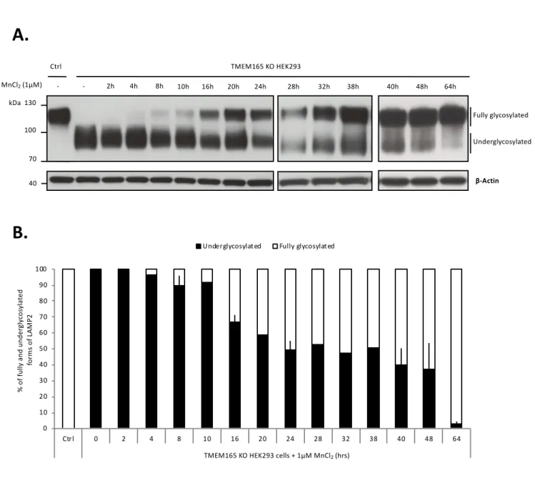
Time course of the rescue of LAMP2 glycosylation by Mn2+
Our previous studies demonstrated that supplementation with low Mn2+ concentrations could
completely suppress LAMP2 glycosylation defects in TMEM165 KO cells (4). This has however been observed for long treatments (48h). The time course of the Mn2+ effect was not evaluated. To
answer this point, a concentration of 1µM of MnCl2 was applied to the cells for increasing times (1h
to 64h) and LAMP2 glycosylation was assessed by western blotting (Fig. 1A). The results show that following Mn2+ treatment of TMEM165 KO cells, fully glycosylated forms of LAMP2 appear after
8h of Mn2+ treatment, yet most LAMP2 remain underglycosylated. Relative quantification of
underglycosylated and fully glycosylated LAMP2 (Fig. 1B) indicates that fully glycosylated LAMP2 progressively increases from 10% of LAMP2 after 8h to 97% after 64h of Mn2+ treatment.
This result is consistent with the slow turnover of LAMP2 estimated to 48h. In further experiments, cells were treated with 1µM MnCl2 for 8 and/or 16h.
Blocking endocytosis in TMEM165 KO cells does not prevent the rescue of LAMP2 glycosylation by Mn2+
The pathways by which Mn2+ enters cells after MnCl2 exposure are unknown. Two different ways
are possible, either by permeating membranes using specific and/or unspecific transporters or via endocytosis and endosome-to-TGN retrograde trafficking (8). To discriminate between these possibilities, cells were treated with different drugs known to interfere with endocytosis such as chloroquine (CQ) (14), a weak base raising the pH of acidic compartments, methyl-β-cyclodextrin (MBCD) that depletes cholesterol from plasma membrane and nocodazole, a microtubule depolymerizing agent (15) (Fig.2 and supplementary Fig. 1 and 2). We first checked the potential effects of these drugs on both the steady-state glycosylation status of LAMP2 in control HEK293 cells and the subcellular localization of LAMP2. No changes in the LAMP2 electrophoresis mobility could be observed in control HEK293 cells after these drug treatments (supplementary Fig. 1A and 2A), suggesting no major alteration of its glycosylation. Moreover, neither nocodazole or methyl-β-cyclodextrin nor chloroquine disrupted the lysosomal localization of LAMP2 (supplementary Fig. 1B and 2B).
10 We also checked the effects of those drugs on the morphology of the Golgi apparatus. Immunofluorescence staining of the Golgi proteins GPP130 and GM130 were performed followed by confocal microscopy analyses (supplementary Fig. 1B and 2B). As already known from the literature, nocodazole fragmented the Golgi apparatus whereas CQ and MBCD had no effect on its morphology.
LAMP2 glycosylation profile in TMEM165 KO HEK293 cells after treatment with or without a combination of those drugs and MnCl2 was then assessed (Fig. 2 and supplementary Fig. 2C). The
observed LAMP2 mobility in TMEM165 KO cells after 1µM MnCl2 treatment was comparable to
the one observed in Fig. 1. 10% of LAMP2 was found to be normally glycosylated after 8h of Mn2+
treatment and 33% after 16h. Cells were then treated with CQ or MBCD in the presence or absence of 1µM MnCl2 for 8h and 16h (Fig. 2). The result shows that CQ and MBCD did not prevent the
rescue of LAMP2 glycosylation after Mn2+ supplementation as 22% and 32% of fully glycosylated
forms respectively are observed after 16h treatment. Similar results were obtained with nocodazole (supplementary Fig.1C, D).
In conclusion, these results clearly highlight that none of these drugs prevents the rescue of LAMP2 glycosylation by Mn2+ in TMEM165 KO cells. This suggests that, at the extracellular concentration
used in our study (1µM), Mn2+ enter TMEM165-defective HEK293 cells through plasma membrane
transporters rather than by endocytosis.
Is the Golgi pump SPCA1 involved in the Mn2+-induced rescue of LAMP2 glycosylation?
After entering TMEM165-defective cells, cytosolic Mn2+ should reach the Golgi lumen to correct
the glycosylation defects. Besides TMEM165, which acts as a key determinant for Golgi Mn2+
homeostasis (4), the SPCA pumps (SPCA1 and SPCA2) are known to be the main suppliers of Mn2+ in the Golgi lumen (11). As SPCA2 is poorly expressed in our cell line, we tested the
contribution of SPCA1 by silencing its gene, ATP2C1 (Fig. 3). SiRNA knockdown was very efficient as 95% of the protein was depleted compared to untreated cells. Surprisingly, the knockdown of ATP2C1 did not prevent the rescue of LAMP2 glycosylation by Mn2+. 37% of
LAMP2 was fully glycosylated after 16h Mn2+ treatment in siATP2C1-treated cells, a level similar
to that observed without ATP2C1 knockdown. SPCA1 is thus not involved in the glycosylation rescue induced by Mn2+ supplementation in TMEM165 KO HEK293 cells.
11
pumps
Given the previous results, we next considered whether the ER could play a role in the observed glycosylation rescue. It had indeed been shown that SERCA pumps are able, in certain conditions, to transport Mn2+ into the ER lumen in addition to Ca2+ (16–18). We thus hypothesized that SERCA
pumps might be involved in the Mn2+ supplementation effect. To address this point, LAMP2
glycosylation status was evaluated by western blotting in cells treated with cyclopiazonic acid (CPA) or thapsigargin (TG), two specific SERCA inhibitors, in presence or absence of 1µM MnCl2
(Fig. 4). We can first notice that CPA as well as TG, did not induce any glycosylation defect on LAMP2 in control HEK293 cells neither after 8 nor 16h of treatment (supplementary Fig. 3). Remarkably, in TMEM165 KO HEK293 cells treated with CPA and 1µM MnCl2, the treatment
strongly delays the rescue of LAMP2 glycosylation by Mn2+ as only 4% of LAMP2 was fully
glycosylated after 8h and 29% after 16h of Mn2+ treatment. To reinforce this result, stronger effects
were obtained after TG treatment (Fig. 4A, B). To confirm these results at the structural level, mass spectrometry of total N-glycans have been performed (Fig. 5). For this, TMEM165 KO HEK293 cells were treated or not with 1µM MnCl2 and with or without TG/CPA. Consistent with our
previous studies, a pronounced hypogalactosylation was seen in TMEM165 KO HEK293 cells, with the accumulation of agalactosylated glycan structures detected at mass-per-charge (m/z) ratios 1661, 1835, 2081 and 2326. Consistent with our previous studies, MnCl2 treatment rescues the
general glycosylation defect as indicated by the decreased abundance of the structures (m/z) 1835, 2081 and 2326 (54% decrease, supplementary Fig. 4). Although TG and CPA treatments in KO TMEM165 cells slightly increase the abnormal agalactosylated glycan structures, such treatments fully prevent the total rescue of TMEM165-associated glycosylation defects by Mn2+ as indicated
by the remaining high abundance of the structures (m/z) 1661, 1835, 2081 and 2326 (supplementary Fig. 4).
To validate these results, we tested whether CPA or TG treatment acted by indirectly altering Mn2+
entry into the cells. Therefore, the total cellular Mn concentration in presence and absence of CPA or TG was evaluated by Inductively Coupled Plasma-Mass Spectrometry (ICP-MS) after MnCl2
supplementation. The result shows that after Mn2+ supplementation, the amount of Mn is
comparable in cells treated or not with CPA or TG (supplementary Fig. 3C). This demonstrates that neither CPA nor TG prevented the cellular Mn2+ entry after MnCl2 supplementation.
In parallel, the effects of CPA and TG on the morphology of the Golgi apparatus were investigated confocal immunofluorescence analysis of the Golgi proteins GPP130 and GM130 (Fig. 4C and supplementary Fig. 3A). No effect was observed on the Golgi apparatus morphology after CPA
12 treatment and only a slight dilatation of the Golgi was shown after TG treatment. Moreover, LAMP2 localization was not disrupted neither by TG nor CPA.
Overall, these results provide pharmacological evidences for the involvement of TG and CPA sensitive pumps in the Golgi N-glycosylation rescue induced by Mn2+ supplementation in
TMEM165 KO HEK293 cells.
Potential involvement of SERCA pump in the Mn2+ -induced rescue of LAMP2 glycosylation
The observed TG and CPA sensitivity led us to investigate the potential role of ER SERCA pump in the Mn2+ supplementation effect. Amongst all SERCA proteins, the SERCA2b isoform is the main
form expressed in HEK cells. SERCA2b was overexpressed for 48h in TMEM165 KO HEK293 cells and LAMP2 glycosylation status was assayed by western blot. The correct SERCA2b expression has been checked by immunofluorescence (data not shown) and western blot analyses in both TMEM165 KO and control HEK293 cells (Fig. 6). Compared to untransfected cells, LAMP2 glycosylation profile is slightly restored in TMEM165 KO HEK293 cells overexpressing SERCA2b (Fig. 6). In order to prove that this slight rescue was not due to potential ER/Golgi Ca2+ homeostasis
changes, the overexpressing cells were treated with 500µM CaCl2 and 1mM sodium pyruvate.
LAMP2 glycosylation rescue is identical to the one obtained without such treatment and suggests that the observed shift is likely not the cause of the SERCA2b Ca2+ pumping activity. Altogether
these results suggest the potential involvement of SERCA2b pump in the Mn2+ rescued
13
DISCUSSION
TMEM165 deficiency was recently found to lead to a type-II CDG associated with strong Golgi glycosylation abnormalities (1). Our previous work has shown that these glycosylation abnormalities in TMEM165 KO cells could result from a lack of Golgi Mn2+ (4). Many Golgi
glycosyltransferases using UDP-sugars as a donor substrate, such as UDP-Gal:N-acetylglucosamine galactosyltransferase I (B4GALT1; EC 2.4.1.22) and UDP-Gal:N-acetylglucosamine β-1,4-galactosyltransferase II (B4GALT2; EC 2.4.1.22), are indeed known to require Mn2+ in their
catalytic site to be fully active. Indeed, as well described by Ramakrishnan et al. (19), Mn2+ first
needs to bind the enzyme in a so-called open conformation to then allow the binding of the nucleotide sugar. Once in this enzyme-Mn2+-nucleotide sugar conformation, the acceptor substrates
bind it and the catalysis can start. Remarkably, we have demonstrated that 1µM MnCl2
supplementation was sufficient to completely suppress the glycosylation defects in TMEM165 KO cells. The underlying mechanism of this glycosylation rescue by Mn2+was however unknown.
To address this point, LAMP2 was used as a reporter glycoprotein to finely study the Mn2+ induced
mechanism of Golgi glycosylation rescue. We have clearly shown that after 8h 1µM MnCl2
treatment, newly synthesized LAMP2 was already fully glycosylated in TMEM165 KO cells. Moreover and in line with its turnover, half of LAMP2 is fully glycosylated after 24h treatment and almost all LAMP2 is fully glycosylated after 64h treatment. This showed that LAMP2 could be used to study glycosylation kinetics and that 1µM MnCl2 was able to totally suppress the
glycosylation defect observed on LAMP2 in TMEM165 KO HEK293 cells.
The first step of this glycosylation rescue induced by 1µM MnCl2 is the Mn2+ uptake at the
plasma membrane. This can be done either by endocytosis or through transporters. Using CQ, nocodazole and MBCD, three drugs known to disrupt endocytosis (14), we have demonstrated that none of these drugs prevented the glycosylation rescue after 1µM MnCl2 exposure in TMEM165
KO cells, then suggesting that Mn2+ does not enter into cells via endocytosis. This can easily be
explained by the presence of several plasma membrane transporters known to import Mn2+. This
includes the divalent metal transporter 1 (DMT1/NRAMP2/SLC11A2) (20, 21), NRAMP1 (22), the transferrin, the transporters SLC30A10/ZNT8 (23), SLC39A8/ZIP8 (7) and SLC30A14/ZIP14 (24, 25). As a consequence, we can reasonably think that Mn2+can use a wide set of transporters to
directly enter into cells, thus making the identification of the involved transporters difficult with possibly different answers depending on the cell type.
14 Once in the cytosol, Mn2+ needs to reach the Golgi lumen to suppress the Golgi
glycosylation defect induced by a lack of TMEM165. In absence of TMEM165, it is likely that Mn2+ supply in the Golgi is achieved via the action of SPCA pumps (SPCA1 and SPCA2) (9, 11, 26). Overexpression of SPCA1 has indeed been shown to increase Mn2+ accumulation into the Golgi
after high Mn2+ concentrations exposure (27). Given SPCA2 is not expressed in HEK cells, we have
depleted SPCA1 by siRNA and analysed the status of LAMP2 glycosylation in TMEM165 KO cells in the presence or absence of 1µM MnCl2. The results unequivocally showed that Mn2+
supplementation could rescue a normal LAMP2 glycosylation in siATP2C1 TMEM165 KO treated cells. This demonstrates that somehow, SPCA1 is not involved in the glycosylation rescue induced by Mn2+ in TMEM165 KO cells. However, since 5% of SPCA1 still remains after siRNA treatment,
we cannot completely exclude that the remaining SPCA1 is sufficient to efficiently transport Mn2+
from the cytosol to the Golgi apparatus to rescue glycosylation.
We then investigated the hypothesis that Mn2+ could reach the ER before being transported
to the Golgi compartment. The effects of specific inhibitors of sarcoplasmic reticulum (SR) calcium ATPase, thapsigargin (TG), and cyclopiazonic acid (CPA) were investigated. Interestingly, we did demonstrate that Mn2+ supplementation could not rescue a correct LAMP2 glycosylation in cells
treated with CPA (100µM) or TG (50nM). Importantly, these used concentrations of CPA and TG did not inhibit the activity of SPCA1. Indeed, Chen et al., recently showed that SPCA1 inhibition by either CPA or TG occurred from 182µM and 7µM, respectively (28). As we demonstrated using ICP-MS that neither CPA nor TG treatment prevented the cellular Mn2+ entry, our results suggests
that the observed Golgi glycosylation rescue induced by Mn2+ supplementation could come from
ER/Golgi uptake via TG and CPA sensitive proteins. To address the potential involvement of SERCA pumps, SERCA2b (the main isoform expressed in HEK cells) overexpression in TMEM165 KO HEK293 cells was performed. Although partial, the LAMP2 glycosylation profile is clearly enhanced then suggesting the involvement of SERCA2b protein in the Mn2+ rescued
glycosylation of LAMP2.
The importance of SERCA pumps in the observed Golgi glycosylation rescue was quite unexpected and the molecular mechanisms by which SERCA pumps are involved in the Mn2+
rescued Golgi glycosylation in TMEM165 KO cells remain unknown. One can expect that under Mn2+ supplementation, cytosolic Mn2+ is directly pumped by SERCA into the ER. Such role has
already been documented in literature. Chiesi et al showed in sarcoplasmic reticulum vesicles that a Ca2+ ATPase could indeed be activated by Mn2+ and was even able to import Mn2+ instead of Ca2+,
15 transport Mn2+ instead of Ca2+ with similar activation energies, same mechanism but with a much
lower affinity (18). Another hypothesis that we can’t exclude would be the crucial importance of ER/Golgi Ca2+ homeostasis in the Mn2+ induced Golgi glycosylation rescue. Some Golgi
glycosyltransferases and particularly the Golgi β4GalT1 (EC 2.4.1.38) enzyme possesses two metal binding sites, the site I that binds Mn2+ with high affinity and site II that binds diverse metal ions
including Ca2+ (18). It could be possible that a decrease in ER/ Golgi Ca2+ homeostasis completely
inhibits, in cellulo, the activity of β4GalT1 even under Mn2+ supplementation.
Overall, this paper shed light on the involvement of thapsigargin and cyclopiazonic acid sensitive pumps, most likely SERCA pump, in the rescue of TMEM165-associated glycosylation defects by Mn2+
16 Acknowledgments
We are indebted to Dounia Mouajjahfor her helpful assistance in mass spectrometry experiments. We also acknowledge Dr Dominique Legrand for the Research Federation FRABio (Univ. Lille, CNRS, FR 3688, FRABio, Biochimie Structurale et Fonctionnelle des Assemblages Biomoléculaires) for providing the scientific and technical environment conducive to achieving this work. We thank the BioImaging Center of Lille, especially Christian Slomianny and Elodie Richard, for the use of the Leica LSM700 and the Leica LSM780.
Conflict of interests None.
Funding
'This work was supported by the French National Research Agency [SOLV-CDG to F.F]; and the Mizutani Foundation for Glycoscience [to F.F].
Author contributions
F. Foulquier and S. Potelle designed the research; M. Houdou, E. Lebredonchel, V. Decool. M. Houdou, S. Potelle and S. Duvet analyzed data; M. Houdou, S. Potelle and F. Foulquier wrote the paper. A. Garat and D. Allorge developed and optimized the ICP-MS analysis and kindly gave us access to the platform for our experiments. D. Legrand, A. Klein, B. Gasnier and M. Ouzzine helped editing the paper and provided useful advices.
17
References
1. Foulquier, F., Amyere, M., Jaeken, J., Zeevaert, R., Schollen, E., Race, V., Bammens, R., Morelle, W., Rosnoblet, C., Legrand, D., Demaegd, D., Buist, N., Cheillan, D., Guffon, N., Morsomme, P., Annaert, W., Freeze, H. H., Van Schaftingen, E., Vikkula, M., and Matthijs, G. (2012) TMEM165 Deficiency Causes a Congenital Disorder of Glycosylation. Am. J. Hum.
Genet. 91, 15–26
2. Zeevaert, R., de Zegher, F., Sturiale, L., Garozzo, D., Smet, M., Moens, M., Matthijs, G., and Jaeken, J. (2013) Bone Dysplasia as a Key Feature in Three Patients with a Novel Congenital Disorder of Glycosylation (CDG) Type II Due to a Deep Intronic Splice Mutation in
TMEM165. JIMD Rep. 8, 145–152
3. Demaegd, D., Foulquier, F., Colinet, A.-S., Gremillon, L., Legrand, D., Mariot, P., Peiter, E., Van Schaftingen, E., Matthijs, G., and Morsomme, P. (2013) Newly characterized Golgi-localized family of proteins is involved in calcium and pH homeostasis in yeast and human cells. Proc. Natl. Acad. Sci. 110, 6859–6864
4. Potelle, S., Morelle, W., Dulary, E., Duvet, S., Vicogne, D., Spriet, C., Krzewinski-Recchi, M.-A., Morsomme, P., Jaeken, J., Matthijs, G., De Bettignies, G., and Foulquier, F. (2016) Glycosylation abnormalities in Gdt1p/TMEM165 deficient cells result from a defect in Golgi manganese homeostasis. Hum. Mol. Genet. 25, 1489–1500
5. Schneider, A., Steinberger, I., Herdean, A., Gandini, C., Eisenhut, M., Kurz, S., Morper, A., Hoecker, N., Rühle, T., Labs, M., Flügge, U.-I., Geimer, S., Schmidt, S. B., Husted, S., Weber, A. P. M., Spetea, C., and Leister, D. (2016) The Evolutionarily Conserved Protein
PHOTOSYNTHESIS AFFECTED MUTANT71 Is Required for Efficient Manganese Uptake at the Thylakoid Membrane in Arabidopsis. Plant Cell 28, 892–910
6. Powell, J. T. and Brew, K. (1976) Metal ion activation of galactosyltransferase. J. Biol. Chem.
251, 3645–3652
7. Park, J. H., Hogrebe, M., Grüneberg, M., DuChesne, I., von der Heiden, A. L., Reunert, J., Schlingmann, K. P., Boycott, K. M., Beaulieu, C. L., Mhanni, A. A., Innes, A. M., Hörtnagel, K., Biskup, S., Gleixner, E. M., Kurlemann, G., Fiedler, B., Omran, H., Rutsch, F., Wada, Y., Tsiakas, K., Santer, R., Nebert, D. W., Rust, S., and Marquardt, T. (2015) SLC39A8
Deficiency: A Disorder of Manganese Transport and Glycosylation. Am. J. Hum. Genet. 97, 894–903
8. Bonifacino, J. S. and Rojas, R. (2006) Retrograde transport from endosomes to the trans-Golgi network. Nat. Rev. Mol. Cell Biol. 7, 568–579
9. He, W. and Hu, Z. (2012) The role of the Golgi-resident SPCA Ca2+/Mn2+ pump in ionic
homeostasis and neural function. Neurochem. Res. 37, 455–468
10. Micaroni, M., Perinetti, G., Berrie, C. P., and Mironov, A. A. (2010) The SPCA1 Ca2+ pump and intracellular membrane trafficking. Traffic Cph. Den. 11, 1315–1333
11. Van Baelen, K., Dode, L., Vanoevelen, J., Callewaert, G., De Smedt, H., Missiaen, L., Parys, J. B., Raeymaekers, L., and Wuytack, F. (2004) The Ca2+/Mn2+ pumps in the Golgi
18 12. Roth, J. A. (2006) Homeostatic and toxic mechanisms regulating manganese uptake, retention,
and elimination. Biol. Res. 39, 45–57
13. Delannoy, C. P., Rombouts, Y., Groux-Degroote, S., Holst, S., Coddeville, B., Harduin-Lepers, A., Wuhrer, M., Elass-Rochard, E., and Guérardel, Y. (2017) Glycosylation Changes Triggered by the Differentiation of Monocytic THP-1 Cell Line into Macrophages. J.
Proteome Res. 16, 156–169
14. Dutta, D. and Donaldson, J. G. (2012) Search for inhibitors of endocytosis. Cell. Logist. 2, 203–208
15. Bayer, N., Schober, D., Prchla, E., Murphy, R. F., Blaas, D., and Fuchs, R. (1998) Effect of Bafilomycin A1 and Nocodazole on Endocytic Transport in HeLa Cells: Implications for Viral Uncoating and Infection. J. Virol. 72, 9645–9655
16. Chiesi, M. and Inesi, G. (1980) Adenosine 5’-triphosphate dependent fluxes of manganese and and hydrogen ions in sarcoplasmic reticulum vesicles. Biochemistry (Mosc.) 19, 2912–2918 17. Chiesi, M. and Inesi, G. (1981) Mg2+ and Mn2+ modulation of Ca2+ transport and ATPase
activity in sarcoplasmic reticulum vesicles. Arch. Biochem. Biophys. 208, 586–592
18. Yonekura, S.-I. and Toyoshima, C. (2016) Mn2+ transport by Ca2+-ATPase of sarcoplasmic reticulum. FEBS Lett. 590, 2086–2095
19. Ramakrishnan, B., Ramasamy, V., and Qasba, P. K. (2006) Structural snapshots of beta-1,4-galactosyltransferase-I along the kinetic pathway. J. Mol. Biol. 357, 1619–1633
20. Au, C., Benedetto, A., and Aschner, M. (2008) Manganese transport in eukaryotes: the role of DMT1. Neurotoxicology 29, 569–576
21. Garrick, M. D., Dolan, K. G., Horbinski, C., Ghio, A. J., Higgins, D., Porubcin, M., Moore, E. G., Hainsworth, L. N., Umbreit, J. N., Conrad, M. E., Feng, L., Lis, A., Roth, J. A., Singleton, S., and Garrick, L. M. (2003) DMT1: a mammalian transporter for multiple metals. Biometals
Int. J. Role Met. Ions Biol. Biochem. Med. 16, 41–54
22. Forbes, J. R. and Gros, P. (2003) Iron, manganese, and cobalt transport by Nramp1 (Slc11a1) and Nramp2 (Slc11a2) expressed at the plasma membrane. Blood 102, 1884–1892
23. Chen, P., Bowman, A. B., Mukhopadhyay, S., and Aschner, M. (2015) SLC30A10: A novel manganese transporter. Worm 4, e1042648
24. Chen, P., Chakraborty, S., Mukhopadhyay, S., Lee, E., Paoliello, M. M. B., Bowman, A. B., and Aschner, M. (2015) Manganese homeostasis in the nervous system. J. Neurochem. 134, 601–610
25. Girijashanker, K., He, L., Soleimani, M., Reed, J. M., Li, H., Liu, Z., Wang, B., Dalton, T. P., and Nebert, D. W. (2008) Slc39a14 gene encodes ZIP14, a metal/bicarbonate symporter: similarities to the ZIP8 transporter. Mol. Pharmacol. 73, 1413–1423
26. Vandecaetsbeek, I., Vangheluwe, P., Raeymaekers, L., Wuytack, F., and Vanoevelen, J. (2011) The Ca2+ Pumps of the Endoplasmic Reticulum and Golgi Apparatus. Cold Spring
19 27. Mukhopadhyay, S. and Linstedt, A. D. (2011) Identification of a gain-of-function mutation in
a Golgi P-type ATPase that enhances Mn2+ efflux and protects against toxicity. Proc. Natl.
Acad. Sci. U. S. A. 108, 858–863
28. Chen, J., De Raeymaecker, J., Hovgaard, J. B., Smaardijk, S., Vandecaetsbeek, I., Wuytack, F., Møller, J. V., Eggermont, J., De Maeyer, M., Christensen, S. B., and Vangheluwe, P. (2017) Structure/activity relationship of thapsigargin inhibition on the purified Golgi/secretory pathway Ca 2+ /Mn 2+ -transport ATPase (SPCA1a). J. Biol. Chem. 292, 6938–6951
20
Legends to figures
Figure 1: Time course of the Mn2+ induced LAMP2 glycosylation rescue. A. Control and
TMEM165 KO HEK293 cells were cultured with 1µM MnCl2 during the indicated times. Cell
culture medium was renewed every 16h. Total cell lysates were prepared, subjected to SDS-PAGE and western blot with the indicated antibodies. B. Relative quantification of fully and underglycosylated forms of LAMP2 (N, number of experiments = 2).
Supplementary figure 1: Effect of chloroquine (CQ) on Golgi morphology, LAMP2 subcellular localization and glycosylation profile. A. Control HEK293 cells were incubated with
CQ (10µM) for 8h or 16h, in combination or not with 1µM MnCl2. Total cell lysates were prepared,
subjected to SDS-PAGE and western blot with the indicated antibodies. B. Immunofluorescence analysis. Control and TMEM165 KO HEK293 cells were incubated with CQ (10µM) for 16h, fixed, permeabilized and labeled with antibodies against GM130, GPP130 and LAMP2 before confocal microscopy visualization. DAPI staining (blue) was performed, showing nuclei.
Supplementary figure 2: Effect of nocodazole on Golgi morphology, LAMP2 subcellular localization and glycosylation profile. A. and C. Control and TMEM165 KO HEK293 cells were
incubated with nocodazole (300nM) for 8h or 16h, in combination or not with 1µM MnCl2. Total
cell lysates were prepared, subjected to SDS-PAGE and western blot with the indicated antibodies.
B. Immunofluorescence analysis. Control and TMEM165 KO HEK293 cells were incubated with
nocodazole (300nM) for 16h, fixed, permeabilized and labeled with antibodies against GM130, GPP130 and LAMP2 before confocal microscopy visualization. DAPI staining (blue) was performed, showing nuclei. D. Relative quantification of fully and underglycosylated forms of LAMP2 (N, number of experiments = 2).
Figure 2: Mn2+ entry into TMEM165 KO HEK293 cells does not rely on endocytosis. A.
Control and TMEM165 KO HEK293 cells were cultured with CQ (10µM) in combination or not with 1µM MnCl2 for 8h or 16h. Total cell lysates were prepared, subjected to SDS-PAGE and
western blot with the indicated antibodies. B. Relative quantification of fully and underglycosylated forms of LAMP2 (N, number of experiments = 2).
Figure 3: ATP2C1 knockdown does not prevent the Mn2+ induced rescue glycosylation of LAMP2 in TMEM165 KO HEK293 cells. A. Control, TMEM165 KO and siATP2C1 HEK293
cells were cultured with 1µM MnCl2 for 16h. Total cell lysates were prepared, subjected to
SDS-PAGE and western blot with the indicated antibodies. B. Quantification of SPCA1 protein expression after normalization with actin. C. Relative quantification of fully and underglycosylated forms of LAMP2.
Figure 4: Involvement of TG and CPA sensitive pumps in the Mn2+ induced rescue of LAMP2 glycosylation. A. Control cells were cultured with either thapsigargine (TG) (50nM) or
cyclopiazonic acid (CPA) (100µM), two SERCA inhibitors, in combination or not with 1µM MnCl2
for 8h or 16h. Total cell lysates were prepared, subjected to SDS-PAGE and western blot with the indicated antibodies. B. Relative quantification of fully and underglycosylated forms of LAMP2 (N,
21 number of experiment = 2). C. Immunofluorescence analysis. TMEM165 KO HEK293 cells were incubated with either TG (50nM) or CPA (100µM) for 16h, fixed, permeabilized and labeled with antibodies against GM130, GPP130 and LAMP2 before confocal microscopy visualization. DAPI staining (blue) was performed, showing nuclei.
Supplementary figure 3: Effect of thapsigargin (TG) and cyclopiazonic acid (CPA)) on Golgi morphology, LAMP2 subcellular localisation and glycosylation profile and manganese uptake. A. Immunofluorescence analysis. Control and TMEM165 KO HEK293 cells were
incubated with either TG (50nM) or CPA (100µM) for 16h, fixed, permeabilized and labeled with antibodies against GM130, GPP130 and LAMP2 before confocal microscopy visualization. DAPI staining (blue) was performed, showing nuclei. B. Control HEK293 cells were incubated with either TG (50nM) or CPA (100µM) for 8h or 16h, in combination or not with 1µM MnCl2. Total
cell lysates were prepared, subjected to SDS-PAGE and western blot with the indicated antibodies.
C. TMEM165 KO HEK293 cells were cultured with either TG (50nM) or CPA (100µM) in
combination with 1µM MnCl2 for 16h. Total cell lysates were prepared as described in Material and
Methods section for ICP-MS analysis and total manganese concentration was measured (N = 2, n, number of samples = 4).
Figure 5: The N-glycosylation defects observed in TMEM165 KO HEK293 cells treated with TG or CPA are not rescued by Mn2+ supplementation. MALDI-TOF-MS spectra of the
permethylated N-glycans from TMEM165 KO HEK293 cells following different treatments. A. No treatment, B. TMEM165 KO HEK293 cells treated with 1µM MnCl2 for 16h, C. and D. TMEM165
KO HEK293 cells treated with 50nM TG in combination or not with 1µM MnCl2 for 16h, E. and F.
TMEM165 KO HEK293 cells treated with 100µM CPA in combination or not with 1µM MnCl2 for
16h. Symbols represent sugar residues as follow: blue square, N-acetylglucosamine; green circle, mannose; yellow circle, galactose; purple diamond, sialic acid; red triangle, fucose. Linkages between sugar residues have been removed for simplicity.
Supplementary figure 4: Quantification of the abnormal structures found in TMEM165 KO HEK293 cells treated with TG or CPA with or without Mn2+. A. Quantification of abnormal
glycan structures observed in TMEM165 KO HEK293 cells following the different indicated treatments. B. Representative glycan structures took into account for the quantification (abnormal structures with mass-per-charge ratios (m/z) 1661, 1835, 2080 and 2326; high mannose structures with mass-per-charge ratios (m/z) 1579, 1783, 1988, 2192 and 2396; complex glycan structures mass-per-charge ratios (m/z) 2227, 2431, 2530, 2605, 2646 and 2891). Symbols represent sugar residues as follow: blue square, N-acetylglucosamine; green circle, mannose; yellow circle, galactose; purple diamond, sialic acid; red triangle, fucose. Linkages between sugar residues have been removed for simplicity.
Figure 6: Potential involvement of SERCA2b pump in the Mn2+ supplementation effect.
Effect of SERCA2b overexpression in control and TMEM165 KO HEK293 cells treated or not with 500µM CaCl2 and 1mM sodium pyruvate on LAMP2 glycosylation. Total cell lysates were
A.
B.
Fig.1
Figure 1: Time course of the Mn2+induced LAMP2 glycosylation rescue. A. Control and TMEM165 KO HEK293 cells were cultured with 1µM MnCl2during
the indicated times. Cell culture medium was renewed every 16h. Total cell lysates were prepared, subjected to SDS-PAGE and western blot with the indicated antibodies. B. Relative quantification of fully and underglycosylated forms of LAMP2 (N, number of experiments = 2).
0 10 20 30 40 50 60 70 80 90 100 Ctrl 0 2 4 8 10 16 20 24 28 32 38 40 48 64 TMEM 165 KO + 1µM MnCl2 (hrs) % of fu lly a nd unde rg ly cos yl at ed fo rm s of L AM P2
Underglycosylated Fully glycosylated
TMEM165 KO HEK293 cells + 1µM MnCl2(hrs)
MnCl2(1µM) - 2h 4h 8h 10h 16h 20h 24h 28h 32h 38h 40h 48h 64h TMEM165 KO HEK293 Ctrl -130 100 70 LAMP2 β-Actin Fully glycosylated Underglycosylated kDa 40
130 100 70 130 100 70 - - 8h 16h - - 8h - - - - 8h 16h 8h -- 8h 16h 8h 16h - - -MnCl2(1µM) MBCD (5mM) CQ (10µM) 16h 16h -Control HEK293 LAMP2 β-Actin kDa 40
A.
B.
Supplementary figure 1: Effect of chloroquine (CQ) and methyl-β-cyclodextrin (MBCD) on Golgi morphology, LAMP2 subcellular localization and glycosylation profile. A. Control HEK293 cells were incubated with CQ (10µM) or MBCD (5mM) for 8h or 16h, in combination or not with 1µM MnCl2. Total cell lysates were prepared, subjected to SDS-PAGE and western blot with the indicated antibodies. B. Immunofluorescence analysis. Control and TMEM165 KO HEK293 cells were incubated with CQ (10µM) or MBCD (5mM) for 16h, fixed, permeabilized and labeled with antibodies against GM130, GPP130 and LAMP2 before confocal microscopy visualization. DAPI staining (blue) was performed, showing nuclei.
Supplementary fig. 1
MBCD No treatment GM130 GPP130 Merge Chloroquine Co nt ro l H EK2 93 LAMP2 TM EM 165 KO H EK293 Merge GM130 GPP130 LAMP2 No treatment Chloroquine MBCD0 20 40 60 80 100 - + - + + + + + 8h treatment 16h treatment % of fu lly a nd u nd er gl yc os yl at ed fo rm s of L AM P2
Underglycosylated Fully glycosylated
Nocodazole (300nM) MnCl2(1µM)
TMEM165 KO HEK293
Supplementary figure 2: Effect of nocodazole on Golgi morphology, LAMP2 subcellular localization and glycosylation profile. A. and C. Control and
TMEM165 KO HEK293 cells were incubated with nocodazole (300nM) for 8h or 16h, in combination or not with 1µM MnCl2. Total cell lysates were prepared, subjected to SDS-PAGE and western blot with the indicated antibodies. B. Immunofluorescence analysis. Control and TMEM165 KO HEK293 cells were incubated with nocodazole (300nM) for 16h, fixed, permeabilized and labeled with antibodies against GM130, GPP130 and LAMP2 before confocal microscopy visualization. DAPI staining (blue) was performed, showing nuclei. D. Relative quantification of fully and underglycosylated forms of LAMP2 (N, number of experiments = 2).
Supplementary Fig. 2
No treatment GM130 GPP130 Merge Nocodazole Co nt ro l H EK 29 3 TM EM 16 5 KO H EK 29 3 Merge GM130 GPP130 LAMP2 LAMP2 No treatment NocodazoleA.
B.
C.
D.
130 100 70 -MnCl2(1µM) Control HEK293 LAMP2 β-Actin 8h - 8h8h - 16h 16h16h Nocodazole (300nM) kDaControl TMEM165 KO HEK293
β-Actin - -- - - 8h 16h - 8h 8h -16h 8h 16h - -LAMP2 16h 16h 130 100 70 MnCl2(1µM) Nocodazole (300nM) Fully glycosylated Underglycosylated kDa 40 40
Fig. 2
A.
B.
Figure 2: Mn2+entry into TMEM165 KO HEK293 cells does not rely on endocytosis. A. Control and TMEM165 KO HEK293 cells were cultured with CQ
(10µM) or MBCD (5mM) in combination or not with 1µM MnCl2for 8h or 16h. Total cell lysates were prepared, subjected to SDS-PAGE and western blot with the indicated antibodies. B. Relative quantification of fully and underglycosylated forms of LAMP2 (N, number of experiments = 2).
130 100 70 16h - 8h 16h 8h 16h 8h -- - - -- -8h CQ (10µM) 16h 16h
Control TMEM165 KO HEK293
LAMP2 β-Actin -MnCl2 (1µM) Fully glycosylated Underglycosylated kDa 40 40 130 100 70 16h - 8h 16h 8h 16h 8h -- - - -- -8h -MBCD (5mM) MnCl2 (1µM) 16h 16h
Control TMEM165 KO HEK293
LAMP2 β-Actin Fully glycosylated Underglycosylated kDa 0 20 40 60 80 100 - CQ ( 10µM) MBCD (5mM) - CQ ( 10µM) (5mM)MBCD + + + + + + 8h treatment 16h treatment % of fu lly a nd unde rg ly cos yl at ed fo rm s of L AM P2
Underglycosylated Fully glycosylated
MnCl2(1µM)
Drug treatment
Fig. 3
A.
Figure 3: ATP2C1 knockdown does not prevent the Mn2+induced rescue glycosylation of LAMP2 in TMEM165 KO HEK293 cells. A. Control, TMEM165 KO
and siATP2C1 HEK293 cells were cultured with 1µM MnCl2for 16h. Total cell lysates were prepared, subjected to SDS-PAGE and western blot with the indicated antibodies. B. Quantification of SPCA1 protein expression after normalization with actin. C. Relative quantification of fully and underglycosylated forms of LAMP2.
B.
C.
0 20 40 60 80 100 - 16h - 16h - - + + TMEM 165 KO HEK293 % of fu lly a nd unde rg ly cos yl at ed fo rm s of L AM P2Underglycosylated Fully glycosylated
siATP2C1 MnCl2(1µM) TMEM165 KO HEK293 0 20 40 60 80 100 - + - + - + - + - - + + - - + + control TMEM 165 KO SP CA 1 re la tiv e ex pr es si on (% ) siATP2C1 MnCl2(1µM), 16h TMEM165 KO HEK293 Control HEK293 130 100 70 100 LAMP2 SPCA1 β-Actin - 16h - 16h - 16h - 16h - - + + - - + +
Control HEK293 TMEM165 KO HEK293
siATP2C1 MnCl2(1µM) Fully glycosylated Underglycosylated kDa 40
Supplementary figure 3: Effect of thapsigargin (TG) and cyclopiazonic acid (CPA)) on Golgi morphology, LAMP2 subcellular localization and glycosylation profile and manganese uptake. A. Immunofluorescence analysis. Control cells were incubated with either TG (50nM) or CPA (100µM) for 16h, fixed,
permeabilized and labeled with antibodies against GM130, GPP130 and LAMP2 before confocal microscopy visualization. DAPI staining (blue) was performed, showing nuclei. B. Control HEK293 cells were incubated with either TG (50nM) or CPA (100µM) for 8h or 16h, in combination or not with 1µM MnCl2. Total cell lysates were prepared, subjected to SDS-PAGE and western blot with the indicated antibodies. C. TMEM165 KO HEK293 cells were cultured with either TG (50nM) or CPA (100µM) in combination with 1µM MnCl2for 16h. Total cell lysates were prepared as described in Material and Methods section for ICP-MS analysis and total manganese concentration was measured (N = 2, n, number of samples = 4).
A.
B.
Supplementary Fig. 3
No treatment GM130 GPP130 Merge Thapsigargin CPA Co nt ro l H EK2 93 LAMP2 β-Actin LAMP2 MnCl2(1µM) CPA (100µM) Control HEK293 -8h 16h 8h -16h 8h 16h 130 100 70 kDa MnCl2(1µM) Control HEK293 8h - 8h 8h -16h 16h 16h 130 100 70 Thapsigargin (50nM) β-Actin LAMP2 kDa 0 50 100 150 200 250 - - TG 50nM CPA 100µM - + + + To ta l m an ga ne se c on ce nt ra tio n (% ) Drug treatment, 16h MnCl2(1µM), 16hC.
40 40Fig. 4
0 20 40 60 80 100 - TG (50nM) CPA (100µM) - TG (50nM) (100µM)CPA + + + + + + 8h treatment 16h treatment % of fu lly a nd u nd er gl yc os yl at ed fo rm s o f L AM P2 Fully glycosylated Underglycosylated Drug treatment MnCl2(1µM) TMEM165 KO HEK293A.
B.
C.
GM130 GPP130TMEM165 KO HEK293Merge LAMP2No tr ea tm en t Th ap sig ar gi n CP A
Figure 4: Involvement of TG and CPA sensitive pumps in the Mn2+induced rescue of LAMP2 glycosylation. A. Control and TMEM165 KO HEK293 cells were
cultured with either thapsigargine (TG) (50nM) or cyclopiazonic acid (CPA) (100µM), two SERCA inhibitors, in combination or not with 1µM MnCl2for 8h or 16h. Total cell lysates were prepared, subjected to SDS-PAGE and western blot with the indicated antibodies. B. Relative quantification of fully and underglycosylated forms of LAMP2 (N, number of experiment = 2). C. Immunofluorescence analysis. TMEM165 KO HEK293 cells were incubated with either TG (50nM) or CPA (100µM) for 16h, fixed, permeabilized and labeled with antibodies against GM130, GPP130 and LAMP2 before confocal microscopy visualization. DAPI staining (blue) was performed, showing nuclei.
kDa
β-Actin
LAMP2
Control TMEM165 KO HEK293
0 0 - - -8h 16h 0 8h 8h 0 16h 8h 16h - -16h 16h 130 100 70 MnCl2(1µM) TG (50nM) CPA (100µM) -- - - -- - -0 8h -0 -16h -16h 8h 16h 8h Fully glycosylated Underglycosylated 40
-Fig. 5
Figure 5: The N-glycosylation defects observed in TMEM165 KO HEK293 cells treated with TG or CPA are not rescued by Mn2+supplementation.
MALDI-TOF-MS spectra of the permethylated N-glycans from TMEM165 KO HEK293 cells following different treatments. A. No treatment, B. TMEM165 KO HEK293 cells treated with 1µM MnCl2for 16h, C. and D. TMEM165 KO HEK293 cells treated with 50nM TG in combination or not with 1µM MnCl2for 16h, E. and F. TMEM165 KO HEK293 cells treated with 100µM CPA in combination or not with 1µM MnCl2for 16h. Symbols represent sugar residues as follow: blue square, N-acetylglucosamine; green circle, mannose; yellow circle, galactose; purple diamond, sialic acid; red triangle, fucose. Linkages between sugar residues have been removed for simplicity.
No treatment 2395 2530 1661 2227 2605 2891 2431 2192 1579 1783 1988 2646 Mass (m/z) % In te ns ity 1835 2080 2326
A.
1µM MnCl2, 16h 2395 2530 1661 2227 2605 2431 2192 1579 1783 1988 2646 Mass (m/z) % In te ns ity 1835 2080 2326B.
50nM TG, 16h 2395 2530 1661 2605 2891 2431 1579 1783 1988 2646 2192 2227 Mass (m/z) % In te ns ity 1835 2080 2326C.
1µM MnCl2 + 50nM TG, 16h 2530 2605 2891 2431 2646 1661 1579 1783 1988 2395 2192 2227 Mass (m/z) % In te ns ity 1835 2080 2326D.
100µM CPA, 16h 2395 2530 2605 2891 2431 1579 1783 1988 2646 2192 2227 Mass (m/z) % In te ns ity 1835 2326 1661 2080E.
1µM MnCl2+ 100µM CPA, 16h 2395 2530 1661 2605 2891 2431 1579 1783 1988 2646 2192 2227 Mass (m/z) % In te ns ity 1835 2080 2326F.
Supplementary figure 4: Quantification of the abnormal structures found in TMEM165 KO HEK293 cells treated with TG or CPA and with or without Mn2+. A. Quantification of abnormal glycan structures observed in TMEM165 KO HEK293 cells following the different indicated treatments. B.
Representative glycan structures took into account for the quantification (abnormal glycan structures with mass-per-charge ratios (m/z) 1661, 1835, 2080 and 2326; high mannose structures with mass-per-charge ratios (m/z) 1579, 1783, 1988, 2192 and 2396; complex glycan structures mass-per-charge ratios m(/z) 2227, 2431, 2530, 2605, 2646 and 2891). Symbols represent sugar residues as follow: blue square, N-acetylglucosamine; green circle, mannose; yellow circle, galactose; purple diamond, sialic acid; red triangle, fucose. Linkages between sugar residues have been removed for simplicity.
Supplementary
fig. 4
- 52% 0 20 40 60 80 100 120 140 - 16h - 16h - 16h - TG (50nM) CPA (100µM) % of a bnor m al g ly ca n st ruc tur es MnCl2, 1µM - 7% - 7% TMEM165 KO HEK293A.
B.
1579 1661 1783 1835 1988 2080 2192 2227 2326 2396 2431 2530 2605 2646 2891 Abnormal glycan structuresFig. 6
Figure 6: Potential involvement of SERCA2b pump in the Mn2+supplementation effect. Effect of SERCA2b overexpression in control and TMEM165 KO
HEK293 cells treated or not with 500µM CaCl2and 1mM sodium pyruvate on LAMP2 glycosylation. Total cell lysates were prepared, subjected to SDS-PAGE and western blot with the indicated antibodies.
100 70 40 130 130 100 130 100 70 LAMP2 SERCA2 β-Actin kDa CaCl2 500µM + sodium pyruvate 1mM
SERCA2b overexpression -48h 24h -24h 48h -48h 24h -24h 48h Control HEK293 TMEM165 KO HEK293
Fully glycosylated Underglycosylated