AH rights reserved
ORIGINAL CONTRIBUTIONS
Incidence of Invasive Cancers following Basal Cell Skin Cancer
Fabio Levi,1 2 Carlo La Vecchia,3 Van-Cong Te,1 Lalao Randimbison,1 and Georges Erler2
To obtain quantitative information on the risk of invasive cancers following a diagnosis of basal cell carcinoma (BCC) of the skin, patients with incident BCC cases listed in the cancer registries of the Swiss cantons of Vaud and Neuchatel between 1974 and 1994 were actively followed up through December 31, 1994, for the occurrence of subsequent invasive neoplasms. Among 11,878 persons with incident BCC who were followed for a total of 76,510 person-years at risk, 1,543 metachronous cancers were observed versus 1,397.9 expected, corresponding to a standardized incidence ratio (SIR) of 1.1 (95% confidence interval (Cl) 1.0-1.2). However, after exclusion of skin cancers (mostly squamous cell carcinoma and melanoma), 975 second primary cancers were observed versus 1,059 expected (SIR = 0.9, 95% Cl 0.8-1.0). Significant excesses were registered for cancer of the lip (SIR = 2.2), for squamous cell skin cancer (SIR = 4.5) and melanoma of the skin (SIR = 2.5), and for non-Hodgkin's lymphoma (SIR = 1.9). The SIRs were also above unity, though not significantly, for cancers of the salivary glands (SIR = 2.8) and the small intestine (SIR = 2.1) and for soft-tissue sarcomas (SIR = 1.7). The SIR for lung cancer was 0.9. The SIRs for salivary gland and skin cancer were appreciably greater below age 70 years. For most sites, particularly for squamous cell cancer and melanoma of the skin, the SIRs remained elevated 5 or more years after BCC diagnosis. The cumulative incidence of squamous cell skin cancer was 13% at 19 years; this stresses the importance of carefully monitoring skin lesions among persons previously diagnosed with BCC. Am J Epidemiol 1998;147:722-6. carcinoma, basal cell; neoplasms; skin neoplasms
Basal cell carcinoma (BCC) is the most common type of skin cancer (1-4). Because of the advanced age at which most cases occur, and because of its favorable prognosis, underregistration is frequent. The cancer registries of the French-speaking Swiss cantons of Vaud and Neuchatel are among the few cancer registries that have paid specific attention to the issue of skin cancer; in these regions, cutaneous lesions that are surgically resected have traditionally been patho-logically examined (1, 5).
We previously described an excess of non-Hodgkin's lymphoma following a diagnosis of BCC (1). However, an excess incidence of other neoplasms Received for publication April 25, 1997, and in final form Sep-tember 15, 1997.
Abbreviations: BCC, basal cell carcinoma; Cl, confidence Inter-val; SIR, standardized incidence ratio.
1 Registre Vaudois des Tumeurs, Instttut UnlversHaire de Medecine Sociale et Preventive, Centre Hospitaller Universitaire Vaudois, Falalses 1, 1011 Lausanne, Switzerland.
2 Registre Neuchatelois des Tumeurs, Les Cadolles, 2000 Neuchatel, Switzerland.
3 Istttuto di Ricerche Farmacologiche "Mario Negri" e Istituto di Statistjca Medica e Biometria, Unlversita degli Studi di Milano, Via Venezian 1, 20133 Milano, Italy.
Reprint requests to Dr. Fabio Levi, Registre Vaudois des Tu-meurs, Instrtut Universitaire de Medecine Sociale et Preventive, Centre Hospitaller Universitaire Vaudote, Falalses 1, Casier 15 CH-1011 Lausanne, Switzerland.
after a diagnosis of BCC has also been reported, including cancers of the lip, salivary glands, larynx, lung, breast, and kidney, in addition to the well-known (but still inadequately quantified) excess of other types of skin cancer (6-9).
To obtain further quantitative information on this question, which has both pathogenic and public health implications, we computed the incidence of second primary cancers among almost 12,000 subjects regis-tered by the Vaud and Neuchatel cancer registries who were followed for a maximum of 20 years.
MATERIALS AND METHODS
Data for the present report were abstracted from the Vaud and Neuchatel cancer registry files, which in-clude all incident cases of malignant neoplasms diag-nosed in these cantons (10, 11). According to the 1990 Swiss census (12), the populations of these areas num-ber approximately 600,000 and 160,000, respectively. In these cantons, cancer registration systems have been in place since 1972, and population-based inci-dence data have been available since 1974. The reg-istries are tumor-based, and multiple primary malig-nancies found in the same person are entered separately. Most cases are registered repeatedly and
from different institutions, which improves the com-pleteness and accuracy of registration. The basic in-formation available from the registries comprises so-ciodemographic characteristics of the patient (age, sex), the primary site and histologic type of the tumor according to the International Classification of
Dis-eases, Ninth Revision (13), and the date of diagnostic
confirmation (histologic or clinical diagnosis). Both passive follow-up (through computer linkage with official mortality data files) and active follow-up (through verification of the vital status of apparently nondeceased cases using registries of current resi-dence) are conducted, and each subsequent item of information concerning an already registered case is used to complete the record of that patient.
After exclusion of synchronous cancers (n = 61), the present series comprised a total of 11,878 histo-logically confirmed BCCs of the skin (13) diagnosed between 1974 and 1994. The age range was 15-100 years (median age, 68 years). These persons were followed up to the end of 1994 for the occurrence of a second primary neoplasm (excluding basal cell skin carcinomas), emigration, or death.
Calculation of expected numbers of cases was based on site-, sex-, age-, and calendar period-specific inci-dence rates, multiplied by the corresponding number of person-years at risk. The significance of the ob-served : expected ratios (standardized incidence ratios (SIRs)) and their corresponding 95 percent confidence intervals was based on the Poisson distribution (14). Cumulative incidence was computed using the stan-dard life table approach (15).
RESULTS
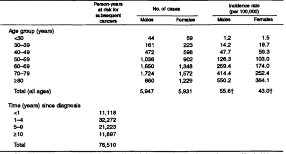
Table 1 gives the distribution of the 11,878 cases of BCC by age group, the corresponding incidence rates for the entire calendar period, and the numbers of person-years at risk in separate strata of time since diagnosis, for a total of 76,510 person-years at risk.
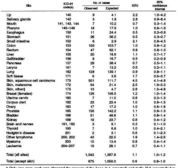
Table 2 gives the observed and expected numbers of all neoplasms and of cancers at selected sites. Overall, 1,543 metachronous cancers were observed versus 1,397.9 expected, corresponding to an SIR of 1.1 (95 percent confidence interval (Cl) 1.0-1.2). However, after exclusion of skin cancers (mostly squamous cell carcinoma and melanoma), 975 second primary can-cers were observed versus 1,059.0 expected (SIR = 0.9, 95 percent CI 0.8-1.0). Significant excesses were registered for cancer of the lip (9 observed, 4.1 ex-pected; SIR = 2.2), for squamous cell skin cancer (501 observed, 111.7 expected; SIR = 4.5) and melanoma of the skin (54 observed, 21.4 expected; SIR = 2.5), for other skin cancers (13 observed, 4.7 expected; SIR = 2.8), and for non-Hodgkin's lymphoma (43 observed, 22.5 expected; SIR = 1.9). The SIRs were also above unity, though not significantly, for cancer of the salivary glands (SIR = 2.8), the small intestine (SIR = 2.1), and the breast (SIR = 1.2) and for soft-tissue sarcoma (SIR = 1.7). The SIR was signif-icantly below unity for cancer of the esophagus (11 observed, 24.4 expected; SIR = 0.5), stomach (26 observed, 56.2 expected; SIR = 0.5), and gallbladder (8 observed, 16.7 expected; SIR = 0.5). The SIR was 0.9 (95 percent Cl 0.8-1.1) for cancer of the lung, 0.5 TABLE 1. Incidence of basal call carcinoma of the t U n , by age group and acoc Vaud and NeucMtel,
Switzerland, 1974-1994*
Age group (years)
<30 30-39 40-49 50-69 60-69 70-79 280
Total (all ages)
Time (years) since dagnosis <1 1-4 5-9 210 Total FMton-yeam 1 risk tor •utnoquent cancan 11,118 32,272 21,223 11,897 76,510 No. of a Male* 44 161 472 1,036 1,650 1,724 860 5,947 net Female* 59 223 598 902 1,348 1,572 1,229 5,931 Incidence rate (par 100,000) Moles 1.2 14.2 47.7 126.3 259.4 414.4 550.2 55.6t Females 1.5 19.7 59.3 103.0 174.0 252.4 364.1 43.0+
* Denominators in 1980: vaud, 529,000; Neuchatel, 158,000. Denominators in 1990: vaud, 602,000; Neuchatel, 164,000.
TABLE 2. Observed and expected numbers of second primary cancers aiI SetSOteO* SIISS aiiai an UDIMI diagnosis of basal osfl carcinoma of ths skin, with corresponding standardized Incidence ratios: Vaud and NeucMtal, Switzerland, 1974-1994
Ste Up Salivary glands Mouth Pharynx Esophagus Stomach Small intestine Colon Rectum Liver Gallbladder Pancreas Larynx Lung Soft tissue CO-9T code(8) 140 142 141, 143, 144 145-148 150 151 152 153 164 155 156 157 161 162 171
Skin, squamous cell carcinoma 173 Skin, melanoma Skin, other}: Breast (females) Uterine cervix Corpus uteri Ovary Prostate Bladder Money
Brain and nerves Thyroid
HodgJdn's disease Other lymphoma Myeloma Leukemia Total (afl sites) Total (except skin)
172 173 174 180 182 183 185 188 189 191, 192 193 201 2 0 0 , 2 0 2 203 204-207 No. of < Observed 9 5 7 18 11 26 6 103 47 20 8 28 7 128 6 501 54 13 126 7 23 17 155 51 18 3 7 2 43 10 19 1,543 975 asa* Expected 4.1 1.8 10.2 17.9 24.4 56.2 2.9 103.7 62.1 18.6 16.7 38.4 13.2 139.1 3.6 111.7 21.4 4.7 108.5 11.0 23.4 17.3 •\42.2 48.6 23.7 9.1 6.8 3.1 22.5 13.4 28.1 1,397.9 1,059.0 StRt Z 2 2.8 0.7 1.0 0.5 0.5 Z 1 1.0 0.8 1.1 0.5 0.7 0.5 0.9 1.7 4.5 2.5 2.8 1.2 0.6 1.0 1.0 1.1 1.1 0.8 0.3 1.0 0.6 1.9 0.8 0.7 1.1 0.9 95% confidence Interval 1.0-4.2 0.9-6.4 0.3-1.4 0.6-1.6 05-0.8 0.3-0.7 0.8-4.5 0.8-1.2 0.6-1.0 0.7-1.7 0.2-0.9 0.5-1.1 0.2-1.1 0.8-1.1 0.6-3.7 4.1-4.9 1.9-3.3 1.5-4.8 1.0-1.4 0.3-1.3 0.6-1.5 0.6-1.6 0.9-1.3 0.8-1.4 0.4-1.2 0.1-1.0 0.4-2.1 0.1-2.3 1.4-2.6 0.4-1.4 0.4-1.1 1.0-1.2 0.8-1.0 * Only one case each was observed for cancers of the bone (1.1 expected) and tests (2.3 expected); two cases were observed for cancers of the nasal cavity (2.4 expected) and four for the eye (2.4 expected).
t ICD-9, International Oasafflcatkm of Diseases, Ninth Revision; SIR, standardized incidence ratio.
$ Four cases of sebaceous adenocardnoma, three cases of malignant carcinoid carcinoma, two cases of
rymphoepithelial carcinoma, one case of apocrine adenocardnoma, one case of Pagefs disease, one small ceD carcinoma, and one "not otherwise specified" carcinoma were included.
(95 percent CI 0.2-1.1) for the larynx, and 0.6 (95 percent CI 0.3-1.3) for the uterine cervix.
Sites of second primary malignancies showing sig-nificant excesses or meaningful patterns are consid-ered further in table 3 by sex, age at BCC diagnosis, and time since diagnosis. The elevated rates of skin neoplasms and non-Hodgkin's lymphoma were similar in males and females. The SIRs for cancers of the lip, salivary glands, and skin were appreciably greater below age 70 years. For most sites, particularly for squamous cell cancer and melanoma of the skin, no consistent pattern of trend was observed with time since BCC diagnosis, and the SIRs remained signifi-cantly above unity 5 or more years after BCC diagno-sis. This indicates that the excess rates cannot be simply explained in terms of increased surveillance
around and after BCC diagnosis.
Figure 1 shows the cumulative incidence of squa-mous cell skin cancer following a diagnosis of BCC. A steady increase was evident up to 19 years after BCC diagnosis, reaching a cumulative incidence of 13 per-cent.
DISCUSSION
The present study, based on more than 11,800 sub-jects diagnosed with BCC and more than 76,000 person-years at risk, and on a population in which skin lesions have long been pathologically examined, con-firms that rates of various types of skin cancer, as well as cancer of the lip and non-Hodgkin's lymphoma, are significantly increased after a diagnosis of BCC.
j
!
1
i
I
i
i
If
gI
u
C « 8.3
i
•I
u> co •- u> co * •« "P io f n f o «l>cJcov<i>CNi4'-:q 2 2- 2. C. £ C. 2 <M » - o d co i s ij i d £ J JO CO CO CM *-° . a> co en co »-; d 2. 2- 2. i 2. ' 8 r - CO O CO O CM O o o, w, o, £ o_ d, CM UJ U , CO ^ CO ^ CO CO ^ * P C O ^ C © ^ C M ^ C O ^ I * CM*r o r S § CO^5 55
n I 8 SJ 8 B "! « n P S. jf. £2- Si C• - g a a
i
3
I
"3 10 YEARS 15 20FIGURE 1. Cumulative incidence (proportion) of squamous cell skin carcinoma (n = 501) after initial diagnosis of basal cell skin carcinoma (n = 11,878), by time Interval, In the cantons of Vaud and Neuchatel, Switzerland, 1974-1994. (Data were obtained from the Vaud and Neuchatel cancer registries.)
The excess incidence of squamous cell and melano-matous skin cancers is probably due to a shared predisposition—phenotypic characteristics and inher-ited cancer susceptibility syndromes, such as nevoid BCC syndrome—and risk factors, i.e., excess expo-sure to sunshine and other sources of ultraviolet radi-ation (16-18). This may explain, at least in part, the excess incidence of non-Hodgkin's lymphoma as well, assuming that ultraviolet radiation causes immunosup-pression (1, 19). More difficult to interpret are the appreciable excesses of salivary gland and skin cancers in patients younger than 70 years. This may reflect a predisposition to these neoplasms or a major role of extensive exposure to selected risk factors at a younger age (9).
The excess rate of sah'vary gland cancer is probably real, since it has also been observed in other data sets (20, 21), although the causes of salivary gland cancer—apart from ionizing radiation—remain largely undefined. The association has generally been attrib-uted to the common embryologic origin of the salivary glands and skin (both are derived from the ectodermal layer of the fetus) (22).
A relation between tobacco use and squamous cell skin cancer has been reported (21, 23). It is therefore interesting to note the absence of an association, in this data set, between BCC and subsequent lung cancer. The lower rates of cancer of the esophagus, stomach, larynx, uterine cervix, and mouth are also of interest. They may reflect different social class correlates or
other general lifestyle factors related to these neo-plasms, since in this population skin and breast cancer tend to have favorable social class correlates, while esophageal, larynx, gastric, and cervical cancer have unfavorable ones (24). The SIR for breast cancer was also moderately elevated in a Danish data set (19).
The absence of excess risk for all neoplasms except skin cancer is also of interest, since a moderately elevated SIR (1.15) was reported from a Danish study (8). This may reflect different correlates of BCC risk in various populations. In any case, there is no con-vincing evidence of a generalized excess of nonskin cancer following BCC.
In conclusion, this study, which was based on a large data set and had a uniquely long period of follow-up, confirms that subjects diagnosed with BCC do not have a generalized excess risk of nonskin neoplasms, with the exception of non-Hodgkin's lym-phoma and cancers of the lip and salivary glands. Given the small numbers of cases observed and the relatively moderate excess risks, this finding cannot be said to have prevention or screening implications, but it may be of some interest in the diagnostic process for these neoplasms. The excess rate of other skin cancers, however, is large enough to suggest a need for careful surveillance of skin lesions in BCC patients.
ACKNOWLEDGMENTS
The authors gratefully acknowledge the contributions of the Swiss, Vaud, and Neuchatel Leagues Against Cancer and of the staffs of the Vaud and Neuchatel Cancer Regis-tries.
REFERENCES
1. Levi F, Randimbison L, Te VC, et al. Non-Hodgkin's lym-phomas, chronic lymphocytic leukaemias and skin cancers. Br J Cancer 1996;74:1847-50.
2. Magnus K. The Nordic profile of skin-cancer incidence: a comparative epidemiological study of the three main types of skin cancer. Int J Cancer 1991;47:12-19.
3. Preston DS, Stem RS. Nonmelanoma cancers of the skin. N Engl J Med 1992.327:1649-62.
4. Franceschi S, Levi F, Randimbison L, et al. Site distribution of different types of skin cancer new aetiological clues. Int J Cancer 1966;67:24-8.
5. Levi F, La Vecchia C, Te VC, et al. Descriptive epidemiology of skin cancer in the Swiss Canton of Vaud. Int J Cancer
1988;42:811-16.
6. Lindelof B, Sigurgeirsson B, Wallberg P, et al. Occurrence of other malignancies in 1973 patients with basal cell carcinoma. J Am Acad Dermatol 1991;25:245-8.
7. Karagas MR, Stukel TA, Greenberg ER, et al. Risk of subse-quent basal cell carcinoma and squamous cell carcinoma of the skin among patients with prior skin cancer. Skin Cancer
Prevention Study Group. JAMA 1992;267:3305-10. 8. Frisch M, Hjalgrim H, Olsen JH, et al. Risk for subsequent
cancer after diagnosis of basal-cell carcinoma: a population-based, epidemiologic study. Ann Intern Med 1996;125: 815-21.
9. Schottenfeld D. Basal-cell carcinoma of the skin: a harbinger of cutaneous and noncutaneous multiple primary cancer. (Ed-itorial). Ann Intern Med 1996;125:852-4.
10. Levi F, Te VC, Randimbison L, et al. Statistics from the registry of the Canton of Vaud, Switzerland, 1988-1992. In: Parkin DM, Whelan SL, Ferlay J, et al. eds. Cancer incidence in five continents. Vol 7. Lyon, France: International Agency for Research on Cancer, 1997:686-9. (IARC Scientific Pub-lication no. 143).
11. Levi F, Portal I, M6an AM, et al. Statistics from the registry of the Canton of Neuchatel, Switzerland, 1988-1992. In: Parkin DM, Whelan SL, Ferlay J, et al, eds. Cancer incidence in five continents. Vol 7. Lyon, France: International Agency for Research on Cancer, 1997:674-7. (IARC Scientific Pub-lication no. 143).
12. Office F6d6ral de la Statistique. Population residante par com-mune et par division territoriale et par groupes d'Sges quin-quennaux, jusqu'a 65 ans et plus, ainsi que selon le sexe. In: Office Fdde'ral de la Statistique. Recensement f&ie'ral de la population 1990, structure de la population: tableaux geographiques. 2nd ed. Beme, Switzerland: Office F&Ie'ral de la Statistique, 1993:86-7. (Statistique de la Suisse, 1: population).
13. World Health Organization. International classification of dis-eases. Manual of the international statistical classification of diseases, injuries, and causes of death. Ninth Revision. Geneva, Switzerland: World Health Organization, 1977. 14. Breslow NE, Day NE. Statistical methods in cancer research.
Vol 2. The analysis of cohort studies. Lyon: International Agency for Research on Cancer, 1987:71. (IARC Scientific Publication no. 82).
15. Peto R, Pike MC, Armitage P, et al. Design and analysis of randomized clinical trials requiring prolonged observation of each patient. II. Analysis and examples. Br J Cancer 1977;35: 1-39.
16. Kricker A, Armstrong BK, English DR. Sun exposure and non-melanocytic skin cancer. Cancer Causes Control 1994;5: 367-92.
17. Zanetti R, Rosso S, Martinez C, et al. The multicentre south European study "Helios." I. Skin characteristics and sunburns in basal cell and squamous cell carcinomas of the skin. Br J Cancer 1996;73:1440-6.
18. Rosso S, Zanetti R, Martinez C, et al. The multicentre south European study "Helios." II. Different sun exposure patterns and the aetiology of basal cell and squamous cell carcinomas of the skin. Br J Cancer 1996;73:1447-54.
19. Adami J, Frisch M, Yuen J, et al. Evidence of an association between non-Hodgkin's lymphoma and skin cancer. BMJ
1995;310:1491-5.
20. Spitz MR, Newell GR, Byers RM, et al. Multiple primary cancer risk in patients with major salivary gland carcinoma. Ann Otol Rhinol Laryngol 1985;94:129-32.
21. Frisch M, Melbye M. New primary cancers after squamous cell skin cancer. Am J Epidemiol 1995;141:916-22. 22. Batsakis JG, Brannon RB, Sciubba JJ. Monomorphic
adeno-mas of major salivary glands: a histologic study of 96 tu-mours. Clin Otolaryngol 1981;6:129-43.
23. De Stefani E, Espandin J, Ronco A, et al. Tobacco smoking and the risk of non-melanoma skin cancer. Tob Control 1995; 4:175-9.
24. Levi F, Negri E, La Vecchia C, et al. Socioeconomic groups and cancer risk at death in the Swiss Canton of Vaud. Int J Epidemiol 1988;17:711-17.