British Journal of Rheumatology 1996;35:446-452
PULMONARY FUNCTION IN RHEUMATOID ARTHRITIS TREATED
WITH LOW-DOSE METHOTREXATE: A LONGITUDINAL STUDY
C. BEYELER, B. JORDI, N. J. GERBER and V. IM HOF*
Department of Rheumatology and *Department of Medicine, Division of Respiratory Diseases, University Hospital, 3010 Berne, Switzerland
SUMMARY
Lung volumes and gas exchange were investigated prospectively in 96 patients with rheumatoid arthritis selected without regard to pulmonary disorders and treated with i.m. methotrexate (MTX) injections [mean weekly dose 13.0 mg (5th-95th percentile (5-95 PC) 7.6-20.8)]. Individual changes over time during MTX treatment [mean duration 2.9 yr (5-95 PC 0.4-5.3)] were assessed by regression analyses in each individual. Forced vital capacity (FVQ remained stable in the majority of patients [mean annual change +0.8% (5-95 PC —8.1 to +14.0) of calculated normal value]. In addition, transfer factor using the indicator gas CO (Tuco) was unaltered in most patients [mean annual change - 2 . 1 % (5-95 PC -16.2 to +11.8) of predicted value]. However, there were significant decreases in the forced expiratory volume in 1 s (FEV,) before and after inhalation of 0.2 mg salbutamol [mean annual change - 0 . 8 % (5-95 PC - 8 . 4 to +3.2) and - 1 . 3 % (5-95 PC - 7 . 8 to +3.9) of the FVC measured, respectively]. In addition, there were significant increases in alveolar-arterial Po, gradients (P(A-.W)J) at rest and after exercise [mean annual change +1.7 mmHg (5-95 PC - 5 . 2 to +12.2) and +1.8 mmHg (5-95 PC -3.5 to 9.0), respectively]. Nevertheless, the amounts were small in view of the reliability of the methods applied and reflect, at least in part, the normal process of ageing. The annual change in FEVi/FVC was negatively correlated with FEV,/FVC at baseline (R, = - 0.46, P < 0.001). The annual change in TUco was also negatively correlated with Ti^o at baseline (Rs = —0.31, P = 0.028). No other risk factors for deterioration of
lung volumes or gas exchange were found, including mean weekly MTX dose, age, gender, smoking, presence of rheumatoid factor and pulmonary function at baseline. We conclude that MTX has no major effect on pulmonary function in the majority of patients and that there is no evidence that patients with pre-existing pulmonary disease are at increased risk for further deterioration of lung function.
KEY WORDS: Pulmonary function, Lung, Rheumatoid arthritis, Methotrexate.
LOW-DOSE weekly methotrexate (MTX) is increasingly used in the treatment of rheumatoid arthritis (RA) [1-3]. MTX has a favourable efficacy to toxicity profile over short and intermediate periods compared with other disease-modifying anti-rheumatic drugs (DMARDs) [4]. Nevertheless, in recent years there have been a growing number of communications about MTX-related pulmonary complications [5, 6]. Among the non-infectious adverse effects, interstitial pneumonitis, lung fibrosis, accelerated pulmonary nodulosis and drug-induced asthma have been reported. Among the pulmonary infections, Pneumo-cystis carinii pneumonia has been described most frequently [7]. Lung fibrosis and nodulosis may be pulmonary manifestations of RA itself [8]. A slowly progressive restrictive pattern and a deterioration of gas exchange may develop. Therefore, it may be very difficult to relate this pathology to MTX treatment or the underlying disease, or both.
There is some controversy about the usefulness of pulmonary function testing in patients with RA treated with low-dose MTX [9-11]. In the present study, we investigated lung volumes and gas exchange prospectively in a cohort of patients with RA with the following aims: (1) to determine the incidence of pulmonary complications requiring MTX with-drawal, (2) to assess pulmonary function over time and Submitted 11 July 1995; revised version accepted 31 January 1996. Correspondence to: Christine Beyeler, Department of Rheumatol-ogy, University Hospital, CH-3010 Berne, Switzerland.
(3) to search for risk factors for a loss of pulmonary function.
PATIENTS AND METHODS Patients
Between September 1988 and July 1992, patients with RA were recruited from the in- and out-patient Department of Rheumatology, University Hospital, Berne. All patients fulfilled the modified ARA criteria [12] and had disease activity sufficient to warrant the introduction of low-dose MTX. Patients were excluded from the study according to the following criteria: renal failure denned by a serum creatinine >1.2 times the upper limit of the reference range; active liver disease with laboratory evidence of liver cell necrosis; patients with a significant history of alcohol consumption and unwillingness to refrain from regular alcohol consumption; active bone marrow disease defined by leucopenia <3OOO/mm3 or thrombopenia
<100 000/m3; patients testing positive for human
immunodeficiency virus (HIV); pregnant women and patients of child-bearing age not practising effective contraception. Informed consent was obtained from all patients at the start of the study.
Methotrexate treatment
Weekly i.m. MTX injections, usually between 10 and 15mg, were started. Every single dose applied was documented on a patient control sheet and the cumulative dose calculated. Regular clinical follow-ups were performed, usually every 3 months or in the case © 1996 British Society for Rheumatology
of deterioration. Appropriate safety investigations included full haematology, liver and renal function tests, and erythrocyte folate concentrations. Weekly MTX doses were repeatedly adjusted according to disease activity, minor side-effects, potential inter-actions with concomitant drug treatment and renal function. Reasons for stopping MTX were carefully documented and classified into the following categories: pulmonary and non-pulmonary adverse effects of MTX, disorders associated with an increased risk for the development of MTX adverse effects, inefficacy of MTX, remission of RA, and disorders not directly related to RA or MTX. In the case of incomplete follow-up of pulmonary function testing, special attention was paid to verification of non-com-pliance due to different reasons in order not to miss any adverse effects of MTX. If MTX was stopped for > 1 month for any reason, results obtained after the interruption were not included in the evaluations. Non-steroidal anti-inflammatory drugs, low-dose oral corticosteroids and other drug treatments were continued as necessary. Folic acid was substituted in the case of decreased erythrocyte folate level.
Pulmonary function tests
Before the introduction of MTX, and every year thereafter, an extensive pulmonary assessment was performed. A history was taken about dyspnoea, cough, expectoration and smoking. An extensive physical examination was carried out, looking for signs of pulmonary and cardiovascular diseases. Detailed pulmonary function tests comprised forced vital capacity (FVC), forced expiratory volume in 1 s (FEV,) before and after inhalation of 0.2 mg salbutamol, FEV,/FVC, residual volume (RV), total lung capacity (TLQ and transfer factor using the indicator gas CO (Tuco). After puncture of the radial artery at rest and after exercise, if possible despite physical disability, arterial blood samples were analysed for partial pressures of oxygen (P^o,) and carbon dioxide (P«,co,), pH, base excess (BE) and CO haemoglobin (HbCO). All tests were performed by experienced staff using the same apparatus. Observed values were compared with those predicted for patients of similar age, sex and height as described by the official statement of the European Respiratory Society [13,14]. Alveolar (A)-arterial (a) Po, gradients, P(A^XOJ, were calculated according to the alveolar gas equation P(A-#W>J (mmHg) = [(barometric pressure (mmHg) 47) x 0.209] (1.25 x P^co, (mmHg)) -P.,o, (mmHg). Values were considered normal if P(A-.XO, (mmHg) was less than two-fifths of the age (in years) of the patient. Smokers were defined by a HbCO 5s 2.0% at rest.
Chest X-rays in two projections were performed by standard techniques. Data collection was stopped in June 1994.
Calculations
The mean weekly MTX dose was estimated by dividing the cumulative dose at the time point of the
last pulmonary function testing by the duration of treatment. Change in pulmonary function over time was assessed by calculations of slopes for each parameter in each individual. In the 22 patients with two measurements only, the slope of the fitted line was calculated. In the 57 patients with three or more values, linear regression analyses were performed. Each individual graph was carefully studied for deviation from linearity (sudden change, curvature). Use of the linear model was judged appropriate for all parameters. The mean coefficients of variations calculated and the percentage of patients with coefficients of variation < 0.2 as an indicator of good predictive power of the linear regressions were as follows: FVC 0.04, 100%; FEV, before inhalation 0.04, 100%; FEV, after inhalation 0.05, 98%; Tuco 0.10, 94%; P(A-«),o, at rest 0.25, 46%; P^-xo, after
exercise 0.35, 28%. Therefore, the precision of the slopes estimated was good for FVC, FEV, before and after salbutamol, and TL,CO, but poor for P(A-.),o, at rest
and after exercise.
Further statistical analyses included calculations of mean, standard deviation (S.D.), 5th and 95th percentiles (5-95 PC), one-group r-test and Spearman's rank correlation R,. The significance level was set at P = 0.05.
RESULTS
The 96 patients with RA included in the study had a mean disease duration of 9.3 yr (s.D. 10.0) and 72 (75%) patients were rheumatoid factor positive. Mean age was 61 yr (s.D. 13.6), 66 were women, 30 were men; mean weight was 64 kg (s.D. 11.3), mean height 162 cm (s.D. 9.2) and mean serum creatinine at entry to the study 82/imol/l (s.D. 17.3) (reference range 44—115). Serum creatinine was 95% of the baseline value (s.D. 13.5) at the end of the study. Thirty-one (32%) patients were smokers.
Lung volumes and gas exchange before treatment with low-dose MTX are summarized in Table I.
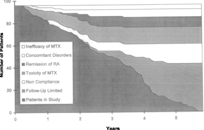
Until the end of the study in July 1994, the mean duration of MTX treatment was 2.9 yr (5-95 PC 0.4-5.3) and mean weekly MTX dose was 13.0 mg (5-95 PC 7.6-20.8). The time course of the number of patients with pulmonary function tests and MTX withdrawal is shown in Fig. 1. Follow-up was incomplete in 18 patients (18.8%) due to different MTX-unrelated causes and MTX was stopped in a total of 35 patients (36.5%). The reason was remission of RA in nine patients (9.4%), concomitant disorders not related to RA or MTX in seven patients (7.3%), inefficacy of MTX in four patients (4.2%), toxicity of MTX in 15 patients (15.6%) with adverse effects of MTX in 10 patients (10.4%) or concomitant disorders necessitating interruption of MTX treatment in order to prevent MTX adverse effects in five patients (5.2%): gastrointestinal intolerance (n = 3), stomatitis (n = 2), 4-fold increase of aminotransferases (n = 1), cutaneous vasculitis (n = 1), accelerated nodulosis (n = 1), post-injection pruritus (n = 1), extended infections {n = 3), infection of a hip prothesis (n = 1) and development of
448 BRITISH JOURNAL OF RHEUMATOLOGY VOL. 35 NO. 5
TABLE I
Lung volumes and gas exchange in 96 patients with rheumatoid arthritis before and during treatment with low-dose methotrexate. For abbreviations and calculation of predicted values, see Patients and methods. The significance levels of the mean annual changes during treatment
with MTX were calculated by the one-group /-test
Parameter FVC
FEVi/FVC before inhalation FEVi/FVC after inhalation Tuco
PIA-^OJ at rest
P(A-^.O, after exercise
Unit ml % predicted % % ml/min/mmHg % predicted mmHg mmHg
Mean values at baseline (5th-95th percentile) 3427 (1930-5240) 117(89-162) 70 (47-87) 73 (50-88) 21.4 (12.9-32.0) 88 (61-115) 19.9 (0.6-34.5) 9.7 ( - 3 . 4 to 29.6)
Mean annual change during treatment with MTX (per year) (5th-95th percentile) - 2 1 ( - 2 9 0 to +316) + 0.8 ( - 8 . 1 to +14.0) - 0 . 8 ( - 8 . 4 to +3.2) - 1 . 3 ( - 7 . 8 to +3.9) - 0 . 6 ( - 4 . 0 to +1.9) - 2 . 1 ( - 1 6 . 2 to +11.8) + 1.7 ( - 5 . 2 to +12.2) + 1.8 ( - 3 . 5 to +9.0) P n.s. n.s. <0.05 < 0.001 <0.01 <0.05 < 0.001 <0.01
respiratory symptoms (n = 2). A 70-yr-old man developed a probable MTX pneumonitis according to Searles' or Carson's criteria [15, 16] within a few days after 6 months of low-dose MTX treatment with a cumulative dose of 405 mg. A 77-yr-old man showed progression of a pre-existing rheumatoid lung disease with progressing restrictive ventilatory defect and deteriorating gas exchange after 16 months of MTX treatment with a cumulative dose of 1305 mg. The relation to MTX treatment is arguable. Therefore, MTX was stopped in two patients because of respiratory complaints, resulting in an incidence of 0.72 withdrawals per 100 patient-years due to pulmonary complications. Individual annual changes in lung volumes and gas exchange during MTX treatment are summarized in
Table I. FVC remained stable over time. In contrast, mean FEVi/FVC decreased little but statistically significantly before and after inhalation (/ = 2.04, P<0.05 and / = 3.35, P< 0.001, respectively). In addition, gas exchange deteriorated with a significant decrease of mean TUCo (' = 2.58, P < 0.01
for observed values and f=1.88, P < 0.05 for measured values related to predicted values) and with significant increases in mean P(A-»,O, at rest and after exercise (/ = 3.22, P < 0.001 and t = 2.68, P < 0.01, respectively).
Risk factors for a loss of pulmonary function during MTX treatment were sought. No effect of gender, smoking and the presence of rheumatoid factor became evident. In addition, no correlations were found
100 80
1
Pati o•s
Numbe r60 J
40 J
20-1•
• D Inefficacy of MTX 1 D Concomitant Disorders 1 • Remission of RA l«Toxicityof MTX 1 • Non Compliance 1 • Follow-Up Limited I • Patients in Study10 T m
I
I
u. •s a c 120 51 0
-
-15-•-FEV1 / FVC after Inhalation at Baseline (%)
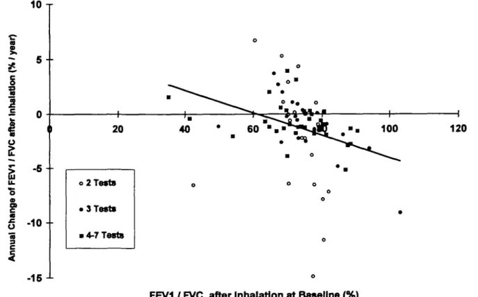
FIG. 2.—Plot of the annual change in FEV./FVC versus FEVi/FVC at baseline after inhalation. Patients with a small number of tests showed a higher variability of the slopes calculated than patients with 4-7 tests.
between individual annual change in lung volumes or gas exchange and age, mean weekly MTX dose or FVC at baseline. In contrast, significant negative correla-tions were revealed between individual annual change in FEV,/FVC and FEV,/FVC at baseline before and after inhalation (both R, = - 0.46, P < 0.001) (Fig. 2).
These findings were upheld for the proportional annual changes of FEV,/FVC in relation to FEV,/FVC at baseline {R. = - 0.39, P < 0.0004 and R. = - 0.27, P = 0.017, respectively). In addition, a negative correlation was found between individual annual change in TL,CO and TUCo at baseline (R, = —0.31,
Reasons for deterioration of FVC Individual change
in FVC (% Number Age Sex predicted per year)
Individual change in FEV./FVC after inhalation (% per year) TABLE II in 10 patients with Individual change in Tua> (% predicted per year)
RA treated with low-dose MTX Individual change
in P(A-U>I after
exercise
(mmHg per year) Reasons for deterioration
MTX related 77 - 25.5 (2) 2 3 4 5 6 7 8 9 10 71 63 80 79 76 79 63 71 55 f f m f f f m m m - 1 1 . 8 ( 2 ) - 8.1 (7) - 8.1 (2) - 7.7 (2) - 7.5 (3) - 7.3 (3) - 6.4 (3) - 6.2 (4) - 6 . 1 (4) 1.0 (2) - 24.7 (2) 5.3(2) 1.6(7) -11.6(2) 1.1 (2) 3.7 (3) - 11 (3) - 0.2 (3) - 2 . 0 ( 4 ) - 3.9 (4) NA NA - 3.4 (2) 4.5 (2) - 0.2 (3) 11.8(2) - 13.5 (3) - 2.7 (4) - 1 1 . 9 ( 4 ) NA Progression of interstitial ( - ) pneumopathy NA Cardiac failure — 0 . 1 ( 2 ) Progression of COPD, -pulmonary embolism NA Progression of pleural — effusions
9.7 (2) Deterioration within normal -range
NA Deterioration within normal — range NA Deterioration of asthma — bronchi ale 1.9 (2) Progression of Caplan's ( —) syndrome^ecurrent pleural effusions NA Progression of COPD -NA Thoracic surgery, deteriora- —
tion of asthma bronchiale NA — not available (number of pulmonary function tests).
450 BRITISH JOURNAL OF RHEUMATOLOGY VOL. 35 NO. 5
P = 0.028), which became insignificant after analysing the proportional annual change in Tuco in relation to TUco at baseline (R. = — 0.22, n.s.). Finally, a negative
correlation was found between the individual annual change in FVC and P(A-.WJ, after exercise at baseline OR. = - 0.35, P = 0.029).
Table II summarizes information about 10 patients with a pronounced loss of FVC, defined by negative annual changes of FVC below the 16th percentile of the whole cohort. All clinical, functional and radiological findings were reviewed and reasons for deterioration postulated. In addition, 10 patients had a deterioration of gas exchange with either a decrease in TL,CO below the 16th percentile (n = 7) or an increase in P(A-«),O,
after exercise above the 84th percentile (n = 3). Reasons were postulated to be deterioration of asthma bronchiale (« = 1), progression of chronic obstructive pulmonary disease (COPD) (n = 1) or not evident (« = 8).
DISCUSSION
The present study assessed pulmonary function prospectively in a cohort of 96 patients with RA selected without regard to pulmonary disorders. The withdrawal rate due to pulmonary complications was low. The prevalence of MTX pneumonitis was 1% after a mean treatment period of 2.9 yr. This rate was at the lower part of the range between 0.3 and 11.6% reported by several retrospective and prospective studies [5]. The incidence of acute MTX pneumonitis was one case per 276 patient-years which was considerably lower than the rates of one case per 26 and 50 patient-years, respectively, observed by retrospective studies [16, 17]. None of the patients developed an opportunistic pulmonary infection. Microscopic examinations and extensive cultures of sputum and bronchoalveolar lavage fluid, and serolog-ical tests for common respiratory viruses, Mycoplasma, rickettsia and Legionella, were all negative in the patients experiencing respiratory symptoms. Two patients showed a worsening of pre-existing interstitial pulmonary disease. In addition, deterioration of asthma bronchiale, progression of COPD, pulmonary embolism, pleural effusions, thoracic surgery and cardiac failure were regarded as being responsible for declines in pulmonary function in some patients. There are little pathognomonic clinical, laboratory or radiological features which allow differentiation between MTX-related pulmonary pathology and underlying diseases. Chronic interstitial pneumonitis with lymphocytic and plasma cell infiltrates as well as fibrosis unrelated to drug therapy can be found in lung biopsies of patients with RA [18]. Exacerbation of this lingering process by MTX is possible. Nevertheless, prevalence and incidence rates of chronic MTX-related interstitial pneumonitis will remain arbitrary and are deliberately not calculated in the present study.
Values of FVC were high at baseline and remained stable throughout the observation period in the majority of our patients. These findings are in agreement with longitudinal assessments of patients
with RA treated with low-dose MTX over 24 and 42 weeks, respectively [10, 11]. Similarly, no significant loss of FVC was found in patients with RA treated with D-penicillamine and various other DMARDs for at least 6 months [19]. In contrast, follow-up studies reported a significant decrease in FVC in patients with RA treated with aurothiomalate for 2 yr and different DMARDs for 8 yr, respectively [20, 21].
Our data revealed a significant annual decrease in FEV,/FVC both before and after inhalation, indicating some increasing airflow obstruction. Patients already with an airflow obstruction at baseline were not at an increased risk of deterioration during MTX treatment. These findings contrast with other longitudinal evaluations, which found no significant changes of FEVi over 24 and 42 weeks of MTX treatment [10, 11]. Nevertheless, a case report gave evidence for the induction of clinical and laboratory features of asthma and an increase in airway responsiveness to metha-choline chronologically related to the weekly intake of 7.5 mg MTX [22]. Another case report described a patient with a history of well-controlled asthma, whose respiratory symptoms reappeared while receiving MTX therapy [23]. However, in unselected patients with RA treated with different DMARDs, the frequency of airflow obstruction and bronchial reactivity was significantly increased compared to controls with minor degenerative joint disorders [24]. Follow-up assessments of patients treated with D-penicillamine, aurothiomalate and other DMARDs gave conflicting results, with only some of them developing obstructive ventilatory defects [19-21]. MTX has been recommended as an effective and safe glucocorticoid-sparing agent in asthma [25, 26]. However, its efficacy remains controversial [27]. The question emerged whether MTX treats or induces asthma [28]. In analogy, the question can be added whether MTX diminishes airways inflammation associated with bronchial narrowing and hyper-reactivity in RA or causes drug-induced airflow obstruction.
Our results showed a deterioration of gas exchange over time reflected by a significant annual decrease in TL,CO and significant annual increases in PCA-IXO, at rest and after exercise. The latter have to be interpreted with caution as values in individual patients revealed considerable variations over time, resulting in high coefficients of variation of the annual changes calculated. In addition, only half of the patients had measurements after exercise because of physical disability. Nevertheless, our findings differ from observations in smaller numbers of patients treated with MTX over shorter periods [10, 11]. On the other hand, follow-up assessments of patients treated with D-penicillamine, aurothiomalate and various other DMARDs gave conflicting outcomes with decreases [19, 20] and increases [21] of TL,CO- Again, our patients with a low TL,CO or high P(A-«),O, at baseline did not have
an increased risk for a further deterioration during MTX therapy.
In summary, the present study illustrates the favourable safety of low-dose MTX treatment in RA.
Withdrawal of MTX due to pulmonary side-effects was rare and represents a justified risk in view of the improvement of the disease in the majority of patients. FVC remained stable once allowance had been made for the physiological effects of ageing. Decreases in expiratory airflow and gas exchange were measured. Although these changes were statistically significant, the amounts were moderate and clinically insignificant in view of the reliability of the methods applied [13, 14] and reflect, at least in part, the normal process of ageing. Considerable interindividual differences were observed. However, we were unable to detect risk factors such as mean weekly MTX dose, age, gender, smoking, the presence of rheumatoid factor and pulmonary function tests at baseline. In our patients, disease activity is changing all the time. Because of this no associations with disease activity were explored. This is in contrast to an attempt in a cross-sectional study in juvenile chronic arthritis [29]. Our study also demonstrates the difficulty and occasional impossibility of categorizing pulmonary abnormalities into pul-monary involvement of RA, concurrent unrelated pulmonary diseases and manifestations of adverse drug reactions. It rather emphasizes the interrelated nature of the clinical presentation of each patient who deserves individual evaluation. Our practical experience during this study showed that pulmonary function tests at baseline helped in discerning the different causes of respiratory symptoms developed during MTX treat-ment. However, in view of the data presented, costly routine measurements of lung volumes and gas exchange no longer seem justified in all patients. Pulmonary function tests before and during MTX treatment are recommended only in the case of a history of lung disease, the presence of respiratory symptoms, abnormal clinical signs or alterations on chest X-rays.
ACKNOWLEDGEMENTS
We thank Dr R. Gerny, Dr F. Koelliker, S. Thomann and all physicians and nurses of the Department of Rheumatology and the Division of Respiratory Diseases for their help with the clinical data collection, and Dr C. Minder for statistical advice. We also thank the staff of the respiratory function laboratory for their careful performance of the tests, and M. Keiser and D. Messer for secretarial assistance. We acknowledge financial support from the Swiss Federal Health Office Research Fund, the University Hospital of Berne, private funds and Lederle (Switzerland).
REFERENCES
1. Bannwarth B, Labat L, Moride Y, Schaeverbeke T. Mcthotrexatc in rheumatoid arthritis; an update. Drugs 1994;47:25-50.
2. Schnabcl A, Gross WL. Low-dose methotrexate in rheumatic diseases—efficacy, side effects and risk factors for side effects. Semin Arthritis Rheum 1994;23: 310-27.
3. Rau R. Methotrexat-Behandlung der chronischen Polyarthritis 1994—cine Uebersicht. Z Rheumatol 1994^3:199-229.
4. Fries JF, Williams CA, Ramey D, Bloch DA. The relative toxicity of disease-modifying antirheumatic drugs. Arthritis Rheum 1993;36:297-306.
5. Barrera P, Laan RFJM, van Riel PLCM, Dekhuijzen PNR, Roerbooms AMT, van de Putte LBA. Methotrex-ate-related pulmonary complications in rheumatoid arthritis. Ann Rheum Dis 1994;53:434-9.
6. Cannon GW. Antirheumatic drug reactions in the lung. Bailliire's Clin Rheumatol 1993;7:147-71.
7. Stenger AAME, Houtman PM, Bruyn GAW, Eggink HF, Pasma HR. Pneumocystis carinii pneumonia associated with low-dose methotrexate treatment for rheumatoid arthritis. ScandJ Rheumatol 1994^3:51-3. 8. Kelly CA. Rheumatoid arthritis: Classical rheumatoid
lung disease. Bailliire's Clin Rheumatol 1993;7:1-16. 9. Velay B, Lamboley L, Massonnet B et al. Prospective
study of respiratory function in rheumatoid arthritis treated with methotrexate. Eur Respir J 1988;l(suppl. 2):371.
10. Crook AD, Furst DE, Helmers RA et al. Methotrexate does not alter pulmonary function in patients with rheumatoid arthritis. Arthritis Rheum 1989;32:S60. 11. Jeurissen MEC, Boerbooms AMT, Festen J, van de Putte
LBA. Pulmonary function tests in rheumatoid arthritis (RA): a double blind study of azathioprine and methotrexate. Arthritis Rheum 1991;34:S90.
12. Arnett FC, Edworthy SM, Bloch DA. The American Association 1987 revised criteria for the classification of rheumatoid arthritis. Arthritis Rheum 1988^1:315-24. 13. Quanjer PH, Tammeling GJ, Cotes JE, Pedersen OF,
Peslin R, Yernault JC. Lung volumes and forced ventilatory flows. Official statement of the European Respiratory Society. Eur Respir J 1993;6(suppl. 16):5-40. 14. Cotes JE, Chinn DJ, Quanjer PH, Roca J, Yernault JC.
Standardization of the measurement of transfer factor (diffusing capacity). Official statement of the European Respiratory Society. Eur Respir J 1993;6(suppl. 16): 41-52.
15. Searles G, McKendry RJR. Methotrexate pneumonitis in rheumatoid arthritis: potential risk factors. Four case reports and a review of the literature. / Rheumatol 1987;14:1164-71.
16. Carson CW, Cannon GW, Egger MJ, Ward JR, Clegg DO. Pulmonary disease during treatment of rheumatoid arthritis with low dose pulse methotrexate. Semin Arthritis Rheum 1987;16:186-95.
17. Carroll GJ, Thomas R, Phatouros CC et at. Incidence, prevalence and possible risk factors for pneumonitis in patients with rheumatoid arthritis receiving methotrex-ate. J Rheumatol 1994;21:51-4.
18. Yousem SA, Colby TV, Carrington CB. Lung biopsy in rheumatoid arthritis. Am Rev Respir Dis 1985;131: 770-7.
19. Haerden J, Coolen L, Dequeker J. The effect of D-penicillamine on lung function parameters (diffusion capacity) in rheumatoid arthritis. Clin Exp Rheumatol 1993;11:509-13.
20. Chakravarty K, Webley M. A longitudinal study of pulmonary function in patients with rheumatoid arthritis treated with gold and D-penicillamine. Br J Rheumatol 1992^1:829-33.
21. Linstow W, Ulrik CS, Kriegbaum NJ, Backer V, Oxhohn P. An 8-year follow-up study of pulmonary function in
452 BRITISH JOURNAL OF RHEUMATOLOGY VOL. 35 NO. 5 patients with rheumatoid arthritis. Rheutnatol Int
1994;14:115-8.
22. Jones G, Mierins E, Karst J. Methotrexate induced asthma. Am Rev Respir Dis 1991;143:179—81.
23. Delclaux B, Whitelaw WA, Derenne JP. Methotrexate and asthma. Chest 1993;103:1307-8.
24. Hassan WU, Keaney NP, Holland CD, Kelly CA. Bronchial reactivity and airflow obstruction in rheuma-toid arthritis. Ann Rheum Dis 1994;53:511—4.
25. Mullarkey MF, Lammert JK, Blumenstein BA. Long-term methotrexate treatment in corticosteroid-dependent asthma. Ann Intern Med 1990;112:557-81.
26. Shiner RJ, Nunn AJ, Chung KF, Ducan MG. Randomised, double blind, placebo controlled trial of
methotrexate in steroid-dependent asthma. Lancet 1990;336:137-40.
27. Erzurum SC, Leff JA, Cochran JE, Ackerson LM, Szefler SJ, Martin RJ. Lack of benefit of methotrexate in severe, steroid-dependent asthma: a double blind, placebo-con-trolled study. Ann Intern Med 1991;114:353-60. 28. Fertel D, Wanner A. Methotrexate: does it treat or
induce asthma [editorial]? Am Rev Respir Dis 1991;143: 1-2.
29. Pelucchi A, Lomater C, Gerloni V, Foresi A, Fantini F, Marazzini L. Lung function and diffusing capacity for carbon monoxide in patients with juvenile chronic arthritis: effect of disease activity and low dose metho-trexate therapy. Clin Exp Rheumatol 1994;12:675-9.