C-reactive protein as a marker for acute coronary
syndromes
F. Mach*f, C. Lovis**, J.-M. Gaspoz*, P.-F. Unger§, M. Bouillie||, P. Urbanf and
W. Rutishauserf
I Cardiology Center, \Clinic of Medicine, ^Emergency Center, \\Central Clinical Chemistry Laboratory, Department of Medicine, University Hospital, Geneva, Switzerland
Background For several years, acute coronary syndromes
have been perceived as causing the most hospital admis-sions, and even hospital mortality. The syndrome of unsta-ble angina frequently progresses to acute myocardial infarction but its pathogenesis is poorly understood, and prognosis determination is still problematic. We tested the hypothesis that measurement of the C-reactive protein in patients admitted for chest pain could be a marker for acute coronary syndromes.
Methods and Results We studied 110 patients admitted
with suspected ischaemic heart disease, but without elevated serum creatine-kinase levels at the time of hospital admission. Patients were subsequently divided into two groups based on their final diagnosis: group 1 comprised patients with unstable angina; group 2 patients with acute myocardial infarction. We measured the C-reactive protein at the time of hospital admission. The concentration of
C-reactive protein was elevated in 59% of the patients with a final diagnosis of acute myocardial infarction, and in 5% of the patients with a final diagnosis of unstable angina,
Conclusion This study indicates that C-reactive protein
levels measured at the time of admission in patients with suspected ischaemic heart disease could be a marker for acute coronary syndromes, and helpful in identifying patients at high risk for acute myocardial infarction. Measurement of C-reactive protein may have practical clinical significance in the management of patients hospital-ized for suspected acute coronary syndromes.
(Eur Heart J 1997; 18: 1897-1902)
Key Words: Acute coronary syndromes, C-reactive
protein.
Introduction
The clinical manifestation of acute coronary syndromes, which include both acute myocardial infarction and unstable angina, may largely depend on the presence and severity of functional factors that transiently and acutely interfere with coronary blood flow, together with an extremely variable degree of coronary athero-sclerosis'11. In the past, several studies have described an increase in C-reactive protein after myocardial infarc-tion, which may reflect an important inflammatory component in this clinical situation'2"41. The syndrome of unstable angina occurs most commonly in the setting of atherosclerotic coronary artery disease and frequently progresses to acute myocardial infarction. Coronary Revision submitted 26 June 1997, and accepted 10 July 1997. •These authors contributed equally to this work.
Correspondence: Francois Mach, Vascular Medicine and
Athero-sclerosis Unit, Department of Medicine, Cardiovascular Division, Brigham and Women's Hospital, Harvard Medical School, 221 Longwood Avenue, LMRC 309, Boston, MA 02115, U.S.A.
thrombosis and vasospasm are known to be implicated in the pathogenesis of myocardial infarction15-61, but the underlying causes of unstable angina are not clear. Although the cell biology of atherosclerosis is not completely defined, inflammation may play a part in unstable angina, as suggested by histological studies of coronary plaques'71, the release of thromboxanes and leukotrienes'8'9', the increased expression of leukocyte receptors in the presence of activated circulating leuko-cytes that may include coronary vasoconstriction, favour thrombotic processes, and activate platelets1'01.
Currently, there is a reduction in the time between the onset of chest pain and hospital admis-sion'11!. As more patients with acute ischaemic heart disease will be hospitalized in the future, predicting the prognosis of these patients very early and managing them accordingly will be crucial. Recently, two studies reported on the possible prognostic value of circulating active-phase C-reactive protein, which may reflect the inflammatory pathogenesis in patients suffering unstable angina'12'131. To characterize the presence of 'active' atherosclerotic lesions in patients admitted with
suspected acute ischaemic heart disease, we measured the value of C-reactive protein at the time of hospital admission and compared it with the final diagnosis of these patients.
Methods
Patients
The study population comprised 110 of 201 consecutive patients admitted to the emergency centre of the Geneva Hospital with chest pain between January and June 1994, and in whom myocardial infarction was suspected according to the clinical and electrocardiographic cri-teria of the Imminent Myocardial Infarction Rotterdam Study'141. There were 90 men and 20 women, with a mean ( ± SD) age of 65 ± 11 years. The criteria for enrollment were the following: chest pain at rest, with at least two episodes in the previous 48 h, or one episode lasting more than 20 min; ST-segment changes on ECG diagnostic of myocardial ischaemia during anginal at-tacks; no elevation of serum creatine kinase and creatine kinase-MB levels at the time of hospital admission. The criteria for exclusion were an interval of more than 24 h since the last ischaemic episode before hospitalization (47 patients), intercurrent inflammatory or neoplastic conditions likely to be associated with an acute-phase response (29 patients), acute myocardial infarction within the previous month (eight patients), valvular heart disease (four patients), and chronic dissecting aortic aneurysm (three patients). All patients were studied prospectively without knowledge of C-reactive protein serum levels. Ninety-eight patients (89%) stayed in the intensive care unit for 2-4 ± 2-4 days after admis-sion. All patients received various combinations of intravenous or oral nitrates, beta-blockers, aspirin, heparin and calcium antagonists. Nineteen patients (17%) were treated with thrombolysis (rTPA) started in the emergency room. Coronary angiography was performed within 2-5 ± 2-2 days of admission in 103 patients (94%), 63 patients (57%) underwent percu-taneous transluminal coronary angioplasty, and 24 (22%) coronary artery bypass grafting.
These 110 patients were divided into two groups based on their final diagnosis: Group 1 comprised 61 patients with a final diagnosis of unstable angina (51 men and 10 women; with a mean ( ± SD) age of 65 ± 11 years), and Group 2, 49 patients with a final diagnosis of acute myocardial infarction (39 men and 10 women; with a mean ( ± SD) age of 66 ± 12 years). The diagnosis of acute myocardial infarction was made by a physician independent of the study, according to the electrocardio-graphic and enzymatic criteria of the World Health Organization for acute myocardial infarction (maximal creatine kinase activity, >270 U . I "1 in men and > 1 7 0 U . l ~ ' in women, or >8% of creatine kinase-MB1151, on the basis of their evolution during the 24 h following the admission. These patients developed their
acute myocardial infarction 12 ± 10 h after admission. Among them, eight patients (16%) had persistent ST elevation on admission.
Study protocol
For all patients, venous blood samples were obtained at the time of hospital admission, every 4 h thereafter during the first day, and daily for the next 3 days. Total creatine kinase and creatine kinase-MB levels were determined routinely when the blood samples were obtained. The first blood samples obtained at the time of hospital admission were stored at - 20 °C and analysed for C-reactive protein and cardiac troponin T at the end of the study. All decisions regarding the care of the patients were made by the physicians in charge, independent of these measurements.
Plasma protein assays
C-reactive protein was assayed by a fluorescence polar-ization automated monoclonal-antibody, solid-phase, sandwich-type enzyme immunoassay (TDxFLx, Abbott Laboratories, North Chicago, IL, U.S.A.)116"181. The range of values detected by the assay is 000 to 26 mg per decilitre, With this method, the median normal value for C-reactive protein, < 0 - 5 m g . d l ~ ' in 95%, and < 10 mg . dl ~ ' in 98% of normal healthy volunteers'19', conforms with a previous report using different meth-ods1201. Cardiac troponin T was measured by an enzyme immunoassay (Boehringer Mannheim, Mannheim, Germany)'2'1. Values >0-20ug.l~' were considered positive for cardiac troponin T in this study. The activity of creatine kinase was measured colorimetrically at 37 °C (NAC, Merck, Darmstadt, Germany) by the stan-dard method of the European Committee for Clinical and Laboratory Standard1221. Creatine kinase isoenzyme MB activity was assessed by an immunoinhibition assay (NAC, Merck, Damstadt, Germany). All three protein immunoassays were performed by technicians who were unaware of the clinical data.
Statistical analysis
Data are presented as the mean ± standard deviation, or counts. A Mann-Whitney test was used to compare variables measured from the patients1231. P values less than 005 (two-tailed) were considered to indicate statistical significance.
Results
The characteristics of the two groups of patients are listed in Table 1. There were no clinical differences between the two groups, which differed only in their
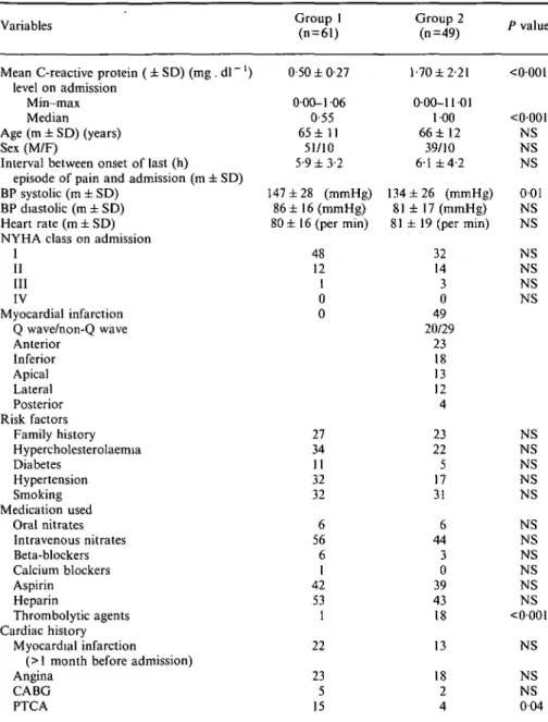
Table 1 Levels of C-reactive protein and population characteristics
Variables
Mean C-reactive protein ( ± SD) (mg . dl ~ ' ) level on admission
M in—max Median Age (m ± SD) (years) Sex (M/F)
Interval between onset of last (h) episode of pain and admission (m ± SD) BP systolic (m ± SD)
BP diastolic (m ± SD) Heart rate (m ± SD) NYHA class on admission
1 II III IV Myocardial infarction Q wave/non-Q wave Anterior Inferior Apical Lateral Posterior Risk factors Family history Hypercholesterolaemia Diabetes Hypertension Smoking Medication used Oral nitrates Intravenous nitrates Beta-blockers Calcium blockers Aspirin Heparin Thrombolytic agents Cardiac history Myocardial infarction
(> 1 month before admission) Angina CABG PTCA Group 1 (n = 61) 0-50 ± 0-27 000-1 06 0-55 65 ± 1 1 51/10 5-9 ±3-2 147 ± 2 8 (mmHg) 8 6 ± 1 6 ( m m H g ) 80 ± 16 (per min) 48 12 1 0 0 27 34 11 32 32 6 56 6 1 42 53 1 22 23 5 15 Group 2 (n=49) 1-70 ±2-21 0-00-11 01 100 66 ± 1 2 39/10 6 1 ±4-2 134 ± 2 6 (mmHg) 81 ± 17 (mmHg) 81 ± 19 (per min) 32 14 3 0 49 20/29 23 18 13 12 4 23 22 5 17 31 6 44 3 0 39 43 18 13 18 2 4 P value <0001 <0001 NS NS NS 001 NS NS NS NS NS NS NS NS NS NS NS NS NS NS NS NS NS <0001 NS NS NS 0 0 4 NS=non-significant
CABG=coronary artery bypass grafting
PTCA = percutaneous transluminal coronary angioplasty.
final diagnosis (group 1: unstable angina; group 2: acute myocardial infarction). Age, sex, cardiovascular risk factors, and baseline therapy were similar. There were no significant differences between the two groups in the number of stenotic coronary vessels, or in the pattern of coronary artery disease demonstrated by angiography. Thirty-eight patients (62%) in group 1 and 25 (51%) in group 2 underwent percutaneous transluminal coronary angioplasty; 12 patients (20%) in group 1 and 12 (24%) in group 2 underwent coronary artery bypass grafting; eight patients died in hospital, all in group 2 (/>=0-004). At the time of admission, all patients had normal creatine kinase and creatine kinase-MB serum levels.
Patients with ultimate confirmed diagnosis
of unstable angina (Group 1)
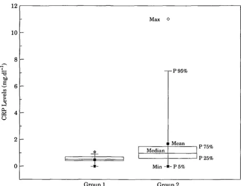
The concentration of C-reactive protein at the time of hospitalization was >0-9 mg . dl~ ' in only three (5%) of the 61 patients (1-3; 1-3 and l - 4 m g . d l ~ ' , respectively), with a final diagnosis of unstable angina (Fig. 1); the mean value of C-reactive protein was 0-50 ± 0-27mg.dl~'. The mean maximal concentrations of total creatine kinase ( 1 2 4 ± 5 9 I U . r ' [range 38-263; reference range, <170IU for women and <270 IU for men]), and creatine kinase-MB (6-1 ± 0-9% [range, 0-7-6; reference range, <8% of the total creatine kinase
12 10 o Max 0 -pP95% Median Min 11 Mean P75% P25% P5% Group 1 Group 2
Figure 1 Elevation of C-reactive protein in patients admitted with suspected acute
coronary syndromes. Group 1: ultimated confirmed diagnosis of unstable angina; Group 2: ultimated confirmed diagnosis of acute myocardial infarction. (/}<0-001 between groups).
if > 170 IU . 1 ' for women and 270IU for men) were normal in all patient samples. The mean concentrations of cardiac troponin T at the time of hospital admission (0-037 ± 007 ug . r ' [range, 0-00-0-37; reference range, 0-00-0-2 ug . 1 ~']) were > 0 - 2 u g . l ~ ' in only three patients (5%). There were no clinical or angiographic differences between patients with levels of C-reactive protein >0-9 mg . dl ~ ' and <0-9 mg . dl ~ '.
Patients with ultimate confirmed diagnosis
of acute myocardial infarction (Group 2)
The concentration of C-reactive protein at the time of hospital admission was ^ O ^ m g . d l "1 in 29 (59%) of the 41 patients with a final diagnosis of acute myocardial infarction, (F<0001 in comparison with Group 1; Fig. 1). The mean value of C-reactive protein was 1 -70± 2-21 mg . d l "1. The mean maximal concen-trations of total creatine kinase were 2150 ± 2507 I U . l "1 [range, 218-8707; reference range, > 170 IU for women and >270 for men], and of creatine kinase-MB 14-9 ± 8-6 IU . 1~' [range, 8-6-51; reference range, >8% of the total creatine kinase if >170 IU . 1" ' for women and >270 for men]. The elevation of creatine kinase and creatine kinase-MB appears 5-2 ± 2-7 h after onset of symptoms. The mean concentrations of cardiac troponin T (0-20±0-49 ug . 1 ~' [range, 000-2-41; reference range, 0-0-2 ug. 1 ~']) were >0-2 jig . 1 ~ ' in seven patients (14%). The elevation of cardiac troponin
T appears 4-9 ± 2-8 h after onset of symptoms. There was no difference in C-reactive protein level between the patients with or without a Q wave acute myocardial infarction. Among the 29 patients with elevated C-reactive protein and acute myocardial infarction, 12 (41%) had severe ST elevation (Pardee wave) suggestive of acute ischaemia at the time of hospitalization. Apart from that, we found no other obvious independent evidence of acute myocardial infarction on admission. There were no clinical or angiographic differences between patients with levels of C-reactive protein > 0 - 9 m g . d l ~ ' and those with < 0 - 9 m g . d l ~ ' . The detailed C-reactive protein levels of both groups are shown in Table 2.
Discussion
The syndrome of unstable angina nearly always occurs in the setting of atherosclerotic coronary artery disease. Increasingly, it is apparent that functional attributes rather than size alone determines the propensity of atherosclerotic plaque to provoke acute coronary events. Inflammation may contribute to weakening of the atherosclerotic plaque and could play a crucial role in the rupture of the fibrous cap, with subsequent exposure of the lipid-core to circulating blood components, result-ing in coronary thrombosis. Previous studies have registered elevated values for acute-phase proteins in patients with unstable angina and acute myocardial
Table Group Group 2 1 2 C-reactive protein (mg. Max 106 1101 Min Mean 000 0-50 000 1-70 dl~') in Median 0-55 100 acute coronary P 99% 106 1101 P 1% 000 000 syndromes P 5 % 000 000 P 95% 0-88 1101 SD 0-27 2-21 Group 1= Patients with ultimate confirmed diagnosis of unstable angina.
Group 2 = Patients with ultimate confirmed diagnosis of acute myocardial infarction. (P=percentile, SD = standard deviation).
infarction'23'. In 1994, Luizzo and colleagues reported the prognostic importance of C-reactive protein for patients with unstable angina'121, and more recently, Thompson and colleagues demonstrated that haemo-static factors and C-reactive protein are predictive markers of acute coronary syndromes'131. We tested the hypothesis that inflammation within the coronary ves-sels may reflect the severity of lesions implicated in plaque rupture. The results of our study confirm the observation that, among patients with acute ischaemic heart disease and no biological marker of myocardial necrosis, the plasma concentration of C-reactive protein at the time of admission was significantly higher in patients in whom an acute myocardial infarction was ultimately diagnosed, which makes it an impor-tant prognostic marker and a useful tool for clinical management.
Compared to previous studies124"261, the percent-age of patient with elevated cardiac troponin T level and unstable angina in our subjects was low. One expla-nation for this finding is the small interval between onset of symptoms and hospital admission in our group of patients (5-6 h). Indeed, the level of cardiac enzymes measured during acute coronary events depends directly on the duration of symptoms. It is unlikely that these patients with unstable angina had less acute disease, since their coronary angiographic findings were similar to the patients with acute infarction. Previous history of percutaneous transluminal coronary angioplasty was higher in patients with unstable angina, but symptoms related to restenosis occurred only in two of 15 (13%), which was similar in the group of patients with acute myocardial infarction (15%). Interestingly, the three patients with elevated cardiac troponin T in this un-stable angina group corresponded to those with a C-reactive protein > 0 - 9 m g . d l ~ ' described above. Finally, the high percentage of patients (41%) with acute myocardial infarction and normal C-reactive protein levels could be due, in out study, to the small interval between onset of symptoms and hospital admission (61 h).
The acute phase of C-reactive protein is a non-specific phenomenon reflecting cytokine-mediated hepatic production triggered by most forms of inflam-mation, infection and tissue injury. Our patients were carefully selected to eliminate intercurrent disorders likely to be associated with an acute-phase response, and similar attention to intercurrent processes will be essential for the practical application of our findings.
The elevation of C-reactive protein in acute coronary syndromes is probably related to 'coronary arteritis', and inflammation may then contribute to both vasospasm and thrombosis. Activated vascular wall cells, such as smooth muscle cells, endothelial cells, macrophages, and T lymphocytes, may play a central role because they produce molecules such as interleukin, adhesion molecules, growth factors, procoagulant activity, that alter both vascular reactivity and thrombogenicity'271. Elevation of C-reactive protein may therefore reflect the central role of inflammation in the pathophysiology of 'active' coronary artery disease. The beneficial effects of aspirin in unstable angina'28"301 could reflect decreased inflammation in addition to decreased platelet aggregation. In agreement with this hypothesis, Ridker and colleagues demonstrated recently that in apparently healthy men, a high baseline plasma C-reactive protein level predicts the risk of future myo-cardial infarction (P<0001), and that the use of aspirin was associated with significant reduction in the risk of acute myocardial infarction (/><002)'3'1.
Early prognostic assessment of myocardial in-farction is a key to optimal therapy and management of patients with this common disease, but entirely depends on the availability of rapid and specific diagnostic tests. Thus, the predictive correlation between C-reactive protein levels at the time of admission and the clinical outcomes may have practical clinical importance in the management of patients hospitalized for acute coronary syndromes, and possible cost-saving potential. In actual clinical practice, a C-reactive protein result can be obtained within 60 min. Our results led us to conclude that, in patients with suspected myocardial infarction, the measurement of C-reactive protein at the time of admission could lead to an improvement in the manage-ment of myocardial ischaemia, increasing prognostic security, and decreasing delay to appropriate triage and treatment. Large prospective studies are necessary to confirm these findings.
References
[1] Fuster V, Badimon L, Badimon JJ, Chesebro JH. The patho-genesis of coronary artery disease and the acute coronary syndromes. N Engl J Med 1992; 326: 242-50.
[2] de Beer FC, Hind DRK, Fox JCM, Allan RM, Maseri A, Pepys MB. Measurements of serum C-reactive protein concen-tration in myocardial ischaemia and myocardial infarction. Br Heart J 1982; 47: 239-43.
[3] Kushner I, Broder ML, Karp D. Control of the acute phase response: serum C-reactive protein kinetics after acute myocardial infarction. J Clin Invest 1978; 61: 235—42. [4] Berk BC, Weintraub WS, Alexander W. Elevation of
C-reactive protein in active coronary artery disease. Am J Cardiol 1990; 65: 168-72.
[5] Falk E. Unstable angina with fatal outcome. Dynamic cor-onary thrombosis leading to infarction and/or sudden death. Circulation 1985; 71: 699-708.
[6] Mandelkom JB, Wolf NM, Singh S et al. Intracoronary thrombus in nontransmural myocardial infarction and un-stable angina pectoris. Am J Cardiol 1983; 52: 1-6.
[7] Ciabattoni G, Silver MD, Mariani F, Guiliano G. Correlation of morphological variables in the coronary atherosclerotic plaque with clinical pattern of ischemic heart disease. Am J Cardiovasc Pathol 1988; 2: 159-72.
[8] Ciabattoni G, Ujang S, Sritara P et al. Aspirin, but not heparin, suppress the transient increase in thromboxane bio-synthesis associated with cardiac catheterization or coronary angioplasty. J Am Coll Cardiol 1993; 21: 1377-81.
[9] Carry M, Korley V, Willerson JT, Weigelt L, Ford-Hutchinson AW, Tagari P. Increase urinary leukotriene excre-tion in patients with cardiac ischemia: in vivo evidence for 5-lipoxygenase activation. Circulation 1992; 85: 230-6. [10] Mazzone A, De Servi S, Ricevuti G et al. Increased expression
of neutrophil and monocyte adhesion molecules in unstable coronary artery disease. Circulation 1993; 88: 358-63. [11] Gaspoz JM, Unger PF, Urban P el al. Delay in management
and treatment of patients with suspected acute myocardial infarction: role of the public, of extra- and intra-hospital structures and transportion methods. Schweiz Med Wochen-schr 1993; 123: 1376-83.
[12] Luizzo G, Biasucci LM, Gallimore JR et al. The prog-nostic value of C-reactive protein and serum amyloid A protein in severe unstable angina. N Engl J Med 1994; 331: 417-24.
[13] Thompson SG. Kienast J, Pyke SD, Haverkate F, van de Loo JC, for the European concerted action on thrombosis and disabilities angina pectoris study group. Hemostatic factors and risk of myocardial infarction or sudden death in patients with angina pectoris. N Engl J Med 1995; 332: 635-41. [14] Van der Does E, Lubsen J, Pool J. Acute coronary events in a
general practice: Objectives and design of the Imminent Myo-cardial Infarction Rotterdam Study. Heart Bull 1976; 7: 1706-10.
[15] Ischaemic heart disease registered: Report of the fifth working group. Copenhagen, Denmark: World Health Organization, 1971.
[16] Popelka SR, Miller DM, Holen JT, Kelso DM. Fluorescence polarization immunoassay II. Analyser for the rapid and
precise measurement of fluorescence polarization using disposable cuvettes. Clin Chem 1981; 27: 1198-201.
[17] Shaffar MR, Stroupe SD. A general method for performing routine clinical chemistry on the Abbott TDx analyser. Clin Chem 1983; 129: 1251.
[18] Dandliker SB, Kelly RJ, Dandliker J, Farquahar J, Levin J. Fluorescence polarization immunoassay. Theory and exper-imental method. Immunochemistry 1973; 10: 219—27. [19] Abbott Laboratories Instruction Manual. Protein C-reactive
TDx®/TDxFLx©. Abbott Laboratories, North Chicago, IL. [20] Shine B, de Beer FC, Pepys MB. Solid phase
radioimmuno-assay for CRP. Clin Chim Acta 1981; 117: 13-23.
[21] Katus HA, Looser S, Hallermayer K et al. Development and in vitro characterization of a new immunoassay of cardiac troponin T. Clin Chem 1992; 38: 386-93.
[22] Neumeier D, Kempes B, Glack B, Knedel M. Verteilung der Creatinkinase-Isoenzyme in Skeletund Hertzmuskulatur des Menschen. J Clin Chem Clin Biochem 1977; 15: 179-80. [23] Dansow-Sanders B, Trapp RJ. Basic and clinical biostatistics.
New York: Lange, 1988: 148-54.
[24] Hamm C, Ravkilde J, Gerhardt W et al. The prognostic value of serum troponin T in unstable angina. N Engl J Med 1992; 327: 146-50.
[25] Katus HA, Ravkilde J, Remppis A et al. Diagnosis efficiency of troponin T measurements in acute myocardial infarction. Circulation 1991; 83: 902-12.
[26] Ravkilde J, Niessen H, Horder M, Thygesen K. Independent prognostic value of serum creatine kinase isoenzyme MB mass, cardiac troponin T and myosin light chain levels in suspected acute myocardial infarction. Analysis of 28 months of follow-up in 196 patients. J Am Coll Cardiol 1995; 25: 574-81.
[27] Liischer TF, Boulanger CM, Yang Z, Noll G, Dohi Y. Interaction between endothelium-derived relaxing and con-tracting factors in health and cardiovascular disease. Circu-lation 1993; 87 (Suppl): V36-V44
[28] Lewis HD, Davis JW, Archibad DG et al. Protective effects of aspirin against acute myocardial infarction and death in men with unstable angina. N Engl J Med 1983; 309: 396-403. [29] Theroux P, Ouimet H, McCans J et al. Aspirin, heparin, or
both to treat acute unstable angina. N Engl J Med 1988; 319: 1105-11.
[30] Thornton MA, Gruentzig AR, Hollman J, King SB, Douglas JS. Coumadin and aspirin in prevention of recurrence after transluminal coronary angioplasty: a randomized study. Circulation 1984; 69: 721-7.
[31] Ridker PM, Cushman M, Stampfer MJ, Tracy RP, Hennekens CH. Inflammation, aspirin, and the risk of cardio-vascular disease in apparently healthy men. N Engl J Med 1997; 336: 973-9.