Puri¢cation and characterization of a
40.8-kDa cutinase in ungerminated conidia of
Botrytis cinerea Pers.: Fr.
Katia Gindro
a*, Roger Pezet
baUniversity of Lausanne, Institute of Systematical Botany and Geobotany, Biology Building, CH-1015 Lausanne, Switzerland b Swiss Federal Research Station for Plant Production of Changins, Route de Duillier, CH-1260 Nyon, Switzerland
Received 4 December 1998; received in revised form 14 December 1998; accepted 15 December 1998
Abstract
Cytoplasmic soluble proteins from ungerminated conidia of Botrytis cinerea exhibited cutinase activity. A 40.8-kDa cutinase was purified to homogeneity from this crude conidial protein extract. This cutinase does not correspond either to constitutive or to induced lytic cutin enzymes already described by other authors. The possible role of this constitutive cutinase in the induction of other cutinolytic proteins in the early stages of infection of plants by B. cinerea is discussed. z 1999 Federation of European Microbiological Societies. Published by Elsevier Science B.V. All rights reserved.
Keywords: Botrytis cinerea; Esterase; Cutinase; [3H]Cutin
1. Introduction
Many plant pathogenic fungi penetrate the un-wounded epidermal layer of their hosts by enzymatic hydrolysis of the cuticle [1]. Attachment of their spores to the plant surface is considered to be crucial for infection. Fungal esterases and particularly ases are involved in these phenomena [2]. A cutin-hydrolyzing enzyme was ¢rst described in Fusarium-solani f. sp. pisi [3]. Cutinases were characterized and puri¢ed from several fungal pathogens [4^7], bacteria
[8], and pollen [9]. A culture ¢ltrate of Botrytis cin-erea Pers.: Fr. contained a cutinase induced by to-mato cutin, which was the sole carbon source [10]. This extracellular cutinase was puri¢ed and the gene was cloned [11]. Recently it was demonstrated that ungerminated conidia of B. cinerea contained a con-stitutive cytoplasmic cutinase [12] which could ini-tiate cutin degradation and release of cutin mono-mers. These fatty acids are known to induce cutinase gene transcription in germinated conidia of F. solani f. sp. pisi and enhance the pathogenicity of this fungus [13]. This paper describes the puri¢cation and characterization of a constitutive cutinase in un-germinated conidia of B. cinerea. The possible role of this enzyme in the early events of infection of grape berries is discussed.
* Corresponding author.
Tel.: +41 (22) 363 43 53; Fax: +41 (22) 362 13 25; E-mail: katia.gindro@rac.admin.ch
2. Materials and methods
2.1. Organism and growth conditions
B. cinerea Pers.: Fr., isolate P69, was cultivated in Petri dishes on oatmeal agar (OMA, Difco). Cultures were placed at 21³C under alternating light and dark (12 h each) for 1 week. Conidia were harvested ac-cording to the method of Pezet and Pont [14] and stored dry at 380³C until required.
2.2. 3H-Labeled tomato cutin preparation
Pure tomato cutin was prepared according to Sal-inas et al. [10] and labeled with [3H]NaBH
4(3.7U109
Bq; 5.2U1011Bq mmol31) according to Koëller et al.
[15]. Cutinase activity was determined in puri¢ed fractions using [3H]tomato cutin according to
Pa-scholati et al. [5].
2.3. Extraction and puri¢cation of a cytoplasmic constitutive cutinase
Cytoplasmic proteins were extracted from 15 g of ungerminated conidia with glass beads according to the method of Van Etten and Freer [16], slightly modi¢ed: bu¡er was replaced by distilled water. The crude protein extract, placed in dialysis tubing, was concentrated on polyethylene glycol (PEG 20 000) to 20 ml, then ¢ltered (Akrodisk Nalgen, 0.2 Wm) and stored at 320³C until use. Crude ex-tract, dialyzed against piperazine-HCl bu¡er (PHB, 10 mM, pH 5.5), was loaded on a Superdex Prep Grade column (Pharmacia, 1.5U30 cm) and eluted with the same bu¡er. Non-speci¢c esterase activity was measured in the eluted fractions using para-ni-trophenylbutyrate (PNB) according to Pascholati et al. [6]. Active fractions were pooled and concentrated to 10 ml on PEG 20 000 as described before, then loaded on a DEAE Sepharose Cl-6B column (Phar-macia, 1.5U10 cm) suspended in PHB. A step gra-dient system of NaCl in the bu¡er (0, 0.05, 0.1, 0.15, 0.2, 0.25, 0.3 M) was used to elute eight fractions. The 0.2 M NaCl fraction contained non-speci¢c es-terase activity. This fraction was dialyzed against PHB, concentrated to 6 ml on PEG 20 000 and 30% (w/v) of (NH4)2SO4 was added. It was loaded
on a butyl Sepharose 4B column (Pharmacia, 1U15
cm) in PHB added with 30% (w/v) of (NH4)2SO4. A
decreasing step gradient system of (NH4)2SO4 (30%
to 0% in 5% steps) was used to elute seven fractions. These fractions were extensively dialyzed against dis-tilled water and concentrated to 4 ml on PEG 20 000 before the PNB assay, where (NH4)2SO4 interferes
strongly. The active fractions (5% and 10% (NH4)2SO4) were concentrated to 2 ml using
micro-dialysis (Microsep, cut-o¡ 10 000). Protein concen-tration was determined using the method of Brad-ford [18] (protein determination kit, Bio-Rad). 2.4. Gel electrophoresis analysis
SDS-PAGE (4%/12.5%) was performed according to Hames [19]. Protein bands were revealed by the silver staining method (Bio-Rad Silver Stain Kit) and esterase activity was revealed on renatured SDS-PAGE by incubation of the gel for 1 h at room temperature in Tris-HCl bu¡er (50 mM, pH 7.0+2% (w/v) Triton X-100) followed by incubation in the same bu¡er with K,L-naphthyl acetate accord-ing to Shaw and Prasad [17]. The molecular mass of cutinase was determined by SDS-PAGE using low-range molecular mass prestained standards (111^ 20.5 kDa, Bio-Rad). The isoelectric point of the pu-ri¢ed protein was determined by IEF analysis on precoated gels (Serva, 3^6 pH gradient, 300 Wm, 125U125 cm, pI marker protein test mixture 9) ac-cording to the manufacturer.
2.5. Immunoblotting
Proteins separated on SDS-PAGE were trans-ferred to PVDF membranes (Immobilon-P, Milli-pore) for 1.5 h at 100 V in electrotransfer bu¡er (Tris 25 mM, glycine 192 mM, methanol 20% v/v, pH 8.3). The membranes were then soaked overnight at 4³C in blocking bu¡er (Tris saline blocking bu¡er (TSBB): Tris-HCl 10 mM pH 7.5; NaCl 100 mM; Tween-20 0.1% (v/v); BSA 5% (w/v)). After three 5-min washes (TSBB without BSA) they were ex-posed for 3 h at room temperature to primary anti-body (10 Wg ml31 in TSBB containing 1% (w/v)
BSA) raised against a constitutive esterase isolated from B. cinerea conidia [20] or an induced cutinase found in the culture ¢ltrate [21] (mAb 14E5 and mAb 21C5 respectively, provided by Dr. A. Schots,
Laboratory of Monoclonal Antibody, Wageningen, The Netherlands). Positive reactions were revealed with a goat anti-mouse IgG conjugated to alkaline phosphatase (Sigma, dilution 1/3000 in TSBB). Al-kaline phosphatase activity was revealed with the fast red/naphthol method (Sigma Fast Red1/Naph-thol AS-MX, N³ F-4523) according to the manufac-turer.
3. Results and discussion
Linskens and Haage [22] ¢rst provided evidence that penetration by B. cinerea in potato leaf cutin
in vitro was linked to a cutinolytic activity. An 18-kDa cutinase was isolated by Salinas [21] in culture ¢ltrates of B. cinerea. More recently, Commeènil et al. [23] described a lipase excreted by B. cinerea which exhibited strong cutinolytic activity. Puri¢ed polyclo-nal antibody raised against this 60-kDa protein sup-pressed B. cinerea lesion formation on tomato leaves. We have demonstrated previously that ungermi-nated conidia of B. cinerea contained a constitutive cytoplasmic cutinase [12], and we have now de-scribed its puri¢cation to homogeneity using gel ¢l-tration, ion exchange and hydrophobic interaction chromatography (Table 1). The 5% (NH4)2SO4
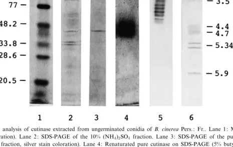
frac-tion separated on butyl Sepharose contained a single Fig. 1. Electrophoretic analysis of cutinase extracted from ungerminated conidia of B. cinerea Pers.: Fr.. Lane 1: Molecular mass stand-ards (silver stain coloration). Lane 2: SDS-PAGE of the 10% (NH4)2SO4 fraction. Lane 3: SDS-PAGE of the pure cutinase (5% butyl
(NH4)2SO4 Sepharose fraction, silver stain coloration). Lane 4: Renaturated pure cutinase on SDS-PAGE (5% butyl (NH4)2SO4
Sephar-ose fraction, K,L-naphthyl acetate coloration). Lane 5: IEF of the pure cutinase (5% butyl (NH4)2SO4Sepharose fraction, K,L-naphthyl
acetate coloration). Lane 6: pI standards (Coomassie coloration).
Table 1
Puri¢cation steps of the constitutive cutinase in ungerminated conidia of B. cinerea Pers.: Fr
Puri¢cation step Volume
(ml) Total protein(Wg Wl31) Total enzyme a
(Wmol min31) Speci¢c activity a
(Wmol min31Wg31) Puri¢cation Yield (%)
Crude extract 20 17.00 24.0 7.0 1.0 100
Superdex 10 0.40 7.9 98.7 14.1 32.5
DEAE (0.2 M NaCl) 6 0.04 3.6 446.0 63.7 14.9
Butyl Sepharose (5% (NH4)2SO4) 2 0.01 1.2 640.0 91.4 5
Butyl Sepharose (10% (NH4)2SO4) 2 0.03 0.7 466.0 66.5 2.9
band with a molecular mass of 40.8 kDa revealed on SDS-PAGE by silver staining (Fig. 1) and with K,L-naphthyl acetate (Fig. 1). This fraction applied to
3H-labeled cutin released ethyl acetate-soluble
radio-active cutin monomers (Table 2). PNB hydrolytic activity (Table 1) and speci¢c cutinase activity (Table 2) were also measured in the 10% (NH4)2SO4
frac-tion (Table 2). Apart from the 40.8-kDa band, other unknown proteins were revealed in this latter frac-tion which will require further puri¢cafrac-tion (Fig. 1). The constitutive 40.8-kDa cutinase does not corre-spond to other cutinases isolated from B. cinerea. Salinas [21] described a 110^111-kDa constitutive es-terase isolated from B. cinerea conidia with an as-sumed cutinolytic activity and a 18-kDa cutinase pu-ri¢ed from culture ¢ltrates. mAb 14E5 raised against this constitutive esterase and mAb 21C5 raised against the excreted cutinase did not reveal the 40.8-kDa cutinase (Fig. 2). However, mAb 14E5 has revealed a 110-kDa protein present in a 2-week-old culture ¢ltrate of B. cinerea (Fig. 2). Sali-nas [21] has described this 110-kDa constitutive con-idial putative cutinase as a membrane-bound pro-tein. The extraction protocols of conidial proteins used in this work did not allow the solubilization of such proteins. We can conclude that ungermi-nated conidia may contain two constitutive cutino-lytic enzymes, one soluble (40.8 kDa) and another bound to membranes or cell walls (110 kDa), which are excreted at conidial germination and produced during mycelial growth. Further research will be nec-essary to elucidate this hypothesis.
Isoelectric focusing analysis of the puri¢ed 40.8-kDa cutinase has revealed seven isoforms with acidic properties (pI 3.5^4.2) (Fig. 1). This constitutive cu-tinase might be excreted during the early events of conidial germination. It could release, through cutin degradation, su¤cient amounts of cutin monomers,
such as 16-hydroxyhexadecanoic acid, to activate the cutA gene expression, described by Van der Vlugt-Bergmans [11], which is responsible for the synthesis of the 18-kDa induced cutinase. Both 40.8-kDa con-Fig. 2. Demonstration by immunoblotting that the 40.8-kDa cuti-nase does not react with mAb 14E5 and mAb 21C5 raised against a constitutive esterase in ungerminated conidia of B. cin-erea and an induced 18-kDa cutinase puri¢ed from culture ¢l-trates of B. cinerea. Lane 1: Prestained molecular mass stand-ards. Lane 2: Detection of an 18-kDa cutinase with mAb 21C5 in 2-week-old cultures of B. cinerea. Lane 3: Detection of a 110-kDa constitutive conidial esterase with mAb 14E5 in 2-week-old cultures of B. cinerea. Lane 4: Pure 40.8-kDa cutinase (5% butyl (NH4)2SO4 Sepharose fraction) is not detected by either mAb
21C5 or mAb 14E5.
Table 2
Radioactivity released from3H-labeled cutin by the pure 40.8-kDa constitutive cutinase contained in the butyl Sepharose (5% (NH4)2SO4)
fraction and in the partially puri¢ed 40.8-kDa cutinase (10% (NH4)2SO4), compared to radioactivity released by cutinolytic activity in the
crude extract
Extract CPM (U105) Speci¢c activity (CPMU104Wg31 protein)
Control 12.0 þ 0.4 0.00 þ 0.00
Crude extract 38.0 þ 0.7 0.21 þ 0.02
Butyl Sepharose (5% (NH4)2SO4) 52.0 þ 0.6 436 þ 2.09
stitutive and 18-kDa induced cutinase could be strongly implicated in early infection processes by B. cinerea preceding latent stages and ¢nal plant col-onization. The role of the 110-kDa cutinase is still unknown. The raising of polyclonal antibodies against the 40.8-kDa cutinase is in progress and such antibodies should be useful to study its role during conidial germination, its development during mycelial growth, and to explain some aspects of the complex enzymatic events of the infection process of B. cinerea.
Acknowledgments
The authors thank Dr. A. Schots (Laboratory of Monoclonal Antibodies, Wageningen Agricultural University, Wageningen, The Netherlands) for pro-viding 14E5 and 21C5 monoclonal antibodies, Drs. A. and T.C. Caesar and Dr. M. Cole for critically reading the manuscript, and Mrs. I. De Groote for valuable technical assistance.
References
[1] Koëller, W. (1990) Plant cuticles: the ¢rst barrier to be over-come by plant pathogens. In: The Fungal Spore and Disease Initiation in Plants and Animals (Cole, G.T. and Hoch, H.C., Eds.), pp. 219^240. Plenum, New York.
[2] Schaëfer, W. (1994) Molecular mechanisms of fungal pathoge-nicity to plants. Annu. Rev. Phytopathol. 32, 461^477. [3] Purdy, R.E. and Kolattukudy, P.E. (1975) Hydrolysis of plant
cuticle by plant pathogens. Puri¢cation, amino acid composi-tion, and molecular weight of two isozymes of cutinase and a non speci¢c esterase from Fusarium solani f. pisi. Biochemistry 14, 2824^2831.
[4] Koëller, W. and Parker, D.M. (1989) Puri¢cation and charac-terization of cutinase from Venturia inaequalis. Phytopathol-ogy 79, 278^283.
[5] Pascholati, S.F., Deising, H., Leite, B., Anderson, D. and Nicholson, R.L. (1993) Cutinase and non-speci¢c esterase ac-tivities in the conidial mucilage of Colletotrichum graminicola. Physiol. Mol. Plant Pathol. 42, 37^51.
[6] Pascholati, S.F., Yoshioka, H., Kunoh, H. and Nicholson, R.L. (1992) Preparation of the infection court by Erysiphe graminis f. sp. hordei: cutinase is a component of the conidial exudate. Physiol. Mol. Plant Pathol. 41, 53^59.
[7] Trail, F. and Koëller, W. (1993) Diversity of cutinases from plant pathogenic fungi: puri¢cation and characterization of
two cutinases from Alternaria brassicicola. Physiol. Mol. Plant Pathol. 42, 205^220.
[8] Sebastian, J. and Kolattukudy, P.E. (1988) Puri¢cation and characterization of cutinase from a £uorescent Pseudomonas putida bacterial strain isolated from phyllosphere. Arch. Bio-chem. Biophys. 263, 77^85.
[9] Maiti, I.B., Kolattukudy, P.E. and Shaykh, M. (1979) Puri¢-cation and characterization of a novel cutinase from Nastur-tium (Tropaeolum majus) pollen. Arch. Biochem. Biophys. 196, 412^423.
[10] Salinas, J., Warnaar, F. and Verhoe¡, K. (1986) Production of cutin hydrolyzing enzymes by Botrytis cinerea in vitro. J. Phy-topathol. 116, 299^307.
[11] Van Der Vlugt-Bergmans, C.J.B., Wagemakers, C.A.M. and Van Kan, J.A.L. (1997) Cloning and expression of the cuti-nase A gene of Botrytis cinerea. Mol. Plant-Microb. Interact. 10, 21^29.
[12] Gindro, K. and Pezet, R. (1997) Evidence for a constitutive cytoplasmic cutinase in ungerminated conidia of Botrytis cin-erea Pers.: Fr. FEMS Microbiol. Lett. 149, 89^92.
[13] Woloshuk, C.P. and Kolattukudy, P.E. (1986) Mechanisms by which contact with plant cuticle triggers cutinase gene expres-sion in the spores of Fusarium solani f. sp. pisi. Proc. Natl. Acad. Sci. USA 83, 1704^1708.
[14] Pezet, R. and Pont, V. (1990) Ultrastructural observations of pterostilbene fungitoxicity in dormant conidia of Botrytis cin-erea Pers. J. Phytopathol. 129, 19^30.
[15] Koëller, W., Allan, C.R. and Kolattukudy, P.E. (1982) Role of cutinase and cell wall degrading enzymes in infection of Pisum sativum by Fusarium solani f. sp. pisi. Physiol. Plant Pathol. 20, 47^60.
[16] Van Etten, J.L. and Freer, S.N. (1978) Simple procedure for disruption of fungal spores. Appl. Environ. Microbiol. 35, 622^623.
[17] Shaw, C.R. and Prasad, R. (1970) Starch gel electrophoresis of enzymes ^ a compilation of recipes. Biochem. Genet. 4, 297^330.
[18] Bradford, M.M. (1976) A rapid and sensitive method for the quanti¢cation of microgram quantities of protein utilising the principle of protein-dye binding. Anal. Biochem. 72, 248^254. [19] Hames, B.D. (1990) One-dimensional polyacrylamide gel elec-trophoresis. In: Gel Electrophoresis of Proteins. A Practical Approach (Hames, B.D. and Rickwood, D., Eds.), pp. 30^38. Oxford University Press, Oxford.
[20] Salinas, J. and Schots, A. (1994) Monoclonal antibodies-based immuno£uorescence test for detection of conidia of Botrytis cinerea on cut £owers. Phytopathology 84, 351^356. [21] Salinas, J. (1992) Function of Cutinolytic Enzymes in the
In-fection of Gerbera Flowers by Botrytis cinerea. PhD Thesis, University of Utrecht, Utrecht.
[22] Linskens, H.F. and Haage, P. (1963) Cutinase-Nachweis in phytopathogene Pilzen. Phytopathol. Z. 48, 306^311. [23] Commeènil, P., Belingheri, L. and Dehorter, B. (1998)
Antili-pase antibodies prevent infection of tomato leaves by Botrytis cinerea. Physiol. Mol. Plant Pathol. 52, 1^14.