Nephrol Dial Transplant (1995) 10: 624-629
Original Article
Nephrology
Dialysis
Transplantation
Reversible acute renal failure from gross haematuria due to
glomerulonephritis: not only in IgA nephropathy and not associated with
intratubular obstruction
G. B. Fogazzi
1, E. Imbasciati
2, G. Moroni
1, A. Scalia
3, M. J. Mihatsch
4and C. Ponticelli
1^ivisione di Nefrologia e Dialisi, Ospedale Maggiore, IRCCS, Milano; 2Servizio di Nefrologia e Dialisi, Ospedale Maggiore, Lodi; 3Servizio di Nefrologia e Dialisi, Ospedale Uboldo, Cernusco sul Naviglio, Italy 4Instittit fur Pathologie,
Basel Universitat, Switzerland
Abstract. Seven patients with acute renal failure due to gross haematuria caused by glomerulonephritis are described. Gross haematuria lasting 4-40 days led to acute impairment of renal function of variable severity (peak plasma creatinine 1.3-12 mg/dl) and duration. While partial recovery of renal function occurred in all patients within few days, complete remission was observed only some months later. Three patients had IgA nephropathy (2 the primary form and 1 nephritis secondary to Schonlein-Henoch purpura), two patients had acute postinfectious glomerulonephritis, and two others had focal necrotizing (pauci-immune) glom-erulonephritis. The glomerular changes seen in the renal biopsy were not enough to explain per se the renal function impairment. Tubular changes, however, were severe and consisted of tubular necrosis, erythro-cyte casts, erythroerythro-cyte phagocytosis by tubular cells, accompanied by interstitial damage (oedema, red-cell extravasation, and inflammatory infiltrates). Study of the renal biopsies by immunofluorescence revealed no retrodiffusion of Tamm—Horsfall protein into the glom-erular Bowman's space, a sign of obstructed tubular flow in any case. It is concluded that acute renal failure due to gross haematuria in glomerulonephritic patients may not occur only in IgA nephropathy, as reported so far, and is not associated with intratubular obstruction.
Key words: acute renal failure; acute tubular necro-sis; glomerulonephritis; gross haematuria; Tamm-Horsfall protein
Introduction
Reversible acute renal failure (ARF) due to gross haematuria in a patient with glomerulonephritis was
Correspondence and offprint requests to: G. B. Fogazzi MD, Divisione di Nefrologia e Dialisi, Ospedale Maggiore, IRCCS, Via Commenda 15, 20122 Milano, Italy.
first described in 1983 by Kincaid-Smith et al. [1]. Since then about 40 similar cases have been reported [2-7], all but one with IgA nephropathy [8]. The cause of ARF in this condition is not yet clear, but it has been hypothesized that it might be due to either intratubular obstruction by erythrocyte casts [1,9] or to a tubulotoxic effect of erythrocyte-derived sub-stances [1,3].
The aims of this study were (1) to describe our experience with ARF due to gross haematuria in glomerulonephritic patients; (2) to evaluate the role of intratubular obstruction in the pathogenesis of the renal dysfunction.
Subjects and methods
Inclusion criteria
The patients included in the study had abrupt renal function impairment (i.e. plasma creatinine increased to at least 50% above the basal value, when available) and typical tubular changes in the renal biopsy (i.e. tubular necrosis, variable amounts of erythrocyte casts, erythrocyte phagocytosis by tubular cells) in the absence of glomerular lesions that could
per se explain the acute renal failure.
Clinical parameters
Sex, age of the patients, and plasma creatinine (basal, peak, and at discharge from hospital) in mg/dl; duration of the episode of gross haematuria, and the time required to obtain full recovery from the ARF.
Pathology
For light-microscopy 4-um sections were obtained from a renal biopsy and stained by H&E, PAS, trichrome (AFOG), silver methenamine, and Perl's Prussian blue method for haemosiderin. Semiquantitative evaluation was done by one of us (IE), based on the parameters reported in Table 2. The severity of the lesions was scored from 0 to + + +. Acute tubular necrosis (ATN) was classified according to Solez
Acute renal failure due to gross haematuna
et al. [10] and semiquantitatively evaluated by summing the
following: intratubular necrotic cells, denuded areas of tubu-lar basement membrane, ruptured tubutubu-lar basement mem-branes, and loss of brush border in proximal tubules, all scored from 0 to 3. When there was enough tissue, electron-microscopy was also performed by one of us (MJM), according to standard procedures. Immunofluorescence was performed by one of us (FGB) on paraffin sections, as reported elsewhere [11]. Sections were stained with fluor-esceinated antisera to IgA, IgG, IgM, Clq, C3 and fibrin (ogen) (Dakopatts, Glostrup, Denmark).
To study intratubular obstruction, biopsy kidney sections were stained with a polyclonal monospecific fluoresceinated sheep anti-human serum to Tamm-Horsfall protein (THp) (Human Uromucoid FITC, The Binding Site, Birmingham Research Park, Birmingham, UK, B15 2SQ), diluted in phosphate-buffered saline (PBS) 1:10. A sign of intratubular obstruction was considered the presence of THp within Bowman's capsule [12-18]. Stain intensity was scored from 0 to + + + . As controls we used 159 renal biopsies from patients with different renal diseases, in 24 of which THp retrodiffusion into the glomerular Bowman's space was found (focal distribution in 19, diffuse in 5, stain intensity ranging from + + to + + + ) [19]. In both negative and positive controls THp was constantly observed in the cytoplasm of the distal tubular segments (intensity of stain + to + +), and in the intratubular casts (intensity of stain + + to + -I- +), which is a usual finding in renal biopsies [20].
Due to the small number of patients, no statistical inferen-tial analysis was attempted.
Results
Seven patients, submitted to renal biopsy from September 1988 to January 1994, fit the inclusion criteria (Table 1). One patient (number 2) has already been described in detail elsewhere [8]. All were men, with ages ranging from 16 to 85 years (mean + SD, 50.8 + 23.5). The duration of gross haematuria ranged from 4 to 40 days (mean + SD: 22.2± 13).
Non-nephrotic proteinuria was present in six patients. Plasma creatinine was normal in the six patients for whom it was available before the appear-ance of gross haematuria. Peak plasma creatinine ranged from 1.3 to 12mg/dl (mean + SD, 6.5 + 3.8). Patient 2 required two haemodialysis sessions before renal function started to recover. Rapid and partial
625
recovery of renal function was seen in all patients, as shown by the levels of plasma creatinine at discharge. In four patients this occurred spontaneously while three of the four patients with necrotizing lesions in the renal biopsy (patients 2, 4, and 7) had been given three (nos 4 and 7) or four (no. 2) i.v. methylpredniso-lone pulses (0.5 g each) followed by oral prednisone 0.5 mg/kg (for 3 weeks in patient 2, 8 weeks in patient 4, 12 weeks in patient 7). In patients 1 to 6 full recovery of renal function occurred over periods ranging from 58 days to 11 months. Patient 1, however, had further episodes of gross haematuria in the months following the discharge. For patient 7, renal function was not normal at the last check (plasma creatinine = 2.5mg/dl), 60 days after peak plasma creatine level. The patient, however, is still under observation at present. There was some correlation between the dura-tion of gross haematuria and the peak plasma creatin-ine levels, but with striking exceptions, as demonstrated by patients 2 and 6.
All biopsies but one were performed while gross haematuria was still present, 3-25 days after its begin-ning (mean + SD = 12.5+ 10). The renal biopsy of patient 3 was performed 4 days after the end of the gross haematuria. After the histological and immuno-histological findings, IgA nephritis was diagnosed in three patients (two primary, one due to Schonlein-Henoch purpura), pauci-immune focal nec-rotizing glomerulonephritis in two patients (necnec-rotizing lesions in 17 and 7% of glomeruli), and acute postinfec-tious glomerulonephritis in another two patients (Table 2). Focal necrotizing glomerular lesions were also found in patients 4 and 5 (40 and 11% of glomeruli). Non-circumferential cellular crescents were observed in four biopsies, involving 4-33% of glomer-uli. ATN was present in all biopsies (Figure 1), with scores ranging from 4 to 9. In all biopsies, erythrocyte casts (+ to + + +) and erythrocyte phagocytosis (+ to + +) were found, as well as interstitial red cell extravasation (+ to + + + ) (Figure 2) and inflam-matory cell infiltrates (+ to + +). Interstitial oedema was present in all but one case (+ to + +) (patient 2). Haemosiderin was found within the cytoplasm of the tubular cells in all biopsies (focal in patients 1, 2, 3, 5; diffuse in the other three) (Figure 3), while it was seen
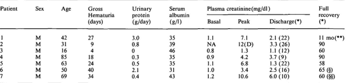
Table 1. Clinical features
Patient Sex Age Gross
Hematuria (days) Urinary protein (g/day) Serum albumin (g/1)
Plasma creatinine (mg/dl) Full Basal 1.1 NA 0.8 0.9 1.1 1.0 1.2 Peak 7.1 12(D) 1.3 4.2 6.8 3.4 10.6 Discharged) 2.1 (22) 3.3 (26) 1.1 (12) 3.7 (9) 1.3(22) 2.5(16) 6.0(10) recovery (*) 11 mo(**) 90 60 90 58 65 (§) 60(§§) M M M M M M M 42 31 16 85 63 50 69 27 9 4 18 24 40 34 3.0 0.8 0 0.3 0.5 2.1 0.4 35 39 46 35 35 31 43
Abbreviations: NA, not available; D, dialysis; mo, months; (*) days after peak plasma creatinine; (**) Several episodes of gross hematuria after discharge; (§) PI creat 1.3 mg/dl, but patient lost to further follow-up; (§§) plasma creatinine 2.5 mg/dl at last check.
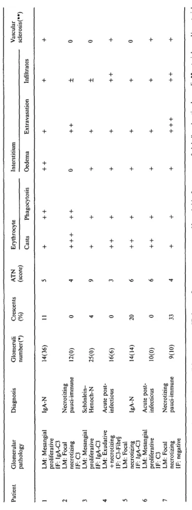
Table 2. Histological findings Patient Glomerular Diagnosis Glomeruli Crescents ATN Erythrocyte Interstitium Vascular pathology number(*) (%) (score) sclerosis(**) Casts Phagocytosis Oedema Extravasation Infiltrates 1 LM: Mesangial proliferative IF: IgA-C3 2 LM: Focal necrotizing IF: C3 3 LM: Mesangial proliferative IF: IgA-C3 4 LM: Exudative
+
necrotizing IF: C3-Fibrs 5 LM: Focal necrotizing IF: IgA-C3 6 LM: Mesangial proliferative IF: C3 7 LM: Focal necrotizing IF: negativeNecrotizing pauci-immune Acute
post-
infectious Acute
post-
infectious Necrotizing pauci-immune
Abbreviations. ATN, acute tubular necrosis; LM, light mlcroscopy; IF, immunofluorescence mlcroscopy; N, nephritis; * percentage of globally sclerotic glomeruli; ** arteriolar and/or arterial sclerosis; § fibrin(ogei1) in the necrotizing areas of the glomerular tufts.
Acute renal failure due to gross haematuria 627
Fig. 1. Denuded basal membranes (arrow) of a tubule the lumen of
which is filled with erythrocytes (patient 2, AFOG stain, original magnification 400 x).
Fig. 2. Severe erythrocyte extravasation into the interstitium
(patient 7, AFOG stain, 400 x).
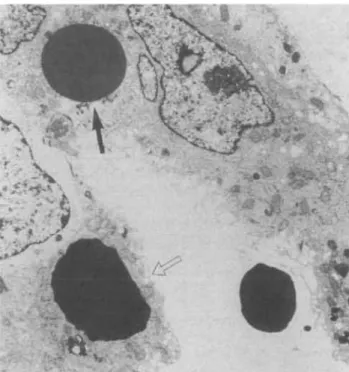
within the interstitium in only two cases (patients 6 and 7). Mild arterial sclerosis was present in four biopsies (patients 1, 4, 6, 7). Electron-microscopy studies (available only for patients 3, 6, and 7) revealed non-specific tubular damage, plus intratubular erythro-cytes in different phases of dissolution. There was also evidence of erythrocyte uptake by the tubular cells (Figure 4), the cytoplasm of which contained increased amounts of lysosomes. All these lesions were unevenly distributed.
There was no close correlation between ATN score
*
•1J
Fig. 3. Haemosiderin (black granules) in the cytoplasm of the
tubu-lar cells (patient 6, Perl's Prussian blue stain, 500 x).
mm-Fig. 4. Erythrocytes (arrows) within the cytoplasm of the tubular
cells by electron-microscopy (patient 3, 8500 x).
and the other histological parameters, as exemplified best by the biopsies of patients 2 and 3. Nor was the ATN score correlated with the duration of gross haem-aturia or with peak plasma creatinine levels. The lack of correlation could also be observed in plot of the two clinical parameters against the amount of erythro-cyte casts. Patients with the necrotizing lesion had, on the average, higher peak plasma creatinine levels than the patients without (8.4 + 3.5 versus 3.9 + 2.9 mg/dl). Retrodiffusion of THp into Bowman's space was never found in any biopsy (Table 3), THp being pre-sent only in the cytoplasm of the distal tubules (6 of 7 biopsies) and in the intratubular casts. There was no extravasation of THp into the interstitium.
628
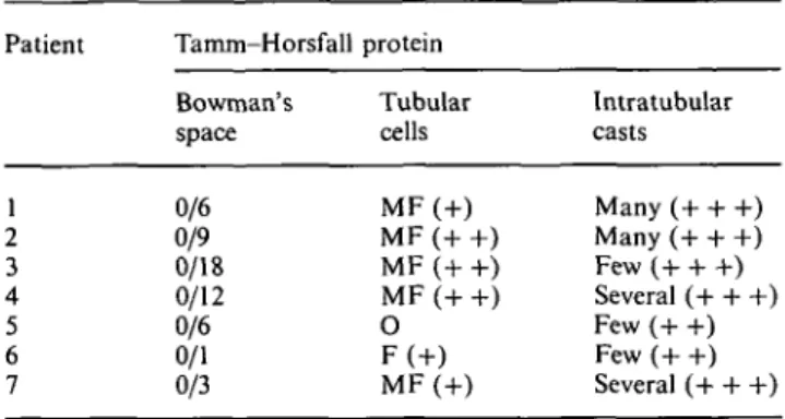
Table 3. Results of the immunofluorescence study with the antiserum
to Tamm-Horsfall protein
Patient Tamm-Horsfall protein Bowman's space 0/6 0/9 0/18 0/12 0/6 0/1 0/3 Tubular cells MF(+) MF (+ +) MF (+ +) MF (+ +) O F(+) MF(+) Intratubular casts Many (+ + +) Many (+ + +) Few (+ + +) Several (+ + +) Few (+ +) Few (+ +) Several (+ + +) Abbreviations: MF, multifocal; F, focal; In parentheses, the intensity of the stain.
Discussion
Acute renal failure from gross haematuria in patients with glomerulonephritis was first reported by Kincaid-Smith et al. [1,2], who demonstrated that the renal dysfunction was due to tubular necrosis which came along with intratubular erythrocyte casts, red-cell pha-gocytosis by tubular cells, and extensive interstitial oedema, rather than to the glomerular changes. Subsequently, Praga et al. [3], prospectively studied 29 episodes of gross haematuria in 21 patients with Berger's disease. Interestingly they found that acute renal failure did not occur in all patients, and that acute renal failure was significantly correlated with a longer duration of gross haematuria, higher amounts of intratubular erythrocyte casts, and a more extensive tubular necrosis. The severity and the duration of acute renal failure varied from patient to patient, but in all cases spontaneous and complete recovery of renal function was seen. Further reports described other similar cases [4-7], some of whom required temporary dialysis before going into remission. All patients but one [8] had IgA nephropathy. Of interest, also Delclaux et al. [7] found that not all patients with gross haematuria developed acute renal failure.
The patients described in this paper are similar to those reported so far in several respects. However, they also have several distinguishing clinical and patholo-gical features. The interval of time before return to normal renal function was longer, on the average, than in the previous reports [1-7]. For case 1 the cause of the slow recovery of the renal function was probably the occurrence of additional episodes of gross haemat-uria in the months after discharge, even though no renal function tests were done when they occurred. Although the tubular damage was the most prominent finding in the renal biopsies of our patients, we cannot exclude that the glomerular lesions may have played a role in determining acute renal failure and/or in delaying renal function recovery. In fact four patients had a focal necrotizing lesion, which was not described in the previous series, and three of them also had globally sclerotic glomeruli. That this may partly
G. B. Fogazzi et al.
account for the acute renal failure is also supported by the finding that these patients had, on average, higher peak plasma creatinine levels than the patients without fibrinoid necrosis in the glomerular tufts. For patients with the necrotizing lesion, a course of corti-costeroids may be justified to obtain or accelerate the repair of the glomerular damage, and therefore to stop the glomerular bleeding, which in turn is responsible for the tubular damage. Some our patients had only mild renal vascular sclerosis and none had nephrotic syndrome. Thus we can reasonably exclude that vascu-lar factors or other mechanisms involved in the acute renal failure seen in nephrotic patients [21] were oper-ating in our cases.
Four patients in our series did not have IgA nephro-pathy, which instead was invariably present in the patients reported by others [1-7]. It is possible, how-ever, that the other investigators deliberately chose to study IgA nephropathy, which is the glomerular disease most commonly associated with gross haematuria [22]. From our findings one can infer that acute renal failure from gross haematuria can be found in all haematuric glomerulonephritides, especially when they are accom-panied by glomerular necrotizing lesions.
At variance with the findings of Praga et al. [3], but in agreement with those of Delclaux et al. [7], we did not find a close correlation between the duration of the gross haematuria and the severity of the renal insufficiency. Interstitial changes were always present in our patients, as in most of the previous series [1,5,6,7].
In spite of the fact that there were intratubular casts in all our biopsies, we were unable to observe retro-diffusion of THp into the glomerular Bowman's space, which has been observed in several other conditions such as obstructive uropathy [13-15], myeloma cast nephropathy [16,17], acute renal failure after acetazol-amide intake [18], or in glomerular diseases associated with tubulointerstitial damage [12]. Since the retro-diffusion of THp into Bowman's space is considered to be a sign of intratubular obstruction, its absence in our patients suggests that this mechanism was not involved in the acute renal failure from gross haemat-uria. This is at variance with a previous hypothesis [1,9]. As an alternative explanation, it has been hypo-thesized that the tubular absorption of haemoglobin or other erythrocyte-derived substances results in tubu-lar damage [1,3], and this has been demonstrated in animals [23,24]. Whether these acute tubulointerstitial lesions, especially when repeated, may lead to chronic renal insufficiency in the long term is still unclear. The available data would suggest that these patients do not have a worse outcome than patients without tubular lesions [7]. However, longer periods of observation and more patients are necessary to draw firm conclusions.
References
1. Kincaid-Smith P, Bennet WM, Dowling JP et al. Acute renal failure and tubular necrosis associated with hematuria due to glomerulonephritis. Clin Nephrol 1983; 19: 206-210.
Acute renal failure due to gross haematuria
2. Kincaid-Smith P, Ryan GB, Dowling JP et al. Acute renal failure in mesangial IgA nephropathy. Conlrib Nephrol 1984; 40: 182-186.
3. Praga M, Gutierrez-Millet V, Navas JJ el al. Acute worsening of renal function during episodes of macroscopic hematuria in IgA nephropathy. Kidney Int 1985; 28: 69-74.
4. Praga M, Costa JR, Shandas GJ et al. Acute renal failure in cirrhosis associated with macroscopic hematuria of glomerular origin. Arch Intern Med 1987; 147: 173-174.
5. Lupo A, Rugiu C, Cagnoli L et al. Acute changes in renal function in IgA nephropathy. Semin Nephrol 1987; 7: 359-362. 6. Lee HS, Pyo HJ, Koh H. Acute renal failure associated with hematuria in IgA nephropathy. Am J Kidnev Dis 12; 1988: 236-239.
7. Delclaux C, Jacquot C, Callard P et al. Acute reversible renal failure with macroscopic haematuria in IgA nephropathy.
Nephrol Dial Transplant 1993; 8: 195-199.
8. Fogazzi GB, Banfi G, Ponticelli C. Acute tubular necrosis caused by gross hematuria in a patient with focal and segmental necrotizing glomerulonephritis. Nephron 1992; 61: 102-105. 9. Ronco P, Dosquet P, Verroust P. La proteine de Tamm-Horsfall.
Presse Med 1988; 17: 1641-1646.
10. Solez K, Morel-Maroger L, Sraer JD. The morphology of 'acute tubular necrosis' in man: analysis of 57 renal biopsies and a comparison with the glycerol model. Medicine 1979; 58: 362-376. 11. Fogazzi GB, Bajetta M, Banfi G et al. Comparison of immuno-fluorescent findings in kidney after snap-freezing and formalin fixation. Pathol Res Pract 1989; 185: 225-230.
12. McGiven AR, Hunt JS, Day WA et al. Tamm-Horsfall protein in the glomerular capsular space. J Clin Pathol 1978; 31: 620-625. 13. Dziukas LJ, Sterzel RB, Hodson CJ et al. Renal localization of Tamm-Horsfall protein in unilateral obstructive uropathy in rats. Lab Invest 1982; 47: 185-193.
14. Solez K, Heptinstall RH. Intrarenal urinary extravasation with formation of venous polyps containing Tamm-Horsfall protein.
J Urol 1978; 119: 180-183.
629 15. Resnick JS, Sisson S, Vernier RL. Tamm-Horsfall protein. Abnormal localization in renal disease. Lab Invest 1978; 38: 550-555.
16. Cohen AH, Border WA. Myeloma kidney. An immunopatho-genetic study of renal biopsies. Lab Invest 1980; 42: 248-256. 17. Rota S, Mougenot B, Baudoin B et al. Multiple myeloma and
severe renal failure: a clinicopathologic study of outcome and prognosis in 34 patients. Medicine 1987; 66: 126-137.
18. Rossert J, Rondeau E, Jondeau G et al. Tamm-Horsfall protein accumulation in glomeruli during acetazolamide-induced acute renal failure. Am J Nephrol 1989; 9: 56-57.
19. Fogazzi GB, Passerini P, Banfi G, Ponticelli C. Study by immunofluorescence ( I F ) on renal biopsies (RB) of the retro-diffusion of Tamm-Horsfall protein (THP) into the glomerular Bowman's space (GBS). JASN 1993; 4: 677 (abstract). 20. Pollack VE, Arbel C. The distribution of Tanun Horsfall
muco-protein (uromucoid) in the human nephron. Nephron 1969; 6: 667-672.
21. Smith JD, Hayslett JP. Reversible renal failure in the nephrotic syndrome. Am J Kidney Dis 1992; 19: 201-213.
22. D'Amico G. Idiopathic IgA mesangial nephropathy. In: Bertani T, Remuzzi G eds. Glomerular Injury 300 Years After Morgagni. Wichtig, Milano 1983: 205-228.
23. Madsen KM, Applegate CW, Tisher CC. Phagocytosis of eryth-rocytes by the proximal tubule of the kidney rat. Cell Tissue
Res 1982; 226: 363-374.
24. Paller MS. Hemoglobin- and myoglobin-induced acute renal failure in rats: role of iron in nephrotoxicity. Am J Physiol 1988; 255: F539-F544.
Editor's note
Please see also Editorial Comment by S. N. Heyman and M. Brezis (pp. 591-593 in this issue).
Received for publication: 14/4/94 Accepted in revised form 10/8/94