Supporting Information
Colloidal Transformations in MS2 Virus Particles:
Driven by pH, Influenced by Natural Organic Matter
Samuel Watts1, Timothy R. Julian2, 3, 4, Katharina Maniura-Weber1, Thomas Graule5, Stefan
Salentinig*1, 6
1Biointerfaces Lab, Empa, Swiss Federal Laboratories for Material Science and Technology, Lerchenfeldstrasse 5, 9014 St. Gallen, Switzerland
2Eawag, Swiss Federal Institute of Aquatic Science, Überlandstrasse 133, 8600 Dübendorf, Switzerland
3Swiss Tropical and Public Health Institute, 4002, Basel, Switzerland
4University of Basel, 4003, Basel, Switzerland
5High Performance Ceramics Lab, Empa, Swiss Federal Laboratories for Materials Science and Technology, Überlandstrasse 129, 8600 Dübendorf, Switzerland
6Department of Chemistry, University of Fribourg, Chemin du Musée 9, 1700 Fribourg, Switzerland
Corresponding author
Material and Methods
Broth preparation:
The broth was prepared by adding 10 g/l of tryptone (Fluka Analytics, St. Louie MO, USA), 1 g/l of select yeast extract (Sigma life science, St. Louie MO, USA), 8g/l of NaCl (≥ 99.5% purity, Sigma-Aldrich Chemie GmbH, Steinheim, Germany), 0.3 g/l of CaCl2 (97% purity, Fluka Analytics, Buchs, Switzerland), 1 g/l Dextrose(Biotechnology grade, Amresco, Solon OH, USA) and 2 mg/l Streptomycin (Fluka Chemie GmbH, Buchs, Switzerland) in ultra-pure water.
Virus dilution buffer:
The virus dilution buffer was prepared by adding 0.78 g/l NaH2PO4∙2H2O (≥ 98.0% purity, Sigma-Aldrich Chemie GmbH, Steinheim, Germany) and 0.58 g NaCl (≥ 99.5% purity, Sigma-Sigma-Aldrich Chemie GmbH, Steinheim, Germany). The pH was equilibrated to 7.0 using NaOH (≥ 99% purity, Carl Roth GmbH, Karlsruhe, Germany) and HCl (ACS reagent grade, Sigma-Aldrich, Buchs, Switzerland).
SDS-PAGE
SDS-Polyacrylamide gel electrophoresis was done as previously described1. In short, 3μl of purified MS2 bacteriophage solution were mixed with 9 μl of buffer composed of 125 mM Tris-HCl, 6% SDS (99% purity, Merck, Darmstadt, Gremany) 30%v/v Glycerol (Fluka, Buchs, Swizerland), 10% 2-Mercaptoethanol (99% purity, Fluka, Buchs, Swizerland), 0.1% w/v Bromphenol blue (Sigma-Aldrich, Steinheim, Germany), and 6 μl H2O and separated on a 13% acrylamide gel. The 10 ml gel was composed of 32.5% v/v of 40% acrylamide solution (Sigma-Aldrich, Steinheim, Germany), 25% v/v 1.5 M Tris-HCl buffer pH 8.8, 41.5% H2O, 1% v/v of 10% solution of SDS (99% purity, Merck, Darmstadt, Germany) to which 5μl TEMED (99% purity, Sigma-Aldrich, Steinheim, Germany) and
50 μl of 10% Ammoniumpersulfate (98% purity, Sigma-Aldrich, Steinheim, Germany) were added. The voltage was set to 200V. The gel was stained with Coomasie blue composed of 2.5 g Brilliant Blue (Sigma-Aldrich, Steinheim, Germany), 450 ml methanol (purity 99.8%, Sigma-Aldrich, Steinheim, Germany), and 100 ml acetic acid (99% purity, Fluka, Buchs, Switzerland) completed to 1l with H2O. The ladder was a PageRuler prestained protein ladder plus (#sm1811, Fermentas, Waltham MA, USA).
Core-shell form factor model
The core-shell model was calculated as follows 2:
𝑃(𝑞) =𝑠𝑐𝑎𝑙𝑒
𝑉 𝐹2(𝑞) + 𝑏𝑎𝑐𝑘𝑔𝑟𝑜𝑢𝑛𝑑 (𝐸𝑞.𝑆1)
Where 𝑠𝑐𝑎𝑙𝑒 allows to scale to the source intensity, is the sample volume, 𝑉 𝑏𝑎𝑐𝑘𝑔𝑟𝑜𝑢𝑛𝑑 is the background signal and 𝐹(𝑞) is defined as follow:
𝐹(𝑞) = 3 𝑉𝑠
[
𝑉𝑐 (𝜌𝑐― 𝜌𝑠) 𝑠𝑖𝑛(𝑞𝑟𝑐)― 𝑞𝑟𝑐𝑐𝑜𝑠(𝑞𝑟𝑐) (𝑞𝑟𝑐)3 + 𝑉𝑠(𝜌𝑠― 𝜌𝑠𝑜𝑙𝑣) 𝑠𝑖𝑛(𝑞𝑟𝑠)― 𝑞𝑟𝑠𝑐𝑜𝑠(𝑞𝑟𝑠) (𝑞𝑟𝑠)3]
(𝐸𝑞. 𝑆2)where is the volume of the whole particle, is the volume of the core, 𝑉𝑠 𝑉𝑐 𝑟𝑠= 𝑟𝑐 is the radius of the particle, is the radius of the core, is the scattering length
+𝑠ℎ𝑒𝑙𝑙 𝑡ℎ𝑖𝑐𝑘𝑛𝑒𝑠𝑠 𝑟𝑐 𝜌𝑐
density of the core, is the scattering length density of the shell, 𝜌𝑠 𝜌𝑠𝑜𝑙𝑣 is the scattering length density of the solvent.
The polydispersity was accounted for using a Gaussian distribution defined as:
𝑓(𝑥) = 1 𝑁𝑜𝑟𝑚𝑒 ―(𝑥 ― 𝑥𝑚𝑒𝑎𝑛) 2 2𝜎2 (𝐸𝑞.𝑆3)
The polydispersity is defined then by
𝑃𝐷 =𝑥 𝜎
𝑚𝑒𝑎𝑛 (𝐸𝑞.𝑆4)
The shell electron density was set to about 130% of the core electron density as shown by deconvolution (figure 1C in the main manuscript) and the polydispersity value taken from the dynamic light scattering (DLS) and set to be the same in the core and shell.
Supporting Figures
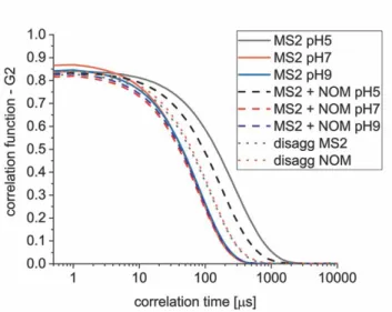
Figure S1: DLS intensity correlation functions of samples reported in this study.
Figure S2: Form factor scattering calculated using the generalized indirect Fourier transformation
(GIFT) method (black line) with the corresponding fit calculated with core-shell form factor model of a spherical core-shell particle using equation S1 (red line). The corresponding parameters are summarized in Table S2.
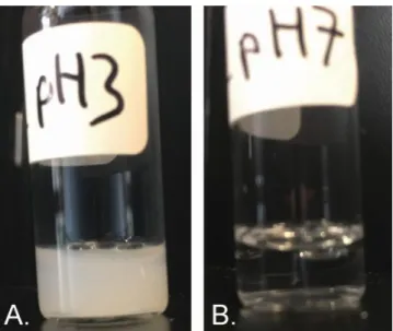
Figure S3: A: MS2 aggregation at pH = 3.0 led to a turbid appearance of the sample due to multiple
scattering effects at large, micrometer sized particles. B: MS2 resuspended spontaneously after pH increased from 3.0 to 7.0 leading to a transparent sample due to the small virus nanoparticles.
Figure S4: Additional cryo-TEM images of disaggregated MS2 at pH = 9.0. Individual viruses
together with virus aggregates (representative ones indicated with blue arrows) and debris (selected ones indicated with red arrows) can be observed.
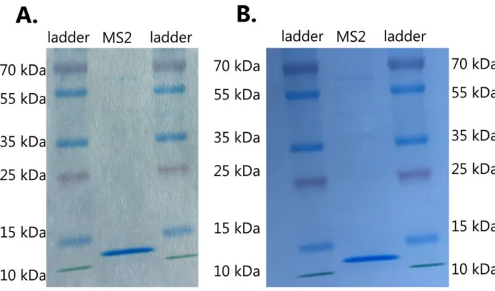
Figure S5: A: SDS-PAGE gel of 1st batch of purified MS2. B: SDS-PAGE gel of 2nd batch of purified MS2. The gels show a single protein band between 10 and 15 kDa corresponding to the size of the coat protein of MS23, hence the MS2 bacteriophage solutions shows no detectable contamination from other proteins.
Supporting Tables
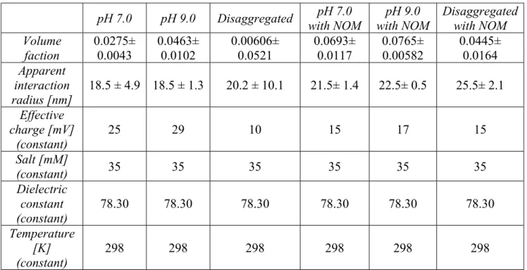
Table S1: Structure factor parameters for the MS2 particles. The effective charge is taken from
ζ-potential measurements, salt concentration is the ionic strength of the virus dilution buffer, the dielectric constant for water at 25°C4.
pH 7.0 pH 9.0 Disaggregated with NOMpH 7.0 with NOMpH 9.0 Disaggregatedwith NOM Volume faction 0.0275± 0.0043 0.0463± 0.0102 0.00606± 0.0521 0.0693± 0.0117 0.0765± 0.00582 0.0445± 0.0164 Apparent interaction radius [nm] 18.5 ± 4.9 18.5 ± 1.3 20.2 ± 10.1 21.5± 1.4 22.5± 0.5 25.5± 2.1 Effective charge [mV] (constant) 25 29 10 15 17 15 Salt [mM] (constant) 35 35 35 35 35 35 Dielectric constant (constant) 78.30 78.30 78.30 78.30 78.30 78.30 Temperature [K] (constant) 298 298 298 298 298 298
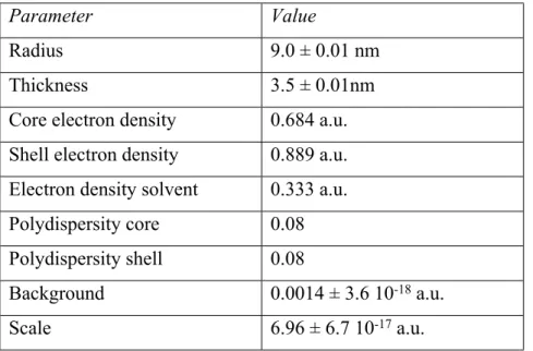
Table S2: The parameters for the spherical core-shell form factor model. The values for radius and
shell thickness of the core-shell particle were optimized to obtain the best possible fit to the form factor scattering obtained from GIFT. The shell electron density is set to 1.3 times the core electron density as estimated from the model-free fitting method by deconvolution of the p(r) function. Polydispersity is estimated from DLS measurements.
Parameter Value
Radius 9.0 ± 0.01 nm
Thickness 3.5 ± 0.01nm
Core electron density 0.684 a.u. Shell electron density 0.889 a.u. Electron density solvent 0.333 a.u. Polydispersity core 0.08 Polydispersity shell 0.08
Background 0.0014 ± 3.6 10-18 a.u.
Scale 6.96 ± 6.7 10-17 a.u.
References
1. Laemmli, U. K., Cleavage of Structural Proteins during the Assembly of the Head of Bacteriophage T4. Nature 1970, 227, 680-685.
2. Guinier, A.; Fournet, G., Small-Angle Scattering of X-Rays. John Wiley and Sons: New York, 1955.
3. Kuzmanovic, D. A.; Elashvili, I.; Wick, C.; O'Connell, C.; Krueger, S., Bacteriophage MS2: Molecular Weight and Spatial Distribution of the Protein and RNA Components by Small-Angle Neutron Scattering and Virus Counting. Structure 2003, 11, 1339-1348.
4. Malmberg, C. G.; Maryott, A. A., Dielectric Constant of Water from 0°C to 100°C. J. Res.