HAL Id: hal-03173303
https://hal.inrae.fr/hal-03173303
Submitted on 18 Mar 2021
HAL is a multi-disciplinary open access archive for the deposit and dissemination of sci-entific research documents, whether they are pub-lished or not. The documents may come from teaching and research institutions in France or abroad, or from public or private research centers.
L’archive ouverte pluridisciplinaire HAL, est destinée au dépôt et à la diffusion de documents scientifiques de niveau recherche, publiés ou non, émanant des établissements d’enseignement et de recherche français ou étrangers, des laboratoires publics ou privés.
bladder
Camille Chagneau, Clémence Massip, Nadège Bossuet-Greif, Christophe
Fremez, Jean-Paul Motta, Ayaka Shima, Céline Besson, Pauline Le Faouder,
Nicolas Cénac, Marie-Paule Roth, et al.
To cite this version:
Camille Chagneau, Clémence Massip, Nadège Bossuet-Greif, Christophe Fremez, Jean-Paul Motta, et al.. Uropathogenic E. coli induces DNA damage in the bladder. PLoS Pathogens, Public Library of Science, 2021, 17 (2), 18 p. �10.1371/journal.ppat.1009310�. �hal-03173303�
the bladder
Camille V. ChagneauID1☯, Cle´mence MassipID1,2☯, Nadège Bossuet-GreifID1,
Christophe FremezID3, Jean-Paul Motta1, Ayaka Shima1, Ce´line BessonID1, Pauline Le
Faouder4, Nicolas Ce´nac
ID1, Marie-Paule Roth1, He´lène CoppinID1, Maxime Fontanie´3,
Patricia Martin1,3, Jean-Philippe NougayrèdeID1*, Eric OswaldID1,2*
1 IRSD, INSERM, Universite´ de Toulouse, INRA, ENVT, UPS, Toulouse, France, 2 CHU Toulouse, Hoˆpital
Purpan, Service de Bacte´riologie-Hygiène, Toulouse, France, 3 VibioSphen, Prologue Biotech, Labège, France, 4 MetaToulLipidomics Facility, INSERM UMR1048, Toulouse, France
☯These authors contributed equally to this work.
*[email protected](JPN);[email protected](EO)
Abstract
Urinary tract infections (UTIs) are among the most common outpatient infections, with a life-time incidence of around 60% in women. We analysed urine samples from 223 patients with community-acquired UTIs and report the presence of the cleavage product released during the synthesis of colibactin, a bacterial genotoxin, in 55 of the samples examined. Uropatho-genic Escherichia coli strains isolated from these patients, as well as the archetypal E. coli strain UTI89, were found to produce colibactin. In a murine model of UTI, the machinery pro-ducing colibactin was expressed during the early hours of the infection, when intracellular bacterial communities form. We observed extensive DNA damage both in umbrella and bladder progenitor cells. To the best of our knowledge this is the first report of colibactin pro-duction in UTIs in humans and its genotoxicity in bladder cells.
Author summary
Colibactin is a bacterial genotoxin suspected of being involved in cancer. So far, the focus has been on colorectal cancer following asymptomatic intestinal carriage of genotoxic Escherichia coli. Here, we show for the first time that this genotoxin is produced in humans, by detecting it in the urine of patients suffering from urinary tract infections. We subsequently show in a rodent UTI model the genotoxic activity of colibactin in the blad-der, including in regenerative cells. These results raise the hypothesis of a role of UTI in bladder cancer, which could be an interesting perspective for future investigation.
Introduction
Urinary tract infections (UTIs) are one of the most common bacterial infections, affecting approximately 150 million individuals each year [1]. UTIs occur most frequently in women, with more than 60% of females diagnosed with a UTI during their lifetime [2]. The severity of
a1111111111 a1111111111 a1111111111 a1111111111 a1111111111 OPEN ACCESS
Citation: Chagneau CV, Massip C, Bossuet-Greif N,
Fremez C, Motta J-P, Shima A, et al. (2021) Uropathogenic E. coli induces DNA damage in the bladder. PLoS Pathog 17(2): e1009310.https://doi. org/10.1371/journal.ppat.1009310
Editor: Matthew A. Mulvey, University of Utah,
UNITED STATES
Received: September 30, 2020 Accepted: January 13, 2021 Published: February 25, 2021
Copyright:© 2021 Chagneau et al. This is an open access article distributed under the terms of the
Creative Commons Attribution License, which permits unrestricted use, distribution, and reproduction in any medium, provided the original author and source are credited.
Data Availability Statement: The authors confirm
that all data underlying the findings are fully available without restriction. Sequencing data presented in the current manuscript have been deposited to the NCBI database, BioProject PRJNA615384 (https://www.ncbi.nlm.nih.gov/ bioproject/PRJNA615384). This project contains the raw reads data deposited in the Sequence Read Archive (SRA) and are publicly available. All other relevant data are within the manuscript and its
these infections ranges from asymptomatic bacteriuria and cystitis,i.e. infections localised to the bladder, to urosepsis, which can be fatal. Recurrences are very frequent, since approxi-mately 30% of women experience a new UTI episode after resolution of the initial infection [2]. In addition to their consequences in terms of morbidity, mortality and associated eco-nomic and societal losses, UTIs are also a major reason for antibiotic treatments and thus strongly contribute to the global issue of antibiotic resistance.Escherichia coli strains, termed uropathogenicE. coli (UPEC) cause approximately 80% of all UTIs. These strains belong mainly to phylogroup B2 in western countries, which is increasingly present in the intestinal microbiota, the reservoir of UPEC [3,4]. UPEC strains produce a large number of virulence factors [5–7]. In particular, several toxins have long been associated with UPEC pathogenicity, such asα-hemolysin and CNF1 toxins [8,9]. More recently, a large proportion of UPEC strains which carrypks pathogenicity island encoding the genotoxin colibactin have been described [10–13].
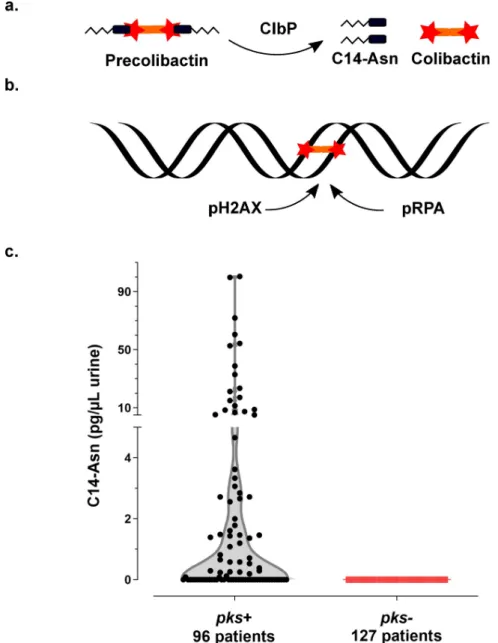
Thepks pathogenicity island, composed of clbA-S genes, encodes a polyketide- non-ribo-somal -peptide (PK-NRP) biosynthesis machinery [14]. Colibactin is first synthesised as an inactive prodrug by ClbN followed by the sequential interventions of multiple Clb enzymes. The ClbP peptidase subsequently cleaves the C14-Asparagine (C14-Asn) motif thereby releas-ing the mature, active form of colibactin with its twin warheads (Fig 1A) [15–17]. The geno-toxin alkylates adenine residues on both strands of DNA, producing DNA interstrand cross-links [18–20]. These highly toxic DNA lesions initiate a DNA damage response, by phosphory-lating replication protein A (pRPA) and phosphoryphosphory-lating the H2AX histone variant (pH2AX) (Fig 1B) [14,18]. Incomplete repair of this DNA damage can result in gene mutations [21].E. coli strains carrying pks island have been shown to promote colon carcinogenesis in different mouse models [22–24]. In epidemiological studies,pks+ E. coli strains are more prevalent in the gut microbiota of patients with colorectal cancer and a distinct mutational signature in human cancer genomes, predominantly colorectal tumours, was recently associated with coli-bactin genotoxic activity, further implicating an involvement of colicoli-bactin-producingE. coli in tumorigenesis [22,23,25,26]. This mutational signature has also been identified in tumours of the urinary tract [26].
Our current study shows that colibactin producing bacteria induce DNA damage in bladder cells, including in urothelial regenerative cells and that colibactin is produced bypks+ UPEC clinical strains isolated from human UTIs.
Results
Evidence of colibactin production in the urine of patients infected with
UPEC
We collected urine samples from 223 adult patients with community-acquired pyelonephritis, cystitis or asymptomatic bacteriuria caused byE. coli at the University Hospital of Toulouse, France. Urine samples were analysed for the presence of C14-Asn, the aminolipid released during the final colibactin maturation step (Fig 1A). In contrast to the highly reactive and unstable colibactin, C14-Asn is stable and can be quantified by LC-MS/MS. C14-Asn was detected in urine samples of one quarter (55/223) of UTI patients, including asymptomatic infections (Fig 1CandS1 Table). We isolatedE. coli strains harbouring genomic pks island from all urine samples which also contained C14-Asn. Conversely, C14-Asn was below the LC-MS/MS detection limits in urine samples of patients infected withE. coli strains which did not carry thepks pathogenicity island.
Funding: This work was supported by grants from
the French National Agency for Research(ANR;
https://anr.fr/) (UTI-TOUL ANR-17-CE35-0010 (EO) and LiBacPain ANR-18-CE14-0039 (NC)) and the French National Institute for Health and Medical Research(INSERM,https://www.inserm.fr/) (two-years grant "poste d’accueil INSERM 2018"(CVC)). The funders had no role in study design, data collection and analysis, decision topublish, or preparation of the manuscript.
Competing interests: The authors have declared
Phylogenetic distribution of
pks island in UPEC strains
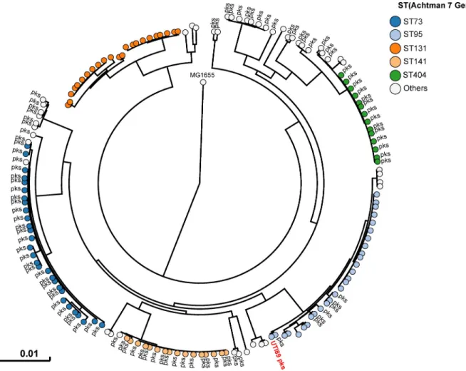
All 225E. coli strains isolated during this sampling campaign were whole genome sequenced. The phylogenetic distribution of theseE. coli isolates was typical of strains which cause UTIs [2]. A majority of strains belonged to the phylogroups B2 (69%) and D (15%) (Fig 2andS1 Table). Forty three percent of strains harbouredpks island. All the pks+ strains belonged to the phylogenetic group B2 and to the most common sequence types (STs) of extra-intestinal
Fig 1. Synthesis of the genotoxin, colibactin, releases the C14-Asn metabolite detected in the urine of patients with UTI. a. Colibactin is first synthesised as a prodrug, precolibactin and then cleaved by ClbP peptidase thereby
releasing the mature genotoxic dimeric colibactin, and C14-Asn. b. Colibactin alkylates both strands of the DNA helix, generating an interstrand cross-link. In response to this DNA damage, the host cell machinery recruits and
phosphorylates the H2AX and RPA proteins. c. Concentration of C14-Asn in human urines according to the presence of apks island in the genome of the corresponding UPEC isolate as determined by LC-MS/MS. All individual data
points are shown on a violin plot, with a two segments linear Y range. C14-Asn was below the LC-MS/MS detection limit in the urine of patients infected with UPEC isolates that did not contain apks island.
pathogenicE. coli such as ST73, ST95, ST141 and ST404 (Fig 2). As expected for extra-intesti-nal pathogenicE. coli, multiple known or suspected virulence genes were also present in the genome of thesepks+ strains (S2 Table).pks island are thus widely present in the typical UPEC strains that cause community-acquired UTIs.
UPEC strains carrying
pks island produce the genotoxin colibactin
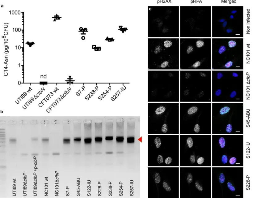
We next sought to confirm thatpks+ UPEC strains expressed colibactin. We readily detected C14-Asn inin vitro cultures of both clinical UPEC isolates and the archetypal strains UTI89 (cystitis collection strain, commonly used in rodent models of UTI) and CFT073 (pyelonephri-tis collection strain) (Fig 3A). Production of C14-Asn depended on a fully functional colibactin assembly line, as mutation of the ClbN synthase gene abrogated its detection (Fig 3A). As for the strain NC101, which has been shown to be pro-carcinogenic in colorectal cancer mouse models, we detected formation of interstrand DNA crosslink by UTI89, but not byclbP mutants not able to maturate colibactin anymore (Fig 3B). The same DNA crosslinking effect was observed with clinicalpks+ UPEC isolates from asymptomatic bacteriuria, cystitis or pyelonephritis cases (Fig 3B). As was shown for NC101, infecting human epithelial cells with
Fig 2. Humanpks+ UPEC belong to major lineages of extraintestinal pathogenic E. coli from phylogroup B2. A phylogenetic tree based on whole
genome analysis was constructed and rooted onE. coli MG1655. Each circle represents an individual UPEC strain. Main Sequence Types are grouped
by colours. The presence of apks island is denoted adjacent to each circle. The archetypal UPEC strain UTI89, which is pks+, was also included in this
tree.
pks+ UPEC strains induced the formation of nuclear pRPA and pH2AX foci, indicating that the DNA of exposed cell is damaged (Figs1Band3C). All together, these results demonstrate that UPEC strainspks island are functional, mediate the synthesis of colibactin and are genotoxic.
Fig 3. The archetypal cystitisE. coli strain UTI89 and clinical pks+ UPEC isolates produce colibactin and induce DNA damage. a. LC-MS/MS quantification of the
C14-Asn cleavage product released following colibactin maturation by the ClbP peptidase in bacterial pellets of UPEC strains UTI89, CFT073 and their respectiveΔclbN mutants (which are not able to synthesise C14-Asn), and four clinical UPEC strains S7, S238, S245, S257. Quantifications were performed in triplicate and represented as a mean± standard error of the mean (SEM). nd: not detected b. Formation of DNA interstrand cross-links upon exposure of linear double stranded DNA to UTI89, NC101 and their respectiveΔclbP mutants as well as seven representative clinical human UPEC isolates (S7, S45, S122, S228, S238, S254, S257), visualised by electrophoresis under denaturing conditions. DNA with interstrand cross-links (red arrow) migrates at an apparent molecular weight of twice that of DNA without cross-links. In the absence of any crosslinking DNA migrates as a single denatured strand. The gel shown is representative of three independent experiments. c. S33p-RPA32 and pH2AX
immunofluorescence staining of HeLa cells, 16 hours after infection with the reference NC101 strain, itsΔclbP mutant or human UPEC strains S45, S122, S228. pH2AX and S33p-RPA32 = pRPA: grayscale. Merged: green = pH2AX; magenta = S33p-RPA32; blue = DAPI; scale bar = 10μm. Human UPEC strains from AS = asymptomatic bacteriuria; IU = cystitis; P = pyelonephritis.
Colibactin is produced during UTIs and induces bladder urothelium DNA
damage
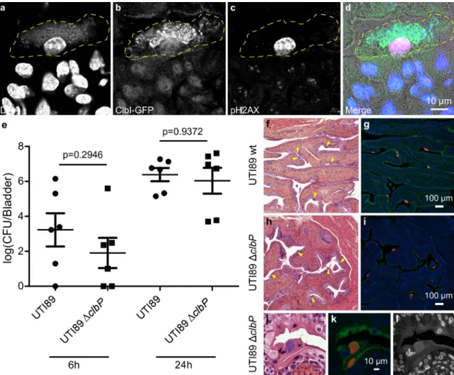
In order to monitor the expression of thepks synthesis machinery during a UTI, we trans-formed the UPEC strain UTI89 with a plasmid expressing a GFP taggedpks island-encoded polyketide ClbI synthase from its endogenous promoter. In bladder tissue collected 6 hours after infection, we observed ClbI-GFP expressing bacteria in intracellular bacterial communi-ties (IBCs) inside superficial umbrella cells which line the lumen of the bladder (Fig 4A–4D andS1). Production of colibactin was further confirmed by the detection of C14-Asn in pooled urines from 8 mice 24 hours after infection (C = 0.66 pg/μL). We next assessed whether the metabolically activepks machinery was associated with DNA damage in bladder cells.
Fig 4. Colibactin is produced during UTI and induces DNA damage. a-d. Confocal microscopic detection of ClbI-GFP expression and
pH2AX in frozen bladder sections, 6 hours post infection with UTI89, hosting the p-clbI-gfp fusion plasmid. The individual channel images
are shown in grayscale, the umbrella cell containing the IBC is circled with a yellow dashed line. In the merged (d) image: blue = DNA, green = ClbI-GFP, magenta = pH2AX, grey = phase contrast. The immunofluorescence staining was repeated three times on different slides, other representative images are shown inS1 Fig. e. Bladder tissueE. coli counts, 6 h and 24 h after infection with wild-type E. coli UTI89
(circles) orΔclbP (squares). Each data point corresponds to one mouse (n = 6), with mean ± standard error of the mean (SEM) shown for each group. Mann-Whitney U test. f-l. Bladder sections 6 hours post inoculation with wild-type UTI89 (f, g) or the isogenicΔclbP mutant (h-l) stained with haematoxylin-eosin (f,h,j), FISH (g,i,k) or DAPI (l, grayscale) all of which detect IBCs (arrows). In the FISH images:
blue = DAPI stain DNA; green = FITC-conjugated wheat germ agglutinin (WGA) stained polysaccharides; red = bacterial cells, labelled with the universal 16S FISH probe. The images are representative of the 5 animals per group observed.
Phosphorylation of H2AX was readily detected in nuclei of umbrella cells containing IBCs, 6 hours after infection with wild-typeE. coli UTI89 with or without the p-clbI-gfp plasmid (Fig 4C and 4DandS1andS2). AclbP mutant unable to produce colibactin was very weakly geno-toxic or not genogeno-toxic at all (S2 Fig), although equally capable of colonising the bladder (Fig 4E) and inducing the formation of IBCs (Fig 4F–4L). These results show that colibactin is expressedin vivo, in the bladder from the very early stages of UTI and can induce DNA damage.
Colibactin induces urothelial cell DNA damage in the regenerative
compartment
The above results show that colibactin expression occurs as soon as 6 hours after infection, with DNA damage mainly in the nuclei of superficial cells. It is well known from mouse mod-els with UTI89 that these superficial cells, especially those containing IBCs, are exfoliated within 6 to 12 hours [27]. The regeneration of the urothelium with a phase of intense prolifera-tion of intermediate and basal cells occurs 24 hours after infecprolifera-tion [27]. In order to study the consequences in terms of genotoxicity for the remaining urothelium of an infection by a coli-bactin-producing strain, we observed pH2AX in bladder sections 24 hours after wild-typeE. coli UTI89 infection. We found significant numbers of positive pH2AX nuclei within the urothelium, generally grouped in patches, including cells from the bottom to the top of the urothelium (Fig 5D–5F). Bladders from mice infected with theclbP mutant did not exhibit such pH2AX positive cells pattern (Fig 5G–5I) and quantification of the number of positive pH2AX nuclei was significantly lower than in individuals infected with the wild-type strain (Fig 5J). pH2AX positive cells nevertheless reappeared after complementation with the wild-typeclbP allele (S3 Fig). The DNA damage was not related to apoptosis as pH2AX+ urothelial cells were not detected with TUNEL apoptosis staining (S4 Fig).
The basal urothelium compartment harbours keratin-14 positive (Krt14+) progenitor cells, which are important for urothelium renewal following injury [28]. There was an increase of Krt14+ cells in urothelial tissue infected with either wild-type orclbP mutant UTI89 24 hours after infection (Figs5KandS5). Interestingly, bladders infected with the genotoxic wild-type UTI89 strain exhibited low but significant numbers of urothelial cells that were positive for both the regenerative cell marker Krt14 and the DNA damage marker pH2AX (Fig 5L–5T). These cells were not positive for Uroplakin, a marker of intermediate and superficial cells (S7 Fig). Thus, during UTI with apks+ UPEC, colibactin induces DNA damage in superficial and basal regenerative urothelial cells.
Discussion
It is now increasingly clear thatpks+ E. coli strains found in the intestinal microbiota may play a role in the aetiology and pathogenesis of colorectal cancer. TheseE. coli strains of phyloge-netic group B2 are often the same ones responsible for UTIs, but to date, no study has been conducted on colibactin and UTIs. To the best of our knowledge the current study is the first to report the presence of a C14-Asn signature, a metabolite of colibactin production, in the urine of patients infected withpks+ UPEC. Thus, colibactin is produced during UTIs, one of the most common human bacterial infections worldwide. We used a mouse model of human UTI, to demonstrate that colibactin is produced and that it induces DNA damage in urothelial cells, including in some (low but significant numbers) bladder Krt14+ progenitor cells.
Our study confirms the frequent occurrence ofpks+ UPEC observed in Europe and the US, irrespective of infection severity [10–13]. This high prevalence in human subjects is in contrast to the apparent lack of selective advantages associated with the production of colibactin in
UTIs mouse models. Our results suggest that colibactin is not essential in our mouse model for the ability of UPEC to colonise the bladder or to form IBCs, whereas colibactin has been shown to play a role in the pathogenicity ofE. coli in extraintestinal infections such as septice-mia or meningitis [29,30]. However, it should be noted that the standard mouse model we used omits a key step in the pathogenesis of UTIs: the domination and emergence from the intestinal reservoir [31].E. coli strains carrying pks island are commonly observed among strains with a greater ability to persist in the intestinal microbiota [3]. In addition to the poten-tial role of colibactin in modulating the intestinal microbiota and promoting gut colonisation, enzymes encoded bypks island are essential for the synthesis of siderophores and siderophore-microcins [32,33]. Siderophores are major determinants in the domination of the intestinal niche [34]. They confer upon strains that produce them the ability to outcompete other bacte-ria for iron, a rare essential nutrient. Moreover, siderophore-microcins, antimicrobial peptides which target phylogenetically-linked enterobacteria help UPEC to implant and proliferate within their intestinal reservoir, a step which precedes UTI [10].
The role ofpks island in UPEC cannot merely be reduced to a determinant of intestinal col-onisation. Indeed, our current study shows that thepks machinery is also active in the urinary tract and even within IBCs. Bacterial multiplication in these structures is intense, with a dou-bling time of nearly 30 min, which requires an efficient and optimised bacterial metabolism [35]. Although colibactin is a small molecule, the metabolic cost of its production,i.e. express-ing and operatexpress-ing the PK-NRP biosynthesis machinery is very high: nearly 1000 times higher than that of peptide synthesis [36]. As UPEC trigger such energetically inexpedient assembly lines during UTIs, one may speculate that they must derive an adaptive benefit from it. Other products of the machinery may possibly confer this advantage. Indeed, there is a wide diversity of metabolites produced by thepks machinery (at least in vitro), ranging from other putative forms of “colibactins” (such as macrocyclic metabolites) to other smaller metabolites, which potentially vary between strains [15,37]. The biological function of these metabolites remains to be elucidated, but some of these metabolites may be relevant to the pathogenesis of infec-tions. We have, for instance, recently described the synthesis of C12-Asn-GABA, by thepks machinery of the probiotic Nissle 1917 strain and shown its digestive pain-relieving activity [38]. The production of such an analgesic metabolite coupled with the increased production of siderophores bypks+ E. coli strains may provide a selective advantage to colonising the urinary tract, in addition to the digestive tract. Future experiments should aim at examining the impact of the variouspks-associated factors on the course of the infection.
Irrespective of the role ofpks island in the virulence of UPECs, the fact remains that the archetypical UPEC strain UTI89 is experimentally genotoxic to mouse bladder cells. Colibac-tin-induced DNA damage can be incompletely repaired, resulting in the accumulation of gene mutations and cell transformation [21,26,39]. We have previously shown that post-exposure
Fig 5. Colibactin induces DNA damage in urothelial cells of the regenerative compartment. a-i.
Immunofluorescence staining of pH2AX on paraffin-embedded bladder sections 24 hours after PBS inoculation (a-c), or UTI89 wild-type (d-f) or UTI89ΔclbP (g-i) infection. The individual fluorescence channel images are shown in grayscale. Merged images: blue = DNA, magenta = pH2AX, grey = phase contrast. Yellow arrows highlight bacteria, white dotted lines represent the basal membrane of the urothelium. Scale bar = 10μm. The experiment was repeated twice on 5 to 8 mice, other representative images are shown inS3 Fig. j-l. pH2AX positive nuclei (j), Krt14 positive cells (k) and Krt14 and pH2AX positive cells (l) were counted objectively using Fiji and CellProfiler softwares on the confocal images from large mosaic of bladder sections. Each dot represents one mouse bladder. Mann Whitney U-test performed between UTI89 wild-type and UTI89ΔclbP. m-t. Paraffin-embedded bladders sections were immuno-stained for pH2AX and Krt14 24 hours post infection with UTI89 wild-type (m-p) or UTI89ΔclbP (q-t). The individual fluorescence channel images are shown in grayscale. Merged images: blue = DNA, magenta = pH2AX, green = Krt14. Cells positive for both Krt14 and pH2AX are circled in pink. White dotted lines highlight the basal membrane of the urothelium. Scale bar = 10μm. Other representative images are shown inS6 Fig.
chromosomal instability to colibactin can persist in daughter cells [21]. DNA damage could also be propagated to contiguous cells by the induction of a senescence-associated secretory phenotype in the affected cells and the production of ROS, that could also explain the pattern of pH2AX positive patches of cells 24 hours after infection [39,40]. In the current study, we observed that some murine bladder progenitor cells (Krt14+) display pH2AX positive nuclei. One could speculate about the long-term consequences of such genotoxic stress on the urothe-lium, especially for patients with recurrent infections. In humans, Krt14 expression character-ises undifferentiated bladder cancers with particularly poor prognoses [41,42]. Rodent bladder cancer models indicate that a significant proportion of cancerous tissue derives from Krt14 positive cells [28]. Thus, we could hypothesise that colibactin damage to Krt14+ cells may initi-ate and propaginiti-ate DNA lesions. In line with this hypothesis, the colibactin mutational signa-ture has recently been identified in colorectal cancers but also in urinary tract cancers [26,43]. Currently the main risk factors for bladder cancers are tobacco and occupational exposure to solvents, which are more frequently investigated than UTIs [44]. However, a large worldwide bladder cancer case control study recently showed that regular UTIs were epidemiologically associated with an increased risk of urinary bladder cancer [45]. We thus suggest thatpks+ UPEC should be now studied as a possible additional risk factor, particularly in cases of chronic and regular infections, irrespective of whether symptoms are present or not. We detected C14-Asn in the urine of patients with asymptomatic bacteriuria, which are usually not treated with antibiotics and can thus persist for periods of up to many years [46]. A better understanding of the consequences of colibactin production could prompt a systematic search forpks island in UPEC isolates or C14-Asn in the urine of patients at risk.
Materials and methods
Ethics statement
According to the French regulations relating to observational database analyses, the collection of clinicalE. coli strains and urine samples did not require specific informed consent. The data were analysed anonymously.
Mouse UTI experiments were performed in accordance with the European directives for the protection of animals used for scientific purposes (2010/63/EU). The local ethics commit-tee from CREFRE-US006 (Regional Centre for Functional Exploration and Experimental Resources) approved the protocol (number CEEA-122 2014–53).
Bacterial strains
The archetypalE. coli strains used in this study were the UPEC strains UTI89 [47], CFT073 [9] and the colitogenicE. coli strain NC101 [22]. TheΔclbP and ΔclbN mutants were constructed using the lambda Red recombinase method [48] with primers IHAPJPN29 and IHAPJPN30 forΔclbP [14] and primers clbN-P1 and clbN-P2 forΔclbN [38]. The pMB702 construct (referred to as p-clbP) was used for complementation of the UTI89 ΔclbP mutant [49]. For the in vivo complementation assay, the UTI89 ΔclbP mutant was transformed with the pCM17-clbP plasmid. Briefly, the pCM17-clbP gene was PCR-amplified from pBRSK-pCM17-clbP [49], with primers clbP-F-Bam_pm (5’-atGGATCCatgacaataatggaacacgttagc-3’) and pBRSK-F-Bam_pm (5’-atG-GATCCcaagctcgga attaaccctc-3’) and cloned into the pCM17 vector [50] BamHI site. The ClbI C-terminal GFP fusion was constructed using the Gibson Assembly kit (New England Biolabs, MA, USA). Briefly, theclbI and gfp genes were amplified by PCR using the following primers:
pK184-clbI-gb1(5’-GATTACGAATTCGAGCTCGGTACCCATGGCAGAGAAT-GATTTTGG-3’); clbI-gfp-gb2 CTTCTCCTTTTCCGCCTCCTCCGCCCTCATTAAT-CATGTCGTTAACTAG-3’); clbI-gfp-gb3
(5’-GATTAATGAGGGCGGAGGAGGCGGAAAAGGAGAAGAACTTTTCACTGG-3’) and gfp-pK184-gb4 (5’-TGCAGGTCGACCTCGAGGGATCCCCTTATTTGTATAGTTCATCC ATGCC-3’). A glycine linker (5’-GGCGGAGGAGGCGGA-3’) was introduced at theclbI and gfp junction for flexibility. PCR amplified fragments and SmaI-digested pK184 vector were assembled according to the manufacturer’s recommendations.
Collection of clinical strains and urines
The collection of 225E. coli strains from urine samples at the Adult Emergency Department of Toulouse University Hospital, France, between July and October 2017, was previously
described [10]. Urine was collected from 223 patients (women and men, under 75 years) with either pyelonephritis (104), symptomatic infections excluding pyelonephritis (cystitis) (83), or asymptomatic bacteriuria (36) without urological comorbidities or catheterisation. All strains were identified asE. coli by matrix-assisted laser desorption/ionisation time-of-flight mass spectrometry (Microflex LT MALDI-TOF MS, Bruker Daltonik GmbH, Germany). Samples of the corresponding urine collected in boric acid tubes were stored at -80˚C until lipid analysis.
C14-Asn quantification
Extraction and quantification of C14-Asn was performed as previously described [38]. Briefly, 5μL internal standard mixture (Deuterium-labelled compound at 400 ng/mL) was added to the bacterial pellets of 24 h DMEM cultures or to 500μL of urine samples before crushing, fol-lowed by addition of cold methanol (MeOH) (15% final volume) and homogenisation. After centrifugation, supernatants were solid phase extracted on HLB plates (OASIS HLB 2 mg, 96-well plate, Waters, Ireland). Following washing with H2O/ MeOH (90:10, v/v) and elution
with MeOH, samples were evaporated twice under N2and finally resuspended in 10μL
MeOH. Separation and quantification of C14-Asn was performed on a high-performance liq-uid chromatography coupled to tandem mass spectrometry system (G6460 Agilent) [38]. Limit of detection was 2.5 pg and limit of quantification was 5 pg.
Sequencing data, sequence alignments and phylogenetic analyses
Whole genome sequencing was performed using the Illumina NextSeq500 Mid Output plat-form (Integragen, Evry, France) to generate 2 x 150 bp paired-end reads, at approximately 80x average coverage. Genomede novo assembly and analysis were performed with the BioNu-merics 7.6 software (Applied Maths) and Enterobase (http://enterobase.warwick.ac.uk/). For SNP-based phylogenetic trees, core genome alignments were generated after mapping raw reads to theE. coli MG1655 genome. The core genome phylogenetic tree was inferred with the Maximum-likelihood algorithm using Enterobase for B2 phylogroup strains. Sequences of all isolates are available in the NCBI Database, Bioproject number PRJNA615384 (https://www. ncbi.nlm.nih.gov/bioproject/PRJNA615384).
Exogenous DNA cross-linking assay
As previously described [18], the pUC19 plasmid, linearised with BamHI, was exposed to bac-teria pre-grown (3 x 10⁶CFU) in DMEM 25 mM Hepes for 40 min at 37˚C. The DNA was purified and 100 ng submitted to denaturing gel DNA electrophoresis (40 mM NaOH and 1 mM EDTA, pH ~12.0). After a neutralisation step, the gel was stained with GelRed and visual-ised under UV using the ChemiDoc Imaging System (BioRad).
pH2AX and pRPA immunofluorescence analysis of post infected HeLa cells
HeLa cells (ATCC CCL2) were cultured and infected in 8-well chamber slides (Labtek), as pre-viously described [18]. 24 hours after passaging, Hela cells were infected with bacteria precul-tured in DMEM 25 mM HEPES at a multiplicity of infection (MOI) of 50, for 4 h. Cells were then washed and incubated overnight in cell culture medium with 100μg/ml gentamicin. Immunofluorescence analysis was then performed as previously described [18]. Briefly, cells were pre-extracted in PBS 0.1%, Triton X-100 and fixed in PBS 4% formaldehyde, permeabi-lised, blocked with MAXblock medium (Active Motif) and stained with antibodies against pH2AX (1:500, JBW301, Millipore) and S33p-RPA32 (1:500, A300-264A, Bethyl), diluted in MAXblock 0.05%, Triton 100. Cells were washed 3 times for 5 min in PBS 0.05%, Triton X-100 and incubated with secondary AlexaFluor 488 or 568 (Invitrogen) diluted 1:500 in MAX-block medium with 1μg/ml DAPI (Sigma). Slides were mounted with Fluoroshield (Sigma) and examined on a Leica SP8 laser scanning confocal microscope in sequential mode with LasX software (Leica), while keeping the same laser and detector settings between different wells. Final images were processed with the ImageJ software.
Mouse UTI model
Female, 6–8 weeks old, C3H/HeN mice (Janvier Labs) were infected transurethrally as previ-ously described [51–53]. Briefly, UPEC were cultivated statically in LB [51] and resuspended to an inoculum of 108CFU in 50μl PBS. Mice under 4% isoflurane anaesthesia were inocu-lated twice at one-hour intervals with a pump that delivered 10μL/s. At 6 or 24 hours, mice were euthanised by cervical dislocation (properly performed by experienced personnel), blad-ders were harvested, homogenised for bacterial enumeration on agar plates, stored in OCT compound at -80˚C or fixed in 4% formaldehyde after filling the bladder prior to paraffin embedding. Each experiment was conducted in duplicate with 5 to 8 mice per group.
Histological and immunofluorescence analyses of mouse bladder tissue
Histological bladder slices were prepared using standard protocols and processed for haema-toxylin-eosin staining then scanned using a Pannoramic 250 scanner (3DHistech). Final images were captured using CaseViewer software (3DHistech).
For pH2AX, Krt14 and Uroplakin (Upk) staining on paraffin-embedded bladders, 5μm thick sections were de-paraffinised and re-hydrated in xylene, ethanol and tap water. Unmask-ing was performed with a 6-min trypsin digestion (Trypsin-EDTA solution, Sigma, T392) fol-lowed by 30 min incubation in citrate buffer pH 6 at 80–95˚C. Sections were then blocked and permeabilised in MaxBlock, 0.3% Triton X100. Slices were stained with anti-pH2AX (Rabbit, Cell Signalling S9718; or Mouse, Millipore #O5-636) at a dilution of 1:200, anti-Krt14 antibod-ies (Chicken, Ozyme BLE906001) at a dilution of 1:250 and anti-Upk III (Rabbit, Abcam, ab157801) at a dilution of 1:800 in MaxBlock, 0.3% Triton X100. Sections were washed in PBS, 0.05% Triton X100 and incubated with secondary antibodies (Alexa 633 Goat Rabbit body, Invitrogen A21071, at a dilution of 1:200 or 1:500; Alexa 555 Goat Chicken anti-body, Invitrogen A32932, at a dilution of 1:500; Tetramethylrhodamine Goat anti-Mouse antibody, Invitrogen T2762, at a dilution of 1:200). Images were acquired as with infected cul-tured cells.
For demonstration of ClbI-GFP reporter expression, to achieve better GFP preservation, we used OCT compound-embedded bladders. Eightμm thick sections were dried on Superfrost plus glass slides, fixed for 15 min in 4% formaldehyde then rinsed with PBS, blocked and per-meabilised with MaxBlock, 0.3% Triton X100. Primary anti-GFP (Rabbit, Abcam 6556) and anti-pH2AX (Mouse, Millipore #O5-636) antibodies were diluted at 1:200 in MaxBlock, 0.3%
triton X100. After washing in PBS, 0.05% Triton X100, slides were incubated with secondary antibodies at 1:200 (Alexa 633 Goat anti-Rabbit antibody (Invitrogen A21071) and Alexa 488 Goat anti-Mouse antibody (Life technologies A11029)) and DAPI.
For pH2AX positive nuclei quantification, the Fiji software was used [54]. Briefly, images were analysed in batch with a macro that run a contrast enhancement (saturation = 0.35) and gaussian blur attenuation (sigma = 2) on the DAPI channel. To create a mask on this channel, “Auto threshold” with Otsu dark was used with holes filling and watershed. The “Analyse parti-cles” command was then used to quantify the pH2AX signal within the nucleus region. For each batch of staining and acquisition, nuclei were counted positive when exhibiting a signal greater than 10 times the average signal on sections of the PBS mouse control analysed in parallel.
For Krt14 positive and Krt14/pH2AX positive cells quantification, the CellProfiler software was used [55]. Briefly, images were analysed in batch with a pipeline that first identify nuclei as primary objects with Otsu thresholding. Cells were defined by 6 pixels expansion from nuclei with “Distance-N” method and then cytoplasm were deducted by subtraction. pH2AX inten-sity in nuclei and Krt14 inteninten-sity in cytoplasm were measured and recorded for each cell. pH2AX positive cells were defined like after Fiji analysis. Krt14 positive cells were counted when exhibiting a signal above 3 times the average signal on the whole bladder section on the mosaic confocal image.
For TUNEL-staining of paraffin-embedded bladders, 5μm thick sections were de-paraffi-nised and re-hydrated in xylene, ethanol and tap water. Sections were then treated by Protein-ase K (15μg/ml) in 10mM Tris/HCl (pH 7.8) at 37˚C for 30 min and then rinsed twice in PBS, as previously already described for paraffin-embedded bladders [56]. As a positive control, slices from PBS inoculated animals were treated with DNAse I (2000 UI in 50 mM Tris-HCl, pH 7.5, 1 mg/ml BSA) for 10 min at room temperature. Slices were then TUNEL-stained using the In Situ Cell Death Detection Kit, TMR red (Roche) according to the manufacturer’s rec-ommendations. Slices were mounted with Fluoroshield containing DAPI and images were acquired as with infected cultured cells.
Fluorescence
in situ hybridisation (FISH) of mouse bladder tissue
Five micron paraffin-embedded bladder sections were de-paraffinised in xylene/ethanol. Sections were incubated in lysozyme solution (10 mg/ml, Sigma, France) for 15 minutes at 37˚C and exposed to 100μl of universal bacterial 16 S fluorescent rRNA probe (Eub338, GCTGCCTC CCGTAGGAGT-Cy5’, Eurofins, France) at a concentration of 5 ng/μl, in hybridisation buffer (20 mM Tris-HCl, pH7.4, 0.9 M NaCl, 0.01% SDS) at 46˚C for 3 hours [57]. Sections were then incu-bated in a 48˚C prewarmed saline-sodium citrate wash buffer (30 mM sodium citrate, 300 mM sodium chloride, pH7.4, Invitrogen, France) for 20 minutes. To stain polysaccharide-rich content, sections were counterstained with FITC-conjugated wheat germ agglutinin (Invitrogen, France) at a 1:1000 dilution in PBS buffer for 30 min. Slides were mounted with Fluoroshield containing DAPI. Images were acquired using a confocal laser scanning microscope (Zeiss LSM 710) and final images were processed and edited with the ImageJ software.
Statistical analyses
Graphical representation and statistical analyses were carried out using GraphPad Prism 8.3. P values were calculated using two-tailed Mann-Whitney U test.
Supporting information
S1 Fig. Immunofluorescence staining of GFP and pH2AX on frozen bladder sections 6 hours post infection with UTI89 p-clbI-GFP (a-l) and wild-type UTI89, non-expressing
GFP (m-p). The individual channel images are shown in grayscale. In the merge images:
blue = DNA, green = ClbI-GFP or GFP, magenta = pH2AX, grey = phase contrast. Umbrella cells containing an IBC are circled with a yellow dashed line, the basal side of the urothelium is delimited with a black dotted line. The images are representative of immunofluorescence staining repeated three times on different slides.
(TIF)
S2 Fig. Colibactin induces DNA damage in umbrella cells containing IBCs at early stages of infection. Immunofluorescence staining of pH2AX on paraffin-embedded bladders
sec-tions 6 hours post-infection with the wild-type UTI89 (a, b) or theΔclbP isogenic mutant (c,d). The individual channel images are shown in grayscale. Umbrella cells containing IBCs are cir-cled with yellow dashed line and urothelium is delimited with green dotted line.
(TIF)
S3 Fig. Colibactin induces DNA damage in urothelial cells. DAPI (DNA: a, d) and
immuno-fluorescence staining of pH2AX (b, e) on paraffin-embedded bladders sections 24 hours post infection by UTI89ΔclbP (a-c) or UTI89ΔclbP+pCM17clbP (d-f). For immunofluorescence, the individual channel images are shown in grayscale. Merged images: blue = DNA, magenta = pH2AX, grey = phase contrast.
(TIF)
S4 Fig. pH2AX positive cells are not apoptotic cells. TUNEL staining of paraffine-embedded
bladder sections 24 hours post PBS inoculation (a-f) or wild-type UTI89 infection (g-j; k-n) in C3H/HeN mice. Sections were treated with DNAse I as a TUNEL positive control (d-f). TUNEL staining of infected bladder sections was compared to pH2AX immunofluorescence on a serial section (j is a section next to g-i and n is next to k-m). The individual fluorescence channel images are shown in grayscale. Merged images: blue = DNA; red = TUNEL; green = pH2AX. Yellow dotted lines represent the basal membrane of the urothelium. Scale bar = 100μm.
(TIF)
S5 Fig. Increase Krt14 positive cell numbers upon UTI. Immunofluorescence staining of
Krt14 and DAPI stained DNA on paraffin-embedded bladders sections 24 hours post PBS inoculation (a-b) or infection by UTI89 wild-type (c-d) or UTI89ΔclbP (e-f). The individual channel images are shown in grayscale. Pink arrows: Areas with Krt14+ cells. Urothelium is delimited with green dotted line. Scale bar = 100μm. Quantification of Krt14 cells is shown in Fig 5K.
(TIF)
S6 Fig. Colibactin induces DNA damage in urothelial cells of the regenerative compart-ment. Paraffin-embedded bladders sections were immuno-stained for pH2AX and Krt14 24
hours post infection with UTI89 wild-type (a-d) or UTI89ΔclbP (e-h). The individual fluores-cence channel images are shown in grayscale. Merged images: blue = DNA,
magenta = pH2AX, green = Krt14. Cells positive for both Krt14 and pH2AX are circled in pink. Scale bar = 10μm.
(TIF)
S7 Fig. Colibactin induces DNA damage in basal Uroplakin-negative cells. a-c.
Immunoflu-orescence staining of Upk and Krt14 on paraffin-embedded bladder sections 24 hours after UTI89 wild-type infection. d-f. Immunofluorescence staining of Upk and pH2AX on a serial section next to the one shown in a-c. Cells positive for pH2AX but negative for Upk are circled with yellow dashed line. The individual fluorescence channel images are shown in grayscale.
Merged images: green = Upk, red = Krt14, magenta = pH2AX, blue = DNA. White dotted lines represent the basal membrane of the urothelium. Scale bar = 10μm. f. Insert: higher mag-nification: green = Upk, magenta = pH2AX, gray = contrast.
(TIF)
S1 Table. Humanpks+ UPEC belong to major lineages of phylogroup B2 ExPEC.
Phy-logroup, Sequence Types and presence of apks island were determined from whole genome sequences of UPEC isolates, along with the detection of C14-Asn from the corresponding urine.
(DOCX)
S2 Table. Characterisation of UPEC strains isolated.
(XLSX)
Acknowledgments
The authors thank Alexandre Perrat, Fre´de´ric Auvray and Laurent Cavalie´ for their help with genomic DNA extraction and sequence analysis and Pauline Dragot for her excellent technical help. We thank the technical microbiology laboratory staff from the Toulouse University Hos-pital for helping to culture and isolateE. coli strains. The authors also wish to thank the staff of the Tri GenoToul imaging facility, Toulouse (particularly Sophie Allart and Danièle Daviaud), the experimental histology facility (CREFRE, Toulouse) and the lipidomic facility of MetaToul, Toulouse. The authors thank the team members for their helpful discussions and critical read-ing of the manuscript and Se´bastien De´jean for his help with data analysis.
Author Contributions
Conceptualization: Jean-Philippe Nougayrède, Eric Oswald.
Data curation: Camille V. Chagneau, Cle´mence Massip.
Formal analysis: Camille V. Chagneau, Jean-Philippe Nougayrède.
Funding acquisition: Patricia Martin, Eric Oswald.
Investigation: Camille V. Chagneau, Cle´mence Massip, Nadège Bossuet-Greif, Christophe Fremez, Jean-Paul Motta, Ce´line Besson, Pauline Le Faouder, Maxime Fontanie´, Patricia Martin, Jean-Philippe Nougayrède.
Methodology: Camille V. Chagneau, Cle´mence Massip, Nadège Bossuet-Greif, Christophe Fremez, Jean-Paul Motta, Pauline Le Faouder, Nicolas Ce´nac, Maxime Fontanie´, Patricia Martin, Jean-Philippe Nougayrède.
Project administration: Eric Oswald.
Resources: Cle´mence Massip, Ayaka Shima, Pauline Le Faouder.
Supervision: Marie-Paule Roth, He´lène Coppin, Maxime Fontanie´, Patricia Martin, Jean-Phi-lippe Nougayrède, Eric Oswald.
Validation: Jean-Philippe Nougayrède, Eric Oswald.
Visualization: Jean-Philippe Nougayrède.
Writing – original draft: Camille V. Chagneau, Cle´mence Massip, Jean-Philippe Nougayrède, Eric Oswald.
References
1. Flores-Mireles AL, Walker JN, Caparon M, Hultgren SJ. Urinary tract infections: epidemiology, mecha-nisms of infection and treatment options. Nat Rev Microbiol. 2015 May; 13(5):269–84.https://doi.org/ 10.1038/nrmicro3432PMID:25853778
2. Klein RD, Hultgren SJ. Urinary tract infections: microbial pathogenesis, host-pathogen interactions and new treatment strategies. Nat Rev Microbiol. 2020 Feb 18.https://doi.org/10.1038/s41579-020-0324-0
PMID:32071440
3. Nowrouzian FL, Oswald E. Escherichia coli strains with the capacity for long-term persistence in the bowel microbiota carry the potentially genotoxic pks island. Microb Pathog. 2012 Sep; 53(3–4):180–2.
https://doi.org/10.1016/j.micpath.2012.05.011PMID:22709536
4. Tenaillon O, Skurnik D, Picard B, Denamur E. The population genetics of commensal Escherichia coli. Nat Rev Microbiol. 2010 Mar; 8(3):207–17.https://doi.org/10.1038/nrmicro2298PMID:20157339 5. Mobley HLT, Donnenberg MS, Hagan EC. Uropathogenic Escherichia coli. EcoSal Plus [Internet]. 2009
Dec 21 [cited 2020 Apr 12]; 3(2). Available from:http://www.asmscience.org/content/journal/ ecosalplus/10.1128/ecosalplus.8.6.1.3.
6. Ulett GC, Totsika M, Schaale K, Carey AJ, Sweet MJ, Schembri MA. Uropathogenic Escherichia coli vir-ulence and innate immune responses during urinary tract infection. Curr Opin Microbiol. 2013 Feb; 16 (1):100–7.https://doi.org/10.1016/j.mib.2013.01.005PMID:23403118
7. Johnson JR, Russo TA. Molecular epidemiology of extraintestinal pathogenic Escherichia coli. EcoSal Plus [Internet]. 2018 Apr 17 [cited 2020 Apr 12]; 8(1). Available from:http://www.asmscience.org/ content/journal/ecosalplus/10.1128/ecosalplus.ESP-0004-2017.https://doi.org/10.1128/ecosalplus. ESP-0004-2017PMID:29667573
8. Mobley HLT. Measuring Escherichia coli gene expression during human urinary tract infections. Pathog Basel Switz. 2016; 5(1).https://doi.org/10.3390/pathogens5010007PMID:26784237
9. Mobley HL, Green DM, Trifillis AL, Johnson DE, Chippendale GR, Lockatell CV, et al. Pyelonephrito-genic Escherichia coli and killing of cultured human renal proximal tubular epithelial cells: role of hemo-lysin in some strains. Infect Immun. 1990 May; 58(5):1281–9. https://doi.org/10.1128/IAI.58.5.1281-1289.1990PMID:2182540
10. Massip C, Chagneau CV, Boury M, Oswald E. The synergistic triad between microcin, colibactin, and salmochelin gene clusters in uropathogenic Escherichia coli. Microbes Infect. 2020 Jan 16.https://doi. org/10.1016/j.micinf.2020.01.001PMID:31954842
11. Krieger JN, Dobrindt U, Riley DE, Oswald E. Acute Escherichia coli prostatitis in previously health young men: Bacterial virulence factors, antimicrobial resistance, and clinical outcomes. Urology. 2011 Jun; 77(6):1420–5.https://doi.org/10.1016/j.urology.2010.12.059PMID:21459419
12. Salvador E, Wagenlehner F, Ko¨hler C-D, Mellmann A, Hacker J, Svanborg C, et al. Comparison of asymptomatic bacteriuria Escherichia coli isolates from healthy individuals versus those from hospital patients shows that long-term bladder colonization selects for attenuated virulence phenotypes. Infect Immun. 2012 Feb; 80(2):668–78.https://doi.org/10.1128/IAI.06191-11PMID:22104113
13. Dubois D, Delmas J, Cady A, Robin F, Sivignon A, Oswald E, et al. Cyclomodulins in urosepsis strains of Escherichia coli. J Clin Microbiol. 2010 Jun; 48(6):2122–9.https://doi.org/10.1128/JCM.02365-09
PMID:20375237
14. Nougayrède J-P, Homburg S, Taieb F, Boury M, Brzuszkiewicz E, Gottschalk G, et al. Escherichia coli induces DNA double-strand breaks in eukaryotic cells. Science. 2006 Aug 11; 313(5788):848–51.
https://doi.org/10.1126/science.1127059PMID:16902142
15. Vizcaino MI, Engel P, Trautman E, Crawford JM. Comparative metabolomics and structural characteri-zations illuminate colibactin pathway-dependent small molecules. J Am Chem Soc. 2014 Jul 2; 136 (26):9244–7.https://doi.org/10.1021/ja503450qPMID:24932672
16. Brotherton CA, Balskus EP. A prodrug resistance mechanism is involved in colibactin biosynthesis and cytotoxicity. J Am Chem Soc. 2013 Mar 6; 135(9):3359–62.https://doi.org/10.1021/ja312154mPMID:
23406518
17. Jiang Y, Stornetta A, Villalta PW, Wilson MR, Boudreau PD, Zha L, et al. Reactivity of an Unusual Ami-dase May Explain Colibactin’s DNA Cross-Linking Activity. J Am Chem Soc. 2019 Jul 24; 141 (29):11489–96.https://doi.org/10.1021/jacs.9b02453PMID:31251062
18. Bossuet-Greif N, Vignard J, Taieb F, Mirey G, Dubois D, Petit C, et al. The colibactin genotoxin gener-ates DNA interstrand cross-links in infected cells. Galan JES Philippe J, editor. mBio. 2018 May 2; 9(2): e02393–17.https://doi.org/10.1128/mBio.02393-17PMID:29559578
19. Wilson MR, Jiang Y, Villalta PW, Stornetta A, Boudreau PD, Carra´ A, et al. The human gut bacterial genotoxin colibactin alkylates DNA. Science. 2019 Feb 15; 363(6428):eaar7785.https://doi.org/10. 1126/science.aar7785PMID:30765538
20. Xue M, Kim CS, Healy AR, Wernke KM, Wang Z, Frischling MC, et al. Structure elucidation of colibactin and its DNA cross-links. Science. 2019 Aug 8;eaax2685.https://doi.org/10.1126/science.aax2685
PMID:31395743
21. Cuevas-Ramos G, Petit CR, Marcq I, Boury M, Oswald E, Nougayrède J-P. Escherichia coli induces DNA damage in vivo and triggers genomic instability in mammalian cells. Proc Natl Acad Sci U S A. 2010 Jun 22; 107(25):11537–42.https://doi.org/10.1073/pnas.1001261107PMID:20534522 22. Arthur JC, Perez-Chanona E, Mu¨hlbauer M, Tomkovich S, Uronis JM, Fan T-J, et al. Intestinal
inflam-mation targets cancer-inducing activity of the microbiota. Science. 2012 Oct 5; 338(6103):120–3.
https://doi.org/10.1126/science.1224820PMID:22903521
23. Dejea CM, Fathi P, Craig JM, Boleij A, Taddese R, Geis AL, et al. Patients with familial adenomatous polyposis harbor colonic biofilms containing tumorigenic bacteria. Science. 2018 02; 359(6375):592–7.
https://doi.org/10.1126/science.aah3648PMID:29420293
24. Dalmasso G, Cougnoux A, Delmas J, Darfeuille-Michaud A, Bonnet R. The bacterial genotoxin colibac-tin promotes colon tumor growth by modifying the tumor microenvironment. Gut Microbes. 2014; 5 (5):675–80.https://doi.org/10.4161/19490976.2014.969989PMID:25483338
25. Buc E, Dubois D, Sauvanet P, Raisch J, Delmas J, Darfeuille-Michaud A, et al. High prevalence of mucosa-associated E. coli producing cyclomodulin and genotoxin in colon cancer. PloS One. 2013 Feb 14; 8(2):e56964.https://doi.org/10.1371/journal.pone.0056964PMID:23457644
26. Pleguezuelos-Manzano C, Puschhof J, Huber AR, Hoeck A van, Wood HM, Nomburg J, et al. Muta-tional signature in colorectal cancer caused by genotoxic pks + E. coli. Nature. 2020 Apr; 580 (7802):269–73.https://doi.org/10.1038/s41586-020-2080-8PMID:32106218
27. Wang J, Batourina E, Schneider K, Souza S, Swayne T, Liu C, et al. Polyploid Superficial Cells that Maintain the Urothelial Barrier Are Produced via Incomplete Cytokinesis and Endoreplication. Cell Rep. 2018 Oct 9; 25(2):464–477.e4.https://doi.org/10.1016/j.celrep.2018.09.042PMID:30304685
28. Papafotiou G, Paraskevopoulou V, Vasilaki E, Kanaki Z, Paschalidis N, Klinakis A. KRT14 marks a sub-population of bladder basal cells with pivotal role in regeneration and tumorigenesis. Nat Commun. 2016 Jun 20; 7(1):11914.https://doi.org/10.1038/ncomms11914PMID:27320313
29. McCarthy AJ, Martin P, Cloup E, Stabler RA, Oswald E, Taylor PW. The genotoxin colibactin is a deter-minant of virulence in Escherichia coli K1 experimental neonatal systemic infection. Infect Immun. 2015 Sep; 83(9):3704–11.https://doi.org/10.1128/IAI.00716-15PMID:26150540
30. Marcq I, Martin P, Payros D, Cuevas-Ramos G, Boury M, Watrin C, et al. The genotoxin colibactin exac-erbates lymphopenia and decreases survival rate in mice infected with septicemic Escherichia coli. J Infect Dis. 2014 Jul 15; 210(2):285–94.https://doi.org/10.1093/infdis/jiu071PMID:24489107
31. Moreno E, Andreu A, Pigrau C, Kuskowski MA, Johnson JR, Prats G. Relationship between Escherichia coli strains causing acute cystitis in women and the fecal E. coli population of the host. J Clin Microbiol. 2008 Aug; 46(8):2529–34.https://doi.org/10.1128/JCM.00813-08PMID:18495863
32. Massip C, Branchu P, Bossuet-Grief N, Chagneau CV, Gaillard D, Martin P, et al. Deciphering the inter-play between the genotoxic and probiotic activities of Escherichia coli Nissle 1917. PLOS Pathog. 2019 Sep 23; 15(9):e1008029.https://doi.org/10.1371/journal.ppat.1008029PMID:31545853
33. Martin P, Marcq I, Magistro G, Penary M, Garcie C, Payros D, et al. Interplay between siderophores and colibactin genotoxin biosynthetic pathways in Escherichia coli. PLoS Pathog. 2013; 9(7):e1003437.
https://doi.org/10.1371/journal.ppat.1003437PMID:23853582
34. Pi H, Jones SA, Mercer LE, Meador JP, Caughron JE, Jordan L, et al. Role of catecholate siderophores in gram-negative bacterial colonization of the mouse gut. PloS One. 2012; 7(11):e50020.https://doi. org/10.1371/journal.pone.0050020PMID:23209633
35. Justice SS, Hung C, Theriot JA, Fletcher DA, Anderson GG, Footer MJ, et al. Differentiation and devel-opmental pathways of uropathogenic Escherichia coli in urinary tract pathogenesis. Proc Natl Acad Sci U S A. 2004 Feb 3; 101(5):1333–8.https://doi.org/10.1073/pnas.0308125100PMID:14739341 36. Amoutzias GD, Chaliotis A, Mossialos D. Discovery Strategies of Bioactive Compounds Synthesized by
Nonribosomal Peptide Synthetases and Type-I Polyketide Synthases Derived from Marine Micro-biomes. Mar Drugs. 2016 Apr; 14(4):80.https://doi.org/10.3390/md14040080PMID:27092515 37. Li Z-R, Li J, Cai W, Lai JYH, McKinnie SMK, Zhang W-P, et al. Macrocyclic colibactin induces DNA
dou-ble-strand breaks via copper-mediated oxidative cleavage. Nat Chem. 2019 Oct; 11(10):880–9.https:// doi.org/10.1038/s41557-019-0317-7PMID:31527851
38. Pe´rez-Berezo T, Pujo J, Martin P, Le Faouder P, Galano J-M, Guy A, et al. Identification of an analgesic lipopeptide produced by the probiotic Escherichia coli strain Nissle 1917. Nat Commun. 2017 Nov 3; 8 (1):1314.https://doi.org/10.1038/s41467-017-01403-9PMID:29101366
39. Cougnoux A, Dalmasso G, Martinez R, Buc E, Delmas J, Gibold L, et al. Bacterial genotoxin colibactin promotes colon tumour growth by inducing a senescence-associated secretory phenotype. Gut. 2014 Dec; 63(12):1932–42.https://doi.org/10.1136/gutjnl-2013-305257PMID:24658599
40. Secher T, Samba-Louaka A, Oswald E, Nougayrède J-P. Escherichia coli producing colibactin triggers premature and transmissible senescence in mammalian cells. PloS One. 2013; 8(10):e77157.https:// doi.org/10.1371/journal.pone.0077157PMID:24116215
41. Lerner SP, McConkey DJ, Hoadley KA, Chan KS, Kim WY, Radvanyi F, et al. Bladder cancer molecular taxonomy: Summary from a consensus meeting. Bladder Cancer Amst Neth. 2(1):37–47.https://doi. org/10.3233/BLC-150037PMID:27376123
42. Volkmer J-P, Sahoo D, Chin RK, Ho PL, Tang C, Kurtova AV, et al. Three differentiation states risk-stratify bladder cancer into distinct subtypes. Proc Natl Acad Sci U S A. 2012 Feb 7; 109(6):2078–83.
https://doi.org/10.1073/pnas.1120605109PMID:22308455
43. Dziubańska-Kusibab PJ, Berger H, Battistini F, Bouwman BAM, Iftekhar A, Katainen R, et al. Colibactin DNA damage signature indicates causative role in colorectal cancer. Nature Medicine. 2020 Jun 1;1–7.
https://doi.org/10.1038/s41591-019-0740-8PMID:31932805
44. Saginala K, Barsouk A, Aluru JS, Rawla P, Padala SA, Barsouk A. Epidemiology of bladder cancer. Med Sci Basel Switz. 2020 Mar 13; 8(1).https://doi.org/10.3390/medsci8010015PMID:32183076 45. Vermeulen SH, Hanum N, Grotenhuis AJ, Castaño-Vinyals G, van der Heijden AG, Aben KK, et al.
Recurrent urinary tract infection and risk of bladder cancer in the Nijmegen bladder cancer study. Br J Cancer. 2015 Feb 3; 112(3):594–600.https://doi.org/10.1038/bjc.2014.601PMID:25429525 46. Lindberg U, Hanson LA, Jodal U, Lidin-Janson G, Lincoln K, Olling S. Asymptomatic bacteriuria in
schoolgirls. II. Differences in escherichia coli causing asymptomatic bacteriuria. Acta Paediatr Scand. 1975 May; 64(3):432–6.https://doi.org/10.1111/j.1651-2227.1975.tb03860.xPMID:1098383 47. Mulvey MA, Schilling JD, Hultgren SJ. Establishment of a persistent Escherichia coli reservoir during
the acute phase of a bladder infection. Infect Immun. 2001 Jul; 69(7):4572–9.https://doi.org/10.1128/ IAI.69.7.4572-4579.2001PMID:11402001
48. Datsenko KA, Wanner BL. One-step inactivation of chromosomal genes in Escherichia coli K-12 using PCR products. Proc Natl Acad Sci. 2000; 97(12):6640–6645.https://doi.org/10.1073/pnas.120163297
PMID:10829079
49. Dubois D, Baron O, Cougnoux A, Delmas J, Pradel N, Boury M, et al. ClbP Is a prototype of a peptidase subgroup involved in biosynthesis of nonribosomal peptides. J Biol Chem. 2011 Oct 14; 286
(41):35562–70.https://doi.org/10.1074/jbc.M111.221960PMID:21795676
50. Morin CE, Kaper JB. Use of stabilized luciferase-expressing plasmids to examine in vivo-induced pro-moters in the Vibrio cholerae vaccine strain CVD103-HgR. FEMS Immunol Med Microbiol. 2009 Oct; 57 (1):69–79.https://doi.org/10.1111/j.1574-695X.2009.00580.xPMID:19678844
51. Hung C-S, Dodson KW, Hultgren SJ. A murine model of urinary tract infection. Nat Protoc. 2009; 4 (8):1230–43.https://doi.org/10.1038/nprot.2009.116PMID:19644462
52. Hannan TJ, Mysorekar IU, Hung CS, Isaacson-Schmid ML, Hultgren SJ. Early Severe Inflammatory Responses to Uropathogenic E. coli Predispose to Chronic and Recurrent Urinary Tract Infection. PLoS Pathog [Internet]. 2010 Aug 12 [cited 2018 Sep 13];6(8). Available from:https://www.ncbi.nlm.nih.gov/ pmc/articles/PMC2930321/
53. Schwartz DJ, Conover MS, Hannan TJ, Hultgren SJ. Uropathogenic Escherichia coli superinfection enhances the severity of mouse bladder infection. PLoS Pathog. 2015 Jan; 11(1):e1004599.https://doi. org/10.1371/journal.ppat.1004599PMID:25569799
54. Schindelin J, Arganda-Carreras I, Frise E, Kaynig V, Longair M, Pietzsch T, et al. Fiji: an open-source platform for biological-image analysis. Nat Methods. 2012 Jul; 9(7):676–82.https://doi.org/10.1038/ nmeth.2019PMID:22743772
55. McQuin C, Goodman A, Chernyshev V, Kamentsky L, Cimini BA, Karhohs KW, et al. CellProfiler 3.0: Next-generation image processing for biology. PLoS Biol. 2018; 16(7):e2005970.https://doi.org/10. 1371/journal.pbio.2005970PMID:29969450
56. Jiang T, Huang Z, Chan JY, Zhang DD. Nrf2 protects against As(III)-induced damage in mouse liver and bladder. Toxicol Appl Pharmacol. 2009 Jun 16; 240(1):8–14.https://doi.org/10.1016/j.taap.2009. 06.010PMID:19538980
57. Motta J-P, Denadai-Souza A, Sagnat D, Guiraud L, Edir A, Bonnart C, et al. Active thrombin produced by the intestinal epithelium controls mucosal biofilms. Nat Commun. 2019 Jul 19; 10(1):3224.https:// doi.org/10.1038/s41467-019-11140-wPMID:31324782