HAL Id: inserm-00123600
https://www.hal.inserm.fr/inserm-00123600
Submitted on 10 Jan 2007
HAL is a multi-disciplinary open access
archive for the deposit and dissemination of
sci-entific research documents, whether they are
pub-lished or not. The documents may come from
teaching and research institutions in France or
abroad, or from public or private research centers.
L’archive ouverte pluridisciplinaire HAL, est
destinée au dépôt et à la diffusion de documents
scientifiques de niveau recherche, publiés ou non,
émanant des établissements d’enseignement et de
recherche français ou étrangers, des laboratoires
publics ou privés.
Does lindane (gamma-hexachlorocyclohexane) increase
the rapid delayed rectifier outward K+ current (IKr) in
frog atrial myocytes?
Martin-Pierre Sauviat, Anthony Colas, Nicole Pages
To cite this version:
Martin-Pierre Sauviat, Anthony Colas, Nicole Pages. Does lindane (gamma-hexachlorocyclohexane)
increase the rapid delayed rectifier outward K+ current (IKr) in frog atrial myocytes?. BMC
Phar-macology, BioMed Central, 2002, 2, pp.15. �inserm-00123600�
BMC Pharmacology 2002,
2 x
Research article
Does lindane (gamma-hexachlorocyclohexane) increase the rapid
delayed rectifier outward K
+
current (I
Kr
) in frog atrial myocytes?
Martin-Pierre Sauviat*
1
, Anthony Colas
1
and Nicole Pages
2
Address: 1Laboratoire d'Optique et Biosciences, Unité INSERM 451, UMR CNRS 7645, Ecole Polytechnique-ENSTA, F-91128 Palaiseau Cedex,
France and 2Laboratoire de Toxicologie, Faculté de Pharmacie, Route du Rhin, 67400 Illkirch, France
E-mail: Martin-Pierre Sauviat* - martin-pierre.sauviat@polytechnique.fr; Anthony Colas - anthony.colas@polytechnique.fr; Nicole Pages - nicole.pages@wanadoo.fr
*Corresponding author
Abstract
Background: The effects of lindane, a gamma-isomer of hexachlorocyclohexane, were studied on
transmembrane potentials and currents of frog atrial heart muscle using intracellular microelectrodes and the whole cell voltage-clamp technique.
Results: Lindane (0.34 microM to 6.8 microM) dose-dependently shortened the action potential
duration (APD). Under voltage-clamp conditions, lindane (1.7 microM) increased the amplitude of the outward current (Iout) which developed in Ringer solution containing TTX (0.6 microM), Cd2+
(1 mM) and TEA (10 mM). The lindane-increased Iout was not sensitive to Sr2+ (5 mM). It was
blocked by subsequent addition of quinidine (0.5 mM) or E-4031 (1 microM). E-4031 lengthened the APD; it prevented or blocked the lindane-induced APD shortening.
Conclusions: In conclusion, our data revealed that lindane increased the quinidine and
E-4031-sensitive rapid delayed outward K+ current which contributed to the AP repolarization in frog atrial
muscle.
Background
Lindane, a gamma-isomer of hexachlorocyclohexane has largely been used as an insecticide and is widely spread in the environment due to the long life time of the molecule [1]. Absorbed by the respiratory, digestive or cutaneous pathways, it accumulates in tissues in the following order: fat > brain > kidney > muscle > lung > heart > spleen > liv-er > blood [2]. Lindane stimulates the synaptic transmis-sion of a large number of muscular and nerve preparations, and suppresses the GABA-activated chloride current [3] by interacting with the receptor GABA-chloride channel complex [4]. Due to the similarity between lin-dane and inositol 1, 4, 5 triphosphate (IP3) [5], it has
been suggested that lindane releases Ca2+ from IP 3
-sensi-tive intracellular stores in macrophages [6] and smooth myometrial muscle cells [7]. Lindane transiently depolar-izes the membrane, opens Ca2+ channels thus increasing
the intracellular Ca2+ concentration, and subsequently
triggers Ca2+-activated K+ current (I
K-Ca) in human sperm
[8]. Lindane (1 microM – 100 microM) does not depress the peak of intracellular Ca2+ transient in guinea pig
my-ocytes, and does not interact directly with the ryanodine receptor Ca2+ release channels from cardiac sarcoplasmic
reticulum vesicles [9]. A Ca2+ release from the
endoplas-mic reticulum, mitochondria and other Ca2+ stores has
been reported in the presence of lindane (0.15 mM) in cat
Published: 10 July 2002
BMC Pharmacology 2002, 2:15
Received: 9 April 2002 Accepted: 10 July 2002 This article is available from: http://www.biomedcentral.com/1471-2210/2/15
© 2002 Sauviat et al; licensee BioMed Central Ltd. Verbatim copying and redistribution of this article are permitted in any medium for any purpose, provided this notice is preserved along with the article's original URL.
BMC Pharmacology 2002, 2 http://www.biomedcentral.com/1471-2210/2/15
kidney cells [10]. Lindane (30 microM) has no effect on the L-type Ca2+ current, but suppresses the activity of large
conductance Ca2+-activated K+ channels and increases the
firing rate of spontaneous action potentials in rat pituitary GH(3) cells [11].
Little is known about the effect of the pesticide on cardiac tissues. The aim of the present work was to study the effect of lindane on the action potential and transmembrane currents of frog auricular heart muscle.
Results
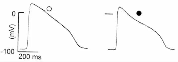
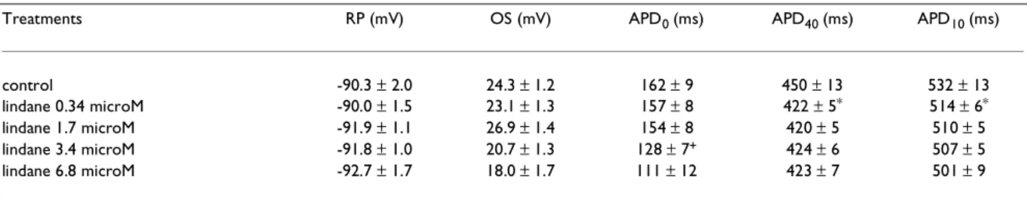
Intracellular recordings of transmembrane potentials show that the addition of lindane (3.4 microM) to the Ringer solution did not alter the RP, decreased the ampli-tude of the OS and shortened the plateau duration (Fig. 1). The effects of lindane on the AP were dose-dependent. Table 1 shows that lindane (0.34 microM to 6.8 microM) did not significantly modify RP; lindane (0.34 microM) slightly but significantly (P < 0.05) shortened APD40 and
APD10 by 6% and 3%, respectively. APD40 and APD10 shortening was not significantly increased by increasing the lindane concentration to 6.8 microM. APD0 was only
significantly shortened (P < 0.05) in the presence of lin-dane 3.4 microM in the Ringer solution (Table 1). Under voltage-clamp conditions, the remaining currents record-ed in the Ringer solution containing TTX (0.6 microM), Cd2+ (1 mM) and TEA (10 mM) (control solution) mainly
corresponded to the leak current and to the background inward rectifier K+ current (I
K1) (Fig. 2A). Current-voltage
relationships plotted for the current measured at the end of the clamp step potential (V) show that the current was inward (Iin) at V more negative than HP and outward (Iout) at V more positive than HP (Fig. 2B). The addition
of lindane (1.7 microM) to the control solution increased Iout but did not alter the tail current (Fig. 2A). Current-voltage relationships of Fig. 2B show that lindane (1.7 mi-croM) increased the amplitude of Iout which developed at membrane potentials more positive than -70 mV.
Subse-quent addition of Sr2+ (5 mM) to the control solution
containing lindane (1.7 µM) decreased the amplitude of Iout in the membrane potential range of -120 mV to +30 mV (Fig. 2B), whereas further addition of quinidine (0.5 mM) to the solution containing both, lindane and Sr2+,
suppressed the remaining Iout whatever the membrane potential tested (Fig. 2B). Lindane (1.7 microM) increased the magnitude of Iout which developed when IK1 was blocked by the addition of Ba2+ (2 mM) to the control
so-lution (Fig. 3A). Current-voltage relationships show that the lindane-increased Iout developed at membrane poten-tials more positive than -20 mV (Fig. 3B). Subsequent ad-dition of E-4031 (1 microM) to the control solution containing lindane blocked the lindane-increased Iout (Fig. 2A) whatever the membrane potential studied (Fig. 3B). The addition of E-4031 (2 microM) to the Ringer so-lution did not modify RP but prolonged APD (Fig. 4Aa) and further addition of lindane (3.4 microM) to the solu-tion containing E-4031 (2 microM) did not modify the APD (Fig. 4Ab). Conversely, the addition of E-4031 (2 croM) to the Ringer solution containing lindane (3.4 mi-croM) lengthened APD0, APD40 and APD10 (Fig. 4B).
Discussion
The present study shows that micromolar concentrations of lindane shortened the action potential duration APD and increases a quinidine and E-4031-sensitive outward current in frog auricle.
Our data show that the shortening of the duration of the repolarizing phase (APD40 and APD10) of the AP is the first significant event occurring in response to the applica-tion of a lindane concentraapplica-tion as low as 0.34 microM. This effect is then followed by a shortening of the plateau duration APD0 which is clearly visible only at a ten times higher concentration.
Voltage-clamp experiments indicate that lindane increases an outward current (Iout). This current develops in the
presence of TEA, known to block the delayed K+ current,
in the control solution and under conditions where Ca2+
current has previously been blocked by Cd2+, suggesting
that a lindane-increased Ca2+ influx may not be directly
involved in the development of Iout. The
lindane-in-creased Iout cannot be attributed to the opening of lin-dane-induced ionic channels since lindane has been shown to be devoid of ionophoretic properties in planar lipid bilayers [9]. Our data show that the lindane-in-creased Iout still persists in the presence of Sr2+ which is
known to block the background IK1[12] and IK-Ca[13]
cur-rents in cardiac tissues. Our findings reveal that quinidine inhibits the effect of lindane on Iout. Quinidine is an open channel blocker of the cardiac rapid delayed rectifier K+
current (IKr) [14–17]. In addition, they show that micro-molar concentrations of E-4031, a specific blocker of IKr
Figure 1
Effect of lindane on spontaneously beating frog auric-ular action potential (AP) AP recorded intracellauric-ularly on
the same auricle in the standard Ringer solution (white cir-cle) before and 5 min after application of lindane (3.4 microM; black circle).
[14,15], prolong the APD in frog auricle, are able to pre-vent or to reverse the APD shortening induced by lindane and in addition suppressed the lindane-increased Iout. These observations indicate that IKr participates to the AP repolarization in frog auricular cells, as in mammalian cardiac tissues [18,19]. This current is sensitive to quini-dine and E-4031 but, as reported in rabbit ventricular cells [20], it is not sensitive to Sr2+ or Ba2+. The data also reveal
that lindane increases IKr. The mechanism by which lin-dane increases IKr is probably not the result of a direct
ac-tivation of the channel IKr, believed to be encoded by the human ether-a-go-go related gene (HERG) [21–25], and which is involved in long QT syndrome, a cardiac disorder characterized by syncope, seizure and sudden death which can be congenital, idiopathic or iatrogenic [26]. HERG K+
channel regulation depends on protein-kinase (PK)-de-pendent pathways. In guinea pig ventricular myocytes, the shift of the activation of HERG K+ channel induced by
phorbol ester involves a PKA-dependent pathway [27]. A PKC-dependent pathway links a G protein-coupled recep-tor that activates phospholipase C to modulate the Herg channel in Xenopus oocytes co-expressing the channel and tyrotropin releasing hormone receptor [28]. Accord-ing to Heath and Terrar [29], IKr is thought to be regulated by PKC which is activated by beta-adrenoceptors stimula-tion in guinea-pig ventricular myocytes. Lindane activates
PKC activity in rat brain and liver tissues [30]. In addition, it has been shown that dynamic regulation of the Herg K+
channels may be achieved via receptor-mediated changes in phosphatidyl inositol bisphosphate (PIP2) concentra-tions; elevated PIP2 accelerated activation and slowed in-activation kinetics [31]. But single exposure of rats to lindane (100 mg / kg) did not cause any significant change in phosphoinositide levels in erythrocyte mem-brane and cerebrum 2 or 24 h after exposure [32].
Conclusions
In conclusion, the results presented show for the first time that the rapid delayed outward current IKr, involved in the repolarization of the cardiac AP, is increased by micromo-lar concentrations of lindane and may be responsible for the alterations of the AP duration induced by the pesti-cide. Although the mechanism by which lindane may in-crease IKr remains to be elucidated, the consequences of
the effect of lindane on IKr are of toxicological interest since this current is involved in cardiac disorder.
Materials and methods
Experiments were performed at 20–21°C on quiescent whole auricle isolated from frog heart and on myocytes isolated enzymatically from the auricle.
Figure 2
Effects of lindane on frog atrial myocytes membrane current Membrane current was recorded on voltage-clamped
frog atrial myocytes bathed in a control Ringer solution containing TTX (0.6 microM), Cd2+ (1 mM) and TEA (10 mM). A).
Superimposed traces of the current (upper traces) elicited by a 170 mV depolarizing step potential applied from HP = -100 mV (lower trace). (white circle) control solution; (black circle) control solution containing lindane (1.7 microM) B). Current-volt-age relationships plotted for the outward current measured at the end (500 ms) of the clamp potential steps, HP = -100 mV. (white circles) control solution; (black circles) control solution containing lindane (1.7 microM); (black triangles) control solu-tion containing lindane and Sr2+ (5 mM); (black squares) control solution containing lindane, Sr2+ and quinidine (0.5 mM).
BMC Pharmacology 2002, 2 http://www.biomedcentral.com/1471-2210/2/15
Solutions
The composition of the frog standard Ringer solution was (mM): NaCl, 110.5; CaCl2, 2; KCl, 2.5; HEPES-NaOH
buffer, 10; pH 7.35. The Ca2+-free solution, used for cells
isolation, was obtained by simple Ca2+ removal and
con-tained 600 mU / ml type I collagenase (Sigma) and 1.5 mU / ml type XIV protease (Sigma). Tetrodotoxin (TTX; 0.6 microM; Sankyo, Japan) and CdCl2 (1 mM) were add-ed to the standard solution to inhibit the peak Na+ current
(INa) and L-type Ca2+ current (I
Ca), respectively.
Tetrae-thylammonium (TEA, Sigma-Aldrich Chimie, Saint Quentin Fallavier, France), quinidine (Sigma-Aldrich Chimie) were used to block delayed K+ current; E-4031
(Alamone, Jerusalem, Israel) was used to inhibit the rapid delayed outward current; SrCl2 (5 mM) and BaCl2 (2 mM) to block the inward rectifying K+ current (I
K1) and the
Ca2+-activated K+ current (I
K-Ca). Lindane (Merck,
Gm-bH) was dissolved in acetone.
Recordings of membrane potentials
Spontaneously beating action potentials (AP) were re-corded on quiescent whole auricle, by means of intracel-lular microelectrodes used in the "floating mode". The tip length (less than 5 mm) of conventional glass microelec-trodes (filled with 3 M KCl, 25–30 Mohms resistance, tip potential less than ± 3 mV) was connected to the input stage of the differential voltage follower by means of a thin Ag / AgCl wire. The following AP parameters were measured: RP: resting membrane potential; OS: over-shoot, AP duration (APD): APD0 duration of the plateau
measured at 0 mV; APD40 and APD10: duration of the AP at the end of the plateau and of the repolarization phase were measured at a membrane potential + 40 mV and + 10 mV higher than RP, respectively [33].
Recordings of membrane currents
Membrane currents were recorded on single myocytes dis-persed by enzymatic digestion of the auricle of frog heart [34]. After isolation of the auricle from the heart, the
ex-ternal epithelial sheet surrounding the auricular tissue was carefully detached and removed. The epithelial-free auri-cle was then pinned at the bottom of an isolating chamber in which the solutions used for the dissociation were maintained at 30°C and gently stirred with a small mag-net. The auricle was successively bathed for 30 min : i) in a Ca2+-free Ringer solution, ii) in a Ca2+-free Ringer
solu-tion containing ethylene glycol tetra acetic acid (EGTA) neutralised with NaOH (0.1 mM), iii) in a Ca2+-free
Ring-er solution then, iv) in a Ca2+-free solution containing
collagenase and protease. All solutions were filtered and oxygenated. When the tissue was digested, the auricle was rinsed twice (10 min) with a Ca2+-free Ringer solution
and then bathed in a standard Ringer solution and kept at 4°C. Before experimentation, cells were dispersed in a Petri dish (outer diameter 33 mm, depth 10 mm, Corn-ing, New-York, USA) filled with Ringer solution (1 ml) by gently shaking the digested auricle. Patch clamp pipettes (Propper Manufacturing glass, id 1.2 mm, wall 0.2 mm, resistance 1.5 to 2.5 Mohms) were filled with a solution containing (mM); KCl, 150; Na2-creatine phosphate, 5; ATP, 5; EGTA neutralised with KOH, 5; HEPES (KOH) buffer, 10; pH = 7.3. The cell current was monitored using an Axopatch 220B amplifier feedback amplifier (Axon In-struments, Foster City, USA). Starting from a holding po-tential (HP) of -100 mV, the membrane popo-tential (V) was displaced in rectangular steps of 10 mV at a rate of 0.2 Hz. Positive potentials correspond to depolarization, positive currents correspond to outward current [34].
Transmembrane potentials and currents were recorded with a Labmaster acquisition card (DMA 100 OEM, Dipsi, Cachan, France), driven by Acquis 1 software linked to the mass storage of a desk computer (AT 80486 DX 33), and displayed on an oscilloscope Nicolet 310 (Nicolet, Madi-son, WI, USA).
Table 1: Effect of lindane on spontaneously beating frog atrial action potential (AP) AP was recorded using intracellular microelectrodes before and after successive and cumulative addition of lindane to the Ringer solution (control).
Treatments RP (mV) OS (mV) APD0 (ms) APD40 (ms) APD10 (ms)
control -90.3 ± 2.0 24.3 ± 1.2 162 ± 9 450 ± 13 532 ± 13
lindane 0.34 microM -90.0 ± 1.5 23.1 ± 1.3 157 ± 8 422 ± 5* 514 ± 6*
lindane 1.7 microM -91.9 ± 1.1 26.9 ± 1.4 154 ± 8 420 ± 5 510 ± 5
lindane 3.4 microM -91.8 ± 1.0 20.7 ± 1.3 128 ± 7+ 424 ± 6 507 ± 5
lindane 6.8 microM -92.7 ± 1.7 18.0 ± 1.7 111 ± 12 423 ± 7 501 ± 9
(RP) resting membrane potential; (OS) amplitude of the overshoot; (APD0) duration of the AP measured at 0 mV; (APD40) and (APD10) duration of
the AP measured at a membrane potential + 40 mV and + 10 mV higher than RP, respectively. The data are mean values ± s. e. mean of 12 AP recorded from 2 different atriums. P < 0.05: * lindane 0.34 microM versus control; + lindane 3.4 microM versus lindane 0.34 microM.
Statistical analysis of data
Numerical data are expressed as mean values ± s. e. mean,
n corresponds to the number of preparations tested. The data were analyzed using the paired Student's t-test using Sigmaplot software (Jandel, Erkrath, Germany) and differ-ences were considered significant at P < 0.05.
List of abbreviations
AP: action potential
APD: action potential duration
APD0: duration of the plateau measured at 0 mV
APD40: duration of the AP at the end of the plateau meas-ured at a membrane potential + 40 mV higher than RP APD10: duration of the AP at the end of the repolarization
phase measured at a membrane potential + 10 mV higher than RP
EGTA: ethylene glycol tetra acetic acid HP: holding potential
IP3: inositol 1, 4, 5 triphosphate IK-Ca: Ca2+-activated K+ current
Figure 3
Effects of E-4031 on the lindane-induced outward current Membrane current was recorded on voltage-clamped frog
atrial myocytes bathed in a control Ringer solution containing TTX (0.6 microM), Cd2+ (1 mM), TEA (10 mM) and Ba2+ (2
mM). A). Superimposed traces of the current elicited by a 170 mV depolarizing step potential applied from HP = -100 mV (c) before and after lindane (1.7 microM) application (traces a) and subsequent addition of E-4031 (1 microM) to the solution taining lindane (traces b); (white circle) control solution: (black circle) control solution containing lindane; (white square) con-trol solution containing lindane and E-4031. B). Current-voltage relationships plotted for the outward current measured at the end (500 ms) of the clamp potential steps, HP = -100 mV. (white circles) control solution; (black circles) control solution con-taining lindane (1.7 microM); (white squares) control solution concon-taining lindane and E-4031 (1 micro M).
BMC Pharmacology 2002, 2 http://www.biomedcentral.com/1471-2210/2/15
Iin: inward current
IK1: inward rectifying K+ current
Iout: outward current
IKr: rapid delayed outward current
microM: micromolar mM: millimolar mm: millimeter ms: millisecond mV: millivolt
mU / ml: milliunit per milliliter OS: overshoot
pA: picoampere
PIP2: phosphatidyl inositol bisphosphate PK: protein kinase
RP: resting membrane potential TEA: tetraethylammonium TTX: tetrodotoxin
V: clamp step potential
References
1. Wauchope RD, Buttler TM, Hornsby AG, Augustijn-Beckers PW, Burt JP: The SCS/ARS/CES pesticide properties database for
environmental decision making. Rev. Environ. Contam Toxicol
1992, 123:1-155
2. Srinivasan K, Radhakrishnamurty R: Studies on the distribution of
beta- and gamma-isomers of hexachlorocyclohexane in rat tissues. J Environ Sci Health B. 1983, 18:401-418
3. Narahashi T: Neuronal ion channels as the target sites of
insec-ticides. Pharmacol & Toxicol 1996, 78:1-14
4. Pomes A, Rodriguez-Farre E, Sunol C: Disruption of
GABA-de-pendent chloride flux by cyclodienes and hexachlorocy-clohexanes in primary cultures of cortical neurons. J Pharmacol Exp Ther 1994, 271:1616-1623
5. Parries GS, Hokin-Nevaverson M: Inhibition of
phosphatidyli-nositol synthase and other membrane-associated enzymes by stereoisomers of hexachlorocyclohexane. J Biol Chem 1985, 260:2687-2693
6. Pinelli E, Campon C, Tronchere H, Chap H, Teissie J, Pipy B:
Ca(2+)-dependent activation of phospholipases C and D from mouse peritoneal macrophages by a selective trigger of Ca2+ influx,
gamma-hexachlorocyclohexane. Biochem Biophys Res Commun
1994, 199:699-705
7. Criswell KA, Stuenkel EL, Loch-Caruso R: Lindane increases
in-tracellular calcium in rat myometrial smooth muscle cells through modulation of inositol 1, 4, 5-triphosphate-sensitive stores. J Pharmacol Exp Ther 1994, 270:1015-1024
8. Silvestroni L, Fiorini R, Palleschi S: Partition of the
organochlo-rine insecticide lindane into human sperm surface induces membrane depolarization and Ca2+ influx. Biochem J 1997,
321:691-698
9. Buck ED, Lachnit WG: In Pessah: Mechanisms of
delta-hex-achloracyclohexane toxicity: I. Relationship between altered ventricular myocyte contractility and ryanodine receptor function. J Pharmacol Exp Ther 1999, 289:477-485
10. Lu CH, Lee KC, Chen YC, Cheng JS, Yu MS, Chen WC, Jan CR:
Lin-dane (gamma-hexachlorocyclohexane) induces internal Ca2+ release and capacitative Ca2+ entry in Madin-Darby
ca-nine kidney cells. Pharmacol Toxicol 2000, 87:149-155
11. Wu SN, Li HF, Chiang HT: Stimulatory effects of
delta-hex-achlorocyclohexane on Ca2+-activated K+ currents in GH(3)
lactotrophs. Mol Pharmacol 2000, 57:865-874
12. Shioya T, Matsuda H, Noma A: Fast and slow blockades of the
in-ward-rectifier K+ channel by external divalent cations in
guinea-pig cardiac myocytes. Pflügers Arch 1993, 422:427-435
13. Coraboeuf E, Carmeliet E: Existence of two transient outward
currents in sheep cardiac Purkinje fibrers. Pflügers Arch 1982, 392:352-359
Figure 4
Effects of E-4031 (1 microM) and lindane (3.4 microM) on the action potential (AP). Superimposed traces of the
AP recorded on frog auricle using intracellular microelectrodes. A. a): AP recorded before and after addition of E-4031 to the Ringer solution; b) further addition of lindane to the solution containing E-4031. B). AP recorded in the Ringer solution con-taining lindane before and after further addition of E-4031.
14. Snyders DJ, Knoth KM, Roberts SL, Tamkun HH: Time-,
voltage-and state-dependent block by quinidine of a cloned human cardiac potassium channel. Mol Pharmacol 1992, 41:322-330
15. Snyders DJ: Functional and pharmacological diversity of
mam-malian potassium channels. A molecular approach: In "Potassi-um channels in normal and pathological conditions". (Edited by: J. Verreecke, PP. van Bogaert, F. Verdonck) L University Press, Leuven 1995,
81-101
16. Snyders DJ: Structure and function of cardiac potassium
chan-nels. Cardiovasc Res 1999, 42:377-390
17. Tseng GN: I(Kr): the hERG channel. J Mol Cell Cardiol 2001,
33:838-849
18. Sanguinetti MC, Jurkiewicz NK: Two components of cardiac
de-layed rectifier K+ current. Differential sensitivity to block by
class III antiarrhythmic agents. J Gen Physiol 1990, 96:195-215
19. Carmeliet E: Voltage- and time-dependent block of the
de-layed K+ current in cardiac myocytes by dofetilide. J Pharmacol
Exp Ther 1992, 262:809-817
20. Paquette T, Clay JR, Ogbaghebriel A, Shier A: Effects of divalent
cations on the E-4031-sensitive repolarization current, IKr, in
rabbit ventricular myocytes. Biophys J 1998, 74:1278-1285
21. Wang Q, Shen J, Li Z, Timothy K, Vincent GM, Priori SG, Schwartz PJ, Keating MT: Cardiac sodium channel mutations in patients
with long QT syndrome, an inherited cardiac arrhythmia. Hum Mol Genet 1995, 4:1603-1607
22. Wang Q, Curran ME, Splawski I, Burn TC, Millholland JM, Van Raay TJ, Shen J, Timothy KW, Vincent GM, de Jager T, Schwartz PJ, Toubin JA, Moss AJ, Atkinson DL, Landes GM, Connors TD, Keating MT:
Po-sitional cloning of a novel potassium channel gene: KVLQT1 mutations cause cardiac arrhythmias. Nat Genet 1996, 12:17-23
23. Barhanin J, Lesage F, Guillemare E, Fink M, Lazdunski M, Romey G:
K(V)LQT1 and lsK (minK) proteins associate to form the I(Ks) cardiac potassium current. Nature 1996, 384:78-80
24. Sanguinetti MC, Jiang C, Curran ME, Keating MT: A mechanistic
link between an inherited and an acquired cardiac arrhyth-mia: HERG encodes the IKr potassium channel. Cell 1995, 81:299-307
25. Sanguinetti MC, Curran ME, Zou A, Shen J, Spector PS, Atkinson DL, Keating MT: Coassembly of K(V)LQT1 and minK (IsK)
pro-teins to form cardiac I(Ks) potassium channel. Nature 1996, 384:80-83
26. Witchel HJ, Hancox JC: Familial and acquired long QT
syn-drome and the cardiac rapid delayed rectifier potassium cur-rent. Clin Exp Pharmacol Physiol 2000, 27:753-766
27. Kiehn J, Karle C, Thomas D, Yao X, Brachmann J, Kübler W: HERG
potassium channel activation is shifted by phorbol esters via protein kinase A-dependent pathways. J Biol Chem 1998, 273:25285-25291
28. Barros F, Gomez-Varela D, Viloria CG, Palomero T, Giraldez T, de la Pena P: Modulation of human erg K+ channel gating by
activa-tion of a G protein-coupled receptor and protein kinase C. J Physiol 1998, 511:333-346
29. Heath BM, Terrar DA: Proteine kinase C enhances the rapidly
activating delayed rectifier potassium current, IKr, through a reduction in C-type inactivation in guinea-pig ventricular myocytes. J Physiol 2000, 522:391-402
30. Bagchi D, Bagchi L, Stohs SJ: Comparative in vitro and in vivo
pro-tein kinase C activation by selected pesticides and transition metal salts. Toxicol Let 1997, 91:31-37
31. Bian J, Cui J, McDonald TV: HERG K(+) channel activity is
regu-lated by changes in phosphatidyl inositol 4,5-bisphosphate. Circ Res 2001, 89:1168-1176
32. Agrawaal D, Subramoiam A, Afaq F: Influence of
hexachlorocy-clohexane on phosphoinisitides in rat erythrocyte mem-branes and brain. Toxicology 1995, 95:135-140
33. Sauviat M-P, Meunier FA, Kreger A, Molgó J: Effects of
trachyni-lysin, a protein isolated from stonefish (Synanceia trachynis) venom, on frog atrial heart muscle. Toxicon 2000, 38:945-959
34. Sauviat M-P, Benoit AG, Debitus C, Pouny I, Laurent D: Alterations
of transmembrane currents in frog atrial heart muscle in-duced by photoexcited gymnochrome a purified from the crinoid, Gymnochrinus richerii. Photochem Photobiol 2001,
74:115-119
Publish with BioMed Central and every
scientist can read your work free of charge
"BioMedcentral will be the most significant development for disseminating the results of biomedical research in our lifetime."
Paul Nurse, Director-General, Imperial Cancer Research Fund
Publish with BMC and your research papers will be:
available free of charge to the entire biomedical community peer reviewed and published immediately upon acceptance cited in PubMed and archived on PubMed Central yours - you keep the copyright
editorial@biomedcentral.com Submit your manuscript here:
http://www.biomedcentral.com/manuscript/