HAL Id: hal-02645480
https://hal.inrae.fr/hal-02645480
Submitted on 29 May 2020
HAL is a multi-disciplinary open access
archive for the deposit and dissemination of
sci-entific research documents, whether they are
pub-lished or not. The documents may come from
teaching and research institutions in France or
abroad, or from public or private research centers.
L’archive ouverte pluridisciplinaire HAL, est
destinée au dépôt et à la diffusion de documents
scientifiques de niveau recherche, publiés ou non,
émanant des établissements d’enseignement et de
recherche français ou étrangers, des laboratoires
publics ou privés.
High bioavailability of bisphenol A from sublingual
exposure.
Véronique Gayrard-Troy, Marlène Z. Lacroix, Séverine Collet, Catherine
Viguié, Alain Bousquet-mélou, Pierre-Louis Toutain, Nicole Picard-Hagen
To cite this version:
Véronique Gayrard-Troy, Marlène Z. Lacroix, Séverine Collet, Catherine Viguié, Alain
Bousquet-mélou, et al..
High bioavailability of bisphenol A from sublingual exposure..
Environmental
Health Perspectives, National Institute of Environmental Health Sciences, 2013, 121 (8), pp.951-6.
�10.1289/ehp.1206339�. �hal-02645480�
High Bioavailability of Bisphenol A from Sublingual Exposure
Véronique Gayrard,1,2 Marlène Z. Lacroix,1,2 Séverine H. Collet,1,2 Catherine Viguié,1,2 Alain Bousquet-Melou,1,2
Pierre-Louis Toutain,1,2 and Nicole Picard-Hagen1,2
1INRA (Institut National de la Recherche Agronomique), UMR1331 (Unité Mixe de Recherche 1331), Toxalim, Research Center in Food
Toxicology, Toulouse, France; 2Université de Toulouse, INPT (Institut National Polytechnique de Toulouse), ENVT (Ecole Nationale
Vétérinaire de Toulouse), EIP (Ecole d’Ingénieurs de Purpan), UPS (Université Paul Sabatier), Toulouse, France
Background: Bisphenol A (BPA) risk assessment is currently hindered by the rejection of reported
higher-than-expected plasma BPA concentrations in humans after oral ingestion. These are deemed incompatible with the almost complete hepatic first-pass metabolism of BPA into its inactive glucurono-conjugated form, BPA glucuronide (BPAG).
oBjectives: Using dogs as a valid model, we compared plasma concentrations of BPA over a 24-hr
period after intravenous, oro gastric, and sub lingual administration in order to establish the absolute bioavailability of BPA administered sub lingually and to compare it with oral bioavailability.
Methods: Six dogs were sublingually administered BPA at 0.05 mg/kg and 5 mg/kg. We compared
the time course of plasma BPA concentrations with that obtained in the same dogs after intravenous administration of the same BPA doses and after a 20-mg/kg BPA dose administrated by oro gastric gavage.
results: The data indicated that the systemic bioavailability of BPA deposited sublingually was
high (70–90%) and that BPA transmucosal absorption from the oral cavity led to much higher BPA internal exposure than obtained for BPA absorption from the gastro intestinal tract. The concen-tra tion ratio of BPAG to BPA in plasma was approximately 100-fold lower following sub lingual administration than after oro gastric dosing, distinguishing the two pathways of absorption. conclusions: Our findings demonstrate that BPA can be efficiently and very rapidly absorbed
through the oral mucosa after sub lingual exposure. This efficient systemic entry route of BPA may lead to far higher BPA internal exposures than known for BPA absorption from the gastro intestinal tract.
keywords: bioavailability, bisphenol A, endocrine disruptor, pharmacokinetic analysis, sub lingual
exposure.
Environ Health Perspect 121:951–956 (2013). http://dx.doi.org/10.1289/ehp.1206339 [Online
12 June 2013]
Introduction
Bisphenol A (BPA) is widely used in its monomeric form in the manufacture of poly carbonate plastics and epoxy resins [European Food Safety Authority (EFSA) 2006]. Vandenberg et al. (2007) suggested that the release of BPA monomers from consumer products leads to the contamination of drink ing water, food, dust, and air, thus providing considerable potential for human exposure to BPA. In support of this suggestion are data reported by Calafat et al. (2008), who found measurable levels of BPA metabolites in > 90% of urine samples from a represen tative cohort of the U.S. population. The principal source of BPA exposure is through the diet and, based on the measurement of urinary concentrations of BPA metabolites as a biomarker of aggregate human exposure levels, the median exposure has been esti mated at only 0.01–0.12 μg/kg/day [Food and Agriculture Organization of the United Nations/World Health Organization (FAO/ WHO) 2010]. The current tolerable daily intake (TDI) is 0.05 mg/kg/day (EFSA 2006).
Widespread human exposure to BPA raises concern among regulatory agencies because of its estrogenic properties in vitro (Wetherill et al. 2007) and in vivo (Richter et al. 2007). The risk assessment for BPA is
controversial because the TDI, which is based on guidelinedriven toxicity studies (Ema et al. 2001; Tyl et al. 2002, 2008), is gener ally higher than doses that produce adverse effects on animals, especially if dosing occurs during the peri natal period (Cabaton et al. 2011; Vandenberg et al. 2008; vom Saal and Hughes 2005).
It is generally assumed that the undesir able effects of BPA are associated with plasma concentrations (internal dose) rather than the administered BPA dose. Thus, some research ers (Vandenberg et al. 2010) have questioned why reportedly high concentrations of uncon jugated BPA in humans (in the nano grams per milli liter range) are not considered by regu latory agencies in the risk assessment process. Other researchers (Mielke and GundertRemy 2009) have noted that the relatively low esti mated BPA daily intake and the observation of an extensive firstpass metabolism of oral BPA into its inactive glucuronoconjugated form (BPAG) (Völkel et al. 2002) are not consistent with those high plasma levels of BPA observed in biomonitoring studies.
Dekant and Völkel (2008) suggested that the high plasma BPA levels reported in humans may be due to arti facts related to sample preparation, storage, over estimation by analytical techniques, or background
contamination from labware or indoor dust. However, there is no little or no direct evi dence for this assertion, and there may be alternative explanations.
Because most BPA exposure in humans occurs via the mouth, we hypothe sized that BPA could be bioavailable sub lingually, which could contribute to higher plasma con centrations. In sub lingual exposure, a sub stance diffuses into the blood through the mucous membrane under the tongue. The sub lingual mucosa is highly vascularized; thus, a substance diffusing across this oral mucosal membrane has direct access to the systemic circulation via capillaries and venous drainage and will avoid firstpass hepatic metabolism (Patel et al. 2011).
In the present study, we used dogs to eval uate the oral transmucosal passage of BPA. The permeability of the buccal membrane is very similar in the dog and human, and thus the dog is a reliable species to assess sub lingual absorption of drugs for human use (Barsuhn et al. 1988). The objectives of the study were
a) to determine the bioavailability of BPA
administered sub lingually; b) to characterize the time course of the plasma BPA concen trations following sub lingual BPA; and c) to compare systemic plasma BPA concentrations as a measure of exposure after sub lingual and conventional oral dosing.
Materials and Methods
Animals. Animals used in this study were treated humanely and with regard for the alleviation of suffering. All animal proce dures were carried out in accordance with the accepted standards of humane animal care under agreement 31–242 for animal experimentation from the French Ministry of Agriculture. The study was conducted in six dogs (two male and four female Beagles; Harlan, Gannat, France). The dogs were Address correspondence to P.L. Toutain, UMR1331 Toxalim, Ecole Nationale Vétérinaire de Toulouse, Laboratoire de Physiologie, 23 chemin des Capelles, BP 87614, 31076 Toulouse cedex 3, France. Telephone: 33 561 193 915. Email: pl.toutain@ envt.fr
Supplemental Material is available online (http:// dx.doi.org/10.1289/ehp.1206339).
We thank E. Jeunesse, L. Boissy, and S. Puel for assistance, and the staff of the dog experimental unit of Toxalim for their involvement in the animal care.
The authors declare they have no actual or potential competing financial interests.
Gayrard et al.
2–3 years of age and weighed 15–19 kg. They were housed in pairs in 12m² rooms, fed a standard diet, and had free access to drink ing water. The animal rooms were illumi nated by artificial light on a 12hr light/dark cycle, and the temperature was maintained at about 20°C. The dogs had access to outdoor exercise areas for about 4 hr/day. Prior to the study day, the animals were fasted overnight and had free access to drinking water. They were given a standard meal 5 hr after dos ing. During sampling periods, the dogs were housed individually in stainless steel cages.
Test material and treatment. BPA and all chemicals were purchased from SigmaAldrich (SaintQuentin, Fallavier, France). For the 5mg/kg BPA dose, solutions were prepared immediately before treatment by dissolving BPA (50 mg/mL) in 1% ethanol/49% pro pylene glycol (vol/vol) for the intra venous (iv) dose, ethanol for the sub lingual bolus, or 40% ethanol/60% water (vol/vol) for sub lingual drops. For the 0.05mg/kg BPA dose, solutions were prepared immediately before treatment by dissolving BPA (0.5 mg/mL) in water containing 1% ethanol for iv and sub lingual administration. For oro gastric adminis tration, BPA (40 mg/mL) was dissolved in 1% ethanol/9% corn oil (vol/vol).
Experimental design and dosing. Experiment 1. The first experiment was divided into two periods separated by 1 week. The dogs received BPA (5 mg/kg) by iv administration one period, and by sub lingual administration during the other period using a cross over design. We chose the 5mg/kg dose of BPA based on the iv dose we esti mated to be required to achieve plasma BPA concentrations greater than the limit of quan tification (LOQ; 1 ng/mL) for a period of about 8–10 hr (i.e., a duration sufficient to observe the terminal phase slope and allow calculation of BPA pharmaco kinetic param eters). We used two different sub lingual modalities of BPA administration: a single bolus and drops. A BPA solution (BPA at 5 mg/kg in ~ 1.3 mL ethanol) was deposited as a single bolus under the tongue of three dogs briefly anesthetized by an iv injection of sodium thiopental (Nesdonal, 11 mg/kg; Merial, Lyon, France). The three other dogs received the same volume and dose of BPA in the form of 20μL drops of an aqueous solution containing 40% ethanol, which was continuously delivered toward the floor of the mouth over a 10min period.
Experiment 2. In a second experiment, BPA was administered at a dose equivalent to the TDI (0.05 mg/kg/day), which was chosen in order to better reflect the maximal pos sible human BPA external exposure and to check the proportionality of BPA pharmaco kinetics with dose. This experiment was divided into three periods, each separated by
1 week. During the first two periods, dogs were adminis tered BPA (0.05 mg/kg) by iv and sub lingual drop adminis tration accord ing to a cross over design as described above. During the third period, BPA (20 mg/kg) was adminis tered by oro gastric intubation. The 20mg/kg BPA dose was selected based on pharmaco kinetic data from a prelimi nary experiment in order to obtain plasma concentrations of unconjugated BPA of the same order as those observed after sub lingual adminis tration of BPA at the TDI dose level.
For both experiments, and for each dog, the iv BPA dose was administered as a bolus via an indwelling 22G catheter under the same conditions of dose, volume, and anes thesia as during the corresponding sublingual adminis tration.
Blood sampling. Serial jugular venous blood samples were collected before all adminis trations, and in the middle (5 min after commencement) and at the end of adminis tration of sub lingual drops. After sub lingual drop adminis tration ended and after iv and sublingual bolus adminis tration, blood samples were collected at 2, 4, 8, 15, 30, 60, 90, 120, 180, and 240 min and every 2 hr for 12 hr (experiments 1 and 2) and at 24 hr for experiment 1. Serial blood samples were obtained at 15, 30, 60, 120, 180, and 240 min, every 2 hr for 12 hr, and at 24 hr after oro gastric BPA adminis tration (experiment 2).
Blood samples were collected into hep arinized poly propylene tubes, immediately chilled on ice, and centrifuged for 10 min at 3,000 × g at 4°C. The supernatant plasma was stored in polypropylene tubes (Eppendorf) at –20°C until assay.
BPA and BPAG assays. BPA and BPAG in plasma samples were simultaneously quanti fied using an Acquity ultra performance liquid chromatograph coupled to a Xevo triple quad rupole mass spectrometer (both from Waters, Milford, MA, USA), according to a previously described method (Lacroix et al. 2011).
Briefly, samples (100 μL) were purified by protein precipitation, diluted with 150 μL aceto nitrile and 50 μL of the internal standards deuterated BPA (BPAd16) and 13Clabeled BPAG (BPAG 13C12), and separated on a C18 column with a water/acetonitrile gradi ent elution. The multiple reaction moni toring transitions used to detect BPA, BPAd16, BPAG, and BPAG 13C12 were 227 > 212, 241 > 142, 403 > 227, and 415 > 239, with collision energies of 28, 20, and 30 eV, respec tively. Chromatographic data were monitored by Targetlynx software (Waters). Blanks and quality control samples were used to monitor potential contamination during analysis and the accuracy and precision of the method.
The mean intra and inter day coefficients of variation for three concentration levels and
for BPA and BPAG, respectively, were < 15%, and the LOQs were validated at 1 ng/mL and 5 ng/mL, respectively.
Pharmacokinetic analysis. Plasma concen tration– time profiles of BPA and BPAG were analyzed according to a non compartmental approach using WinNonlin® Professional, version 5.3 (Pharsight Corporation, Cary, NC, USA). From the plasma BPA and BPAG concentration–time data in indi vidual dogs, we derived the maxi mum con centration (Cmax) and the time to maximum concentration (Tmax). The areas under the BPA and BPAG plasma concentration–time curves (AUClast) and the areas under the first moment curves (AUMClast) were calculated using the linear trapezoidal rule from dosing time to the last sampling time. The mean resi dence time (MRT) was calculated as the ratio of AUMClast:AUClast.
In deriving the BPA pharmaco kinetic parame ters (AUClast, AUMClast, Cmax, Tmax, and MRT), we did not include the first 8 min immediately after sub lingual adminis tration ended because high plasma BPA levels encountered during this lag time may reflect the immediate input of BPA drained from the tongue into the jugular vein (the site of blood sampling) before it is diluted in the general circu lation. The AUClast values were normalized by the corresponding BPA dose.
For each dog, we determined the abso lute bioavailability of BPA administered sub lingually (both methods) at 5 mg/kg as the ratio of the normalized BPA AUClast for the sub lingual route to the equivalent AUClast for the iv route. For the two BPA doses (0.05 and 5 mg/kg), we defined the extent of BPA sub lingual absorption as the ratio of the BPAG AUClast values obtained for the sub lingual route to the equivalent BPAG AUClast for the iv route. For 0.05 mg/kg BPA sub lingual dos ing, we considered this latter value to be an appropriate measure of the absolute bioavail ability of BPA based on the assumption that BPAG is not formed at the site of adminis tration or by a first pass effect. For compu tation of oral bioavailability and absorption rate, we consider only the iv dose of 5 mg/kg.
Statistical analyses. All results are pre sented as mean ± SD. Student’s ttest and SYSTAT12 software (Systat Software Inc., Chicago, IL, USA) were used to analyze dif ferences in mean BPA and BPAG pharmaco kinetic parameters (AUClast, Cmax, Tmax, and MRT) by route of administration.
Results
BPA was not detected in any of the control samples obtained before BPA adminis tration. The values for pharmaco kinetic parameters of BPA and BPAG obtained for different doses via different routes (iv, sub lingual, and oro gastric) of BPA adminis tration are presented in
Tables 1 and 2. As noted above, calculations of pharmaco kinetic parameters omitted the BPA plasma concentrations obtained during and up to 8 min after the end of sub lingual adminis tration of BPA.
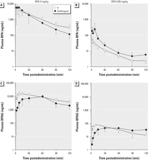
Experiment 1: iv and sub lingual dosing (BPA 5 mg/kg). Because the two sub lingual adminis tration methods (bolus and drops) gave comparable results, they were combined into one data set. The time course of mean plasma BPA concentrations after sub lingual BPA adminis tration was very similar to that obtained in the same dogs after iv adminis tration (Figure 1A); however, the plasma concentrations during the first minutes after sub lingual application were higher than those obtained after iv administration.
For the 5mg/kg BPA sublingual dose, the mean BPA Tmax was 13 ± 9 min (mean ± SD) (Figure 1A, Table 1). The mean BPA Cmax for this sub lingual dose was not significantly dif ferent from the corresponding value obtained after iv adminis tration (7,296 ± 1,615 ng/mL for iv vs. 6,443 ± 3,910 ng/mL for sub lingual;
p = 0.6; Table 1). The BPA MRT was not sig
nificantly different according to the iv versus sub lingual routes of administration (69 ± 13 vs. 73 ± 33 min; p = 0.7; Table 1).
For BPAG, the mean Cmax did not sig nificantly differ by route of adminis tration (15,657 ± 6,426 vs. 11,808 ± 10,419 ng/mL for iv vs. sub lingual, respectively; p = 0.2, Table 2). However, the BPAG Tmax was delayed (Figure 1C, Table 2) compared with the iv route (16 ± 7 vs. 35 ± 13 min for iv vs. sub lingual; p = 0.04).
The mean BPA AUClast (normalized to administered dose) in response to sub lingual adminis tration of BPA at 5 mg/kg was lower than that obtained after iv adminis tration (p = 0.04; Table 1, Figure 2A), whereas the corresponding mean BPAG AUClast values were not significantly different. The mean BPA bioavailability for the high sub lingual dose was 70 ± 31%. This high systemic bio availability was confirmed by the mean ratio of BPAG AUC values (81 ± 18% for sub lingual dosing), which is also an estimate of the systemic bioavailability, provided that the BPAG is not formed at the administration site or by a firstpass effect, which seems to be a reasonable assumption for a direct buccal absorption.
Experiment 2: iv and sub lingual dosing (BPA 0.05 mg/kg). BPA was not detected by about 2 hr after iv or sub lingual BPA adminis tration (0.05 mg/kg), whereas BPAG plasma levels in three dogs remained > LOQ for 8–10 hr after BPA administration.
Following sub lingual applications of BPA at 0.05 mg/kg, the BPA plasma levels were more variable and higher than those obtained in the same dogs after iv administration of the same dose (Figure 1B).
Table 1. Mean (± SD) values for pharmacokinetic parameters of BPA after iv, sublingual, and orogastric
BPA dosing. Pharmacokinetic parametera iv Sublingual Orogastric 0.05 mg/kg 5 mg/kg 0.05 mg/kg 5 mg/kg 20 mg/kg Cmax (ng/mL) 64 ± 36 7,296 ± 1,615 249 ± 331 6,443 ± 3,910 47 ± 20 Tmax (min) 2 ± 0 3 ± 1 10 ± 4 13 ± 9 20 ± 8
AUClast (× 103 ng/min/mL) 1 ± 0 221 ± 54 2 ± 1 145 ± 44* 6 ± 2
MRT (min) NC 69 ± 13 NC 73 ± 33 112 ± 37
BPA bioavailability (%) NA NA NC 70 ± 31 0.72 ± 0.28 Abbreviations: NA, not applicable; NC, not calculated.
aThe first 8 min following the end of sublingual adminis tration were not taken into account in deriving the BPA pharmaco
kinetic parameters. *p ≤ 0.05, compared with iv administration of the same BPA dose by Student’s ttest.
Table 2. Mean (± SD) values for pharmacokinetic parameters of BPAG after iv, sublingual, and orogastric
BPA dosing. Pharmacokinetic parameter iv Sublingual Orogastric 0.05 mg/kg 5 mg/kg 0.05 mg/kg 5 mg/kg 20 mg/kg Cmax (ng/mL) 78 ± 38 15,657 ± 6,426 46 ± 20* 11,808 ± 10,419 30,777 ± 13,604 Tmax (min) 12 ± 4 16 ± 7 35 ± 20 35 ± 13* 38 ± 18 AUClast (× 103 ng/min/mL) 8 ± 5 2,884 ± 776 7 ± 5 2,355 ± 893 6,081 ± 1,935
MRT (min) NC 417 ± 65 NC 562 ± 164 501 ± 200
BPA absorption and/or
bioavailability (%) NA NA 90 ± 26 81 ± 18 54 ± 19 Abbreviations: NA, not applicable; NC, not calculated.
*p ≤ 0.05, compared with iv administration of the same BPA dose by Student’s ttest.
Figure 1. Semilogarithmic plots of mean (± SD) plasma concentrations (ng/mL) of BPA (A,B) and
BPAG (C,D) versus time after a single iv (n = 6) or sublingual (n = 6) administration of BPA at 5 mg/kg (A,C) and 0.05 mg/kg (B,D). Time 0 represents the end of BPA administration.
Time postadministration (min)
Plasma BP A (ng/mL) Plasma BP A (ng/mL) Plasma BP AG (ng/mL) Plasma BP AG (ng/mL)
Time postadministration (min)
Time postadministration (min) Time postadministration (min)
1 10 100 1,000 10,000 1 10 100 1,000 10,000 0 30 60 90 120 BPA 5 mg/kg BPA 0.05 mg/kg 1 10 100 1,000 10,000 100,000 1 10 100 1,000 10,000 100,000 0 30 60 90 120 0 30 60 90 120 0 30 60 90 120 iv Sublingual
Gayrard et al.
The mean Tmax, not counting the 8 min immediately after sub lingual BPA adminis tration (0.05 mg/kg) ended, was 10 ± 4 min (Figure 1B, Table 1). The mean BPA Cmax after sub lingual administration was more vari able than the corresponding value obtained after iv administration (64 ± 36 ng/mL vs. 249 ± 331 ng/mL, for iv vs. sub lingual administration; Table 1). For BPAG, the mean Cmax after sub lingual dosing was lower than that after iv administration (78 ± 38 for iv vs. 46 ± 20 ng/mL for sub lingual;
p = 0.03; Table 2). However, the BPAG Tmax after sub lingual adminis tration was delayed (35 ± 20 min; Figure 1D, Table 2) compared with the iv route (12 ± 4 min; p = 0.06).
As reflected by the mean BPA AUClast, the systemic BPA exposure resulting from sub lingual dosing of BPA at 0.05 mg/kg was more variable and higher than that obtained after iv adminis tration and was thus not considered for bioavailability computation.
The mean BPA sub lingual bioavailability for the 0.05 mg/kg dose was 90 ± 26%, as computed by the mean ratio of the BPAG AUClast values obtained for the sub lingual route to the equivalent BPAG AUClast for the iv route.
Oro gastric BPA dosing (20 mg/kg). The mean Cmax and Tmax values of plasma BPA observed after oro gastric administration of BPA at 20 mg/kg were 47 ± 20 ng/mL and 20 ± 8 min, respectively. The mean BPA bio availability was 0.72 ± 0.28%. This value was lower than the mean ratio of BPAG AUC val ues (54 ± 19%), showing that BPA was rather well absorbed by the gastro intestinal tract but that most absorbed BPA is metabolized by a firstpass effect at the hepatic level. A major difference between the two modalities of oral adminis tration (oro gastric vs. sub lingual) was the BPAG:BPA plasma molar concentration ratio. During the first 120 min after sub lingual administration of BPA at 5 mg/kg, the mean BPAG:BPA ratio ranged from 1:1 to 13:1. During the 120 min that followed sub lingual dosing with BPA at 0.05 mg/kg, this ratio ranged between 1:1 and 6:1. This was almost 100 times lower than the value obtained after BPA absorption from the gastro intestinal tract
after oro gastric dosing, which ranged from 237:1 to 634:1 over the same time period (Figure 2B).
Discussion
Much of the concern regarding BPA safety in humans has centered on the adverse effects of BPA in experimental animal studies, in which blood concentrations of BPA were close to those of unconjugated BPA (in the nanograms permilliliter range) that have been reported in numerous human biomonitoring surveys (Vandenberg et al. 2010). However, these high BPA concentrations are considered to be erroneous and have been discounted for risk assessment purposes a) because of their deemed incompatibility with the low BPA esti mated daily intake, which is mainly through the diet (FAO/WHO 2010); and b) because of the tenet based on oral pharmaco kinetic data in humans, which indicate extensive hepatic firstpass glucu roni dation of BPA leading to inactivation of almost all ingested BPA (Völkel et al. 2002).
To our knowledge, the present study is the first to demonstrate that BPA deliv ered sub lingually is almost totally bioavail able. Indeed, this pathway of BPA absorption allows hepatic firstpass glucuronidation to be bypassed, leading to much higher internal BPA exposures than those obtained after con ventional oral administration.
In our study we used an in vivo dog model to establish the systemic uptake of sublingually administered BPA. The rele vance of this model is supported by similarity of the mechanisms of drug transport and of histology of the dog buccal mucosa compared with human buccal mucosa (Barsuhn et al. 1988), which is not the case for rats where the buccal epithelium is keratinized (Shojaei 1998). In the present study, we selected the jugular vein as the site of blood sampling because it allows the col lection of blood from the venous drainage of the tongue. Thus, the fact that jugular blood BPA concentrations were transiently higher after sub lingual administration than were those obtained after iv administration of the same dose suggests a rapid and efficient pas sage of BPA by the transmucosal oral route.
The disadvantage of this blood collection site is that the corresponding plasma BPA con centrations do not properly reflect systemic exposure during the sublingual absorption phase (Sohlberg et al. 2013), that is, before mixing of the jugular blood with systemic blood. For this reason, in the evaluation of the systemic exposure to BPA and in deriv ing the BPA AUC values for BPA sub lingual dosing, in the kinetic analysis we discounted the plasma BPA concentrations measured in jugular blood during the BPA adminis tration itself and during the 8min immediately after the sub lingual application ended. We consid ered this delay sufficient to not bias the bio availability estimation because the BPA MRT values did not differ between the iv and sub lingual routes, indicating a very short buccal (sublingual) absorption phase of about a few minutes. In a separate experiment performed on two of the six dogs used in this study, we collected blood samples in parallel from the jugular and the cephalic veins after BPA sub lingual dosing at both 0.05 and 0.5 mg/kg BPA doses. We observed that after sub lingual dosing with BPA at these doses, plasma BPA concentrations in the jugular vein were higher and more variable than corresponding con centrations in the cephalic vein during the first 60 min and 15 min after dosing, respectively [see Supplemental Material, Figure S1 (http:// dx.doi.org/10.1289/ehp.1206339)]. Systemic BPA exposures estimated from blood samples taken from the cephalic vein represented about 57% and 94% of that estimated from jugular blood samples obtained from 8 min follow ing the completion of BPA sub lingual dos ing at 0.05 and 0.5 mg/kg, respectively (see Supplemental Material, Table S1); this result indicates that for the 5mg/kg BPA dose, the BPA bioavailability (70%) was properly calcu lated from jugular blood BPA concentrations obtained from the 8 min following the com pletion of BPA administration. This finding was supported by the high extent of BPA bio availability computed using systemic exposure to BPAG (81%). Indeed, bioavailability can be also determined by the AUC ratio of the metabolite, provided that the metabolite is not formed at the site of adminis tration or by a firstpass effect (Cutler 1981; Weiss 1990); that seems to be a reasonable assumption for a direct buccal (sub lingual) absorption.
The mean absolute BPA bioavailabil ity resulting from sub lingual administration (70%) computed using plasma BPA con centrations after the administration of BPA at 5 mg/kg showed high bioavailability. For this experiment, we used an ethanol vehicle (40–100% ethanol) and a highly concen trated dosing solution for sub lingual adminis tration; thus, a vehicle effect facilitating the sub lingual absorption cannot be ruled out. To check the rele vance of our findings with the
Figure 2. Mean (± SD) BPA AUClast and BPAG AUClast normalized for the actual administered dose (A) and
semilogarithmic plot of the mean ratio of BPAG:BPA molar concentrations versus time (B). The numbers above the bars represent the mean value of the BPAG:BPA molar concentrations.
0.000 0.001 0.002 0.003 0.004 0.005 0.000 0.010 0.020 0.030 0.040 AUC BP AG AUC BP A iv Sublingual Orogastric 0.1 100.0 15 30 60 120 BP AG:BP A
Time (min postadministration) Dose route 3 4 6 11 1 3 9 13 255 472 634 237
5mg/kg BPA dose, in our second experiment, we administered BPA in an aqueous solu tion containing 1% ethanol and a 100times lower BPA dosage, corresponding to the TDI (0.05 mg/kg). In that experiment, the fact that BPA was no longer detected about 2 hr after iv and sub lingual BPA administration prevented accurate evaluation of the terminal slope and of two BPA pharmaco kinetic parameters (AUClast and bioavailability) that were more accu rately evaluated after the administration of the 5mg/kg dose. However, the systemic expo sure observed after sub lingual dosing of BPA at 0.05 mg/kg, compared with that obtained after iv administration of the same BPA dose, clearly indicates that the findings obtained for the high BPA dose are consistent with those obtained with a lower dose level. In addition when considering BPAG, the bioavailability of BPA after administration of a low BPA dose (0.05 mg/kg) can be properly computed and was 90%.
The physicochemical properties of BPA, namely, its moderate water solubility (LogP of 3.3), and its relative low molecular weight (228), are likely to explain its penetration across the sub lingual membrane and may explain the high extent of BPA absorption.
The use of this in vivo canine model showed that the extensive uptake of BPA fol lowing sub lingual applications, which bypasses the hepatic firstpass glucuronidation mecha nism, may lead to a internal BPA exposure about 100fold higher than that obtained after oro gastric administration of the same external BPA dose. The markedly increased internal BPA exposure resulting from trans mucosal absorption highlights the possible limitations of those studies in which BPA was administered as a single oral bolus (Doerge et al. 2010a, 2010b). These limitations were discussed by Sieli et al. (2011), who reported some differences in BPA internal exposures in mice after exposure through the diet versus a single oral bolus. The results of the present study suggest that the presence of BPA in food may increase the internal exposure to bioactive BPA in animals and humans compared with a single bolus oral administration, although in rodents the totally keratinized oral mucosal lining (Shojaei 1998) may limit trans mucosal BPA absorption. Currently, the results of Teeguarden et al. (2011) do not support sub lingual absorption as a major contributor of dietary BPA to a much higher than expected human internal exposure. The conditions controlling absorption after sub lingual dosing in our experimental design may be different from those prevailing during oral exposure to BPA contained in food or dust. The potential contribution of sub lingual absorption of BPA to high blood unconjugated concentrations (in the nanogrampermilliliter range) must be evaluated through biomonitoring surveys
designed to integrate both dietary and non dietary sources of BPA, including the poten tial non dietary ingestion route associated with handtomouth activity. Indeed, a meta analysis addressing the question of mouth ing behaviors in children have shown that the frequency of handtomouth activity (i.e., up to 28 contacts per hour) is an important variable for exposure assessments (Xue et al. 2007). Considering the potential non food sources of BPA, it is important to note that a significant amount of BPA can be released from resinbased dental materials, estimated at 13 μg and 30 mg of BPA in the average and the worst case scenarios, respectively, after one full crown restoration of a molar (Geens et al. 2012; Van Landuyt et al. 2011) and that BPA present in thermal papers may be taken in orally through direct contact of unwashed hands with the mouth (Geens et al. 2012).
Another major finding of the pres ent study is that the two pathways of BPA systemic availability (i.e., with or without a hepatic firstpass effect) can be easily distin guished by taking into account the plasma BPAG:BPA molar concentration ratio. After BPA entry into the systemic circulation by the sub lingual route, this ratio was about 100 times lower than that obtained after oro gastric administration. This ratio obtained after oro gastric dosing in dogs is consistent with data on oral pharmaco kinetics in humans and non human primates that showed that the peak serum concentrations of unconjugated BPA after oral administration are approxi mately 0.2–1% (Völkel et al. 2002) and 0.1–3% of the total (unconjugated plus conju gated) BPA (Doerge et al. 2010b; Taylor et al. 2011; Tominaga et al. 2006). The remark ably lower BPAG:BPA ratio obtained after sub lingual administration justifies the claim of differences relating to systemic absorp tion by passing hepatic metabolizing enzymes. These data suggest that unconjugated BPA concentrations in human serum associated with a BPAG:BPA plasma concentration ratio of < 10 are achievable. It follows that such data do not have to be attributed to sample contamination. Therefore, recent data indi cating that BPAG is not abundant in human serum relative to total BPA levels (Kosarac et al. 2012) should be reevaluated in light of the present results demonstrating a possible direct systemic entry of BPA from sub lingual absorption.
Conclusions
The finding that BPA can be efficiently and very rapidly absorbed by the sub lingual route suggests that that sub lingual absorption of BPA that enters the mouth from both dietary and non dietary sources may contribute to much higher internal exposure to the uncon jugated form of BPA than would be expected
after the passage through the gastro intestinal tract. Our study further shows that the BPAG:BPA plasma concentration ratio clearly differentiates the routes of BPA entry into to the systemic circulation with or without bypassing the liver. This finding is likely to have major implications for the interpretation of human biomonitoring data; such inter pretation should take into account that BPA concentrations in blood cannot directly be extrapolated from the BPAG levels by assum ing a systematic extensive hepatic firstpass effect under all circumstances.
RefeRences
Barsuhn CL, Olanoff LS, Gleason DD, Adkins EL, Ho NF. 1988. Human buccal absorption of flurbiprofen. Clin Pharmacol Ther 44:225–231.
Cabaton NJ, Wadia PR, Rubin BS, Zalko D, Schaeberle CM, Askenase MH, et al. 2011. Perinatal exposure to environ mentally relevant levels of bisphenol A decreases fertil ity and fecundity in CD1 mice. Environ Health Perspect 119:547–552.
Calafat AM, Ye X, Wong LY, Reidy JA, Needham LL. 2008. Exposure of the U.S. population to bisphenol A and 4tertiaryoctylphenol: 2003–2004. Environ Health Perspect 116:39–44.
Cutler D. 1981. Assessment of rate and extent of drug absorption. Pharmacol Ther 14:123–160.
Dekant W, Völkel W. 2008. Human exposure to bisphenol A by biomonitoring: methods, results and assessment of environ mental exposures. Toxicol Appl Pharmacol 228:114–134. Doerge DR, Twaddle NC, Vanlandingham M, Fisher JW. 2010a.
Pharmacokinetics of bisphenol A in neonatal and adult SpragueDawley rats. Toxicol Appl Pharmacol 247:158–165. Doerge DR, Twaddle NC, Woodling KA, Fisher JW. 2010b.
Pharmacokinetics of bisphenol A in neonatal and adult rhesus monkeys. Toxicol Appl Pharmacol 248:1–11. EFSA (European Food Safety Authority). 2006. Opinion
of the Scientific Panel on Food Additives, Flavourings, Processing Aids and Materials in Contact with Food on a request from the Commission related to 2,2bis(4 hydroxyphenyl)propane (bisphenol A). EFSA J 428:13–75. Available: http://www.efsa.europa.eu/fr/scdocs/doc/428. pdf [accessed 28 June 2012].
Ema M, Fujii S, Furukawa M, Kiguchi M, Ikka T, Harazono A. 2001. Rat twogeneration reproductive toxicity study of bisphenol A. Reprod Toxicol 15:505–523.
FAO/WHO (Food and Agriculture Organization of the United Nations/World Health Organization). 2010. Joint FAO/ WHO Expert Meeting to Review Toxicological and Health Aspects of Bisphenol A: Summary Report Including Report of Stakeholder Meeting on Bisphenol A. Available: http://www.who.int/foodsafety/chem/chemicals/BPA_ Summary2010.pdf [accessed 28 June 2012].
Geens T, Aerts D, Berthot C, Bourguignon JP, Goeyens L, Lecomte P, et al. 2012. A review of dietary and nondietary exposure to bisphenolA. Food Chem Toxicol 50:3725–3740. Kosarac I, Kubwabo C, Lalonde K, Foster W. 2012. A novel
method for the quantitative determination of free and conjugated bisphenol A in human maternal and umbilical cord blood serum using a twostep solid phase extraction and gas chromatography/tandem mass spectrometry. J Chromatogr B Analyt Technol Biomed Life Sci 898:90–94. Lacroix MZ, Puel S, Collet SH, Corbel T, PicardHagen N,
Toutain PL, et al. 2011. Simultaneous quantification of bisphenol A and its glucuronide metabolite (BPAG) in plasma and urine: applicability to toxicokinetic investiga tions. Talanta 85:2053–2059.
Mielke H, GundertRemy U. 2009. Bisphenol A levels in blood depend on age and exposure. Toxicol Lett 190:32–40. Patel VF, Liu F, Brown MB. 2011. Advances in oral trans
mucosal drug delivery. J. Control Release 153:106–116. Richter CA, Birnbaum LS, Farabollini F, Newbold RR, Rubin BS,
Talsness CE, et al. 2007. In vivo effects of bisphenol A in laboratory rodent studies. Reprod Toxicol 24:199–224. Shojaei AH. 1998. Buccal mucosa as a route for systemic drug
Gayrard et al.
Sieli PT, Jašarevic E, Warzak DA, Mao J, Ellersieck MR, Liao C, et al. 2011. Comparison of serum bisphenol A concentrations in mice exposed to bisphenol A through the diet versus oral bolus exposure. Environ Health Perspect 119:1260–1265. Sohlberg E, Halldin MM, Annas A, Königsson K, Jansson B,
Pehrson R, et al. 2013. The impact of the site of blood sam pling on pharmacokinetic parameters following sub lingual dosing to dogs. J Pharmacol Toxicol Methods 67:1–4. Taylor JA, vom Saal FS, Welshons WV, Drury B, Rottinghaus G,
Hunt PA, et al. 2011. Similarity of bisphenol A pharmaco kinetics in rhesus monkeys and mice: relevance for human exposure. Environ Health Perspect 119:422–430. Teeguarden JG, Calafat AM, Ye X, Doerge DR, Churchwell MI,
Gunawan R, et al. 2011. Twentyfour hour human urine and serum profiles of bisphenol A during highdietary exposure. Toxicol Sci 123:48–57.
Tominaga T, Negishi T, Hirooka H, Miyachi A, Inoue A, Hayasaka I, et al. 2006. Toxicokinetics of bisphenol A in rats, monkeys and chimpanzees by the LCMS/MS method. Toxicology 226:208–217.
Tyl RW, Myers CB, Marr MC, Sloan CS, Castillo NP,
Veselica MM, et al. 2008. Twogeneration reproductive toxicity study of dietary bisphenol A in CD1 (Swiss) mice. Toxicol Sci 104:362–384.
Tyl RW, Myers CB, Marr MC, Thomas BF, Keimowitz AR, Brine DR, et al. 2002. Threegeneration reproductive tox icity study of dietary bisphenol A in CD SpragueDawley rats. Toxicol Sci 68:121–146.
Vandenberg LN, Chahoud I, Heindel JJ, Padmanabhan V, Paumgartten FJ, Schoenfelder G. 2010. Urinary, circu lating, and tissue biomonitoring studies indicate wide spread exposure to bisphenol A. Environ Health Perspect 118:1055–1070.
Vandenberg LN, Hauser R, Marcus M, Olea N, Welshons WV. 2007. Human exposure to bisphenol A (BPA). Reprod Toxicol 24:139–177.
Vandenberg LN, Maffini MV, Schaeberle CM, Ucci AA, Sonnenschein C, Rubin BS, et al. 2008. Perinatal exposure to the xenoestrogen bisphenolA induces mammary intraductal hyperplasias in adult CD1 mice. Reprod Toxicol 26:210–219. Van Landuyt KL, Nawrot T, Geebelen B, De Munck J,
Snauwaert J, Yoshihara K, et al. 2011. How much do
resinbased dental materials release? A metaanalytical approach. Dent Mater 27:723–747.
Völkel W, Colnot T, Csanady GA, Filser JG, Dekant W. 2002. Metabolism and kinetics of bisphenol A in humans at low doses following oral administration. Chem Res Toxicol 15:1281–1287.
vom Saal FS, Hughes C. 2005. An extensive new literature concerning lowdose effects of bisphenol A shows the need for a new risk assessment. Environ Health Perspect 113:926–933.
Weiss M. 1990. Use of metabolite AUC data in bioavailability studies to discriminate between absorption and firstpass extraction. Clin Pharmacokinet 18:419–422.
Wetherill YB, Akingbemi BT, Kanno J, McLachlan JA, Nadal A, Sonnenschein C, et al. 2007. In vitro molecular mecha nisms of bisphenol A action. Reprod Toxicol 24:178–198. Xue J, Zartarian V, Moya J, Freeman N, Beamer P, Black K,
et al. 2007. A metaanalysis of children’s handtomouth frequency data for estimating nondietary ingestion exposure. Risk Anal 27:411–420.