A Comparison of Floral Structures of Anisophylleaceae and Cunoniaceae and the Problem
of their Systematic Position
MERRAN L. MATTHEWS*{, PETER K. ENDRESS{, JUÈRG SCHOÈNENBERGER{{ and ELSE MARIE FRIIS{
{Institute of Systematic Botany, University of Zurich, Zollikerstrasse 107, CH-8008 Zurich, Switzerland and {Department of Palaeobotany, Swedish Museum of Natural History, Box 50007, S-104 05 Stockholm, Sweden
Received: 27 March 2001 Returned for revision: 10 May 2001 Accepted: 7 June 2001
Flowers of Anisophyllea (Anisophylleaceae, Cucurbitales) and Ceratopetalum (Cunoniaceae, Oxalidales) are surpris-ingly similar in appearance. To date, these families have never been interpreted as closely related, and even in present molecular (rbcL) studies they appear in dierent orders of eurosids I (APG, Annals of the Missouri Botanical Garden 85:531±553, 1998). In this investigation, ¯owers of selected taxa of both families are morphologically and anatomically compared. In addition, previous work on the two families is reviewed. The results strongly emphasize the great similarity in all ¯oral organs. Some special similarities include the occurrence of trimerous ¯owers, isomerous organ whorls (including the gynoecium), valvate sepals, digitate petals, obdiplostemony, incurved ®laments in bud with similar anthers, similar pollen, similar nectaries, carpels with free styles, a canal in the centre of each individual carpel as well as in the centre of the entire gynoecium along the symplicate zone, and similar ovules with a slit-shaped micropyle. In addition, recently recovered Late Cretaceous ¯oral fossils that share features of both families further emphasize a potential close relationship. However, if more extensive molecular studies are performed in the future that support the current disparate position of the two families, then an explanation of the biological/ functional similarities in ¯oral structure should be attempted: speci®cally, whether this suite of features is a symplesiomorphy for basal rosids, or an autapomorphy for each family. # 2001 Annals of Botany Company Key words: Anisophylleaceae, Cucurbitales, Cunoniaceae, eudicots, ¯oral structure, molecular systematics, Myrtales, Oxalidales, Saxifragales.
INTRODUCTION
Although the elucidation of the phylogenetic tree of angio-sperms has made rapid progress in the last decade based on molecular analyses, and on combined molecular and structural analyses of a large number of taxa, the position of a number of angiosperm families is still unclear, or at least not well supported in the cladograms (Chase et al., 1993; APG, 1998; Nandi et al., 1998; Savolainen et al., 2000;Soltis et al., 2000).
It is our aim to contribute to the understanding of such unresolved branching areas of the phylogenetic tree by comparative structural studies of ¯owers in critical groups of eudicots. In this investigation we highlight a potential unrecognized relationship of an elusive family, the Aniso-phylleaceae. Anisophylleaceae were long classi®ed with Rhizophoraceae (e.g. Baillon, 1862; reviews in Cronquist, 1983; and Juncosa and Tomlinson, 1988a), and although separated by some authors (®rst by Ridley, 1922), it was only much later that a separate position of Anisophyllea-ceae was established, following detailed structural studies (Tobe and Raven, 1987,1988a,b;Dahlgren, 1988). Earlier, members of Anisophylleaceae had also been considered as belonging to the Euphorbiaceae (Ducke, 1932) or Olaca-ceae (Croizat, 1939a,b).
In surveying groups with dissected petals in eudicots, P.K.E. found a number of surprising similarities between ¯owers of Anisophylleaceae and certain Cunoniaceae, which prompted us to examine more closely the ¯oral structures of these two families. Independently, J.S. and E.M.F. recovered ¯oral fossils of the Late Cretaceous from Sweden, which exhibit a number of features common to both families, and thus seem to have an ambiguous position (SchoÈnenberger et al., 2001). Anisophylleaceae have not been considered to be closely related to Cunoniaceae, probably because of the dissimilarity in vegetative parts (although their former aliation with Rhizophoraceae was not hindered by vegetative dierences of a similar magni-tude). Only a general connection with Saxifragales (includ-ing Cunoniaceae) was considered byBaehni and Dansereau (1939a,b).Cronquist (1981,1983) andThorne (1992)placed Anisophylleaceae in a broadly circumscribed Rosales s.l.,
Dahlgren (1983) in Cornales. Tobe and Raven (1988b)
found closest relationships with Myrtales.Takhtajan (1997)
mentioned `some remote anities with the Cunoniales'. In the morphological/chemical analysis of rosids by Huord (1992), Anisophylleaceae plus Rhizophoraceae were sister to Paracryphia/Dilleniaceae/Theaceae, and this entire complex was sister to a clade consisting of Cunoniaceae and Fagales. In rbcL trees, Cunoniaceae appear in Oxalidales, which is quite remote from Cucurbitales where Anisophylleaceae appear, although both families are in eurosids I sensuAPG
Annals of Botany 88: 439±455, 2001
doi:10.1006/anbo.2001.1494, available online at http://www.idealibrary.com on
0305-7364/01/090439+17 $35.00/00 # 2001 Annals of Botany Company
* For correspondence: Fax 00 41 1 634 84 03, e-mail mmatthews@ access.unizh.ch
(1998) (Chase et al., 1993; Soltis and Soltis, 1997; APG, 1998;Savolainen et al., 2000). The position of Anisophyl-leaceae in Cucurbitales was also assumed and discussed in two additional studies that concentrated on Rhizophoraceae and Anisophylleaceae, both based on rbcL sequences (Setoguchi et al., 1999;Schwarzbach and Ricklefs, 2000); in addition, it appears in a study on Corynocarpaceae, also based on rbcL analyses (Wagsta and Dawson, 2000). There are previous studies on the ¯oral structure for both families, but comparative studies and comparative discussions encompassing both families are lacking (Anisophylleaceae:
Tobe and Raven, 1987,1988a,b; Cunoniaceae:Mauritzon, 1939; Bensel and Palser, 1975; Dickison, 1975a,b, 1989;
Govil and Saxena, 1976; Prakash and McAlister, 1977;
Kennedy and Prakash, 1981).
The present study deals primarily with extant taxa and their relationships, while the fossil ¯ower is described and discussed in detail in the accompanying paper by
SchoÈnenberger et al. (2001). These two studies are examples of how structural and palaeobotanical investigations may contribute to the discussion of regions of the phylogenetic tree that are not well resolved by molecular studies.
MATERIALS AND METHODS
A morphological and anatomical analysis of ¯oral buds and open ¯owers was performed on selected taxa of Anisophylleaceae and Cunoniaceae:
Anisophylleaceae
Anisophyllea disticha Baill., A.M. Juncosa s.n., October 1981, Brunei, male buds (Figs 1M and 7); A. Kocyan AK970124/1/01, Singapore, female buds; M.L. Matthews MM002 (Figs 23 and 25), MM003, MM004 (Figs 1A-L
and 37), MM005 (Fig. 13), November 2000, Singapore, male and female buds and open ¯owers.
Combretocarpus rotundatus Dans., A.M. Juncosa s.n., 27 October 1981 A, Brunei, buds.
Polygonanthus amazonicus Ducke, s.nom., s.n. (received by A.M. Juncosa), Brazil, male buds.
Cunoniaceae
Ceratopetalum gummiferum Sm., P.K. Endress 6344, cultivated old Botanic Garden, Brisbane, Australia, buds and open ¯owers.
Davidsonia pruriens F. Muell., W. Forstreuter, s.n., Botanic Garden, University of Marburg, buds and open ¯owers (Figs 5,26,28and36); P.K. Endress 4248, northern Queensland, Australia, buds (Fig. 10). Note: we conceive D. pruriens in the broad sense, based onBange (1952)and not onHarden and Williams (2000), whose description of D. pruriens s.str. does not fully correspond with our material (Endress 4248). The style length of our material corresponds to that of the subtropical D. jerseyana, although it was collected in tropical Queensland.
Gillbeea adenopetala F. Muell., P.K. Endress 4273, buds, P.K. Endress 9073, open ¯owers, northern Queensland, Australia.
The following taxa of Cunoniaceae were also used for some aspects:
Acsmithia davidsonii (F. Muell.) Hoogland, A.K. Irvine 1212, northern Queensland, Australia, buds.
Cunonia lenormandii Brongn. et Gris, P.K. Endress 6096, New Caledonia, buds.
Schizomeria whitei Mattf., P.K. Endress 4209, northern Queensland, Australia, buds and open ¯owers.
Preserved material, ®xed in FAA and stored in 70 % ethanol, was used for light (LM) and scanning electron (SEM) microscopy. For serial microtome sections, two techniques were applied: (1) specimens were dehydrated in an ethanol and Histo-clear II series and embedded in paraplast, then sectioned with a conventional rotary microtome; the 10 mm thick sections were stained with Astrablue and safranin; (2) specimens were embedded in Kulzer's Technovit (2-hydroethyl methacrylate), as described inIgersheim (1993)andIgersheim and Cichocki (1996), and sectioned with a Microm HM 355 rotary microtome and conventional microtome knife type D. The mostly 5 mm thick sections were stained with ruthenium red and toluidine blue (Weber and Igersheim, 1994). All sections were mounted in Histomount. For SEM studies, specimens were dehydrated in ethanol and acetone, critical-point dried, and sputter-coated with gold. All vouchers and the permanent slides of the microtome sections are deposited at the Institute of Systematic Botany of the University of Zurich (Z).
RESULTS
Floral structure is described for three species from both families with especially striking similarities. For Anisophyl-lea disticha (AnisophylAnisophyl-leaceae) and Ceratopetalum gummi-ferum (Cunoniaceae), descriptions are given in full. Where a feature is shared by one, or both, of the other taxa described within the same family, their initials are given following that feature. Thus for Anisophylleaceae, Com-bretocarpus rotundata and Polygonanthus amazonicus are denoted by CR and PA, respectively, and for Cunoniaceae, Davidsonia pruriens and Gillbeea adenopetala are denoted by DP and GA, respectively. Where all three species share the aforenamed feature, then (all) is used following the shared feature. The descriptions are based on advanced ¯oral buds, in which male meiosis has taken place. This stage was preferred over a study of anthetic ¯owers as the perianth organs are still in an upright position. In this way, entire ¯owers could be studied in transverse section (TS), which was important as both sepals and petals provided unusually interesting features for comparison. The gynoe-cium at anthesis was also studied when material was available. The ¯owers are generally described from the top, downward.
Anisophyllea disticha (Anisophylleaceae)
Morphology. Flowers are unisexual, organs of the opposite gender relatively well developed (PA); 3- to 4-merous; obdiplostemonous (Fig. 1A±L) (all) (¯owers with slight deviations to this basic pattern were also found;
440
Matthews et al.ÐComparison of Floral Structures of Anisophylleaceae and Cunoniaceae
Figs 1Mand25). Sepals have a broad base (all), they are involute-valvate, the involute parts of adjacent sepals postgenitally coherent by cuticular (and cellular) dentation; the involute part of the sepal tip is papillate. The ¯ower bud appears 3±4-angular because of the valvate aestivation of the sepals (Fig. 1A±E). Petals have a narrow base (all), they
are short, digitate, with ®ve upward-directed lobes, shaped like a hand with ®ve ®ngers, or with three lobes (the two outermost reduced) (Fig. 7); each petal halfway surrounds the stamen of the same ¯oral sector (Fig. 1C); petal margins with sparse unicellular hairs (PA). Stamens are arranged in two series (all), episepalous stamens are slightly longer and
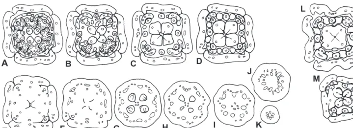
FIG. 2. Combretocarpus rotundatus. Floral bud, TS series. A, Level of anthers. B, Below anthers. C, Symplicate region of style at level of nectaries.
D, Transition between superior and inferior region; base of nectaries. E, Upper inferior region at level of placenta. F, Level of ovules. G, Level of locules below ovules. H±K, Below locules, showing rearrangement of vascular bundles. Bar 1 mm.
FIG. 3. Polygonanthus amazonicus. Male ¯oral bud, TS series. A, Level of episepalous anthers. B, Level of epipetalous anthers. C, Level of
nectaries. D, Transition between superior and inferior region; base of nectaries. E, Upper inferior region with ununited sepal margins and rudimentary ovary locules. F±G, Below locules, showing rearrangement of vascular bundles. Bar 1 mm.
FIG. 1. Anisophyllea disticha. A±K, Female ¯oral bud, TS series. A, Level of anthers. B, Level of epipetalous anthers. C, Below level of anthers
and above nectaries. D, Level of nectaries. E, Upper inferior region with ununited sepal margins. F, Upper inferior region at level of placenta. G, Level of ovules. H, Level of locules below ovules. I±K, Below locules, showing rearrangement of vascular bundles. L, Female ¯ower at anthesis,
TS at level of nectaries. M, Male ¯oral bud, TS at level of nectaries. Bars 1 mm.
®laments broader and thicker than epipetalous stamens (all), or all equal; anthers are sagittate (CR), dorsi®xed and introrse (all), with or without a short connective tip; incumbent in male ¯ower buds, strongly recumbent (approx. 908) in female ¯ower buds (because of the large stigmas); ®laments in advanced buds are longer than anthers and incurved. The gynoecium has an inferior ovary (Fig. 1A±H) (all); carpels are free in the superior region, united in the inferior region; the stigmas in female ¯owers are large, expanded, not papillate, decurrent on the ventral side, the ventral slit is only evident in the lower half of the free part of the carpels; in male ¯owers the carpel apex is punctiform, unicellular-papillate, the ventral slit extends up to the apex; there is a small gap in the centre between the united carpels extending down into the inferior region (Fig. 1D, E and M) (CR, Fig. 31); a compitum seems to be absent, as the centre of the symplicate zone does not appear to be part of the pollen tube transmitting tracts, which are restricted to the inner angle of the ventral slit of each carpel (CR); the ovary is apparently largely synascidi-ate, symplicate only in the uppermost region; placentation is axile (all), in the synascidiate zone; each carpel with a single, laterally attached ovule that ®lls the locule (Fig. 1G); only in the micropylar region is there a small gap, and this gap is ®lled with secretion produced by the placental/ funicular area adjacent to the micropyle; ovules are unitegmic, crassinucellar, anatropous (Tobe and Raven, 1987), syntropous (for term, see Endress, 1994), with the micropyle directed sideward and appearing as a longitudi-nal slit (Fig. 1G) (CR, Fig. 2E and F), the result of the
integument which has two lateral ¯anks, pressed together (Fig. 37). The nectary disc forms a bulge between each of the stamens (interstaminal) (Fig. 7) and is irregularly continuous or discontinuous (depending on the available space) between the adaxial side of the ®laments (intrastam-inal) and the gynoecium (in male ¯owers interstaminal portions fuse with intrastaminal portions and then with stamen ®laments, Figs 1M and 25; in female ¯owers interstaminal portions fuse with stamen ®laments, intras-taminal portions, if present, fuse with the gynoecium,
Fig. 1L). In the transition region from the superior to the inferior part, the stamens and the median parts of the sepals fuse higher up with the gynoecium than the petals and the lateral parts of the sepals (Figs 1E and 23) (all: CR,
Fig. 2D; PA, Fig. 3D and E). Thus a ¯oral cup is not formed (all).
Anatomy. Sepals have three main (and approx. three smaller, secondary) vascular bundles and three vascular traces in the ¯oral base (Fig. 1A±K). Petals with one vascular trace that divides into ®ve bundles to serve the ®ve lobes. Stamens have a single vascular bundle and a single trace (all). Carpels have a dorsal median bundle that extends up to the stigmatic region and a ventral median bundle (serving the ovule) in the lower synascidiate part. In the ¯oral base, the trace of each episepalous stamen fuses with the median trace of the sepal of the same radius (all); the trace of each epipetalous stamen fuses with the trace of the petal of the same radius, and with this joint bundle then fuse the lateral traces of the adjacent two sepals (PA). Thus
FIG. 4. Ceratopetalum gummiferum. Floral bud, TS series. A, Level of epipetalous anthers. B, Level of episepalous anthers. C, Level of nectary
disc. D, Level of ¯oral cup with ununited sepal margins. E, Transition between superior and inferior region; level of upper symplicate zone. F, Symplicate region of ovary at level of ovules. G, Synascidiate region of ovary at level of placenta. H, Below placenta at base of locules. I±J, Below
locules, showing rearrangement of vascular bundles. Bar 1 mm.
this peripheral bundle system consists of six or eight (in 3-merous or 4-merous ¯owers) main bundles and a number of smaller bundles located in between, which together form a reticulate pattern. Below the ovary locules, the ventral median carpel bundles join the peripheral bundle complex. The nectary does not contain any vascular strands.
Histology. The epidermis of the abaxial side of the sepals shows two striking dierentiations: (1) most cells have a thickened, mucilaginous inner tangential wall (this dier-entiation is absent in the inferior part of the ¯ower but present, in addition, in the lower superior region of the gynoecium); (2) stomata are raised on protrusions (Fig. 13) (all; CR,Fig. 14). Scattered unicellular hairs are present on the abaxial sepal surface. Although nectaries have stomata, the epidermis shows the same histological dierentiation as the underlying secretory region and may also be secretory
(PA). Tanniferous cells occur scattered in the outer epidermis of the sepals and petals, and in a more-or-less continuous layer in the anthers, on the ventral side of the free part of the carpels and the stigma (also in male ¯owers), and at the periphery of the ovules. Cells with oxalate druses are sparse in the epidermis.
Combretocarpus rotundatus (Anisophylleaceae)
Morphology. Flowers are bisexual, 3-merous (more rarely 4-merous,Fig. 9), obdiplostemonous (Fig. 2A±K) (¯owers with deviations to this pattern also found, e.g. Fig. 9). Sepals are valvate, in the lower part somewhat revolute-valvate; the ¯anks of adjacent sepals are united by short unicellular hairs (at the outer edges perhaps also by secretion) (Fig. 11). The ¯ower appears distinctly 3-angular because of the revolute-valvate aestivation of the sepals.
FIG. 5. Davidsonia pruriens. Floral bud, TS series. A, Level of free sepals. B, Level of anthers and styles (styles re¯exed, therefore appearing twice
in this section). C, Upper symplicate region of ovary. D, Synascidiate region of ovary at level of placenta. E, Locules below level of ovules; nectary portions present. F, Transition between superior and inferior region; level of nectary disc. G, Base of ovary locules. H±K, Below locules, showing
rearrangement of vascular bundles. Bar 1 mm.
Petals have two or three terete lobes or are simple, terete; they are shorter than stamens or occasionally lacking. Anthers have a blunt connective protrusion, they are incumbent in bud (Figs 9, 17 and18) and the connective is very thin; ®laments in bud already longer than anthers and incurved at the top. Carpels are free in most of the superior region, united in the inferior and lowermost superior region (Fig. 2A±G) (PA, Fig. 3A±E); the ventral slit extends up to the stigma, with a narrow canal in the inner angle (Fig. 35); the stigma is punctiform, smooth, secretory (Fig. 33). The ovary is symplicate in the upper two-thirds, synascidiate in the lower third of its length. A compitum seems to be absent, as the central gap present between the united carpels has a tanniferous epidermis. At the lower end of the symplicate zone each carpel has two laterally attached ovules that ®ll the locule except for its lowermost part (Fig. 2E and F); occasional small gaps between ovule and locular wall in the micropylar area are ®lled with secretion. The nectary disc is only slightly raised and does not protrude between the ®laments (Fig. 27).
Anatomy. Sepals have three main (and up to 12 or more smaller, secondary) vascular bundles and several vascular traces in the ¯oral base (Fig. 2A±K). Petals have one vascular trace that divides into three bundles to serve the three lobes. Carpels with a dorsal median bundle that extends up to the stigmatic region and a ventral median bundle in the synascidiate region, which is divided into two lateral bundles in the symplicate region, serving the two ovules; these lateral bundles are not present above the placentae. Part of the lateral sepal bundles fuses with this bundle complex; however, the outermost branches of the sepal bundles fuse with the central vascular system only
lower down, where a stele is formed in the centre of the ¯oral base. Somewhat higher up, but below the locules, all other vascular bundles, i.e. those of petals, episepalous stamens, and dorsal and ventral carpel bundles, form a stelar ring, together with the bundle complexes that consist of median sepal bundles and episepalous stamen bundles.
Histology. Multicellular, uniseriate and multiseriate, peltate hairs are present on the abaxial surface of the sepals. These hairs are probably secretory as they are covered by an extracellular substance still present in the microtome sections. Similar hairs are also present on the pedicel (Fig. 39). Tanniferous cells are present in the periphery (epidermis) of all ¯oral organs, including the ovules (also in the ventral slits in the centre of the superior syncarpous part of the gynoecium), and also scattered in layers below the epidermis; hairs are also tanniferous. Cells with oxalate druses are present in sepals, the connective of anthers, within the gynoecium in the lower superior region, the area surrounding the locules and the nucellus. Unicellular and uniseriate-pluricellular, non-ligni®ed hairs are present on the lower superior part of the gynoecium. The epidermis of the nectary contains stomata, and epidermal cells are tanniferous; in addition, unicellular, non-ligni®ed hairs are present (Fig. 27).
Polygonanthus amazonicus (Anisophylleaceae)
Morphology. Flowers are unisexual (only male ¯owers were available for study), 4-merous, obdiplostemonous (Fig. 3A±G). Sepals are valvate, postgenitally united by cuticular (and cellular) indentation of the epidermis, and are involute at the tip. The ¯ower appears 4-angular because of
FIG. 6. Gillbeea adenopetala. Floral bud, TS series. A, Level of episepalous anthers and styles. B, Level of epipetalous anthers and symplicate region of ovary. C, Synascidiate region of ovary at level of placenta. D, Below level of placenta. E, level of nectary disc. F, Transition from superior to inferior region; level of ununited sepal margins below locules. G±H, Inferior region, showing rearrangement of vascular bundles.
Bar 1 mm.
the valvate aestivation of the sepals (Fig. 3A±E). Petals are entire, trullate, with a papillate abaxial surface and hairs toward the margins; each petal halfway surrounds the base of the stamen ®lament of the same ¯oral sector (Fig. 3C and D). Stamen ®laments are papillate; anthers X-shaped, with-out a connective tip; ®laments in bud are already much longer than anthers, and incurved to such an extent that the anthers are turned 1808 (Fig. 21). In the male ¯owers studied, four narrow ovary locules are present (Fig. 3E) and ovules are lacking; however, even in the absence of ovules, the ovary has a relatively long synascidiate zone. The rudi-mentary stigma is unicellular-papillate. In female ¯owers a
single anatropous, crassinucellar, bitegmic ovule is present in each carpel (Tobe and Raven, 1987). The nectary disc forms a protrusion between each of the stamens and behind each stamen (Fig. 21). These protrusions are basally continuous, and are fused with the stamens slightly higher up than with the other adjacent organs (Fig. 3C and D).
Anatomy. Sepals have three main (and two smaller, secondary) vascular bundles and three to ®ve vascular traces in the ¯oral base (Fig. 3A±G). Petals have one vascular trace that divides into three or ®ve bundles in the broadest part. Carpels with a dorsal vascular bundle that
FIGS7±8. Floral bud, sepals removed, viewed from the side. Bar 300 mm. Fig. 7. Anisophyllea disticha, male. Fig. 8. Ceratopetalum
gummiferum. n, Nectary; f, ®lament; p, petal.
FIGS9±10. Floral bud, sepals and petals (if present) removed, from above. Arrowhead indicates style. Bar 500 mm. Fig. 9. Combretocarpus
rotundatus. Fig. 10. Davidsonia pruriens. a, Anther.
FIGS11±12. TS of ¯oral bud, postgenital coherence of sepal margins by unicellular hairs (arrowhead). Bar 100 mm. Fig. 11. Combretocarpus rotundatus. Fig. 12. Ceratopetalum gummiferum.
extends up toward the apex, and a ventral median bundle that ends below the locule (in the male ¯owers available). In the ¯oral base, the trace of each episepalous stamen fuses with the median trace of the sepal of the same radius; the trace of each epipetalous stamen fuses with the trace of the petal of the same radius, and to this joint bundle then fuse the lateral traces of the adjacent two sepals. Each dorsal carpel bundle also joins the bundle complex of the same
sector. This results in eight major peripheral bundle complexes. Towards the base, the four ventral carpel bundles split into several smaller ones and irregularly join the peripheral bundle complex.
Histology. The adaxial sepal surface contains scattered cells with a thickened, mucilaginous inner tangential wall (Fig. 29); such cells also occur sparingly in the adaxial
FIGS13±16. Raised stoma on abaxial side of sepal (arrowhead). Fig. 13. Anisophyllea disticha. Bar 25 mm. Fig. 14. Combretocarpus rotundatus,
LS. Bar 50 mm. Fig. 15. Ceratopetalum gummiferum. Bar 25 mm. Fig. 16. Ceratopetalum gummiferum, LS. Bar 50 mm. FIGS17±20. Stamens; arrowhead indicates connective protrusion. Bar 200 mm. Fig. 17. Combretocarpus rotundatus, from dorsal side. Fig. 18.
Combretocarpus rotundatus, from ventral side. Fig. 19. Schizomeria whitei, from dorsal side. Fig. 20. Schizomeria whitei, from ventral side. FIGS21±22. Floral bud, from the side. Arrowhead indicates incurving of ®lament. Bar 250 mm. Fig. 21. Polygonanthus amazonicus, male.
Fig. 22. Cunonia lenormandii. a, Anther; f, ®lament.
hypodermis, and in the epidermis of the abaxial surface and the margin. The epidermis of the rudimentary gynoecium (in male ¯owers), including the `stigma', and the lower part of the nectary lobes are tanniferous. Cells with oxalate druses were not found.
Ceratopetalum gummiferum (Cunoniaceae)
Morphology. Flowers are bisexual (all), 5-merous (GA) (but gynoecium 2-merous, DP), (ob)diplostemonous (all) (a distinction between diplostemonous and obdiplostemonous is problematic because the gynoecium is not isomerous with
the androecium whorls) (Fig. 4A±J). Sepals have a broad base (all), they are revolute-valvate. The revolute parts of adjoining sepals are postgenitally coherent by hairs (Fig. 12) and (towards the periphery) probably also by cuticular dentations. The ¯ower bud appears 5-angular because of the valvate aestivation of the sepals (Fig. 4A and B). Petals have a narrow base (GA), they are digitate, with ®ve upward-directed lobes, shaped like a hand with ®ve ®ngers (Fig. 8). Stamens are in two series (all), episepalous stamens longer than epipetalous (alternisepalous) stamens, and ®laments broader and thicker (all). Anthers are sagittate (DP), dorsi®xed, and introrse (all), with a broad
FIGS23±24. Floral bud, TS of upper inferior region. Arrowheads indicate ununited sepal margins. Bar 200 mm. Fig. 23. Anisophyllea disticha,
male. Fig. 24. Ceratopetalum gummiferum. p, Petal.
FIGS25±26. Floral bud, TS of lower superior region. Intrastaminal (arrow) and interstaminal (arrowheads) nectary portions. Bar 200 mm.
Fig. 25. Anisophyllea disticha, male. Fig. 26. Acsmithia davidsonii. g, Gynoecium; f, ®lament; s, sepal.
FIGS27±28. Interstaminal nectary portion with unicellular hairs. Bar 100 mm. Fig. 27. Combretocarpus rotundatus. Fig. 28. Davidsonia
pruriens. f, Filament.
connective tip; ®laments already longer than anthers in bud, incurved (all); ®laments of episepalous stamens even more incurved with the eect that their anthers are hidden behind and below the epipetalous ones in bud (Fig. 8). The gynoecium has a largely inferior ovary (Fig. 4A±H); carpels
are free in the superior region, united in the lowermost superior and in the inferior region; the ventral slit extends up to the stigma (all); stigma punctiform to slightly capitate, unicellular-papillate (Fig. 34) (GA); the ovary is synascidiate in approximately its lower half, symplicate
FIGS29±30. TS of adaxial surface of sepal, showing distinctive epidermal cells with a thickened, mucilaginous inner tangential wall (white
asterisk; cell lumen indicated by black asterisk). Bar 50 mm. Fig. 29. Polygonanthus amazonicus. Fig. 30. Gillbeea adenopetala. FIGS31±32. TS of superior symplicate region of gynoecium showing internal gap between the fused styles (arrowhead), surrounded by
tanniferous epidermis. Bar 50 mm. Fig. 31. Combretocarpus rotundatus. Fig. 32. Gillbeea adenopetala.
FIGS33±34. Carpel tip with stigma and ventral slit (arrowhead). Bar 100 mm. Fig. 33. Combretocarpus rotundatus, advanced ¯oral bud.
Fig. 34. Ceratopetalum gummiferum, at anthesis.
FIGS35±36. TS of style, showing stylar canal ®lled with secretion (arrowhead) and ventral slit (arrow). Bar 100 mm. Fig. 35. Combretocarpus
rotundatus, advanced ¯oral bud. Fig. 36. Davidsonia pruriens, at anthesis.
FIGS37±38. Ovule at anthesis. Arrowheads indicate extent of longitudinal micropylar slit, arrow indicates attachment region of the ovule. Bar 100 mm. Fig. 37. Anisophyllea disticha. Fig. 38. Ceratopetalum gummiferum.
FIGS39±40. TS of pedicel, showing multicellular, peltate hair. Bar 25 mm. Fig. 39. Combretocarpus rotundatus. Fig. 40. Acsmithia davidsonii.
448
Matthews et al.ÐComparison of Floral Structures of Anisophylleaceae and Cunoniaceae
above (Fig. 4E±H) (GA,Fig. 6B±E); there is a small gap in the centre between the united carpels in the uppermost symplicate zone; a compitum may be present in the lower symplicate zone; placentation is axile, partly in the symplicate, partly in the synascidiate zone (Fig. 4F and G) (all; DP,Fig. 5D; GA,Fig. 6C); four ovules per carpel; ovules arranged in two basipetally divergent lines (all); ovules bitegmic and crassinucellar (all), hemianatropous (GA) (see also Mauritzon, 1939), intermediate between syntropous and antitropous (for terminology seeEndress, 1994); the micropyle is formed by both integuments (all) (or, in some ovules at least in part, by the inner integument); it has the shape of a longitudinal slit (Fig. 38) (all). The nectary disc forms a protrusion between each of the stamens and is continuous behind the attachment of the ®laments, and also descends somewhat on the inner slope of the ¯oral cup (Figs 4Cand8). There is a shallow ¯oral cup around the lowermost part of the superior region of the ovary (Fig. 4D). Perianth organs and stamens fuse ®rst in the innermost part of the ¯oral periphery, and the median part of the sepals fuses higher up with the androecium than the lateral parts of the sepals and the petals (Figs 4Dand24).
Anatomy. Sepals have three main (and up to approx. six smaller secondary) vascular bundles and three vascular traces in the ¯oral base (Fig. 4A±J) (GA, Fig. 6A±H). Petals have one vascular trace that divides into ®ve bundles to serve the ®ve petal lobes. Stamens have a single vascular bundle and a single trace (all). Carpels have a dorsal bundle that extends up to the stigmatic region (DP); in the apocarpous region two lateral bundles are also present but do not extend as high as the dorsal one; the upper syncarpous region has four lateral bundles, and the lower one, six. The two lateralmost bundles of each carpel are centrally positioned in the septum and serve the ovules; in the synascidiate zone these four bundles belong to both carpels, and form a vascular complex. In the ¯oral base, the trace of each episepalous stamen fuses with the median trace of the sepal of the same radius, the trace of each epipetalous stamen fuses with the trace of the petal of the same radius, and this joint bundle also fuses with the lateral traces of the adjacent two sepals (GA). Thus this peripheral bundle system consists of ten major bundle complexes. The lateral carpellary bundles also join these bundle complexes. Below the ovary locules, the central vascular complex of the gynoecium described above splits into several bundles which join the peripheral bundle system. The nectary has many small phloem strands, which join neighbouring larger, vascular bundles, primarily the stamen traces.
Histology. Unicellular, ligni®ed hairs are present near the margin of the adaxial side of the sepals, where neighbour-ing sepals are contiguous (Fig. 12). Stomata are raised on protrusions on the abaxial surface of the sepals (Figs 15
and16) (DP). The nectary is of the mesophyll type (in the terminology ofVogel, 1977); it has stomata, and the cell layers below the epidermis contain dense cytoplasm, more so in the protruding lobes than on the inner slope of the ¯oral cup. Tanniferous cells are present in most of the tissue
of the sepals (all), petals (GA), stamen ®laments (all), anther epidermis and connective (GA), and carpels (all), and in the inner epidermis of the inner integument of the ovules (GA). Cells with oxalate druses are abundant (these cells are not tanniferous), especially on the adaxial side of the mesophyll of the sepals, in the connective of the anthers, in the ovary wall around the locules, in the nectary, and in the ¯oral cup.
Davidsonia pruriens (Cunoniaceae)
Morphology. Flowers are bisexual, 5- or 4-merous, (ob)diplostemonous (Fig. 5A±K). Sepal aestivation is valvate (somewhat revolute) and in the upper part of the calyx sepals are postgenitally connected by interlocking hairs, while in the lower part they are congenitally united. The ¯ower appears 5- or 4-angular (Figs 5A±D and 10). Petals are lacking. Episepalous ®laments are narrower and thinner than alternisepalous ones. The position of stamens is not irregular asMoody and Huord (2000)describe; the only deviation from the regular two-whorled pattern is the occasional occurrence of double or triple positions of stamens (two or three stamens side by side instead of one) in the episepalous position, which was observed in our material (collection by W. Forstreuter) and which also appears to be present in Fig. 19 of Moody and Huord (2000). In our collection, Endress 4248, we only found regularly (ob)diplostemonous ¯owers (Fig. 10). Dickison (1975a) also mentioned regular positions. Anthers have a narrow connective tip and a very narrow attachment point to the ®lament; ®laments form a short loop in bud. The gynoecium has a slightly inferior ovary (approx. 1/4); carpels are free in the stylar region, united in the ovarial region (Fig. 5B±G) (GA); styles are long and slender with a narrow canal in the inner angle; stigmas are slightly capitate, unicellular-papillate; ovary largely synascidiate, symplicate only in the uppermost region; a compitum is present (united ventral slit of carpels ®lled with secretion) (GA); ®ve to eight ovules present per carpel, arranged in two basipetally divergent lines in the symplicate zone and more towards the median plane in the synascidiate zone (Fig. 5C and D) (see also Bange, 1952); ovules are anatropous and syntropous; the micropyle has the shape of a longitudinal (often somewhat open) slit (GA). At anthesis, the stylar canals and the ovary locules are ®lled with secretion (Fig. 36), which seems to be produced by the inner epidermis of the style and by a conspicuously dierentiated secretory area in the placental/funicular region. The nectary disc protrudes with a portion between each of the stamens and (irregularly) a small portion behind each stamen (Figs 5E, Fand28). All nectary portions and ®lament bases unite to form a continuous ring around the ovary (Fig. 5F). Below this, the ring fuses with the ovary and with the calyx at about the same level. Thus a ¯oral cup is not formed.
Anatomy. Sepals have three or ®ve main bundles in the free part, the lateralmost bundles of neighbouring sepals more-or-less unite in the congenitally united region (and approx. four smaller secondary bundles in each sepal in the
Matthews et al.ÐComparison of Floral Structures of Anisophylleaceae and Cunoniaceae
449
congenitally united part), and ten vascular traces for the entire calyx in the ¯oral base (from each sepal, a median bundle, and from each two neighbouring sepal margins, a synlateral) (Fig. 5A±K). In addition to a dorsal bundle, carpels have two lateral bundles, and a basipetally increasing number of secondary laterals (up to approx. ten on each side), which anastomose with each other. The two lateral bundles of each carpel serve the ovules, and they unite to form a cylindrical complex below the placenta. The nectary disc is supplied by numerous small phloem strands, which connect with the nearest stamen and carpel bundles. In the ¯oral base the traces of the episepalous sectors unite as do those of the alternisepalous sectors. The cylindrical vascular complex in the centre of the gynoecium opens into a number of small complexes, which join the peripheral bundle system. Lower down in the ¯oral base the entire vascular system forms a more or less cylindrical stele.
Histology. Unicellular, ligni®ed hairs are present on both surfaces of the sepals and along their valvate margins; they are also present on the ovary and nectary disc (Fig. 28). We did not ®nd stomata on the nectary nor is the tissue distinguished by richness in cytoplasm; this may indicate that the `nectary' disc is not functional. Tanniferous tissue is predominant in the sepals, stamen ®laments, styles and stigmas, at the periphery of the ovary and ovules, and on the inner slope of the nectary; the hairs are also tanniferous. Cells with oxalate druses were not found.
Gillbeea adenopetala (Cunoniaceae)
Morphology. Flowers are bisexual, 5-merous (but gynoe-cium 3-merous), (ob)diplostemonous (Fig. 6A±H). Sepals are valvate (involute at the tip), somewhat unequal in size, with an apparent quincuncial pattern: the outermost (®rst) is the largest, the other four are successively smaller; they are postgenitally coherent by hairs. The ¯oral bud appears roundish, and not 5-angular, in spite of the valvate sepal aestivation (Fig. 6A±F). Petals are short, the upper part is broad with two lateral tips that are incurved in bud, the lateral tips each with a cup-shaped ending that is secretory (Endress, 1994), the lower part with hairs at the margins. Anthers are X-shaped, and without a connective tip. The gynoecium has a largely superior ovary (Fig. 6A±E). In the centre of the gynoecium a gap remains for a short distance at the level where the carpels unite (Fig. 32). The stigma appears to be secretory. (Three to) four ovules are present in each carpel (only two according toDickison, 1975a; four to ®ve according toRozefelds and Pellow, 2000). Ovules are syntropous or deviating from this pattern. Stylar canals, ovary locules and the micropylar area of the ovules are ®lled with secretion already in advanced bud; at least part of this secretion may be produced by secretory hairs in the placental region. The nectary is a ¯at, continuous disc between the androecium and the gynoecium and between the stamens (Fig. 6E). It ®rst fuses with the stamens and then with the median part of the sepals (Fig. 6F). Thus, a ¯oral cup is not formed. The lateral parts of the sepals with valvate ¯anks are still free at a level where all other ¯oral
organs are united, and thus can be recognized as ®ve furrows in the periphery of the solid ¯oral base (Fig. 6G). Anatomy. Petals have one vascular trace that divides into three to ®ve bundles (Fig. 6A±H). Carpels have three main vascular bundles: a dorsal median bundle that extends up to about the level where the carpels become free, and two lateral bundles that extend still higher into the free part of the carpels; in the synascidiate zone the lateral bundles join those of adjacent carpels to form synlaterals, which serve the ovules; in the syncarpous region additional minor bundles form a network in the carpel walls. Below the ovary locules, the dorsal and minor lateral carpel bundles join the traces of the other ¯oral organs. The traces of the episepalous stamens join the median sepal traces; the traces of the epipetalous stamens join the petal traces, and they are also joined by the outermost sepal traces; the sepal traces that are between the median and the outermost lateral traces either join the petal or the median sepal traces. The synlateral carpel bundles are the lowermost to join the bundle system in the ¯oral base.
Histology. The outer surface of the sepals is covered with both stellate and unicellular ligni®ed hairs; on the inner surface, scattered unicellular hairs also occur. In many epidermal cells of the adaxial sepal surface, the inner tangential wall is thickened and mucilaginous (Fig. 30), such cells also occur sparingly in the abaxial epidermis. Below the adaxial epidermis there are two to three layers of smaller cells, all with thickened walls. Although nectaries have stomata, the epidermis shows the same histological dierentiation as the underlying secretory region and may also be secretory. Oxalate druses are present in those hypodermal layers of the adaxial side of the sepals that have thickened cell walls (see above), in petals, stamen ®laments, on the connective side of the anthers, in the periphery of the nectary disc, and in the ¯oral base.
DISCUSSION
Structural similarities in ¯owers of Anisophylleaceae and Cunoniaceae
The initial morphological similarities observed by P.K.E. between Anisophyllea disticha (Anisophylleaceae) and Cer-atopetalum gummiferum (Cunoniaceae), and clearly dis-played in Figs 7 and 8, were further supported and expanded by the anatomical and histological similarities detailed in this paper. Both families have ¯owers with a small diameter (51 cm), except for the female ¯owers of Polygonanthus which are longer (Tobe and Raven, 1988a), and some Cunoniaceae with brush ¯owers or with large petals (Bauera and Eucryphia) (Dickison, 1975b). Bisexual ¯owers are present in both families, and in Anisophyllea and Polygonanthus, where ¯owers are unisexual, the organs of the opposite gender are present but not functional. Similarly, the Late Cretaceous ¯owers of Platydiscus are small and possess bisexual organization (SchoÈnenberger et al., 2001).
Flower merism is also similar between the two families. Although ¯owers are mostly 4- or 3-merous (rarely 5-merous) in all ¯oral whorls in Anisophylleaceae (see also
Ding Hou, 1958;Tobe and Raven, 1988a), and mostly 5-merous (in perianth and androecium) in Cunoniaceae, some members of the Cunoniaceae have 4-merous ¯owers (Acsmithia, Aistopetalum, Spiraeanthemum; Kanehira and Hatusima, 1942; Hoogland, 1960, 1979, 1987; Dickison, 1975a) (including the fossil Platydiscus; SchoÈnenberger et al., 2001) and others, 3-merous ¯owers (Vesselowskya p.p.) (in Vesselowskya the gynoecium is 2-merous) (Engler, 1928; Dickison, 1989; Rozefelds et al., 2001). Most noteworthy is the presence of 3-merous ¯owers in both families, as this feature is relatively rare in eudicots (reviewed inEndress, 1996). In addition, although the gynoecium in Anisophylleaceae normally has the same number of organs as the outer ¯oral whorls, exceptional ¯owers with fewer carpels were found in Anisophyllea and Combretocarpus. Thus, these ¯owers exhibited the same condition as is commonly present in Cunoniaceae. Among Cucurbitales, in which Anisophylleaceae appear in rbcL studies (Savolainen et al., 2000), ¯owers with a trimerous perianth and gynoecium occur in Begoniaceae and Datiscaceae, but in these families a polymerous androecium is present, in contrast to Anisophylleaceae.
Anisophylleaceae and most Cunoniaceae have ¯owers with two whorls of stamens; the episepalous stamens are commonly longer, thicker and broader than the epipetalous ones, and if the gynoecium is isomerous with the other organs, the carpels have an epipetalous position, i.e. the androecium is obdiplostemonous (if the gynoecium is not isomerous with the androecium whorls a distinction between diplostemonous and obdiplostemonous is proble-matic). Also in the fossil Platydiscus, the carpels have an epipetalous position (SchoÈnenberger et al., 2001). Although obdiplostemony occurs in a number of eudicots (such as Oxalidales) (e.g.Ronse Decraene and Smets, 1995), it is not present in other Cucurbitales (only Coriariaceae has two stamen whorls but is not obdiplostemonous).
Sepals are valvate in some genera of both families (revolute-valvate in Combretocarpus, Ceratopetalum and Davidsonia), and they are postgenitally connected in bud. The same diverse methods of coherence have evolved in parallel: connection by hairs (Combretocarpus, Davidsonia, Gillbeea); by cuticular (and cellular) dentation of the epidermis (Anisophyllea, Polygonanthus, Acsmithia); and by hairs and probably also cuticular dentation (Cerato-petalum) (Figs 11and12). In the fossil Platydiscus, sepals are also valvate (SchoÈnenberger et al., 2001). The sepal tips are involute (and thus located inside the closed ¯oral bud) and papillate (Anisophyllea, Acsmithia). Valvate sepals are not present in other Cucurbitales, except for tepal pairs in Cucurbitaceae (Cronquist, 1981). In addition, in both families, at the ¯oral base, the sepals commonly fuse with the inner ¯oral organs ®rst in their median areas, while the sepal ¯anks remain free and descend as ¯anges for some distance (Figs 23and24).
A number of special features of the epidermis of the sepals are common in Cunoniaceae and Anisophylleaceae. These include the presence of peltate, secretory hairs with
multicellular, uniseriate or multiseriate stalks on the abaxial sepal surface, and also on the pedicel in Combretocarpus (see alsoTobe and Raven, 1988a), and in the fossil Platydiscus (SchoÈnenberger et al., 2001); similar uniseriate hairs are present on the pedicel in Acsmithia (Figs 39and40). Stellate hairs occur in Gillbeea (see also Engler, 1928, for other Cunoniaceae). Stomata on the abaxial surface of the sepals are conspicuously raised like short chimneys in representa-tives of both families, such as Anisophyllea, Combretocarpus, Polygonanthus, Ceratopetalum and Davidsonia (Figs 13±16). In the sepals of members of both families, distinctive epidermal cells with a thickened, mucilaginous, inner tangential wall were found. In Polygonanthus and Gillbeea, such cells occur on the adaxial sepal surface, and are more scattered on the abaxial surface (Figs 29 and 30); in Anisophyllea, they are present on the abaxial sepal surface. To our knowledge, this feature in ¯owers has not received attention at an angiosperm-wide level. Should it be found to be rare, it would be an additional indicator of a closer relationship between the two families. However, similar cells are known from foliage leaves of various angiosperms, including Cunoniaceae (Hallier, 1903; Dickison, 1975c;
Gregory, 1998). For reviews of other families see Napp-Zinn (1973, p. 185) andMetcalfe (1979, pp. 65, 66, 198, 199).
Eschrich (1995, p. 74) illustrates such cells in foliage leaves of Calluna (Ericaceae); in other publications they are not ®gured in detail.
Petals show parallel variation patterns of striking similarity in both families. Both families appear to have a similar plasticity in petal morphology. Large, showy petals with broad plates are conspicuously lacking (except for Bauera and Eucryphia in Cunoniaceae). In one form or another, divided petals are common (Anisophyllea, Com-bretocarpus, Poga, Anodopetalum, Ceratopetalum, Gillbeea, Platylophus, Schizomeria) (Tobe and Raven, 1988a;Barnes and Rozefelds, 2000; this study). The most striking form is digitate petals, which may look like a hand with ®ve ®ngers that halfway surround the epipetalous stamen in bud (Anisophyllea, Ceratopetalum) (this study) (Figs 7 and 8). The number of `®ngers' varies in both families. In some taxa the ®ngers have a thickened tip (Anisophyllea p.p., Poga;Tobe and Raven, 1988a; Gillbeea). The thickened tips are suggested to be secretory in Anisophylleaceae (Tobe and Raven, 1988a); in Gillbeea, secretion was shown by
Endress (1994), who argued that they may function as pseudonectaries. In other taxa the petals are small and trullate (Polygonanthus, Caldcluvia, cf.Schlechter, 1914, as Opocunonia). Reduction may also be expressed in lability of petal shape: in Combretocarpus, petals may be lobed or unlobed, occasionally very small or missing. In some Cunoniaceae, petals are absent. Presence or absence may be labile within a genus, such as Ceratopetalum (Hoogland, 1960), or at a higher level: petaliferous and apetalous genera are irregularly distributed in the major clades of the family (based on the morphological cladistic study byHuord and Dickison, 1992). Unicellular hairs on petal margins were found in Anisophyllea, Polygonanthus and Gillbeea (for the latter see alsoRozefelds and Pellow, 2000).
Features of the androecium also show strong similarity in members of both families. Anthers are dorsi®xed, introrse,
Matthews et al.ÐComparison of Floral Structures of Anisophylleaceae and Cunoniaceae
451
commonly sagittate; they have a connective tip in Aniso-phyllea, Combretocarpus, Ceratopetalum, Davidsonia and Schizomeria (Figs 17±20). The connective is thin in most taxa. The attachment point of the anther to the ®lament is also thin in many taxa, therefore the anthers are versatile, and tend to break o easily (for Cunoniaceae, cf. also
Endress and Stumpf, 1991). This combination of features does not occur in other Cucurbitales, except for Coryno-carpaceae. Similar long stamen ®laments are present in advanced buds of both families. In the extreme case, they form conspicuous loops over the level of the anthers, such as in Polygonanthus (this study) and Cunonia (Endress and Stumpf, 1991) (Figs 21and22). Filaments are also incurved in the fossil Platydiscus (SchoÈnenberger et al., 2001). In addition, in both families, pollen is commonly 3-colporate and often prolate, the exine reticulate, with lumina of meshes diminishing in size toward the apertures (Hideux and Ferguson, 1976;Vezey et al., 1988), and also in the Late Cretaceous Platydiscus (SchoÈnenberger et al., 2001). Mature pollen is bi-nuclear in both families (Gardner, 1975;Tobe and Raven, 1987).
The nectaries of Anisophyllea and Ceratopetalum are especially similar when viewed from the side (Figs 7and8). In both families, the nectaries form hemispherical bulges which protrude between the stamen ®laments, and are more-or-less connected behind these ®laments (also in Anisophylleaceae the bulges are not always completely separated from each other, in contrast toTobe and Raven, 1988a) (Figs 25and26). Stomata are present on the nectary surface in all taxa studied, except for Davidsonia. Although the presence of stomata seems to be common in disc nectaries of eudicots (Endress, 1994), it is worth mention-ing. Unicellular (non-secretory) hairs on nectaries were found in Combretocarpus and Davidsonia (Figs 27and28). They are also present in the Late Cretaceous Platydiscus (SchoÈnenberger et al., 2001). Among other Cucurbitales, nectaries are either lacking or, if present (Cucurbitaceae, Corynocarpaceae), they are located at other sites on the ¯ower than in Anisophylleaceae.
In the stylar region, the carpels are free in all taxa, and simple (unbranched). These features are not in agreement with most Cucurbitales. The stigma is commonly small, restricted to the apex, punctiform or slightly capitate, unicellular-papillate, and the ventral slit reaches the stigmatic region (Figs 33 and 34). Only in few taxa of both families is the stigma decurrent on the ventral side (Anisophyllea, Vesselowskya;Dickison, 1989; this study). A stylar canal in the inner angle of the ventral slit of each carpel is present in Combretocarpus, Davidsonia and Gillbeea (for distribution in Cunoniaceae, see alsoHuord and Dickison, 1992) (Figs 35and36). In addition, there is a hole or canal in the centre of the gynoecium at the level where the carpels unite, which may extend over a shorter or longer distance (Anisophyllea, Combretocarpus, Polygo-nanthus, Ceratopetalum, Gillbeea) (Figs 31 and 32). This feature is also present in the Late Cretaceous Platydiscus (SchoÈnenberger et al., 2001). A compitum seems to be lacking in Anisophylleaceae, but is present in Cunoniaceae. Unicellular hairs are present on carpels in the lower region of the superior part of the gynoecium, thus on the ovary or
the lower part of the style, in Combretocarpus, Acsmithia, Davidsonia and other Cunoniaceae. Ovaries are inferior in Anisophylleaceae and almost inferior in some Cunoniaceae (Ceratopetalum, Pullea) (although more often semi-inferior or superior in Cunoniaceae), and semi-inferior in the fossil Platydiscus (SchoÈnenberger et al., 2001); they are conspicu-ously synascidiate in both families, commonly in at least half the length of the syncarpous part. Placentae are axile (but commonly parietal in Cucurbitales), with commonly pendant ovules. The entire ovules (or at least the micropylar region) are immersed in heavy secretion, which is formed by prominently dierentiated tissue with swollen cell walls in the placental/funicular region (Anisophyllea, Combretocarpus, Davidsonia, Gillbeea).
Each carpel has one (Anisophyllea, Acsmithia p.p.) or two ovules (Combretocarpus, several Cunoniaceae); only in several Cunoniaceae (and in the fossil Platydiscus;
SchoÈnenberger et al., 2001) are more than two ovules present. Ovules are bitegmic in Poga and Polygonanthus (Tobe and Raven, 1987, 1988b), and in all Cunoniaceae studied so far, such as Bauera, Ceratopetalum, Cunonia, Schizomeria (Mauritzon, 1939), Weinmannia (Govil and Saxena, 1976), Callicoma (Kennedy and Prakash, 1981), Davidsonia and Gillbeea (this study); only in Anisophyllea and Combretocarpus are they unitegmic (Tobe and Raven, 1987). They are crassinucellar in all genera of Anisophyl-leaceae (Tobe and Raven, 1987) and in all Cunoniaceae studied to date, such as Bauera, Ceratopetalum, Schizo-meria, Weinmannia (Mauritzon, 1939), Weinmannia (Govil and Saxena, 1976), Callicoma (Kennedy and Prakash, 1981), and the taxa of this study. The ovules are anatropous in all Anisophylleaceae and most Cunoniaceae (in Cuno-niaceae more rarely hemianatropous: in Ceratopetalum, Schizomeria, Mauritzon, 1939; and Gillbeea, this study); they are more-or-less syntropous (for terminology see
Endress, 1994) in Anisophylleaceae and at least partly in Cunoniaceae. In bitegmic ovules the micropyle is formed by both integuments (for Anisophylleaceae:Tobe and Raven, 1987,1988b; for Cunoniaceae:Mauritzon, 1939;Govil and Saxena, 1976;Kennedy and Prakash, 1981; this study). The outer, or the only integument forms two lateral lobes, which result in a conspicuous longitudinal slit in the micropylar region (Anisophyllea, Ceratopetalum, Davidso-nia) (Figs 37and38).
The ¯oral vascular pattern is similar in both families (for Anisophylleaceae see also Tobe and Raven, 1988a; for Cunoniaceae, Dickison, 1975a). Although some com-ponents may be common in eudicots, the sum of the pattern is noteworthy. The sepals have three main vascular bundles and three vascular traces in the ¯oral base; the petals and stamens only one. The traces of the petals and epipetalous stamens fuse with the neighbouring lateral sepal traces. In the synascidiate zone, below the placenta, the lateral (ventral) carpel bundles form a narrow cylindrical vascular complex in both families. Tanniferous tissue is abundant in ¯owers of both families. Cells with oxalate druses are common (not found in Polygonanthus and Davidsonia).
Fruits are diverse in both families. Although many genera in Cunoniaceae have dehiscent fruits, there are also genera with indehiscent fruits, which are drupaceous in
452
Matthews et al.ÐComparison of Floral Structures of Anisophylleaceae and Cunoniaceae
Aistopetalum, Davidsonia and Schizomeria, as is common in Anisophylleaceae. A similar kind of fruit that is indehis-cent, one-seeded (developed from a several-ovuled ovary), and with three prominent longitudinal wings, occurs in both families (Combretocarpus, Gillbeea); however, it develops from an inferior ovary in the former and from a superior ovary in the latter (Ding Hou, 1958, for Anisophylleaceae;Dickison, 1984; andDoweld, 1998, for Cunoniaceae). Even if this particular fruit shape is autapomorphous in both taxa, which is most probable, it is of interest as it indicates the result of a similar potential for dierentiation of form in both families.
Systematics
Based on our present structural investigation of repre-sentatives of Anisophylleaceae and Cunoniaceae, a global discussion on the mutual position of these families seems timely. Many of the structural features of Anisophylleaceae described in this paper do not fully correspond with those common to other members of Curcurbitales, the order in which the family is currently placed (APG, 1998). To date, the systematics of both families has not been thoroughly studied at a molecular level and no comparisons between the two families exist. Only two (Anisophyllea, Combreto-carpus) of the four genera of Anisophylleaceae were considered by rbcL analyses of the family (Setoguchi et al., 1999; Schwarzbach and Ricklefs, 2000), and for Cunoniaceae, molecular studies have considered at most three genera (Soltis and Soltis, 1997;Savolainen et al., 2000;
Soltis et al., 2000). The actual number of genera belonging to Cunoniaceae is currently uncertain; Huord and Dickison (1992) considered the family to consist of 24 genera including Eucryphia, Aphanopetalum, Bauera and Brunellia. In their analysis, Gillbeea and Spiraeanthemum (Acsmithia) appear as members of the basalmost clade of the family. However, according to D. Soltis (pers. comm.), Aphanopetalum is better placed in Saxifragales (see also Endress and Stumpf, 1991; Dickison et al., 1994), and Brunellia appears as sister of a clade formed by Cephalo-taceae, Cunoniaceae and Elaeocarpaceae. According to
Moody and Huord (2000), Davidsonia, once considered to belong in its own family, is now better placed in Cunoniaceae.
Anisophylleaceae have long been placed in Rhizophor-aceae, in spite of vegetative dierences such as spiral vs. opposite leaves, and stipules absent vs. present. Vegetative dierences of a similar magnitude appear to be present in Anisophylleaceae and Cunoniaceae. However, on closer inspection, these dierences are not as distinct as they ®rst seem. In Anisophylleaceae, at least in Anisophyllea disticha, minute secretory appendages are present at the location of stipules (Vincent and Tomlinson, 1983; Juncosa and Tomlinson, 1988b; Dengler et al., 1989), while among Cucurbitales, stipules are lacking in Cucurbitaceae, Datis-caceae, Tetramelaceae (Cronquist, 1981), and in Cunonia-ceae, Davidsonia has spiral phyllotaxis (Dickison and Rutishauser, 1990). In addition, sieve tube plastids of the S-type are found in Anisophylleaceae and Cunoniaceae (Behnke, 1988) (but also in Cucurbitales). With respect to
wood anatomy, Anisophylleaceae are considered the most divergent family among Cucurbitales (Carlquist and Miller, 2001).
The many general and special similarities in ¯oral structure between Anisophylleaceae and Cunoniaceae indicate either a much closer relationship than hitherto assumed, or an amazing convergent evolution. In the ®rst case, one would have to assume that the previous rbcL results have given a false phylogenetic signal. Especially with regard to their ¯oral structure, Anisophylleaceae ®t much better with Oxalidales (together with Cunoniaceae) than with Cucurbitales. This view is further emphasized by the discovery of well preserved fossil ¯owers of Platydiscus from the Late Cretaceous that correspond to both families (SchoÈnenberger et al., 2001). It would certainly be important to study critically the position of Anisophyllea-ceae, based on more nucleotide sequences than rbcL. Another problem is the phylogenetic topology among members of basal rosids. The position of Saxifragales is still uncertain (D. Soltis, pers. comm.). Thus, potential relationships between Cunoniaceae, Anisophylleaceae and Saxifragales require further testing by more extended molecular and structural analyses.
In the second case, if Anisophylleaceae (Cucurbitales) and Cunoniaceae (Oxalidales) are con®rmed to be correctly placed by results of the current rbcL analyses, a study to elucidate the function and evolutionary signi®cance of these similar ¯oral structures should be performed. Pertinent questions would then be: (1) do the two families exhibit a suite of plesiomorphic features of rosids, which was established in the Late Cretaceous and retained in the two families, while it was lost in other, closely related families, or (2) was this suite of features present in early rosids and later lost, reappearing in Anisophylleaceae and Cunonia-ceae, or (3) could the character combination be a suite of functionally related traits that have convergently evolved in both families as autapomorphies?
ACKNOWLEDGEMENTS
For ®xed ¯owers we thank A.M. Juncosa, A. Kocyan and W. Forstreuter. P.K.E. is indebted to B.P.M. Hyland and B. Gray for their support in the ®eld in northern Queensland and for pickled material, and to P. Morat for support in New Caledonia. We thank D.E. Soltis for unpublished information on Saxifragales and Brunellia. R. Siegrist is thanked for some of the microme sections, U. Jauch for support with the SEM. This study is part of a project of P.K.E., ®nancially supported by the Swiss National Foundation (grant nr. 3100-059149.99/1); it also pro®ted from work supported by the Swedish Natural Science Research Council (EMF).
LITERATURE CITED
APG (The Angiosperm Phylogeny Group). 1998. An ordinal classi®-cation for the families of ¯owering plants. Annals of the Missouri Botanical Garden 85: 531±553.
Baehni C, Dansereau P. 1939a. Polygonanthus, genre de SaxifragaceÂes. Bulletin de la SocieÂte Botanique de France 86: 183±186.
Baehni C, Dansereau P. 1939b. La position systeÂmatique du genre Polygonanthus. Bulletin de la SocieÂte Botanique de Suisse 49: 415±416.
Baillon H. 1862. Observations sur les aniteÂs du Macarisia et sur l'organisation de quelques RhizophoreÂes. Adansonia 3: 15±41. Bange GGJ. 1952. A new family of dicotyledons: Davidsoniaceae.
Blumea 7: 293±296.
Barnes RW, Rozefelds AC. 2000. Comparative morphology of Anodopetalum (Cunoniaceae). Australian Systematic Botany 13: 267±282.
Behnke H-D. 1988. Sieve-element plastids and systematic relationships of Rhizophoraceae, Anisophylleaceae, and allied groups. Annals of the Missouri Botanical Garden 75: 1387±1409.
Bensel CR, Palser BF. 1975. Floral anatomy in the Saxifragaceae sensu lato. IV. Baueroideae and conclusions. American Journal of Botany 62: 688±694.
Carlquist S, Miller RB. 2001. Wood anatomy of Corynocarpaceae is consistent with cucurbitalean placement. Systematic Botany 26: 54±65.
Chase MW, Soltis DE, Olmstead RG, Morgan D, Les DH, Mishler BD et al. 1993. Phylogenetics of seed plants: an analysis of nucleotide sequences from the plastid gene rbcL. Annals of the Missouri Botanical Garden 80: 528±580.
Croizat L. 1939a. Une nouvelle sous-famille des OlacaceÂes au BreÂsil. Bulletin de la SocieÂte Botanique de France 86: 5±7.
Croizat L. 1939b. Polygonanthus, not a genus of the Saxifragaceae. Journal of the Arnold Arboretum 20: 443±445.
Cronquist A. 1981. An integrated system of classi®cation of ¯owering plants. New York: Columbia University Press.
Cronquist A. 1983. Some realignments in the dicotyledons. Nordic Journal of Botany 3: 75±83.
Dahlgren RMT. 1983. General aspects of angiosperm evolution and macrosystematics. Nordic Journal of Botany 3: 119±149. Dahlgren RMT. 1988. Rhizophoraceae and
AnisophylleaceaeÐsum-mary statement, relationships. Annals of the Missouri Botanical Garden 75: 1278±1295.
Dengler NG, Ritland CE, Donnelly PM. 1989. Leaf development and primary vascular organization in shoots of Anisophyllea disticha. American Journal of Botany 76: 1326±1343.
Dickison WC. 1975a. Studies on the ¯oral anatomy of the Cunonia-ceae. American Journal of Botany 62: 433±447.
Dickison WC. 1975b. Floral morphology and anatomy of Bauera. Phytomorphology 25: 69±76.
Dickison WC. 1975c. Leaf anatomy of Cunoniaceae. Botanical Journal of the Linnean Society 71: 275±294.
Dickison WC. 1984. Fruits and seeds of the Cunoniaceae. Journal of the Arnold Arboretum 65: 149±190.
Dickison WC. 1989. Comparisons of primitive Rosidae and Hamame-lidae. In: Crane PR, Blackmore S, eds. Evolution, systematics, and fossil history of the Hamamelidae. Introduction and `lower' Hamamelidae, Vol. 1. Oxford: Clarendon Press, 47±73.
Dickison WC, Rutishauser R. 1990. Developmental morphology of stipules and systematics of the Cunoniaceae and presumed allies. II. Taxa without interpetiolar stipules and conclusions. Botanica Helvetica 100: 75±95.
Dickison WC, Hils MH, Lucansky TW, Stern WL. 1994. Comparative anatomy and systematics of woody Saxifragaceae Aphanopetalum Endl. Botanical Journal of the Linnean Society 114: 167±182. Ding Hou. 1958. Rhizophoraceae. Flora Malesiana Ser. 1, 5: 429±493. Doweld AB. 1998. The carpology and taxonomic relationships of Davidsonia (Davidsoniacae). Edinburgh Journal of Botany 55: 13±25.
Ducke A. 1932. Neue Gattungen aus der Hylaea Brasiliens. Notizblatt des Botanischen Gartens Berlin-Dahlem 11: 345±347.
Endress PK. 1994. Diversity and evolutionary biology of tropical ¯owers. Cambridge: Cambridge University Press.
Endress PK. 1996. Homoplasy in angiosperm ¯owers. In: Sanderson MJ, Huord L, eds. Homoplasy: the recurrence of similarity in evolution. San Diego: Academic Press, 303±325.
Endress PK, Stumpf S. 1991. The diversity of stamen structures in `Lower' Rosidae (Rosales, Fabales, Proteales, Sapindales). Botan-ical Journal of the Linnean Society 107: 217±293.
Engler A. 1928. Cunoniaceae. In: Engler A, Prantl K, eds. Die natuÈrlichen P¯anzenfamilien, 2nd edn, 18a. Leipzig: Engelmann, 229±262, 486.
Eschrich W. 1995. Funktionelle P¯anzenanatomie. Berlin: Springer. Gardner RO. 1975. A survey of the distribution of binucleate and
trinucleate pollen in the New Zealand ¯ora. New Zealand Journal of Botany 13: 361±366.
Govil CM, Saxena NP. 1976. Anatomy and embryology of Weinmannia fraxinea Sm. (Cunoniaceae). Journal of the Indian Botanical Society 55: 219±226.
Gregory M. 1998. Cunoniaceae. In: Cutler DF, Gregory M, eds. Anatomy of the dicotyledons, Vol. IV Saxifragales. Oxford: Clarendon Press, 10±27.
Hallier H. 1903. UÈber die VerwandtschaftsverhaÈltnisse bei Engler's Rosalen, Parietalen, Myrti¯oren und in anderen Ordnungen der Dikotylen. Abhandlungen aus dem Gebiete der Naturwissenschaften herausgegeben vom Naturwissenschaftlichen Verein in Hamburg 18: 1±98.
Harden GJ, Williams JB. 2000. A revision of Davidsonia (Cunonia-ceae). Telopea 8: 413±428.
Hideux MJ, Ferguson IK. 1976. The stereostructure of the exine and its evolutionary signi®cance in Saxifragaceae sensu lato. In: Ferguson IK, Muller J, eds. The evolutionary signi®cance of the exine. London: Academic Press, 327±377.
Hoogland RD. 1960. Studies in the Cunoniaceae. I. The genera Ceratopetalum, Gillbeea, Aistopetalum, and Calycomis. Australian Journal of Botany 8: 318±341.
Hoogland RD. 1979. Studies in the Cunoniaceae. II. The genera Caldcluvia, Pullea, Acsmithia, and Spiraeanthemum. Blumea 25: 481±505.
Hoogland RD. 1987. Studies in the Cunoniaceae. IV. Further notes on New Caledonian Acsmithia. Adansonia 9: 393±397.
Huord L. 1992. Rosidae and their relationships to other non-magnoliid dicotyledons: a phylogenetic analysis using morpho-logical and chemical data. Annals of the Missouri Botanical Garden 79: 218±248.
Huord L, Dickison WC. 1992. A phylogenetic analysis of Cunonia-ceae. Systematic Botany 17: 181±200.
Igersheim A. 1993. The character states of the Caribbean monotypic Strump®a (Rubiaceae). Nordic Journal of Botany 13: 545±559. Igersheim A, Cichocki O. 1996. A simple method for microtome
sectioning of prehistoric charcoal specimens embedded in 2-hydroxyethyl methacrylate (HEMA). Review of Paleobotany and Palynology 92: 389±393.
Juncosa AM, Tomlinson PB. 1988a. A historical and taxonomic synopsis of Rhizophoraceae and Anisophylleaceae. Annals of the Missouri Botanical Garden 75: 1278±1295.
Juncosa AM, Tomlinson PB. 1988b. Systematic comparison and some biological characteristics of Rhizophoraceae and Anisophyllea-ceae. Annals of the Missouri Botanical Garden 75: 1296±1318. Kanehira R, Hatusima S. 1942. The Kanehira-Hatusima 1940
collection of New Guinea plants. VII. Botanical Magazine. Tokyo 61: 105±119.
Kennedy MJ, Prakash N. 1981. A morphological and embryological study of Callicoma serratifolia Andr. (Cunoniaceae). Australian Journal of Botany 29: 721±731.
Mauritzon J. 1939. Contributions to the embryology of the orders Rosales and Myrtales. Lunds Universitets Aarsskrift, n.F., Avd. 2. 35: 1±121.
Metcalfe CR. 1979. The leaf: general topography and ontogeny of the tissues. In: Metcalfe CR, Chalk L, eds. Anatomy of the dicotyledons, 2nd edn, Vol. 1. Oxford: Clarendon Press, 63±75. Moody M, Huord L. 2000. Floral development and structure of
Davidsonia (Cunoniaceae). Canadian Journal of Botany 78: 1034±1043.
Nandi OI, Chase MW, Endress PK. 1998. A combined cladistic analysis of angiosperms using rbcL and non-molecular data sets. Annals of the Missouri Botanical Garden 85: 137±212.
Napp-Zinn K. 1973. Anatomie des Blattes. II. Angiospermen A,1. Handbuch der P¯anzenanatomie, Spezieller Teil VIII 2A. Berlin: Borntraeger.
Prakash N, McAlister EJ. 1977. An embryological study of Bauera capitata with comments on the systematic position of Bauera. Australian Journal of Botany 25: 615±622.
Ridley HN. 1922. The ¯ora of the Malay Peninsula, Vol. 1. London: Reeves.
Ronse Decraene LP, Smets EF. 1995. The distribution and systematic relevance of the androecial character oligomery. Botanical Journal of the Linnean Society 118: 193±247.
Rozefelds AC, Pellow B. 2000. A new species of Gillbeea (Cunoniaceae) from north-eastern Queensland, Australia. Nordic Journal of Botany 20: 435±441.
Rozefelds AC, Barnes RW, Pellow B. 2001. A new species and comparative morphology of Vesselowskya (Cunoniaceae). Austra-lian Systematic Botany 14: 175±192.
Savolainen V, Fay MF, Albach DC, Backlund A, van der Bank M, Cameron KM, Johnson SA, Lledo MD, Pintaud J-C, Powell M, Sheanhan MC, Soltis DE, Soltis PS, Weston P, Whitten WM, Wurdack KJ, Chase MW. 2000. Phylogeny of the eudicots: a nearly complete familial analysis based on rbcL gene sequences. Kew Bulletin 55: 257±309.
Schlechter R. 1914. Die Cunoniaceae Papuasiens. Botanische JahrbuÈ-cher fuÈr Systematik 52: 139±166.
SchoÈnenberger J, Friis EM, Matthews ML, Endress PK. 2001. Cunoniaceae in the Cretaceous of Europe: evidence from fossil ¯owers. Annals of Botany 88: 423±437.
Schwarzbach AE, Ricklefs RE. 2000. Systematic anities of Rhizo-phoraceae and Anisophylleaceae, and intergeneric relationships within Rhizophoraceae, based on chloroplast DNA, nuclear ribosomal DNA, and morphology. American Journal of Botany 87: 547±564.
Setoguchi H, Kosuge K, Tobe H. 1999. Molecular phylogeny of Rhizophoraceae based on rbcL gene sequences. Journal of Plant Research 112: 443±455.
Soltis DE, Soltis PS. 1997. Phylogenetic relationships in Saxifragaceae sensu lato: a comparison of topologies based on 18S rDNA and rbcL gene sequences. American Journal of Botany 84: 504±522. Soltis DE, Soltis PS, Chase MW, Mort ME, Albach DC, Zanis M,
Savolainen V, Hahn WH, Hoot SB, Fay MF, Axtell M, Swensen SM, Prince LM, Kress WJ, Nixon KC, Farris JS. 2000. Angiosperm phylogeny inferred from a combined data set of 18S rDNA, rbcL, and atpB sequences. Botanical Journal of the Linnean Society 133: 381±461.
Takhtajan A. 1997. Diversity and classi®cation of ¯owering plants. New York: Columbia University Press.
Thorne RF. 1992. Classi®cation and geography of the ¯owering plants. Botanical Review 58: 225±348.
Tobe H, Raven PH. 1987. Systematic embryology of the Anisophyllea-ceae. Annals of the Missouri Botanical Garden 74: 1±26. Tobe H, Raven PH. 1988a. Floral morphology and evolution in
Anisophylleaceae. Botanical Journal of the Linnean Society 98: 1±25.
Tobe H, Raven PH. 1988b. Additional notes on the embryology of Polygonanthus (Anisophylleaceae) and relationships of the family. Annals of the Missouri Botanical Garden 75: 1425±1428. Vezey EL, Shah VP, Skvarla JJ, Raven PH. 1988. Morphology and
phenetics of Rhizophoraceae pollen. Annals of the Missouri Botanical Garden 75: 1369±1386.
Vincent JR, Tomlinson PB. 1983. Architecture and phyllotaxis of Anisophyllea disticha (Rhizophoraceae). Garden's Bulletin Singa-pore 36: 3±18.
Vogel S. 1977. Nektarien und ihre oÈkologische Bedeutung. Apidologie 8: 321±335.
Wagsta SJ, Dawson MI. 2000. Classi®cation, origin, and patterns of diversi®cation of Corynocarpus (Corynocarpaceae) inferred from DNA sequences. Systematic Botany 25: 134±149.
Weber M, Igersheim A. 1994. `Pollen buds' in Ophiorrhiza (Rubiaceae) and their role in pollenkitt release. Botanica Acta 107: 257±262.