Cortical excitability changes induced by deafferentation of the contralateral hemisphere
12
0
0
Texte intégral
(2) Deafferentation-induced excitability changes deafferentation leads to rapid changes of receptive ®elds in the somatosensory cortex in both hemispheres (Calford and Tweedale, 1990). In humans, ischaemic nerve block (INB) implemented by in¯ating a tourniquet around the forearm results in acute, reversible deprivation of somatosensory input and in welldescribed functional changes in the contralateral motor cortex (Brasil-Neto et al., 1992, 1993; Ridding and Rothwell, 1995; Ziemann et al., 1998a; McNulty et al., 2002). The purpose of the present study was to determine if INB of one hand also leads to reorganizational changes in the motor cortex contralateral to the deafferented one.. Material and methods Subjects. Fourteen healthy volunteers [aged 34.1 6 1.5 years, ®ve females, nine males; all but one subject were right-handed (Edinburgh Inventory; Old®eld, 1971)] participated in the experiments. All subjects gave their written informed consent according to the declaration of Helsinki [http://ohsr.od.nih.gov/helsinki.php3 or (World Medical Association declaration of Helsinki, 1997)] and the NINDS Institutional Review Board approved the study protocol.. Recording and stimulation procedures. Subjects lay comfortably on a bed with cushions to support both their arms and head. EMG was recorded from silver± silver chloride electrodes positioned in a belly-tendon montage on the skin overlying the target muscles. After ampli®cation and bandpass ®ltering (50 Hz±2 kHz) (Counterpoint Electromygraph: Dantec Electronics, Skovlunde, Denmark), the EMG signal was digitized (sampling rate 5 kHz) and fed to a PC for off-line analysis of the waveforms. Transcranial magnetic stimulation (TMS) was applied using either a ®gure-of-eight (mean loop diameter 7 cm, peak magnetic ®eld strength 2.2 T) (Experiments I, III, IV and V) or a round-shaped (9 cm, 2 T) coil (Experiment II) connected to a Magstim 200 magnetic stimulator (Magstim, Whitland, Dyfed, UK). The magnetic coil was placed tangentially over both sides of the scalp with the handle pointing backward and perpendicular to the presumed direction of the central sulcus, ~45° to the midsagittal line. In this position, the induced current is optimal to activate the corticospinal tract transsynaptically (Werhahn et al., 1994). The round coil was placed tangentially ~1±2 cm posterior to Cz on the midsagittal line with the current ¯owing counter-clockwise. The optimal coil position to elicit maximal motor evoked potential (MEP) responses was determined in each individual and marked with an ink pen to ensure stability throughout the experiment. The resting motor threshold (RMT) was determined at the optimal scalp position for activating the biceps brachii (Bic) and ®rst dorsal interosseus (FDI) muscles. 1403. (Experiments I, III and IV) bilaterally as well as for the tibialis anterior (TA) muscle (Experiment II). RMT was de®ned as the minimal stimulus intensity (as a percentage of the maximal stimulator output) that produced MEPs >50 mV peak-to-peak in amplitude in at least ®ve out of 10 trials. The intensity of TMS stimulation was set at 50% above the RMT of the Bic muscles (Experiments I, III and IV) or at 30% above the RMT of the TA muscles (Experiment III). Activation of the pyramidal tract at the level of the brainstem (brainstem electrical stimulation; BES) was achieved by using high-voltage electrical stimuli (100 ms pulse, 225± 750 mV, Digitimer D 180) delivered through electrodes ®xed over the mastoid processes (Ugawa et al., 1991). This technique has been used by different laboratories in the past (see, for example, Brasil-Neto et al., 1994; Rothwell et al., 1994; Stefan et al., 2000) to investigate subcortical changes in motoneuronal excitability. In contrast to TMS, BES elicits only singleÐas opposed to multipleÐdescending corticospinal volleys and presumably activates and recruits spinal motor units in a different order and with a different gain (Touge et al., 2001). To de®ne threshold to BES, the stimulus intensity was increased from 20% (relative to the maximum stimulator output) in increments of 2.5%. Threshold for BES was de®ned as the lowest stimulus intensity that evoked MEP amplitudes of at least 50 mV in both FDI muscles. Experiments were performed with the target muscles at rest. Relaxation was monitored with audiovisual feedback. Additionally, we used a specially designed `conditional triggering' system. This technique automatically holds the triggering of TMS pulses if EMG activity >40 mV peak-topeak in amplitude is detected in the 1000 ms period preceding the stimulus (Kaelin-Lang and Cohen, 2000). The use of this system allowed an improved signal to noise ratio in this study relative to previous reports of plasticity associated with INB (Brasil-Neto et al., 1992; Ridding and Rothwell, 1995; Ziemann et al., 1998a).. Intervention and experimental protocols Experiment I. Ten subjects participated in Experiment I (35.8 6 1.6 years of age, ®ve females, ®ve males; all but one subject was righthanded) designed to identify excitability changes in muscles contralateral to deafferentation during INB. Recordings were made bilaterally from both Bic and FDI muscles. A blood pressure tourniquet was placed just below the right elbow and in¯ated to 220 mmHg to induce INB. Tourniquet pressure was kept constant during the experiment. Measures of corticomotor excitability were obtained before (baseline) and during INB and following tourniquet de¯ation. TMS was delivered through a ®gure-of-eight magnetic coil placed at the optimal scalp position for activation of FDI. MEPs were recorded with interstimulus intervals randomly ranging between 6 and 8 s before tourniquet in¯ation (n = 60), at 15 min (INB15; n = 40) and late (28.5 6 1.0 min) after the.
(3) 1404. K. J. Werhahn et al.. start of tourniquet in¯ation (INBlate; n = 40). Stimulus intensity was adjusted to 150% of the RMT of the Bic muscles. The INBlate measures were obtained under the condition of a complete motor block and complete light touch anaesthesia (as measured using von Frey mono®laments). Immediately following INBlate measurements, the tourniquet was de¯ated (36.5 6 1.0 min). MEP amplitudes (n = 40) were measured subsequently 10, 30 and 60 min after tourniquet de¯ation (termed post10, post30 and post60). Light touch sensation normalized within 3 min, and paraesthesiae subsided completely within 10 min after tourniquet de¯ation.. Experiment II. This experiment was performed to investigate the speci®city of the excitability increase observed in Bic and FDI contralateral to INB. Six subjects (34.3 6 1.8 years of age, one female, ®ve males; one subject left-handed) were studied, three of whom had been studied in Experiment I. In addition to Bic and FDI, surface EMG recordings were made from the pectoralis (Pec) and TA muscles bilaterally. TMS was delivered through a round coil with the current ¯owing counter-clockwise placed optimally to elicit MEPs of similar amplitudes on both sides of the body (baseline MEP amplitudes: FDI R 3.95 6 0.8 and FDI L 3.34 6 0.8 mV; Bic R 0.41 6 0.07 and Bic L 0.43 6 0.12 mV; Pec R 0.97 6 0.24 and Pec L 1.14 6 0.24 mV; TA R 0.85 6 0.23 and TA L 0.72 6 0.23 mV). The mean (6SD) stimulus intensity during recordings was 98 6 3% of maximum stimulator output (133 6 14% of RMT in TA, 190 6 30% in Bic and 150 6 25% in Pec). INB was induced as described for Experiment I. MEPs were also recorded at baseline (50 trials), INB10, INB20, INBlate (33.8 6 1.6 min) into ischaemia and at post10 and post20 (30 trials each) after the end of INB.. Experiment III. This experiment was performed on ®ve subjects (35 6 2.6 years of age, two females, three males) to investigate the site of the effect. Recordings were made from the FDI muscles bilaterally. A ®gure-of-eight shaped magnetic coil was used for TMS stimulation delivered to the motor cortex using the same parameters as described in Experiment I. MEPs were recorded from FDI after random magnetic stimulation of the motor cortex (TMS, 30 trials) or BES (10 trials) before tourniquet in¯ation (baseline), during INB (INB15), late into INB (INBlate: 33.5 6 0.8 min) and 10 min after tourniquet de¯ation (post10). To investigate the role of interhemispheric interactions during INB, we measured interhemispheric inhibition in a subgroup of subjects (n = 4) using a previously described paired-pulse paradigm (Ferbert et al., 1992; Hanajima et al., 2001). In brief, two coils were placed over the optimal scalp position for FDI for both hemispheres. The coil over the left, deafferented sensorimotor cortex served as conditioning coil and that over the right cortex as test coil. Trials with paired. pulses (n = 12) in which the conditioning stimulus preceded the test stimulus by 10 ms were intermixed randomly with trials in which the test stimulus was given alone (n = 12). Stimulus intensities of the test pulse were adjusted to elicit responses in the left FDI of ~2 mV peak-to-peak amplitude at baseline. The intensity of the conditioning TMS stimulus was 1.3 3 RMT. Interhemispheric inhibition was measured before, 15 and 40 (40.2 6 1.0) min after tourniquet in¯ation as well as 15 min after its de¯ation.. Experiment IV. The purpose of this experiment was to investigate the role of GABAergic in¯uences on corticomotor excitability changes elicited by INB. In ®ve subjects (aged 34 6 2.4 years), we tested the effect of a single oral dose of lorazepam (LZP) [7chloro-5-(o-chlorophenyl)-1,3-dihydo-3-hydroxy-2H-1,4benzodiazepine-2-one], a short-acting benzodiazepine that causes cell hyperpolarization by enhancing Cl± currents via GABAA receptors (Macdonald, 1995) on MEP amplitude changes elicited by INB in a crossover double-blind placebocontrolled design. Sessions were separated by at least 1 week to avoid any interference from the long-term effects of LZP. Subjects received either placebo or LZP (0.038 mg/kg body weight, 2.65 6 0.4 mg, mean 6 SD) on average 2 h 34 min 6 18 min before starting the baseline recordings. At this time, blood levels are expected to be within the therapeutic range and remain there for 3±5 h (Greenblatt et al., 1993). A pharmacokinetic computer simulation that assumes a two-compartment model with ®rst-order oral absorption (Gupta et al., 1990) was performed for each subject (considering individual LZP dose and body weight) to estimate concentrations before and after INB (WinNonlin v1.5, Pharsight, Mountain View, Calif., USA). Results indicated that LZP concentrations exceeded 42.9 ng/ml, well above therapeutic levels (Greenblatt et al., 1989) in all individuals and intervals tested (range 43±56 mg/l) including pre-INB measurements. Recordings were made from FDI and Bic bilaterally at baseline, at INB15, INBlate (duration of INB: placebo 44.2 6 1.0 min, LZP 43.2 6 1.0 min) and post10. Stimulation parameters and INB induction procedures were identical to those described for Experiment I. Additionally, we documented the intensity and affective reaction to the tourniquet-related discomfort in the right forearm on an analogue visual scale (Gracely et al., 1978). This measurement was shown previously to have good internal consistency, reliability, objectivity and discrimination between the sensorial and affective components of pain (Gracely, 1999). Subjects judged discomfort levels immediately after the last TMS measurement before tourniquet de¯ation.. Experiment V. To examine changes of corticomotor excitability with chronic deafferentation, we measured resting and active.
(4) Deafferentation-induced excitability changes. 1405. Fig. 1 Effect of deafferentation of the right hand by ischaemic nerve block (INB) on MEP amplitudes in small hand muscles bilaterally (A) and in the biceps brachii (Bic) immediately proximal to the tourniquet (B). The upper half shows EMG raw data from the left and right ®rst dorsal interosseus (FDI) and the right Bic muscles of one representative subject. The lower half depicts the time course of the effect, the shaded areas illustrating the period of INB. Data represent group means 6 SEM. *P < 0.01 in post hoc testing.. motor thresholds (de®ned as the intensity needed to evoke MEPs in the tonically contracting FDI of at least 100 mV in ®ve of 10 consecutive trials), and the size of 20 MEPs in the resting FDI at different TMS stimulus intensities (100±160% of RMT, order pseudo-randomized) in eight patients with upper limb amputation (44.4 6 3.5 years, three females, ®ve males, duration of amputation 8.8 6 2.2 years) and compared the results with a group of aged-matched healthy control subjects (n = 8, mean age 35.0 6 2.8 years, seven males, one female). In both groups, we also recorded compound muscle action potentials (M-waves) in FDI following supramaximal electrical stimulation of the ulnar nerve at the wrist. We constructed stimulus±response (S±R) curves and a non-linear curve ®tting by plotting the size of the MEPs in FDI, expressed as a percentage of maximum M-wave in each subject, at different stimulation intensities relative to RMT (Devanne et al., 1997; Ridding and Rothwell, 1997).. Statistical analysis. The size of the MEPs evoked by TMS and BES was determined by measuring MEP amplitudes from peak to peak. The peak-to-peak amplitudes of the MEPs at each time point were averaged and expressed as a percentage. of pre-ischaemic values. All data were tested for normal distribution (Shapiro±Wilk test of normality) and homogeneity of variance (Bartlett's c2). For non-parametric data, we used Friedman two-way analysis of variance (ANOVA) by ranks to assess the main effect of intervention and time. In the case of signi®cant c2 values, post hoc pairwise comparisons were performed by the Wilcoxon signed-rank test with correction for multiple comparisons. Group differences of normally distributed data such as RMT or demographic data of the subjects were analysed using paired or unpaired twoway t statistics. Statistical comparison of the S±R curves was performed using repeated-measures ANOVA with group and intensities as within-subject factors. For post hoc pairwise comparisons, the Newman±Keuls procedure was performed. For calculation of the slope, plateau and stimulus intensity where responses reached 50% of maximum size (termed mid-size intensity), a non-linear curve ®tting using the Levenberg±Marquardt algorithm with the Boltzmann equation of the S±R curves was calculated. Use of this method of curve ®tting for analysing TMS S±R curves has been described elsewhere (Devanne et al., 1997; Kaelin-Lang and Cohen, 2000). Unless stated otherwise, results are expressed as mean 6 standard error of the mean (SEM). Results were considered signi®cant at a level of P < 0.05..
(5) 1406. K. J. Werhahn et al.. Table 1 Change (mean % 6 SE, n = 10) of MEP size relative to baseline during and following INB of the right forearm Contralateral FDI Ipsilateral Bic Ipsilateral FDI. INB15. INBlate. Post10. Post30. Post60. 41.7 6 11.7* 128.5 6 43* ±5.3 6 7.0. 50.6 6 11.6* 176.1 6 43.8* ±89.8 6 4.1*. 24.5 6 11.6 47.4 6 22.6 ±6.8 6 10.4. 25.4 6 12.9 68.7 6 30.3 12.2 6 6.7. 20.2 6 9.7 48.5 6 21.4 21.3 6 7.8. *P < 0.01 Wilcoxon signed-ranked, conditioned on signi®cant ANOVA.. Fig. 2 Amplitudes of MEPs in eight different muscles: bilateral FDI, Bic, pectoralis major (Pec) and tibialis anterior (TA) during right hand deafferentation (shaded area). Deafferentation resulted in a signi®cant increase of MEP amplitudes in the Bic ipsilateral and in the FDI contralateral to the tourniquet. Data represent group means. *P < 0.01 in post hoc testing.. Results Effects of deafferentation on corticomotoneuronal output. Tourniquet in¯ation leading to INB resulted progressively in numbness and paraesthesiae in the deafferented hand. MEP amplitude from the right FDI decreased as a function of INB [c2 (5) 31.4, P < 0.0001, Fig. 1A]. Approximately 15 min following tourniquet in¯ation, MEP amplitudes from the FDI below the tourniquet began decreasing (relative to baseline) in half of the subjects (by 5.3 6 7%, range ±44 to +23%, Fig. 1A). Thereafter, tourniquet in¯ation led to complete anaesthesia and substantially reduced MEP amplitudes (by 89.8%, P = 0.0051) maximal at ~30 min (30.5 6 3.3 min, INBlate, Fig. 1A). As previously reported, tourniquet in¯ation led to larger MEP responses in the right Bic muscle, immediately proximal to the tourniquet [c2 (5) 26.8, P < 0.0001, Fig. 1B]. This increase was already evident at INB15 (increase of 128.5% relative to baseline, P = 0.0051) and became maximum at INBlate (by 176.1%, P = 0.0051) (Table 1) in the absence of changes in RMT. Tourniquet de¯ation resulted in a rapid return of MEP amplitudes to baseline levels.. In addition to changes in cortical excitability of muscles ipsilateral to INB (right arm), tourniquet in¯ation resulted in larger MEP amplitudes in the left FDI contralateral to the ischaemic hand [c2 (5) 18.7, P < 0.005, Fig. 1A]. MEP amplitudes in the left FDI contralateral to the tourniquet started to increase 15 min after in¯ation (by 41.7%, P = 0.0051, Fig. 1A), well before complete motor block was achieved but when hypoaesthesia in the right hand was already advanced. Thereafter, tourniquet in¯ation resulted in substantially increased MEP amplitudes in the left FDI (by 50.6 6 1.6%, P = 0.001, Fig. 1A) that began returning to baseline levels following tourniquet de¯ation. To investigate the focality of this effect, we studied the consequences of INB of the right hand on MEP amplitudes recorded from multiple muscle groups in the arms (FDI and Bic), thorax (Pec) and legs (TA) (Fig. 2). As in the previous experiment, tourniquet in¯ation resulted in motor block in the right FDI below the tourniquet (by 98.9%, Fig. 2) and in increased MEP amplitudes in the left FDI [by 31.2, 35.5 and 50.5% at INB10, INB20 and INBlate, respectively; c2 (5) 16, P < 0.01], and in the right Bic immediately proximal to the tourniquet [to 110.1, 105.2 and.
(6) Deafferentation-induced excitability changes. 1407. Fig. 3 Left FDI MEP elicited by TMS and BES (A) in a representative subject at baseline (dotted lines) and at right hand INBlate (solid lines). (B) Time course of the effect. Data represent group means 6 SEM. *P < 0.01 in post hoc testing.. 125.9% at INB10, INB10 and INBlate, respectively; c2 (5) 12.3, P < 0.05, Fig. 2]. In contrast, this intervention did not elicit signi®cant changes in contralateral Bic, ipsi- and contralateral Pec, and TA muscles. In an additional control experiment, a tourniquet was applied around the calf (below the knee) to elicit anaesthesia of the right foot and further investigate the in¯uence of alertness and discomfort. Tourniquet in¯ation (n = 3) resulted in complete cutaneous anaesthesia (loss of light sensation at the plantar surface of the second toe) and motor block (absence of peripheral M-waves in the extensor digitorum communis muscles when stimulating the peroneal nerve). No signi®cant changes were detected in MEP amplitudes at INBlate (INB duration 37.7 6 0.6 min) in FDI (right 91 6 12.1%, left 98.7 6 13%; mean 6 SD) or Bic (right 116.6 6 10.1%, left 109.9 6 24.1%) muscles relative to baseline.. Effects of deafferentation on responses elicited by BES and TMS. In this experiment, we investigated the effects of INB on MEP elicited by TMS and BES. INB caused a signi®cant decrease in MEP amplitudes elicited by TMS (by 93%) and BES (by 88%) in the right FDI below the tourniquet. In the left FDI, as in the previous experiments, INB resulted in increased MEP amplitudes to TMS [by 67 6 24.6%; c2 (3) 9.2, P < 0.05] in the absence of MEP amplitude changes to BES [by 2 6 12.9%; not signi®cant (ns), Fig. 3]. Therefore, right INB resulted in MEP amplitude changes in the left FDI elicited by TMS but not by BES.. Paired pulse stimulation revealed a signi®cant [F(3,4) = 4.61, P < 0.05] decrease of interhemispheric inhibition during INB compared with pre-INB measurements (size of conditioned MEPs as a percentage of test size: baseline 61.2 6 8.7%, INB15 73.7 6 9.7%, INBlate 87.8 6 10.9% and INBpost 76.4 6 10.8%; post hoc P < 0.05 for INBlate).. GABAergic in¯uences on excitability changes in left FDI and right Bic. In the placebo session, right hand INB resulted in an increase in left FDI MEP amplitudes of 59.8% at INBlate in reference to baseline [3.82 6 0.8 and 2.39 6 0.57 mV respectively, c2 (3) 10.2, post hoc P = 0.017]. In the LZP session, left FDI MEP amplitude decreased by 8.1% at INBlate in reference to baseline (2.28 6 0.27 and 2.48 6 0.4 mV, respectively, ns]. Therefore, a single oral dose of LZP blocked the enhancing effect of right hand INB on left FDI MEP amplitude identi®ed in the placebo session (Fig. 4A). Note that baseline MEP amplitudes in left FDI were comparable in both placebo and LZP sessions. In the placebo session, right hand INB resulted in an increase in right Bic MEP amplitudes of 72.2% at INBlate in reference to baseline [0.62 6 0.16 and 0.36 6 0.12 mV, respectively, c2 (3) 10.5, post hoc P = 0.015]. In the LZP session, right Bic MEP amplitude increased by only 21.7% at INBlate in reference to baseline (0.56 6 0.17 and 0.46 6 0.1 mV respectively, ns). Therefore, a single oral dose of LZP substantially decreased the enhancing effect of right hand INB on right Bic MEP amplitude identi®ed in the placebo session (Fig. 4B). In contrast, LZP did not in¯uence the effects of INB on right FDI (Fig. 4C). The duration of.
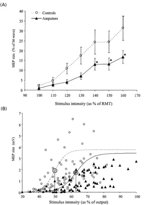
(7) 1408. K. J. Werhahn et al.. Fig. 4 MEP amplitudes from left FDI (A), right biceps brachii (Bic) (B) and right FDI (C) at baseline (dotted bars) and INBlate (®lled bars) in the placebo and lorazepam (LZP) sessions. MEP amplitude changes at INBlate relative to baseline are shown in the insets. Note that LZP did not modify the reduction in right FDI MEP amplitudes secondary to motor block (C). In contrast, LZP substantially attenuated the increase in left FDI (A) and right Bic (B) MEP amplitudes. Data represent group means = SEM. *P < 0.05 in post hoc testing.. INB, rating of tourniquet-related discomfort as well as TMS stimulus intensities and RMTs were comparable in both placebo and LZP sessions.. Effects of chronic deafferentation on ipsilateral corticomotoneuronal output. In amputee patients, there was a signi®cant increase in motor thresholds with stimulation over the non-deafferented hemisphere both in relaxed (48.5 6 1.8 and 39.8 6 1.2% for amputees and controls, respectively; two-tailed t test P < 0.01) and active (38.5 6 1.9 and 32.8 6 1.2%; P < 0.05) muscles compared with age-matched controls. In parallel, S±R curves were signi®cantly depressed in amputees [interaction group 3 intensity F(2, 8) = 2.5, P < 0.05; post hoc P < 0.05 for intensities of 140±160% RMT] compared with controls (Fig. 5A). Analysis using non-linear curve ®tting revealed a signi®cant increase in the intensity needed to obtain mid-size MEP responses in amputees (62.9 6 2.0 and 50.0 6 1.5%; P < 0.001), leading to a shift of the R±C curve to the right (Fig. 5B). In contrast, the slope and plateau intensities of the curves were not signi®cantly different between the groups (P > 0.2).. Discussion. The main ®ndings of this study are that INB, in addition to eliciting functional excitability changes in the deafferented cortex, resulted in a focal increase of excitability in the hand motor representation homotopic to the deafferented one, and that this form of plasticity is in¯uenced by interhemispheric. interactions and GABAergic function and is balanced in the setting of chronic deafferentation.. Effects of hand deafferentation on the excitability of the contralateral motor cortex. Hand deafferentation resulted in larger MEP amplitudes in the Bic muscle proximal to the tourniquet, a result consistent with previous experiments demonstrating increased Bic S±R curves during forearm INB (Ridding and Rothwell, 1997). This effect is short lasting (Brasil-Neto et al., 1992; Ziemann et al., 1998a) and indicative of an increased excitability of cortical body part representations immediately proximal to the deafferented one (Brasil-Neto et al., 1992, 1993). Our ®nding that pre-medication with LZP substantially attenuated these excitability changes raises the hypothesis of GABAergic inhibition being an operating mechanism. This proposal is consistent with the short duration of the effect (Ziemann et al., 1998a) and is supported further by the ®nding of rapid decreases in the concentration of GABA and GABA/creatine ratios in the human sensorimotor cortex contralateral to an acutely deafferented hand (Levy et al., 1999).. Effects of hand deafferentation on the excitability of the ipsilateral hand motor representation. Hand deafferentation resulted in increased excitability of the ipsilateral hand motor representation. The increase in MEP size may be due to a net increase in TMS-evoked corticospinal motor output, an increased synchronization of.
(8) Deafferentation-induced excitability changes. 1409. Fig. 5 Stimulus±response curves in FDI in the intact arm of upper limb amputees (n = 8) and healthy controls (n = 8). (A) Mean (6SEM) MEP amplitudes expressed as a percentage of the maximum peripheral M-wave plotted against stimulus intensities expressed as a percentage of RMT illustrating the depression of S±R curves in amputees relative to healthy controls. *P < 0.05 in post hoc testing. (B) Comparison of the averaged size of the MEP amplitudes (mV) in amputees and controls. Stimulus intensities are expressed as a percentage of the maximum output of the device. Each point represents the average of 20 MEP responses in amputees (triangles) or controls (open circles) at different intensities. Note the shift of the MEP amplitudes in amputees towards higher intensities. The lines represent the mean non-linear curve ®tting for amputees (solid line) or controls (dotted line), the large shaded circle indicating the mid-size intensity in each group.. the TMS-evoked corticomotor volleys (resulting in a reduced phase cancellation) or both. This effect started rapidly, terminated shortly after the afferent input from the hand was restored, and was topographically speci®c since the excitability of upper arm representations in the same limb (Bic) remained unaffected, as did extremity or thoracic representations. Previous studies in our own laboratory showed a nonsigni®cant trend toward increased excitability of the hand. motor representation ipsilateral to INB (see ®g. 2 in BrasilNeto et al., 1992). This trend was more evident in the present study because we evaluated a greater number of subjects and collected more trials per subject. Furthermore, we used an automated system for conditional triggering of TMS pulses that secured delivery of these pulses in the absence of EMG activation (Kaelin-Lang and Cohen, 2000), consequently decreasing data variability..
(9) 1410. K. J. Werhahn et al.. It is unlikely that unspeci®c alertness or discomfort-related factors explain these results since they were focal (not present in thorax and lower extremity muscles) and they did not in¯uence brainstem responses, usually affected by attentional ¯uctuations (Gandevia et al., 1997). Moreover, INB applied to the right lower leg failed to modulate MEP amplitudes in FDI and Bic consistent with a focal effect. Additionally, tonic pain elicits an effect opposite to that described here: attenuation of MEP amplitudes in muscles close to the painful site and lack of change of MEP amplitudes in muscles distant from the painful site in the opposite limb (Farina et al., 2001; Le Pera et al., 2001). INB resulted in marked excitability changes demonstrated with TMS but not with transmastoidal electrical stimulation at the level of the brainstem. Motor responses evoked by BES originate in descending corticospinal volleys elicited at the level of the pyramidal decussation (Ugawa et al., 1991), while those evoked by TMS are the consequence of predominantly transsynaptic stimulation of pyramidal tract neurones in the motor cortex (Amassian et al., 1990; Rothwell et al., 1991; Di Lazzaro et al., 1998). Therefore, responses to TMS that are in¯uenced by activity in the motor cortex (Rothwell, 1997) changed markedly in our study, while those evoked by BES did not, suggesting that the main site of this effect is cortical. Minor additional involvement of spinal mechanisms cannot be ruled out given the methodological differences between the two forms of stimulation (Touge et al., 2001). The precise site within the cortex where this interaction occurs remains to be determined. There are sparse direct interhemispheric connections linking primary motor (Jenny, 1979; Jones and Powell, 1969b) and sensory (Jones and Powell, 1969b; Jones et al., 1979; Killackey et al., 1983) cortices and it has been proposed that they could exert inhibitory in¯uences on homotopic sites in the contralateral hemisphere (Asanuma and Okuda, 1962; Ferbert et al., 1992; Di Lazzaro et al., 1999; Hanajima et al., 2001). Such connections could be the substrate for the transfer of INBinduced plasticity changes between homologous body part representations. For example, the reduction of interhemispheric inhibitory drive reported in this study could lead to disinhibition of contralateral motor areas. Several lines of evidence are consistent with this interpretation. Acute deafferentation of one hand region leads to an increased cerebral blood ¯ow in the contralateral as well as the ipsilateral motor representation (Sadato et al., 1995). Induction of a virtual lesion by cooling of a cortical representation elicits receptive ®eld changes in homotopic areas of the contralateral hemisphere (Calford and Tweedale, 1990; Clarey et al., 1996). Acute neocortical lesions result in increases in excitability in homotopic areas of the contralateral hemisphere (Buchkremer-Ratzmann et al., 1996; Neumann-Haefelin and Witte, 2000). Finally, acute deafferentation in primates and ¯ying foxes leads to rapid changes of receptive ®elds in the ipsilateral cortex (Calford and Tweedale, 1990) that are, similar to our results, balanced. after long-term deafferentation. The ®nding of decreased excitability in the motor cortex contralateral to the remaining hand of amputees may re¯ect the re-establishment of a loss of interhemispheric balance of excitation following the amputation, consistent with competition models of cortical processing (Kinsbourne, 1977; Rauschecker, 1997). Alternative interhemispheric anatomical pathways mediating this effect include those linking the supplementary motor areas, which have denser commissural projections between the hand representations than the primary motor cortex (Jones and Powell, 1969a; Gould et al., 1986), or those linking somatosensory areas 1 and 2 (Jones et al., 1979; Disbrow et al., 2001). In the latter case, the transferred information could be transmitted to area 4 through point-topoint corticocortical connections (Jones and Powell, 1969a; Jones et al., 1978; Pons and Kaas, 1986). Finally, it is less likely that ipsilateral somatosensory pathways (Noachtar et al., 1997) play a role, since they are diffuse and could not explain the focality of our results. The transient increase in excitability of the hand motor representation ipsilateral to an acutely deafferented hand was blocked by pre-medication with LZP, an allosteric modulator of the GABAA receptor. GABAergic manipulations effectively modulate rapid plasticity in the animal (Jacobs and Donoghue, 1991; Castro-Alamancos et al., 1995) and human (Ziemann et al., 1998b; BuÈte®sch et al., 2000) motor cortex. Results from animal studies support this association. Increases in cortical excitability induced by transient ischaemia in contralateral homotopic areas are paralleled by decrease in GABAergic inhibition (Neumann-Haefelin et al., 1995) and GABAA receptor density (Witte and Stoll, 1997). In summary, our ®ndings demonstrate that excitability changes in a human motor representation are associated with functional modi®cations in homotopic representations of the contralateral hemisphere that are in¯uenced by GABAergic function. This transfer of excitability across hemispheric boundaries may play a role in compensatory processes that follow injury to the motor pathways.. Acknowledgements. We wish to thank Aaron H. Burstein, Clinical Pharmacokinetics Research Laboratory, Clinical Centre Pharmacy Department, NIH, for the simulation of drug pharmacokinetics, and M. Hallett and S. P. Wise for their comments. B.B. was supported by a grant from the Deutsche Forschungsgemeinschaft (DFG). References Amassian VE, Quirk GJ, Stewart M. A comparison of corticospinal activation by magnetic coil and electrical stimulation of monkey motor cortex. Electroencephalogr Clin Neurophysiol 1990; 77: 390±401. Asanuma H, Okuda O. Effects of transcallosal volleys on pyramidal tract cell activity of the cat. J Neurophysiol 1962; 25: 198±208..
(10) Deafferentation-induced excitability changes Brasil-Neto JP, Cohen LG, Pascual-Leone A, Jabir FK, Wall RT, Hallett M. Rapid reversible modulation of human motor outputs after transient deafferentation of the forearm: a study with transcranial magnetic stimulation. Neurology 1992; 42: 1302±6. Brasil-Neto JP, Valls-Sole J, Pascual-Leone A, Cammarota A, Amassian VE, Cracco R, et al. Rapid modulation of human cortical motor outputs following ischaemic nerve block. Brain 1993; 116: 511±25. Brasil-Neto JP, Cohen LG, Hallett M. Central fatigue as revealed by postexercise decrement of motor evoked potentials. Muscle Nerve 1994; 17: 713±9. Buchkremer-Ratzmann I, August M, Hagemann G, Witte OW. Electrophysiological transcortical diaschisis after cortical photothrombosis in rat brain. Stroke 1996; 27: 1105±11. BuÈte®sch CM, Davis BC, Wise SP, Sawaki L, Kopylev L, Classen J, et al. Mechanisms of use-dependent plasticity in the human motor cortex. Proc Natl Acad Sci USA 2000; 97: 3661±5. Calford MB, Tweedale R. Immediate and chronic changes in responses of somatosensory cortex in adult ¯ying-fox after digit amputation. Nature 1988; 332: 446±8. Calford MB, Tweedale R. Interhemispheric transfer of plasticity in the cerebral cortex. Science 1990; 249: 805±7. Castro-Alamancos MA, Donoghue JP, Connors BW. Different forms of synaptic plasticity in somatosensory and motor areas of the neocortex. J Neurosci 1995; 15: 5324±33. Chen R, Corwell B, Yaseen Z, Hallett M, Cohen LG. Mechanisms of cortical reorganization in lower-limb amputees. J Neurosci 1998; 18: 3443±50. Clarey JC, Tweedale R, Calford MB. Interhemispheric modulation of somatosensory receptive ®elds: evidence for plasticity in primary somatosensory cortex. Cereb Cortex 1996; 6: 196±206. Cohen LG, Bandinelli S, Findley TW, Hallett M. Motor reorganization after upper limb amputation in man. A study with focal magnetic stimulation. Brain 1991; 114: 615±27. Devanne H, Lavoie BA, Capaday C. Input±output properties and gain changes in the human corticospinal pathway. Exp Brain Res 1997; 114: 329±38. Di Lazzaro V, Oliviero A, Pro®ce P, Saturno E, Pilato F, Insola A, et al. Comparison of descending volleys evoked by transcranial magnetic and electric stimulation in conscious humans. Electroencephalogr Clin Neurophysiol 1998; 109: 397±401. Di Lazzaro V, Oliviero A, Pro®ce P, Insola A, Mazzone P, Tonali P, et al. Direct demonstration of interhemispheric inhibition of the human motor cortex produced by transcranial magnetic stimulation. Exp Brain Res 1999; 124: 520±4. Disbrow E, Roberts T, Poeppel D, Krubitzer L. Evidence for interhemispheric processing of inputs from the hands in human S2 and PV. J Neurophysiol 2001; 85: 2236±44. Farina S, Valeriani M, Rosso T, Aglioti S, Tamburin S, Fiaschi A, et al. Transient inhibition of the human motor cortex by capsaicininduced pain. A study with transcranial magnetic stimulation. Neurosci Lett 2001; 314: 97±101. Ferbert A, Priori A, Rothwell JC, Day BL, Colebatch JG, Marsden. 1411. CD. Interhemispheric inhibition of the human motor cortex. J Physiol (Lond) 1992; 453: 525±46. Gandevia SC, Wilson LR, Inglis JT, Burke D. Mental rehearsal of motor tasks recruits alpha-motoneurones but fails to recruit human fusimotor neurones selectively. J Physiol (Lond) 1997; 505: 259± 66. Gilbert CD, Wiesel TN. Receptive ®eld dynamics in adult primary visual cortex. Nature 1992; 356: 150±2. Gould HJ 3rd, Cusick CG, Pons TP, Kaas JH. The relationship of corpus callosum connections to electrical stimulation maps of motor, supplementary motor, and frontal eye ®elds in owl monkeys. J Comp Neurol 1986; 247: 297±325. Gracely RH. Pain measurement. [Review]. Acta Anaesthesiol Scand 1999; 43: 897±908. Gracely RH, McGrath P, Dubner R. Validity and sensitivity of ratio scales of sensory and affective verbal pain descriptors: manipulation of affect by diazepam. Pain 1978; 5: 19±29. Greenblatt DJ, Ehrenberg BL, Gunderman J, Scavone JM, Tai NT, Harmatz JS, et al. Kinetic and dynamic study of intravenous lorazepam: comparison with intravenous diazepam. J Pharmacol Exp Ther 1989; 250: 134±40. Greenblatt DJ, Scavone JM, Harmatz JS, Engelhardt N, Shader RI. Cognitive effects of beta-adrenergic antagonists after single doses: pharmacokinetics and pharmacodynamics of propranolol, atenolol, lorazepam, and placebo. Clin Pharmacol Ther 1993; 53: 577±84. Gupta SK, Ellinwood EH, Nikaido AM, Heatherly DG. Simultaneous modeling of the pharmacokinetic and pharmacodynamic properties of benzodiazepines. I: lorazepam. J Pharmacokinet Biopharm 1990; 18: 89±102. Hanajima R, Ugawa Y, Machii K, Mochizuki H, Terao Y, Enomoto H, et al. Interhemispheric facilitation of the hand motor area in humans. J Physiol (Lond) 2001; 531: 849±59. Jacobs KM, Donoghue JP. Reshaping the cortical motor map by unmasking latent intracortical connections. Science 1991; 251: 944±7. Jenny AB. Commissural projections of the cortical hand motor area in monkeys. J Comp Neurol 1979; 188: 137±45. Jones EG. Cortical and subcortical contributions to activitydependent plasticity in primate somatosensory cortex. [Review]. Annu Rev Neurosci 2000; 23: 1±37. Jones EG, Powell TP. Connexions of the somatic sensory cortex of the rhesus monkey. I. Ipsilateral cortical connexions. Brain 1969a; 92: 477±502. Jones EG, Powell TP. Connexions of the somatic sensory cortex of the rhesus monkey. II. Contralateral cortical connexions. Brain 1969b; 92: 717±30. Jones EG, Coulter JD, Hendry SH. Intracortical connectivity of architectonic ®elds in the somatic sensory, motor and parietal cortex of monkeys. J Comp Neurol 1978; 181: 291±347. Jones EG, Coulter JD, Wise SP. Commissural columns in the sensory-motor cortex of monkeys. J Comp Neurol 1979; 188: 113± 35..
(11) 1412. K. J. Werhahn et al.. Kaas JH, Merzenich MM, Killackey HP. The reorganization of somatosensory cortex following peripheral nerve damage in adult and developing mammals. [Review]. Annu Rev Neurosci 1983; 6: 325±56. Kaas JH, Krubitzer LA, Chino YM, Langston AL, Polley EH, Blair N. Reorganization of retinotopic cortical maps in adult mammals after lesions of the retina. Science 1990; 248: 229±31. Kaelin-Lang A, Cohen LG. Enhancing the quality of studies using transcranial magnetic and electrical stimulation with a new computer-controlled system. J Neurosci Methods 2000; 102: 81±9. Kelahan AM, Doetsch GS. Time-dependent changes in the functional organization of somatosensory cerebral cortex following digit amputation in adult raccoons. Somatosens Res 1984; 2: 49±81. Killackey HP, Gould HJ 3rd, Cusick CG, Pons TP, Kaas JH. The relation of corpus callosum connections to architectonic ®elds and body surface maps in sensorimotor cortex of New and Old World monkeys. J Comp Neurol 1983; 219: 384±419. Kinsbourne M. Hemi-neglect and hemisphere rivalry. Adv Neurol 1977; 18: 41±9. Kolarik RC, Rasey SK, Wall JT. The consistency, extent, and locations of early-onset changes in cortical nerve dominance aggregates following injury of nerves to primate hands. J Neurosci 1994; 14: 4269±88. Le Pera D, Graven-Nielsen T, Valeriani M, Oliviero A, Di Lazzaro V, Tonali PA, et al. Inhibition of motor system excitability at cortical and spinal level by tonic muscle pain. Clin Neurophysiol 2001; 112: 1633±41. Levy LM, Ziemann U, Chen R, Cohen L. Rapid modulation of GABA in human cortical plasticity demonstrated by magnetic resonance spectroscopy [abstract]. Neurology 1999; 52 (6 Suppl 2): A88. Macdonald RL. Benzodiazepines. In: Levy RH, Mattson RH, Meldrum BS, editors. Antiepileptic drugs. 4th edn. New York: Raven Press; 1995. p. 695±703. McNulty PA, Mace®eld VG, Taylor JL, Hallett M. Cortically evoked neural volleys to the human hand are increased during ischemic block of the forearm. J Physiol 2002; 538: 279±88. Merzenich MM, Kaas JH. Reorganization of mammalian somatosensory cortex following peripheral nerve injury. Trends Neurosci 1982; 5: 434±6. Neumann-Haefelin T, Witte OW. Periinfarct and remote excitability changes after transient middle cerebral artery occlusion. J Cereb Blood Flow Metab 2000; 20: 45±52. Neumann-Haefelin T, Hagemann G, Witte OW. Cellular correlates of neuronal hyperexcitability in the vicinity of photochemically induced cortical infarcts in rats in vitro. Neurosci Lett 1995; 193: 101±4. Noachtar S, Luders HO, Dinner DS, Klem G. Ipsilateral median somatosensory evoked potentials recorded from human somatosensory cortex. Electroencephalogr Clin Neurophysiol 1997; 104: 189±98.. Old®eld RC. The assessment and analysis of handedness: the Edinburgh inventory. Neuropsychologia 1971; 9: 97±113. Pons TP, Kaas JH. Corticocortical connections of area 2 of somatosensory cortex in macaque monkeys: a correlative anatomical and electrophysiological study. J Comp Neurol 1986; 248: 313±35. Pons TP, Garraghty PE, Ommaya AK, Kaas JH, Taub E, Mishkin M. Massive cortical reorganization after sensory deafferentation in adult macaques. Science 1991; 252: 1857±60. Qi HX, Stepniewska I, Kaas JH. Reorganization of primary motor cortex in adult macaque monkeys with long-standing amputations. J Neurophysiol 2000; 84: 2133±47. Rajan R. Receptor organ damage causes loss of cortical surround inhibition without topographic map plasticity. Nat Neurosci 1998; 1: 138±43. Rasmusson DD. Reorganization of raccoon somatosensory cortex following removal of the ®fth digit. J Comp Neurol 1982; 205: 313± 26. Rauschecker JP. Mechanisms of compensatory plasticity in the cerebral cortex. [Review]. Adv Neurol 1997; 73: 137±46. Ridding MC, Rothwell JC. Reorganisation in human motor cortex. Can J Physiol Pharmacol 1995; 73: 218±22. Ridding MC, Rothwell JC. Stimulus/response curves as a method of measuring motor cortical excitability in man. Electroencephalogr Clin Neurophysiol 1997; 105: 340±4. Rothwell JC. Techniques and mechanisms of action of transcranial stimulation of the human motor cortex. [Review]. J Neurosci Methods 1997; 74: 113±22. Rothwell JC, Thompson PD, Day BL, Boyd S, Marsden CD. Stimulation of the human motor cortex through the scalp. [Review]. Exp Physiol 1991; 76: 159±200. Rothwell J, Burke D, Hicks R, Stephen J, Woodforth I, Crawford M. Transcranial electrical stimulation of the motor cortex in man: further evidence for the site of activation. J Physiol 1994; 481: 243± 50. Sadato N, Zef®ro TA, Campbell G, Konishi J, Shibasaki H, Hallett M. Regional cerebral blood ¯ow changes in motor cortical areas after transient anesthesia of the forearm. Ann Neurol 1995; 37: 74±81. Silva AC, Rasey SK, Wu X, Wall JT. Initial cortical reactions to injury of the median and radial nerves to the hands of adult primates. J Comp Neurol 1996; 366: 700±16. Stefan K, Kunesch E, Cohen LG, Benecke R, Classen J. Induction of plasticity in the human motor cortex by paired associative stimulation. Brain 2000; 123: 572±84. Touge T, Gerschlager W, Brown P, Rothwell JC. Are the aftereffects of low-frequency rTMS on motor cortex excitability due to changes in the ef®cacy of cortical synapses? Clin Neurophysiol 2001; 112: 2138±45. Ugawa Y, Rothwell JC, Day BL, Thompson PD, Marsden CD. Percutaneous electrical stimulation of corticospinal pathways at the level of the pyramidal decussation in humans. Ann Neurol 1991; 29: 418±27. Werhahn KJ, Fong JK, Meyer BU, Priori A, Rothwell JC, Day BL,.
(12) Deafferentation-induced excitability changes et al. The effect of magnetic coil orientation on the latency of surface EMG and single motor unit responses in the ®rst dorsal interosseous muscle. Electroencephalogr Clin Neurophysiol 1994; 93: 138±46. Witte OW, Stoll G. Delayed and remote effects of focal cortical infarctions: secondary damage and reactive plasticity. [Review]. Adv Neurol 1997; 73: 207±27. World Medical Association declaration of Helsinki. Recommendations guiding physicians in biomedical research involving human subjects. J Am Med Assoc 1997; 277: 925±6.. 1413. Ziemann U, Corwell B, Cohen LG. Modulation of plasticity in human motor cortex after forearm ischemic nerve block. J Neurosci 1998a; 18: 1115±23. Ziemann U, Hallett M, Cohen LG. Mechanisms of deafferentationinduced plasticity in human motor cortex. J Neurosci 1998b; 18: 7000±7.. Received January 2, 2002. Revised January 8, 2002. Accepted January 11, 2002.
(13)
Figure


Documents relatifs