An engineered IN-1 F
abfragment with improved affinity for the
Nogo-A axonal growth inhibitor permits immunochemical
detection and shows enhanced neutralizing activity
M.Fiedler1, C.Horn1,2, C.Bandtlow3, M.E.Schwab4and
A.Skerra1,5
1Lehrstuhl fu¨r Biologische Chemie, Technische Universita¨t Mu¨nchen, Freising-Weihenstephan, Germany,3Institut fu¨r medizinische Chemie und Biochemie, Universita¨t Innsbruck, Austria and4Institut fu¨r Hirnforschung, Universita¨t und Dept. Biologie, ETH, Zu¨rich, Switzerland
2Present address: Biozentrum, Universita¨t Frankfurt am Main, Germany 5To whom correspondence should be addressed.
E-mail: [email protected]
The myelin axonal growth inhibitor NI-220/250 (Nogo-A) has attracted considerable attention in elucidating the mechanisms that account for the lack of plasticity in the adult central nervous system. The cognate monoclonal antibody IN-1, which was obtained prior to the molecular characterization of its Nogo-A antigen, has played a crucial role in this respect. However, this murine IgM/κ antibody does not only provide an inappropriate format for in vivo studies, its low antigen affinity has also hampered the thorough structure–function analysis of its neutralizing effect toward the Nogo-A inhibitor on a molecular basis. We describe here the affinity maturation of a bacterially produced functional IN-1 Fabfragment via protein
engin-eering. A soluble fragment of Nogo-A derived from the central exon 3 of its gene, which was prepared by secretion into the periplasm of Escherichia coli, served as a target in these experiments. After repeated cycles of site-directed random mutagenesis and screening, the mutant II.1.8 of the IN-1 Fabfragment was obtained, carrying five side chain
substitutions within CDR-L3. Its dissociation constant for the complex with the recombinant Nogo-A fragment was determined in surface plasmon resonance measurements as approximately 1 µM. The affinity of the unmutated IN-1 Fab fragment was 8-fold lower. The engineered Fab
fragment appeared to be well suited for the specific detec-tion of Nogo-A in immunochemical assays and for the histochemical staining of myelin-rich tissue sections. Most importantly, its concentration-dependent neutralizing effect on the Nogo-A inhibitory activity was significantly enhanced in cell culture. This study confirms Nogo-A to be the antigen of the IN-1 antibody and it demonstrates increased potential of the engineered Fab fragment as a reagent for
promoting axonal regeneration in vivo.
Keywords: affinity maturation/axonal regeneration/
bacterial expression/neuronal inhibitor/random mutagenesis
Introduction
The very limited capacity of the adult central nervous system (CNS) for axonal regeneration is a phenomenon of broad and ongoing scientific as well as medical interest (Ramon y Cajal, 1928; Horner and Gage, 2000). In contrast, sprouting and elongation of lesioned axons readily occurs in the peripheral nervous system (PNS). Inhibitory effects and non-permissible
properties of CNS tissue, in particular of CNS myelin and oligodendrocytes, probably contribute considerably to the restriction of neuronal regeneration and plasticity. In vitro, CNS myelin and oligodendrocyte membranes induce growth cone collapse (Bandtlow et al., 1990).
Based on earlier observations of the inhibitory effect of CNS myelin on neurite outgrowth (Caroni and Schwab, 1988a; Spillmann et al., 1997, 1998) the myelin-associated neurite growth inhibitor NI-220 (Spillmann et al., 1998), later called Nogo-A (Huber and Schwab, 2000), was identified in bovine spinal cord tissue as a predominant protein of oligodendrocytes that prevents axonal growth. The corresponding cDNAs from rat and man were recently described (Chen et al., 2000; GrandPre´ et al., 2000; Prinjha et al., 2000). The nogo gene encodes three distinct proteins, Nogo-A, Nogo-B and Nogo-C, which apparently arise by alternative splicing and/or promoter usage. Of those, just the full-length Nogo-A transcript is specifically expressed in oligodendrocytes and hence is made mainly responsible for their neuronal growth inhibitory activity (Spillmann et al., 1998; Chen et al., 2000).
The murine monoclonal IgM/κ antibody IN-1 was originally raised against the homologous protein NI-250 from rat CNS myelin (Caroni and Schwab, 1988b) and cross-reacts both with the bovine and human protein (Spillmann et al., 1997, 1998). IN-1 was shown to effectively neutralize the inhibitory activity
in vitro (Bandtlow et al., 1990; Spillmann et al., 1998) and in vivo, giving rise to long-distance regeneration and improved
plastic changes of injured CNS fibre tracts (Schnell and Schwab, 1990; Z’Graggen et al., 1998). The variable domain cDNAs of this antibody were cloned from the hybridoma cell line, followed by the bacterial production of the corresponding
recombinant murine Fab fragment, whose functionality was
demonstrated in vitro (Bandtlow et al., 1996). A partially
humanized IN-1 Fab fragment was produced by Escherichia
coli fermentation and shown to successfully promote
regenera-tion of corticospinal axons in adult rats after spinal cord lesion
in vivo (Broesamle et al., 2000). The recombinant IN-1 Fab fragment also induced significant elongation of injured cochlear fibres upon intrathecal treatment (Tatagiba et al., 2002) and a pronounced sprouting response of Purkinje cells after injection into the intact adult cerebellum (Buffo et al., 2000).
Unfortunately, the IN-1 antibody as well as its recombinant Fab fragment exhibit rather low antigen affinity, suggesting that the IgM antibody was a product of the early immune response (Bandtlow et al., 1996). Its light chain was found to be encoded by a Vκgermline gene devoid of somatic mutations. In the case of the IgM, the poor affinity is partially compensated by an avidity effect due to its decavalent nature, which is attributable to a more general mechanism in immunology, even though at the expense of relaxed specificity in the early humoral response. Consequently, the apparently weaker binding activity of the monovalent Fabfragment necessitates higher concentra-tions in order to provoke comparable in vitro activity (Bandtlow
Nevertheless, in comparison with the intact IgM the use of the recombinant Fabfragment is attractive for several reasons.
First, the much smaller Fab fragment was shown to possess
high stability in buffer solution and to exert efficient tissue penetration (Bandtlow et al., 1996; Broesamle et al., 2000).
Secondly, the Fab fragment can be produced at high yields
and quickly purified to homogeneity using a well optimized bacterial expression system (Fiedler and Skerra, 1999, 2001a,b). Finally, its recombinant format permits the applica-tion of protein engineering techniques not only to improve its antigen-binding activity but also to humanize the mouse-specific immunoglobulin sequences for medical use.
Here we report the improvement of the functional affinity of the recombinant IN-1 Fabfragment by virtue of introducing a small set of amino acid substitutions into its antigen-binding site. In order to perform cycles of random mutagenesis and selection for enhanced binding activity the presumed extracel-lular domain of the Nogo-A antigen (Chen et al., 2000; Huber and Schwab, 2000) was produced in a bacterial expression system, thus also permitting molecular characterization of the cognate antigen–antibody interaction for the first time.
Materials and methods
General DNA methodology
DNA manipulations were performed according to standard techniques (Sambrook et al., 1989). Vectors, genes and bacterial strains were from the authors’ collection. DNA polymerases, modifying and restriction enzymes were from generic vendors (New England Biolabs, Frankfurt/Main, Germany; Promega, Mannheim, Germany; Stratagene, Amsterdam, Netherlands). Oligodeoxynucleotides for PCR and DNA sequencing were purchased from Interactiva (Ulm, Germany). PCR reactions were performed using a standard protocol, employing phosphorothioate primers in conjunction with Pfu DNA poly-merase (Skerra, 1992). Site-directed mutagenesis was carried out with single-stranded plasmid DNA following the method of Kunkel et al. (Kunkel et al., 1987) according to a standardized procedure (Geisselsoder et al., 1987). All plasmid construc-tions and mutagenesis experiments were confirmed by restric-tion analysis, followed by DNA sequencing of relevant regions on an ABI-Prism™ 310 Genetic Analyzer (Perkin-Elmer/Applied Biosystems, Weiterstadt, Germany) using the BigDye™ terminator kit.
Vector construction for Nogo fragments
A 2.3 kb Nogo-A gene fragment was amplified from the cloned cDNA (Chen et al., 2000) via PCR with the primers 5⬘-GCT CAG CGG CCG AGA CCC TTT TTG CTC TTC CTp(S)G-3⬘ (the EagI restriction site is underlined) and 5⬘-GCT TTT AAC TAT 5⬘-GCT GCC CAT TTC TGp(S)T-3⬘. The single PCR product was digested with EagI, purified from a 1% agarose gel, and inserted into the multiple cloning region of pASK111 (Vogt and Skerra, 2001), which had been cut with BsaI (resulting in a sticky end compatible with EagI) as well as Eco47III, yielding pASK111-NiFr1. This vector leads to the production of a mature protein with a molecular mass of 85.0 kDa, including the Strep-tag (Skerra and Schmidt, 2000) at the C-terminus, after processing of the OmpA signal peptide fused in-frame to the N-terminus. pASK111-NiFr2 was constructed from pASK111-NiFr1 by precisely deleting the N-terminal 59 codons from the cloned Nogo-A gene fragment via site-directed mutagenesis using the oligodeoxy-nucleotide 5⬘-GGT ATC CAT GTT CTT TAA AAG AGG
CCT GCG CTA CGG TAG C-3⬘. Furthermore, Cys residues
were replaced by Ser via site-directed mutagenesis with single-stranded DNA prepared from pASK111-NiFr2 using appro-priate oligodeoxynucleotide primers.
The C-terminal Strep-tag encoded on pASK111-NiFr2 was exchanged for a His6affinity tag by site-directed mutagenesis
with the oligodeoxynucleotide 5⬘-CAC TTC ACA GGT CAA
GCT TAT TAA TGG TGA TGG TGA TGG TGA GCG CTT TTA ACT ATG CTG CCC-3⬘. A KasI restriction site was concomitantly introduced at the 5⬘-end of the cloned Nogo-A structural gene using the oligodeoxynucleotide 5⬘-GGT ATC CAT GTT CTT TAA AAG AGG CGC CCT GCG CTA CGG TAG C-3⬘ (the KasI recognition site is underlined), resulting in the vector pASK111-NiFr3. The region encoding the Nogo-A fragment together with the His6-tag was finally subcloned via KasI and NsiI (cutting within the vector, downstream of
the Camr gene) on pASK-IBA4 (Skerra and Schmidt, 2000),
which provided the coding region for an N-terminal Strep-tag II directly downstream of the OmpA signal sequence. The resulting vector was dubbed pASK111-NiFr4.
Vector construction for Fab fragments
The IN-1 Fabfragment and its mutants were produced utilizing the vectors pASK88, pASK106 or pASK107. All of them encode a chimeric Fabfragment with variable domains derived from the mouse monoclonal antibody IN-1 (Bandtlow et al., 1996) and human constant domains belonging to the subclass IgG1/κ (Schiweck and Skerra, 1995). Secretion into the oxidiz-ing milieu of the bacterial periplasm is ensured by the presence of signal peptides at the N-termini of both chains (Skerra, 1994b) and transcription of the artificial dicistronic operon is under tight control of the chemically inducible tetp/° (Skerra, 1994a). pASK88 (Schiweck and Skerra, 1995) was used for soluble expression and purification via the His6-tag attached to the C-terminus of the heavy chain (Skerra, 1994b; Fiedler and Skerra, 2001a), whereas pASK107 (M.Fiedler and A.Skerra, unpublished results) provided the Strep-tag II for streptavidin affinity purification (Skerra and Schmidt, 2000) instead. pASK106 codes for a Fabfragment similar to pASK88 but with an albumin-binding domain (ABD) appended to the C-terminus of the light chain (Ko¨nig and Skerra, 1998). The variable domain genes were exchanged between the differing vector formats using conserved restriction sites as described (Skerra, 1994b).
Single amino acid exchanges within the IN-1 Fabfragment or its mutants were introduced by site-directed mutagenesis. For this purpose single-stranded DNA of the corresponding vectors pASK88-IN1 or pASK88-I.2.6 was used in conjunction with appropriate oligodeoxynucleotide primers. Random amino acid substitutions were introduced into the variable domain (VL) gene of the IN-1 light chain at defined positions via PCR by means of degenerate oligodeoxynucleotide primers (without the phosphorothioate modification) in conjunction with Taq DNA polymerase. Amplification was performed on pASK85-IN1 with the originally cloned genes (Bandtlow et al., 1996) as template. The forward primer 5⬘-GAC ATT GAG CTC
ACC CAG TCT CCA GCA ATC ATG KCT GC-3⬘ (SstI
restriction site underlined) was used in all experiments whereas
the oligodeoxynucleotide 5⬘-GCG CTT CAG CTC GAG CTT
GGT CCC AGC TCC GAA CGT MNN AGG MNN MNN TAA CACATT TTG ACA GTA-3⬘ (XhoI restriction site underlined) served as backward primer for randomizing the CDR-L3 positions L93, L94 and L96 at the first stage of
the affinity maturation process. The second mutagenesis cycle was performed with pASK88-I.2.6(L96V) as template and the
oligodeoxynucleotide 5⬘-GCG CTT CAG CTC GAG CTT
GGT CCC AGC TCC GAA CGT AAC CGG CAC CCG
MNN MNN ATT TTG ACA GTA ATA CGT TGC-3⬘ as
second primer for randomizing the positions L91 and L92 together with fixed mutations at L93, L94 and L96. In each case, a single PCR product was obtained, purified from a 1% agarose gel, and cut with SstI and XhoI. The resulting DNA fragment of approximately 300 bp was ligated with the likewise cut vector backbone of pASK106-IN1 (see above). Colonies obtained after transformation of CaCl2-competent E.coli K-12 JM83 cells (Yanisch-Perron et al., 1985) were directly subjected to the filter-sandwich colony-screening assay.
Bacterial protein production
Cultures of E.coli JM83 transformed with the appropriate expression vector were grown in 2 l of Luria–Bertani (LB) medium supplemented with the corresponding antibiotic (Amp for derivatives of vectors pASK88, pASK106 and pASK107, or Cam for pASK111) at 22°C and 200 r.p.m. Gene expression was induced at an optical density (OD) of 0.5 at 550 nm by addition of 200µg/l anhydrotetracycline (aTc; Acros Organics, Geel, Belgium) in the case of the Fabfragments or of 400µg/ l aTc in the case of Nogo-A fragments. After 3 h induction the bacteria were harvested by centrifugation and the periplasmic protein fraction was prepared as described (Skerra and Schmidt,
2000). For the production of Nogo-A fragments 200 µg/ml
lysozyme was added to the cell fractionation buffer (50 mM
NaPi, pH 7.5, 500 mM sucrose, 1 mM EDTA).
Nogo-A fragments NI-Fr1 and NI-Fr2 were purified from the periplasmic protein extract via the Strep-tag fused to their C-termini employing streptavidin affinity chromato-graphy (Skerra and Schmidt, 2000), whereby elution was effected under mild conditions in the presence of D-desthiobi-otin. After dialysis against a chromatography buffer (50 mM
NaPi, pH 7.5, 150 mM NaCl, 1 mM EDTA) and the
concentra-tion of the eluate (Vivaspin 15, MWCO 30 kDa; Greiner, Frickenhausen, Germany), further purification was achieved by gel filtration on a Superdex™ 200 prep grade column (Pharmacia, Uppsala, Sweden) using Dynamax™ SD-300 HPLC equipment (Rainin, Woburn, MA). NI-Fr4 was first
purified by means of the His6-tag via immobilized metal
affinity chromatography (IMAC) (Skerra, 1994b) using 50
mM NaPi, pH 7.5, 1 M NaCl as the chromatography buffer
and a linear elution gradient from 0 to 75 mM imidazole·HCl in the same buffer. The specifically eluted protein fraction was then subjected to streptavidin affinity chromatography as above. The recombinant IN-1 Fabfragments were purified either by
IMAC via the His6-tag fused to the C-terminus of their
heavy chain (Fiedler and Skerra, 2001a) or, when using pASK107 (see above), via streptavidin affinity chromatography (Schlapschy and Skerra, 2001). IMAC was also performed under FPLC conditions using a POROS MC/M column (0.46⫻10 cm; PerSeptive Biosystems, Wiesbaden, Germany)
charged with Zn2⫹ ions and Dynamax™ SD-300 HPLC
equipment (Rainin) operating at a flow rate of 2.0 ml/min. A 12.5 ml aliquot of periplasmic extract from a 2 l E.coli culture
dialysed against 50 mM NaPi, pH 7.5, 500 mM betaine was
applied to the column and, after washing with dialysis buffer, elution was effected by application of a linear gradient of
200 mM imidazole·HCl, pH 7.5, 50 mM NaPi, 500 mM betaine
against dialysis buffer. This method enabled a 5-fold quicker
purification compared with the conventional procedure (Fiedler and Skerra, 2001a), yielding recombinant Fabfragments with
an apparent purity of⬎95% as estimated from SDS–PAGE.
The yields of purified recombinant proteins from 2 l shaker-flask experiments were highly reproducible but varied
between 0.04 and 0.8 mg l–1 OD–1 for the different F
ab fragments and between 0.1 and 0.3 mg l–1OD–1for the Nogo-A fragments. Nogo-After purification the proteins were stored in
PBS (4 mM KH2PO4, 16 mM Na2HPO4, 115 mM NaCl)
containing 0.1 mM EDTA at 4°C for up to several weeks. Protein purity was checked by SDS–PAGE using 0.1% (w/ v) SDS, 10 or 15% (w/v) polyacrylamide gels (Fling and Gregerson, 1986) stained with Coomassie brilliant blue. The concentration of the purified recombinant proteins was deter-mined using calculated absorption coefficients at 280 nm (Gill and von Hippel, 1989) of 0.41 ml mg–1 cm–1for the Nogo-A fragments and of 1.8 ml mg–1cm–1for the IN-1 F
abfragments, respectively.
Filter-sandwich colony-screening assay
This assay was carried out based on published procedures (Skerra et al., 1991; Schlehuber et al., 2000). Transformed
E.coli JM83 cells harbouring the pASK106 vector encoding
Fab fragments fused with the ABD (see above) were plated
on a hydrophilic membrane (GVWP, 0.22 µm; Millipore,
Bedford, MA), placed on a Petri dish with LB/Amp agar, such that approximately 500 colonies were obtained, and incubated at 37°C for 8–9 h. In the meantime, a hydrophobic
membrane (Immobilon™-P, 0.45 µm; Millipore) was coated
with 10 mg/ml human serum albumin (HSA; Sigma, Deisenhofen, Germany) in PBS for 4 h and blocked with 3% (w/v) BSA (Roth, Karlsruhe, Germany), 0.5% (v/v) Tween-20 in PBS. The membrane was washed twice with PBS, soaked
in LB/Amp containing 200µg/ml aTc, and placed on an LB/
Amp agar plate supplemented with 200 µg/ml aTc. The first
membrane, carrying tiny colonies of the transformed cells, was then placed onto the second (hydrophobic) membrane. The filter sandwich was incubated for 16 h at 22°C. During
this period the mutated IN-1 Fab fragments were secreted—
and partially released from the colonies by leakage from the bacterial periplasm—and finally immobilized on the lower membrane via complex formation between HSA and ABD.
The first membrane with the still viable colonies was transferred to a fresh LB/Amp agar plate and stored at 4°C. The second membrane was washed three times in PBS containing 0.1% (v/v) Tween-20 (PBS/T) and the immobilized Fabfragments, each in a spot corresponding to the position of the original colony, were probed for antigen binding. To this end recombinant Nogo-A fragment was labelled at a molar ratio
of 5:1 with digoxigenin-3-O-methylcarbonyl-ε-aminocaproic
acid N-hydroxy-succinimide ester (Roche Diagnostics, Mannheim, Germany) and applied to the membrane for 1 h at
a concentration of 30 or 50 µg/ml in PBS/T. After washing
three times with PBS/T the membrane was incubated for 1 h with 0.75 U/ml anti-digoxigenin Fabfragment conjugated with alkaline phosphatase (Roche Diagnostics) in 10 ml of PBS/T. The membrane was finally washed twice with PBS/T and twice with PBS and the signals were developed using standard chromogenic substrates as described (Schlehuber et al., 2000). Colonies corresponding to signals with an intensity above average were identified, recovered from the first membrane, and propagated for further analysis of their recombinant gene products.
Enzyme-linked immunosorbent assay (ELISA)
ELISA was carried out in a 96-well microtitre plate (Becton Dickinson, Heidelberg, Germany) at ambient temperature with incubation steps of 1 h unless otherwise stated. Three washing steps with PBS/T were used after each incubation, and residual liquid was removed thoroughly. The wells were coated for 4 h with 50µl of a solution of NI-Fr2 at concentrations between 180
and 200 µg/ml in PBS buffer and then blocked with 200 µl
of 3% (w/v) BSA, 0.5% (v/v) Tween-20 in PBS. After washing, 50µl of the purified recombinant Fabfragment was applied as a dilution series in PBS/T. The wells were then incubated with
50 µl of anti-human Cκ antibody conjugated with alkaline
phosphatase (Sigma), diluted 1:1000 in PBS/T. Signals were finally developed in the presence of p-nitrophenyl phosphate (Voss and Skerra, 1997). Enzymatic activity was measured at 25°C as the change in absorbance at 405 nm/min with a SpectraMAX 250 instrument (Molecular Devices, Sunnyvale, CA). The data were corrected for background values deter-mined in wells that were merely coated with BSA and fitted by non-linear least squares regression as described (Voss and Skerra, 1997).
Real-time surface plasmon resonance (SPR) measurements
Molecular interaction analysis was performed using a Biacore-X® system equipped with the NTA-sensor chip® (Biacore AB, Uppsala, Sweden). PBS containing 0.005% (v/v) surfactant P20 was used as continuous flow buffer as well as for dilution of proteins. Analysis was performed at 25°C using a flow rate
of 35 µl/min. For each measurement the derivatized chip
surface was charged with 70 µl of 0.5 mM NiSO4, followed
by immobilization of NI-Fr4 via its His6-tag in one of the two flow channels by applying 70µl of a 50 µg/ml solution of the purified recombinant protein. Then, the Fabfragment (produced by means of the vector pASK107 and purified via the Strep-tag II) was injected at a defined concentration (between 0.25
and 6.8 µM) for 2 min, followed by buffer flow for 4 min.
The chip surface was regenerated using 70 µl of 0.35 M
EDTA, pH 8.0 in flow buffer prior to the next measurement.
Each time-dependent binding isotherm of the Fab fragment
was corrected for the background signal that was detected in the flow channel without NI-Fr4 using BIAevaluation software (Version 3.0). Resonance unit values for the bound Fab fragment at equilibrium for each applied concentration were then deduced and fitted (Voss and Skerra, 1997) by non-linear least squares regression using an equation of the type
y⫽ a * x / (b ⫹ x). Immunohistochemistry
Cryosections (12 µm) of rat brain (Rattus norvegicus) were
fixed for 10 min using ice-cold ethanol. The following incuba-tion steps were then each performed for 1 h at room temperature in a humid chamber using PBS. Unless otherwise stated slides were washed for 5 min with PBS. After blocking with
4% (w/v) BSA, the Fabfragment (produced using the pASK88
vector type and purified via the His6-tag) was applied at a concentration of 100µg/ml. After three washing steps, bound
Fab fragment was detected with an anti-human Cκ antibody
alkaline phosphatase conjugate (Sigma), diluted 1:100. The sections were then washed three times with TBS (25 mM Tris–HCl, pH 7.4, 145 mM NaCl, 3 mM KCl) and staining was performed using the Fast Red kit (Roche Diagnostics). The microscopic slides were photographed on an Axiophot microscope (Carl Zeiss, Jena, Germany) using 20-fold magni-fication.
Neurite outgrowth assay
Neurite growth-modulating properties of the different Fab
fragments were tested on 4-well plastic dishes (Greiner, Nu¨rtingen, Germany) coated with recombinant Nogo-A.
Briefly, test wells were coated for 20 min with 100 µg/ml
poly-L-lysine, washed with Hank’s balanced salt solution
(HBSS; Life Technologies, Basel, Switzerland) and coated for
2 h with 15 or 30 µg/ml of recombinant rat Nogo-A (entire
extracellular domain refolded from bacterial inclusion bodies; T.Oertle et al., manuscript in preparation). Recombinant Nogo-A was omitted in the wells serving for control. Nogo-After aspiration, the wells were washed with Dulbecco’s modified Eagle’s medium (Life Technologies) containing 10% (v/v) fetal calf serum (Life Technologies) and blocked in the same medium for 20 min at 37°C.
Cerebellar cell cultures were prepared from rat on post-natal day 7/8. Cells were dissociated by combined trituration and trypsinization and purified on Percoll gradients as described (Hatten, 1985). The cerebellar granule cells were plated in chemically defined neurobasal medium supplemented with B27 and 0.2 mM glutamine, 100 U/ml penicillin, and 0.1 mg/ ml streptomycin (Life Technologies). To assess the neutraliza-tion of inhibitory acitivity, substrate-coated wells were first
incubated with 100 µg/ml of the different recombinant Fab
fragments dialysed against NaCl/Pi (137 mM NaCl, 2.7 mM
KCl, 1.5 mM KH2PO4, 8 mM Na2HPO4, pH 7.4) for 20 min at 37°C. The wells were then washed briefly with HBSS and cells were applied in the presence of the Fabfragments.
Assays were stopped after 24 h in culture by adding 4% (w/v) formalin buffered with NaCl/Pi. For assaying the inhibitory substrate properties, the proportion of total cells bearing neurites longer than the diameter of the cell body (indicating that neurite outgrowth was successfully initiated) was determined. Under control conditions, i.e. in the absence of recombinant Nogo-A, 70% of the cerebellar granule neurons formed processes. Quantification of neurite lengths was performed on cultures monitored with a Zeiss Axiophot microscope. Phase contrast pictures were acquired with a 12-bit digital CCD camera (Visicam Visitron, Germany) and analysed using Metamorph software (Universal Imaging Corporation, West Chester, PA). For each well, the longest neurites of at least 100 isolated neurons were measured and averaged. Three wells were investigated for each experimental condition.
Results
Bacterial synthesis of a soluble Nogo-A domain
Although the precise transmembrane topology of the Nogo-A protein has yet to be experimentally determined, its primary structure—as deduced from the cDNA sequence (Chen et al., 2000)—reveals several characteristic features (Figure 1). The rat full-length protein comprises 1163 amino acids. Its N-terminal region does not reveal a secretory signal sequence as would be expected for a normal class I membrane protein (von Heijne and Gavel, 1988). Instead it is highly negatively charged. In particular, the amino acid stretch from positions 31 to 50 is almost entirely (with the exception of a single Pro) composed of Glu and Asp residues. The acidic sequence is followed by a region rich in Pro, Ala and—to a lesser extent— also Ser and Gly residues, which reaches from position 61 to 171, i.e. directly up to the end of exon 1, which is followed by a very small exon 2 (T.Oertle, et al., manuscript in
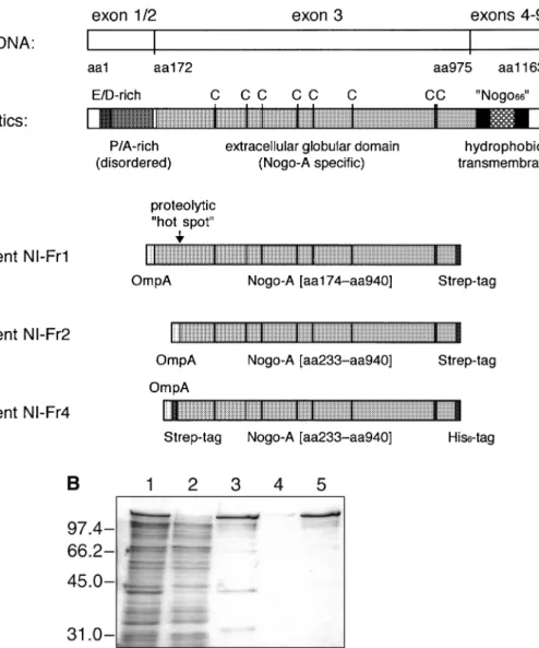
Fig. 1. Recombinant Nogo-A fragments prepared in this study. (A) Structural characteristics of the native neurite growth inhibitor Nogo-A and of the
recombinant fragments derived from it. (B) SDS–PAGE analysis of bacterially produced NI-Fr4. Periplasmic protein extract from E.coli JM83 harbouring pASK111-NIFr4 (lane 1); through of IMAC column (lane 2); eluted protein from IMAC column as applied to the streptavidin column (lane 3); flow-through of streptavidin column (lane 4); purified protein after streptavidin affinity chromatography (lane 5). Molecular sizes are indicated at the left.
preparation). Thus, the domain encoded by exon 1 is likely not to adopt a regular secondary structure. In contrast, the large Nogo-A-specific exon 3 reveals an average amino acid composition similar to that of globular proteins. Furthermore, it exhibits altogether eight Cys codons, whereas otherwise this amino acid is absent in the full-length protein. This feature might point towards the presence of disulphide bonds in the exon 3 region as they are frequently found in extracellular domains of membrane proteins. The C-terminal domain encoded by exons 4–9 (for the precise exon structure refer to T.Oertle, et al., manuscript in preparation) is short again, comprising altogether 188 residues. It includes two stretches of 36 (residues 990–1025) and 35 (residues 1092–1126) amino acids, respectively, with a high content of hydrophobic side chains, thus probably constituting transmembrane regions.
From the structural point of view the protein fragment encoded by exon 3 is most likely folded as a globular domain and should carry the biological active site. Consequently, we chose the polypeptide comprising residues 174–940 (containing 767 residues, i.e. 66% of full-length Nogo-A) for production as a recombinant protein. The gene fragment was amplified
from the cDNA and subcloned on a standard vector for protein production in E.coli. Using appropriate PCR primers, the Nogo-A fragment was precisely fused at its N-terminus (i.e. in front of residue 174) to the OmpA signal peptide, thus effecting secretion into the bacterial periplasm, where efficient disulphide bond formation is favoured by an oxidizing redox environment. At the C-terminus (i.e. following residue 940) the fragment was fused with the Strep-tag affinity peptide, conferring binding activity towards streptavidin for simplified purification (Skerra and Schmidt, 2000). Transcription of the resulting hybrid gene was under tight control of the tetracycline promoter/operator (Skerra, 1994a). The resulting protein was denominated NI-Fr1 (Figure 1A).
Upon induction of gene expression NI-Fr1 was readily liberated from the periplasmic protein fraction of E.coli and purified by streptavidin affinity chromatography in one step. SDS–PAGE analysis revealed that approximately 50% of the recombinant protein comprised a product with the proper length whereas 50% corresponded to a series of smaller polypeptides, probably representing proteolytic degradation products (data not shown). In particular, there appeared one
prominent band just underneath that for the major recombinant protein. Both bands were subjected to N-terminal sequencing. The upper band yielded the sequence Glu–Thr–Leu–Phe–Ala, which resulted from the precise cleavage of the OmpA signal peptide. The lower band started with the amino acids Ser– Phe–Lys–Glu–His, i.e. at a position 59 codons downstream within the cloned sequence (beginning at residue 233 in the full-length primary structure). Its appearance was most likely due to the action of a bacterial protease and might indicate that the N-terminal part of the chosen Nogo-A fragment still belongs to a polypeptide segment devoid of well defined structure.
In order to achieve better homogeneity of the gene product, the first 59 residues of the mature polypeptide chain were deleted from the cloned coding region, leading to NI-Fr2 (Figure 1A). This protein was readily produced in the periplasm of E.coli, with similar yields as the former version but clearly a reduced degradation pattern. The possible presence of structural disulphide bonds in the recombinant protein was investigated by individually substituting all eight Cys residues (corresponding to positions 323, 403, 443, 536, 574, 676, 885 and 890 in the full-length Nogo-A sequence) with Ser via site-directed mutagenesis. The eight mutant Nogo-A fragments were produced in E.coli as before. However, it was not possible to recover the mutants Cys323→Ser and Cys885→Ser from
the periplasmic protein fraction, while the mutants
Cys443→Ser and Cys890→Ser gave rise to significantly diminished yields after Strep-tag purification when compared with the wild-type protein. In contrast, the other four mutants were produced at similar amounts as the original Nogo-A fragment. These observations indicate that at least some of the Cys residues are important for folding and may be involved in cystine crosslinks.
The wild-type NI-Fr2 protein still gave rise to certain truncated products, which was considered undesirable for precise binding measurements (see below). Notably, for the bovine bNI-220 susceptibility towards proteolysis had been observed before (Spillmann et al., 1998). Consequently, a doubly tagged version of the recombinant protein was prepared using an otherwise identical expression system. First, the Strep-tag at the C-terminus was exchanged for a His6-tag (yielding NI-Fr3 as an intermediate construct; data not shown), and, secondly, the Strep-tag was inserted at the N-terminus again, downstream of the OmpA signal peptide. Interestingly, the yield of bacterially produced soluble protein, termed NI-Fr4 (Figure 1A), was found to be significantly higher (by a factor of 2.5, approaching 300 µg l–1 OD–1). NI-Fr4 was isolated from the periplasmic protein fraction in two steps by IMAC followed by streptavidin affinity chromatography. This protein was essentially pure, just a minor fraction of truncated poly-peptide chains was still detectable (Figure 1B).
In vitro affinity maturation of the IN-1 Fab fragment
Initial experiments on the detection of natural Nogo-A as well as recombinant Nogo-A fragments on western blots or on tissue sections by means of the bacterially produced IN-1 Fab fragment revealed relatively weak signals (Bandtlow et al., 1996), indicating that the antigen affinity was poor. From the cloning of the variable domain genes of the IN-1 monoclonal antibody and analysis of their sequences it was evident that
the VH domain had undergone somatic hypermutation while
the VLdomain appeared to be a newly defined mouse germline gene (Bandtlow et al., 1996). A computer modelling study
was carried out based on a human anti-thyroid peroxidase autoantibody (Chacko et al., 1996; PDB entry 1VGE) and a murine anti-phenylarsonate antibody (Rose et al., 1990; PDB entry 6FAB), both of which have sets of complementarity-determining regions (CDRs) with the same lengths and canonical structure determinants (Chothia et al., 1989) as IN-1 and share a high amino acid sequence similarity with it. This analysis suggested that the CDR-L3 of IN-1 and, to a lesser extent, its CDR-L1, were the most promising target regions for protein engineering towards improved antigen recognition. In particular, residue L96 and also residue L32 (in CDR-L1) were expected to be exposed close to the centre of the combining site and thus likely to be involved in contacts with the antigen.
Within CDR-L1 both IN-1 and 1VGE have an Ala residue at position L32 whereas 6FAB carries a Phe. On the other hand, IN-1 as well as 6FAB carry an Arg at position L96 (in CDR-L3) while 1VGE exhibits a Leu. Therefore, the structural
consequences of the amino acid exchanges AL32→F and
RL96→L within the V
Ldomain of IN-1 were modelled, resulting in their identification as potential paratope residues. The corresponding single amino acid exchanges in the recombinant
Fab fragment were introduced by site-directed mutagenesis
(Fiedler and Skerra, 1999), followed by production in E.coli and purification via IMAC (Fiedler and Skerra, 2001a). A test for neutralizing biological activity in the 3T3 fibroblast assay for inhibition of cell spreading on a CNS myelin substrate (Bandtlow et al., 1996) revealed that the mutant RL96→L had a slightly improved activity. In contrast, the mutant AL32→F had mostly lost its neutralizing activity when compared with the wild-type IN-1 Fabfragment (data not shown). Therefore, the IN-1(L32F) F
ab fragment was used as a negative control
during in vivo regeneration experiments of lesioned cortico-spinal tract fibres in rats (Broesamle et al., 2000).
In order to perform functionally more complex changes within the paratope of the IN-1 antibody a cluster of three amino acids in CDR-L3 corresponding to positions L93, L94 and L96, was now subjected to targeted random mutagenesis. All 20 side chains were allowed for substitution in each position, followed by screening for improved binding of the recombinant Nogo-A fragment via a filter-sandwich colony-screening assay (Skerra et al., 1991; Schlehuber et al., 2000). For this purpose, a genetic random library was prepared by
PCR amplification of the IN-1 VL gene using a degenerate
primer that carried the corresponding mixed base positions (see Materials and methods). The mutagenized gene fragment was recloned on the expression vector pASK106-IN1 (encoding
a Fab fragment fused with an ABD to the C-terminus of its
light chain; Ko¨nig and Skerra, 1998). E. coli JM83 was transformed with the ligation mixture and the cell suspension was plated on four filter membranes, placed on top of agar plates, thus screening approximately 2000 colonies in parallel. The antigen-binding activity of the bacterially secreted mutants of the IN-1 Fabfragment, which became functionally immobilized on a second membrane soaked with HSA (see Materials and methods), was probed by incubation with digoxigenin-labelled NI-Fr2, followed by detection with an anti-digoxigenin Fab fragment conjugated with alkaline phos-phatase. Colonies that gave rise to staining signals above average were recovered, propagated, and their plasmids were isolated for DNA sequence analysis. Out of 31 clones that were investigated, 12 plasmids were identified carrying functional VL genes (for the mutations see Table I). Otherwise, frameshift
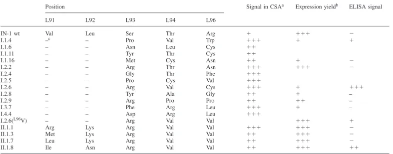
Table I. Mutants obtained from affinity maturation of the IN-1 Fabfragment
Position Signal in CSAa Expression yieldb ELISA signal
L91 L92 L93 L94 L96
IN-1 wt Val Leu Ser Thr Arg ⫹ ⫹⫹⫹ ⫺
I.1.4 –c – Pro Val Trp ⫹⫹⫹ ⫹ ⫹
I.1.6 – – Asn Leu Cys ⫹⫹
I.1.11 – – Tyr Thr Cys ⫹⫹
I.1.16 – – Met Cys Asn ⫹⫹ ⫹ ⫺
I.2.2 – – Arg Thr Asn ⫹⫹⫹ ⫹⫹⫹ ⫺
I.2.4 – – Gly Thr Phe ⫹⫹⫹
I.2.5 – – Pro Cys Val ⫹⫹⫹
I.2.6 – – Arg Val Cys ⫹⫹⫹ ⫹ ⫹⫹⫹
I.2.8 – – Tyr Ala Gly ⫹⫹ ⫹ –
I.2.9 – – Arg Pro Pro ⫹⫹ ⫹⫹ –
I.3.7 – – Phe Arg Leu ⫹⫹⫹ ⫹ –
I.4.4 – – Asp Arg Leu ⫹⫹⫹
I.2.6(L96V) – – Arg Val Val ⫹⫹⫹ ⫹
II.1.1 Arg Lys Arg Val Val ⫹⫹⫹ ⫹⫹⫹ ⫺
II.1.3 Met Lys Arg Val Val ⫹⫹ ⫹⫹⫹ ⫺
II.1.7 Leu Lys Arg Val Val ⫹⫹ ⫹⫹⫹ ⫺
II.1.8 Ile Asn Arg Val Val ⫹⫹ ⫹⫹⫹ ⫹⫹
aFilter-sandwich colony-screening assay. bIn E.coli JM83 using the vector pASK88. cNo exchange.
mutations or internal amber termination codons were abundant. This finding suggests a competition between antigen-complexing signals of higher affinity mutants and non-specific binding by conformationally defective or toxic mutagenesis products during the colony-screening assay.
For soluble production of the recombinant Fabfragments in a standard format (i.e. without the ABD domain but still having a His6tag fused to the C-terminus of the heavy chain) the mutagenized VLgene cassettes from seven selected clones (Table I) were subcloned on pASK88-IN1 (Fiedler and Skerra, 1999). The mutants were produced in shaker flask cultures and isolated from the periplasmic cell fraction in one step via IMAC. All Fabfragments contained the light and heavy chains in stoichiometric composition and quantitatively linked via a disulphide bond. Antigen-binding activity of the mutant Fab fragments was subsequently tested by ELISA using the recom-binant NI-Fr2 for coating of the microtitre plate wells (Figure 2). Almost no binding signal above background was obtained with the recombinant wild-type IN-1 Fabfragment, illustrating its low antigen affinity. In contrast, the mutant I.2.6 (Figure 2A) gave rise to a clearly detectable and concentration-dependent binding signal. No significant signal was obtained in a control experiment with BSA serving as antigen. Hence, the mutant I.2.6 was the protein of choice for further affinity maturation experiments.
Unfortunately, the I.2.6 mutant of the IN-1 Fab fragment was produced as a soluble protein in E.coli at a much lower level, with a relative yield of 5% after purification (0.04 mg l–1 OD–1 versus 0.8 mg l–1 OD–1 for the wild-type IN-1 F
ab fragment). Obviously, the free Cys residue that occurred at the exposed position L96 within CDR-L3 had a deleterious influence on the folding efficiency of the Ig fragment and a concomitant toxic effect on the bacterial host cell, as had been similarly observed in other cases (Ostermeier et al., 1995; Schmiedl et al., 2000). Following earlier substitution experi-ments concerning position L96 (see above), attempts were made to replace the Cys residue in the I.2.6 mutant by small apolar side chains like those of Ala, Val, Met, Leu and Ile.
The substitutions were introduced by site-directed mutagenesis
and all corresponding recombinant Fab fragments were
pro-duced and purified as before, resulting in yields that were similar again to the wild-type IN-1 Fab fragment. However, when binding activity towards the recombinant NI-Fr2 antigen was tested in an ELISA, all these mutants gave rise to significantly lower signals than the original I.2.6 Fabfragment. Merely the replacement CysL96→Val resulted in a detectable binding behaviour (Figure 2) and was therefore used as the basis for the second affinity maturation cycle.
CDR-L3 forms a connecting loop between two neighbouring β-strands such that the positions L91 and L92 are in close spatial proximity with L96. Hence, in order to structurally compensate a possible misfit at position L96—due to the exchange of Cys by Val—the positions L91 and L92 within CDR-L3 of the I.2.6(L96V) F
ab fragment were subjected to
targeted random mutagenesis and the filter-sandwich colony-screening assay was performed again. This time the stringency of selection was raised by lowering the concentration of the recombinant antigen (a mutant of NI-Fr2 devoid of Cys574
and Cys676) from 50 to 30 µg/ml. From screening
approxi-mately 1000 colonies spread on two filter membranes, 16 clones were identified according to their pronounced colour signals. In contrast with the previous experiment, all of them carried plasmids encoding functional mutants of the I.2.6(L96V) Fab fragment. The VL gene cassettes of four clones (Table I)
were subcloned on pASK88-IN1 and the corresponding Fab
fragments were produced and purified as before. One of them, the II.1.8 Fabfragment (Figure 2A), exhibited clearly improved
binding activity over the I.2.6(L96V) mutant in an ELISA
(Figure 2B), even though its affinity was still lower than that of the original I.2.6 mutant carrying the free Cys residue. Nevertheless, the yield of the II.1.8 mutant was 12-fold higher upon expression in E.coli and thus close to that of the recombinant wild-type IN-1 Fabfragment (0.5 versus 0.8 mg l–1 OD–1, respectively).
Functional analysis of engineered Fabfragments
For a detailed analysis of the antigen-binding activity and application in immunohistochemistry as well as cell culture
Fig. 2. Structural and functional characteristics of engineered IN-1 Fab fragments. (A) Amino acid sequence of the VLdomain (Kabat database accession no. 029919; Martin, 1996) of the monoclonal antibody IN-1 (Bandtlow et al., 1996) together with the substitutions introduced in the course of affinity maturation. CDRs are underlined according to the definition by Kabat et al. (Kabat et al., 1991), while amino acid positions are numbered consecutively. The mutations obtained by exchange of residues within CDR-L1 (Fiedler and Skerra, 1999) and CDR-L3 (this study) are marked with bold letters below the wild-type sequence. (B) Comparison of antigen-binding activity of engineered Fabfragments in an ELISA. The wells of a microtitre plate were coated with recombinant NI-Fr2 and purified Fabfragments were applied in decreasing concentration. Bound Fabfragment was detected by means of a goat anti-human Cκ antibody conjugated with alkaline phosphatase, followed by chromogenic reaction.
assays, the different engineered versions of the IN-1 Fab
fragment were produced in E.coli in shaker flask cultures and purified by IMAC to homogeneity (Figure 3A). The thermodynamic affinity for the recombinant Nogo-A fragment NI-Fr4 was determined both for the II.1.8 mutant and for the wild-type IN-1 Fabfragment using the method of real-time SPR on a Biacore-X® system. Preliminary experiments revealed considerable difficulties in immobilizing the recombinant Nogo-A fragments NI-Fr2 as well as NI-Fr4 to sensor chips carrying the conventional carboxy-methylated dextran matrix (CM5 sensor chip®) via the standard amine coupling procedure (O’Shannessy et al., 1992). Most likely, the extremely low pI of the Nogo-A fragment (calculated to be 4.2 for NI-Fr2 using
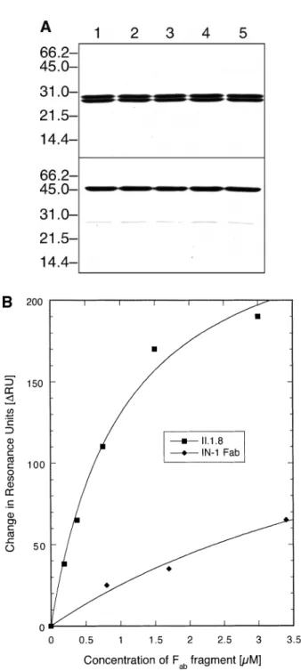
Fig. 3. Antigen affinity determination for the wild-type IN-1 Fabfragment and its mutant. (A) SDS–PAGE analysis of purified recombinant Fab fragments prepared in this study. Fabfragments were produced in E.coli JM83 harbouring the corresponding derivative of the vector pASK88 and purified by IMAC. Samples in the upper part were reduced with β-mercaptoethanol prior to gel electrophoresis whereas those in the lower part were kept unreduced: IN-1 (wild-type) Fabfragment (lane 1); AlaL32→Phe mutant (lane 2); I.2.6 mutant (lane 3); I.2.6(L96V) mutant (lane 4); II.1.8 mutant (lane 5). Molecular sizes are indicated at the left. All Fabfragments appear as a homogeneous protein with stoichiometric presence of the light and heavy chains and show quantitative formation of their interchain disulphide bond. (B) Measurement of the concentration-dependent interaction between the IN-1 Fabfragment and its optimized mutant II.1.8 with the recombinant Nogo-A fragment NI-Fr4 (NTA-sensor chip® charged with 285 to 305∆RU) by SPR. Equilibrium values (difference in resonance units,∆RU) determined after subtraction of the background signal in the absence of NI-Fr4 were plotted against the applied concentration of wild-type IN-1 Fabfragment or its II.1.8 mutant and finally fitted by non-linear regression.
the GCG program package; Devereux et al., 1984) led to electrostatic repulsion within the acceptable range of pH conditions.
Therefore, a Ni/NTA-derivatized sensor chip (NTA-sensor chip®; Nieba et al., 1997) was chosen, to which NI-Fr4 was immobilized via its C-terminal His6-tag (Figure 1A; see Materials and methods). In order to prevent binding of the recombinant Fabfragments to the same surface, their variable domain genes were subcloned on the vector pASK107, thus permitting purification—with comparable efficiency—via the
Strep-tag II instead of the His6-tag (see Materials and methods). The Fabfragments were applied to the sensor chip, each time charged with fresh antigen, at different concentrations and the amount of bound Ig fragment was measured under equilibrium conditions. In this way, binding isotherms were obtained for the wild-type and engineered Fabfragments (Figure 3B), from which dissociation constants were deduced. The KDvalue for
the recombinant wild-type IN-1 Fab fragment was 7.8⫾ 1.9
µM. In contrast, the dissociation constant for its II.1.8 mutant was 1.04 ⫾ 0.18 µM, i.e. 8-fold better. Control experiments with an unrelated protein, recombinant cystatin (Auerswald
et al., 1989) carrying a His6-tag (A.Skerra, unpublished results), that was used instead of the Nogo-A fragment for coating of the sensor chip, confirmed the absence of unspecific binding (data not shown).
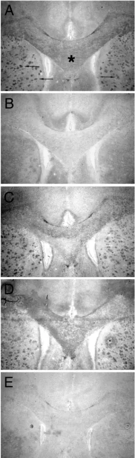
The engineered II.1.8 Fab fragment was also employed for the detection of natural Nogo-A by immunohistochemistry. Figure 4 shows cross-sections of adult rat brain which were stained with different recombinant Fabfragments, followed by a secondary antibody conjugated with a reporter enzyme. The II.1.8 mutant specifically stained the myelinated regions, especially the Corpus callosum and transected fibre bundles of the Capsula interna in the Corpus striatum. The staining pattern is similar in morphology and intensity to the one
obtained with a recombinant Fab fragment derived from the
monoclonal antibody 8-18C5, which is directed against the major oligodendrocyte glycoprotein MOG (Linington et al., 1984). The staining with the recombinant wild-type IN-1 Fab fragment was very weak under the present conditions of
fixation. An unrelated recombinant anti-CD30 Fab fragment
derived from the HRS-3 antibody (Engert et al., 1990) gave only background staining. These results demonstrate that the affinity of the II.1.8 mutant of the IN-1 Fabfragment has been raised to a sufficient extent in order to detect the Nogo-A antigen in standard immunochemical experiments. Analogous results were obtained using immunofluorescence microscopy (data not shown).
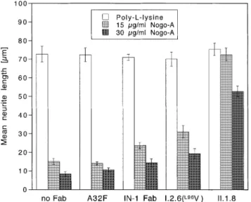
Finally, the engineered Fab fragments were tested for their neutralizing effect on Nogo-A substrate properties using a cell culture assay. As shown in Figure 5, neurite outgrowth of cerebellar granule cells was severely reduced when recom-binant Nogo-A was used as a substrate. In contrast, in its absence, poly-L-lysine promoted extensive attachment of
granule cells, as well as robust neurite growth with an average
neurite length of approximately 70 µm in 70% of adherent
cells. In this in vitro bioassay functional neutralization of the inhibitory Nogo-A substrate was observed at different degrees for the various engineered Fabfragments (Figure 5). While the
recombinant wild-type IN-1 Fab fragment revealed partial
neutralization of Nogo-A activity, as previously demonstrated (Bandtlow et al., 1996), introduction of the mutation
AlaL32→Phe into the V
L domain completely abolished this
effect. In contrast, the mutants I.2.6(L96V) and, in particular,
Fig. 4. Specific staining of myelin-rich regions in the rat brain using the
IN-1 Fabfragment and its engineered mutants. (A) Staining was performed with an anti-MOG Fabfragment (H.Zander et al., to be published); the myelinated, MOG-positive Corpus callosum is marked by an asterisk and myelinated fibres of the Capsula interna in the Corpus striatum are indicated by arrows. (B) Staining with wild-type IN-1 Fabfragment. (C) I.2.6(L96V) Fab fragment. (D) II.1.8 Fabfragment. (E) anti-CD30 Fabfragment (M.Schlapschy et al., to be published). Bound Fabfragment was detected with a goat anti-human Cκantibody conjugated with alkaline phosphatase and revealed using the Fast Red procedure.
Fig. 5. Stepwise improvement of the biological activity of the IN-1 Fab fragment during affinity maturation as determined in an in vitro neurite outgrowth assay. The columns show the mean neurite lengths of granula cells from the rat cerebellum cultured on a recombinant Nogo-A substrate— or just on poly-L-lysine as a control—whose inhibitory properties were neutralized in the presence of the IN-1 Fabfragment and its engineered mutants (applied at 100µg/ml). Error bars correspond to standard deviations from triplicate experiments.
II.1.8, exhibited significantly stronger neutralizing effects, as revealed by their better fibre growth-promoting activities, even when the concentration of the inhibitory material was raised. None of the applied Fabfragments exerted an effect on neurite outgrowth of cerebellar granule cells under control conditions, i.e. in the absence of Nogo-A. Notably, the stepwise improve-ment of the biological activity of the mutants I.2.6(L96V) and
II.1.8 in comparison with the wild-type IN-1 Fab fragment
correlated nicely with their relative increase in antigen affinity observed in the ELISA experiment (Figure 2B).
Discussion
In spite of its considerable potential as a reagent for studying the biological role and distribution of the Nogo-A antigen and for neutralizing its inhibitory effect on axonal growth in tissue culture as well as animal experiments, the use of the monoclonal antibody IN-1 has so far been hampered by its apparently low antigen affinity. In the present study we were able to quantify the intrinsic affinity of its recombinant Fabfragment to be in the 105M–1 range. The remarkably high dissociation constant
that was measured between the IN-1 Fab fragment and the
bacterially produced Nogo-A domain explains several observa-tions from previous studies.
First, the antibody fragment had to be applied at a relatively high concentration, typically 5 mg/ml (corresponding to approximately 100µM), in order to provoke a biological effect (Bandtlow et al., 1996; Broesamle et al., 2000). The original monoclonal antibody was similarly effective at a 5–10-fold lower concentration, which is in agreement with what has to be expected from the strong avidity effect of an IgM as soon as it interacts with an immobilized antigen at a high surface density. Unfortunately, the precise concentration of the IgM was rarely used as an experimental parameter because of the difficulties associated with its purification from the hybridoma supernatant.
Secondly, the use of IN-1 in immunochemical detection experiments, like western blotting, immunohistochemistry,
fluorescence microscopy and immunoprecipitation, was
limited, and the original recombinant Fab fragment was
even dissatisfactory in this respect. In such experiments an initial incubation period allowing for antibody–antigen association is usually followed by several washing steps, where the Ig protein (or the solubilized antigen) may be quickly lost by a bleeding effect if the kinetics of dissociation are fast. This usually happens if the thermodynamic dissociation constant is high and it was also observed here in the course of our Biacore measurements (data not shown). Unfortunately, as a general implication of reaction kinetics this phenomenon cannot be overcome by simply raising the concentration of the applied complexing partner.
The fundamental nature of this problem explains why it took an increased effort and repeated cycles of mutagenesis
in order to finally engineer a derivative of the IN-1 Fab
fragment with 8-fold enhanced affinity. The II.1.8 mutant
possesses a KD value equal to 1 µM, which is obviously
sufficient to yield clear antigen-binding signals in an ELISA and to effect specific staining in immunohistochemistry on brain sections. The preceding mutant I.2.6 might actually perform even better, yet its exposed Cys residue at position L96 within CDR-L3, which apparently plays a crucial role, prevents its practical application due to poor expression in
E.coli. In this respect, one might consider switching to another
host organism. However, unpaired exposed Cys residues have a general tendency of being post-translationally modified in eukaryotic cells (Lyons et al., 1990) and they provide a problem during storage because of susceptibility towards oxidation. Nevertheless, the mutant II.1.8 IN-1 Fab fragment with its well defined antigen-binding activity presents a promis-ing startpromis-ing point for future affinity maturation attempts, where the mutagenesis could be focused at hypervariable regions
apart from CDR-L3, possibly even within the VHdomain.
The present investigation also sheds light on aspects of the structure and biological function of the Nogo-A antigen. First, our data provide evidence that the epitope for the IN-1 antibody (or its Fab fragment, respectively) resides within the protein domain encoded by exon 3. This exon is exclusively present in the splice form called Nogo-A, which constitutes a potent neurite growth inhibitory protein (Chen et al., 2000; Prinjha
et al., 2000). Since the IgM as well as its recombinant Fab fragment effectively neutralize the inhibitory effect of CNS myelin on axonal growth (Bandtlow et al., 1996; Broesamle
et al., 2000) we conclude that the exon 3 domain is involved
in the inhibitory activity of Nogo-A. This interpretation is in agreement with observations from immunocytochemical staining of live oligodendrocytes with an unrelated exon 3-specific antibody (Huber and Schwab, 2000) and with func-tional assays based on an exon 1–3 fragment that was expressed as an IgG Fcfusion protein (Prinjha et al., 2000). However, an inhibitory effect on axonal regeneration was recently reported for a region called ‘Nogo-66’, which is found between the two hydrophobic, presumably transmembrane, segments within the C-terminal domain of Nogo-A (GrandPre´ et al., 2000; Fournier et al., 2001). Therefore, regardless of the current model of its transmembrane topology, it remains possible that both the exon 3 region and the Nogo-66 region inhibit axon growth, possibly via differing receptors.
The fact that the exon 3 moiety of Nogo-A was readily secreted from E.coli, including efficient processing of the bacterial signal peptide that was attached to its N-terminus, and recovered as a soluble protein from the periplasmic space,
would be in agreement with the notion of its nature as an extracellular domain. We found that several of its eight Cys residues are important for proper folding, indicating that the large domain may be adapted to an oxidizing extracellular environment instead of the reducing cytosolic milieu. Even though the native full-length Nogo-A lacks a conventional signal peptide, which normally drives translocation of an N-terminal extracellular domain of a transmembrane protein across the membrane bilayer (von Heijne, 1996), peculiar post-translational mechanisms are known which may determine unusual topologies (Broome-Smith et al., 1994; Lu et al., 1998). Nevertheless, it remains to be seen from future experi-ments whether different orientations could exist for this atypical
membrane protein. The engineered IN-1 Fab fragment with
its improved affinity should provide a helpful reagent in this respect.
The thorough characterization of the molecular interaction
between the recombinant IN-1 Fab fragment, as well as its
mutant II.1.8 with improved binding properties, and Nogo-A constitutes an important step towards the elucidation of the biological role of this signalling molecule. The identification of cognate receptors on growing neurites is obviously one of the next goals. But there is also strong interest in substances which can neutralize the inhibitory activity of Nogo-A. Once its active site is precisely mapped within the rather large polypeptide chain, small cognate antagonist compounds may be identified. However, the engineered IN-1 Fabfragment itself provides a promising drug candidate, too. Its slower diffusion, compared with a low molecular weight compound, together with its moderate dissociation constant, enables site-directed delivery on the one hand, but also prevents undesired neuronal plasticity effects—remote from the site of application—due to a quick dilution effect, on the other. Finally, the engineered IN-1 Fab fragment described here offers excellent starting conditions for the so-called humanization of its murine frame-work sequences with the final goal of generating a potential therapeutic agent.
Acknowledgements
The authors wish to thank Hilke Zander for help in the histological staining experiments and production of the recombinant anti-MOG Fabfragment and Martin Schlapschy for providing the anti-CD30 Fabfragment. This work was supported by the Deutsche Forschungsgemeinschaft Grant SK 33/2-1.
References
Auerswald,E.A., Genenger,G., Assfalg-Machleidt,I., Kos,J. and Bode,W. (1989) FEBS Lett., 243, 186–192.
Bandtlow,C.E., Zachleder,T. and Schwab,M.E. (1990) J. Neurosci., 10, 3837–3848.
Bandtlow,C.E., Schiweck,W., Tai,H.-H., Schwab,M.E. and Skerra,A. (1996) Eur. J. Biochem., 241, 468–475.
Broesamle,C., Huber,A.B., Fiedler,M., Skerra,A. and Schwab,M.E. (2000) J. Neurosci., 20, 8061–8068.
Broome-Smith,J.K., Gnaneshan,S., Hunt,L.A., Mehraein-Ghomi,F., Hashemzadeh-Bonehi,L., Tadayyon,M. and Hennessey,E.S. (1994) Mol. Membr. Biol., 11, 3–8.
Buffo,A., Zagrebelsky,M., Huber,A.B., Skerra,A., Schwab,M.E., Strata,P. and Rossi,F. (2000) J. Neurosci., 20, 2275–2286.
Caroni,P. and Schwab,M.E. (1988a) J. Cell Biol., 106, 1281–1288. Caroni,P. and Schwab,M.E. (1988b) Neuron, 1, 85–96.
Chacko,S., Padlan,E.A., Portolano,S., McLachlan,S.M. and Rapoport,B. (1996) J. Biol. Chem., 271, 12191–12198.
Chen,M.S., Huber,A.B., van der Haar,M.E., Frank,M., Schnell,L., Spillmann,A.A., Christ,F. and Schwab,M.E. (2000) Nature, 403, 434–439. Chothia,C., Lesk,A.M., Tramontano,A., Levitt,M., Smith-Gill,S.J., Air,G., Sheriff,S., Padlan,E.A., Davies,D., Tulip,W.R., Colman,P.M., Spinelli,S., Alzari,P.M. and Poljak,R.J. (1989) Nature, 342, 877–883.
Devereux,J., Haeberli,P. and Smithies,O. (1984) Nucleic Acids Res., 12, 387–395.
Engert,A., Martin,G., Pfreundschuh,M., Amlot,P., Hsu,S.M., Diehl,V. and Thorpe,P. (1990) Cancer Res., 50, 2929–2935.
Fiedler,M. and Skerra,A. (1999) Protein Expr. Purif., 17, 421–427. Fiedler,M. and Skerra,A. (2001a) In Kontermann,R. and Du¨bel,S. (eds),
Antibody Engineering. Springer Verlag, Heidelberg, pp. 243–256. Fiedler,M. and Skerra,A. (2001b) Gene, 274, 111–118.
Fling,S.P. and Gregerson,D.S. (1986) Anal. Biochem., 155, 83–88.
Fournier,A.E., GrandPre´,T. and Strittmatter,S.M. (2001) Nature, 409, 341–346. Geisselsoder,J., Witney,F. and Yuckenberg,P. (1987) BioTechniques, 5,
786–791.
Gill,S.C. and von Hippel,P.H. (1989) Anal. Biochem., 182, 319–326. GrandPre´,T., Nakamura,F., Vartanian,T. and Strittmatter,M. (2000) Nature,
403, 439–444.
Hatten,M.E. (1985) J. Cell Biol., 100, 384–396. Horner,P.J. and Gage,F.H. (2000) Nature, 407, 963–970. Huber,A.B. and Schwab,M.E. (2000) Biol. Chem., 381, 407–419.
Kabat,E.A., Wu,T.T., Perry,H.M., Gottesman,K.S. and Foeller,C. (1991) Sequences of Proteins of Immunological Interest, 5th edn. National Institutes of Health, Bethesda, MD.
Ko¨nig,T. and Skerra,A. (1998) J. Immunol. Methods, 218, 73–83.
Kunkel,T.A., Roberts,J.D. and Zakour,R.A. (1987) Methods Enzymol., 154, 367–382.
Linington,C., Webb,M. and Woodhams,P.L. (1984) J. Neuroimmunol., 6, 387–396.
Lu,Y., Xiong,X., Helm,A., Kimani,K., Bragin,A. and Skach,W.R. (1998) J. Biol. Chem., 273, 568–576.
Lyons,A., King,D.J., Owens,R.J., Yarranton,G.T., Millican,A., Whittle,N.R. and Adair,J.R. (1990) Protein Eng., 3, 703–708.
Martin,A.C.R. (1996) Proteins: Struct. Funct. Genet., 25, 130–133. Nieba,L., Nieba-Axmann,S.E., Persson,A., Ha¨ma¨la¨inen,M., Edebratt,F.,
Hansson,A., Lidholm,J., Magnusson,K., Karlsson,A.F. and Plu¨ckthun,A. (1997) Anal. Biochem., 252, 217–228.
O’Shannessy,D.J., Brigham-Burke,M. and Peck,K. (1992) Anal. Biochem.,
205, 132–136.
Ostermeier,C., Essen,L.-O. and Michel,H. (1995) Proteins: Struct. Funct. Genet., 21, 74–77.
Prinjha,R., Moore,S.E., Vinson,M., Blake,S., Morrow,R., Christie,G., Michalovich,D., Simmons,D.L. and Walsh,F.S. (2000) Nature, 403, 383–384.
Ramon y Cajal,S. (1928) Degeneration and Regeneration of the Nervous System. Hafner, New York.
Rose,D.R., Strong,R.K., Margolies,M.N., Macolm,G.L. and Pestko,G.A. (1990) Proc. Natl Acad. Sci. USA, 87, 338–342.
Sambrook,J., Fritsch,E.F. and Maniatis,T. (1989) Molecular Cloning: A Laboratory Manual, 2nd edn. Cold Spring Harbor Labotatory Press, Cold Spring Harbor, NY.
Schiweck,W. and Skerra,A. (1995) Proteins: Struct. Funct. Genet., 23, 561–565.
Schlapschy,M. and Skerra,A. (2001) In Kontermann,R. and Du¨bel,S. (eds), Antibody Engineering. Springer Verlag, Heidelberg, pp. 292–306. Schlehuber,S., Beste,G. and Skerra,A. (2000) J. Mol. Biol., 297, 1105–1120. Schmiedl,A., Breitling,F., Winter,C.H., Queitsch,I. and Du¨bel,S. (2000) J.
Immunol. Methods, 242, 101–114.
Schnell,L. and Schwab,M.E. (1990) Nature, 343, 269–272. Skerra,A. (1992) Nucleic Acids Res., 20, 3551–3554. Skerra,A. (1994a) Gene, 151, 131–135.
Skerra,A. (1994b) Gene, 141, 79–84.
Skerra,A. and Schmidt,T.G.M. (2000) Methods Enzymol., 326A, 271–304. Skerra,A., Dreher,M.L. and Winter,G. (1991) Anal. Biochem., 196, 151–155. Spillmann,A.A., Amberger,V.R. and Schwab,M.E. (1997) Eur. J. Neurosci.,
9, 549–555.
Spillmann,A.A., Bandtlow,C.E., Lottspeich,F., Keller,F. and Schwab,M.E. (1998) J. Biol. Chem., 273, 19283–19293.
Tatagiba,M., Rosahl,S., Gharabaghi,A., Blo¨mer,U., Brandis,A., Skerra,A., Samii,M. and Schwab,M.E. (2002) Acta Neurochir. (Wien), 144, 181–187. Vogt,M. and Skerra,A. (2001) J. Mol. Recognit., 14, 79–86.
von Heijne,G. (1996) Prog. Biophys. Mol. Biol., 66, 113–139. von Heijne,G. and Gavel,Y. (1988) Eur. J. Biochem., 174, 671–678. Voss,S. and Skerra,A. (1997) Protein Eng., 10, 975–982.
Yanisch-Perron,C., Vieira,J. and Messing,J. (1985) Gene, 33, 103–119. Z’Graggen,W.J., Metz,G.A.S., Kartje,G.L., Thallmair,M. and Schwab,M.E.
(1998) J. Neurosci., 18, 4744–4757.