HAL Id: hal-02387991
https://hal.uca.fr/hal-02387991
Submitted on 30 Nov 2019
HAL is a multi-disciplinary open access
archive for the deposit and dissemination of
sci-entific research documents, whether they are
pub-lished or not. The documents may come from
teaching and research institutions in France or
abroad, or from public or private research centers.
L’archive ouverte pluridisciplinaire HAL, est
destinée au dépôt et à la diffusion de documents
scientifiques de niveau recherche, publiés ou non,
émanant des établissements d’enseignement et de
recherche français ou étrangers, des laboratoires
publics ou privés.
Distributed under a Creative Commons Attribution - NonCommercial - NoDerivatives| 4.0
International License
breast cancer, including recommendations to assess TILs
in residual disease after neoadjuvant therapy and in
carcinoma in situ: A report of the International
Immuno- Oncology Biomarker Working Group on
Breast Cancer
Maria Dieci, Nina Radosevic-Robin, Susan Fineberg, Gert van den Eynden,
Nils Ternes, Frédérique Penault-Llorca, Giancarlo Pruneri, Timothy
D'alfonso, Sandra Demaria, Carlos Castaneda, et al.
To cite this version:
Maria Dieci, Nina Radosevic-Robin, Susan Fineberg, Gert van den Eynden, Nils Ternes, et al..
Up-date on tumor-infiltrating lymphocytes (TILs) in breast cancer, including recommendations to assess
TILs in residual disease after neoadjuvant therapy and in carcinoma in situ: A report of the
Interna-tional Immuno- Oncology Biomarker Working Group on Breast Cancer. Seminars in Cancer Biology,
Elsevier, 2018, 52, pp.16 - 25. �10.1016/j.semcancer.2017.10.003�. �hal-02387991�
Contents lists available atScienceDirect
Seminars in Cancer Biology
journal homepage:www.elsevier.com/locate/semcancer
Review
Update on tumor-in
filtrating lymphocytes (TILs) in breast cancer, including
recommendations to assess TILs in residual disease after neoadjuvant
therapy and in carcinoma in situ: A report of the International
Immuno-Oncology Biomarker Working Group on Breast Cancer
☆
Maria Vittoria Dieci
a,b,⁎, Nina Radosevic-Robin
c,d, Susan Fineberg
e,f, Gert van den Eynden
g,h,
Nils Ternes
i,j, Frederique Penault-Llorca
c,d,k, Giancarlo Pruneri
l,m, Timothy M. D
’Alfonso
n,
Sandra Demaria
o, Carlos Castaneda
p, Joselyn Sanchez
p, Sunil Badve
q, Stefan Michiels
i,j,
Veerle Bossuyt
r, Federico Rojo
s, Baljit Singh
t, Torsten Nielsen
u, Giuseppe Viale
v,
Seong-Rim Kim
w, Stephen Hewitt
x, Stephan Wienert
y, Sybille Loibl
z, David Rimm
r,
Fraser Symmans
A, Carsten Denkert
B, Sylvia Adams
C, Sherene Loi
D, Roberto Salgado
E,F,G, on
behalf of the International Immuno-Oncology Biomarker Working Group on Breast Cancer
aDepartment of Surgery, Oncology and Gastroenterology, University of Padua, Padua, Italy bMedical Oncology 2, Veneto Institute of Oncology IOV– IRCCS, Padua, Italy
cDepartment of Surgical Pathology and Biopathology, Jean Perrin Comprehensive Cancer Centre, Clermont-Ferrand, France dInserm/University of Auvergne U1240, Clermont-Ferrand, France
eMontefiore Medical Center, Bronx, NY, USA fThe Albert Einstein College of Medicine, Bronx, NY, USA
gMolecular Immunology Unit, Institut Jules Bordet, Université Libre de Bruxelles, Brussels, Belgium hDepartment of Pathology, GZA Ziekenhuizen, Antwerp, Belgium
iService de Biostatistique et d'Epidémiologie, Gustave Roussy, B2 M, RdC,114 rue Edouard-Vaillant, 94805, Villejuif, France jCESP, Fac. de médecine– Univ. Paris-Sud, Fac. de médecine – UVSQ, INSERM, Université Paris-Saclay, Villejuif, 94805, France kSchool of Medicine, University of Auvergne, Clermont-Ferrand, France
lDivision of Pathology, Fondazione IRCCS Istituto Nazionale Tumori, Milan, Italy mUniversity of Milan, School of Medicine, Milan, Italy
nDepartment of Pathology and Laboratory Medicine, Weill Cornell Medicine, New York, NY, USA oDepartment of Radiation Oncology, Weill Cornell Medicine, New York, NY, USA
pDepartment of Research, Instituto Nacional de Enfermedades Neoplasicas, Lima 15038, Peru qDepartment of Pathology and Laboratory, Medicine, Indiana University, Indianapolis, USA rDepartment of Pathology, Yale University School of Medicine, New Haven, USA sPathology Department, IIS, Fundacion Jimenez Diaz, UAM, Madrid, Spain tDepartment of Pathology, Coney Island Hospital, NY, USA
uGenetic Pathology Evaluation Centre, Department of Pathology and Laboratory Medicine, University of British Columbia, Vancouver, Canada vDepartment of Pathology, Istituto Europeo di Oncologia, University of Milan, Milan, Italy
wNational Surgical Adjuvant Breast and Bowel Project Operations Center/NRG Oncology, Pittsburgh, PA, USA
xExperimental Pathology Laboratory, Laboratory of Pathology, Center for Cancer Research, National Cancer Institute, NIH, Bethesda, MD, USA ydlw Laborsoftware UG (haftungsbeschränkt) Lübben, Germany
zGerman Breast Group, Neu-Isenburg, Germany
ADepartment of Pathology, University of Texas M.D. Anderson Cancer Center, Houston, USA BInstitute of Pathology, Charité Universitätsmedizin Berlin, Berlin, Germany
CNew York University Medical School, Perlmutter Cancer Center, NY, USA
DDepartment of Medical Oncology, Peter MacCallum Cancer Centre, Melbourne, Australia
ETranslational Breast Cancer Genomic and Therapeutics Laboratory, Peter Mac Callum Cancer Center, Victoria, Australia FBreast Cancer Translational Research Laboratory, Jules Bordet Institute, Brussels, Belgium
GDepartment of Pathology, GZA, Antwerp, Belgium
http://dx.doi.org/10.1016/j.semcancer.2017.10.003
Received 5 September 2017; Accepted 4 October 2017
☆See supplement list 6.
⁎Corresponding author at: Department of Surgery, Oncology and Gastroenterology, University of Padova, Division of Medical Oncology 2, Istituto Oncologico Veneto IRCCS, Via
Gattamelata 64, 35128 Padova, Italy.
E-mail addresses:mariavittoriadieci@gmail.com,mariavittoria.dieci@unipd.it(M.V. Dieci).
Seminars in Cancer Biology 52 (2018) 16–25
Available online 09 October 2017
1044-579X/ © 2017 The Authors. Published by Elsevier Ltd. This is an open access article under the CC BY-NC-ND license (http://creativecommons.org/licenses/BY-NC-ND/4.0/).
A R T I C L E I N F O
Keywords: Tumor-infiltrating lymphocytes Breast cancer Neoadjuvant Residual disease Ductal carcinoma in situ Residual cancer burdenA B S T R A C T
Morphological evaluation of tumor-infiltrating lymphocytes (TILs) in breast cancer is gaining momentum as evidence strengthens the clinical relevance of this immunological biomarker. TILs in the post-neoadjuvant re-sidual disease setting are acquiring increasing importance as a stratifying marker in clinical trials, considering the raising interest on immunotherapeutic strategies after neoadjuvant chemotherapy. TILs in ductal carcinoma in situ, with or without invasive carcinoma, represent an emerging area of clinical breast cancer research. The aim of this report is to update pathologists, clinicians and researchers on TIL assessment in both the post-neoadjuvant residual disease and the ductal carcinoma in situ settings. The International Immuno-Oncology Working Group proposes a method for assessing TILs in these settings, based on the previously published International Guidelines on TIL Assessment in Breast Cancer. In this regard, these recommendations represent a consensus guidance for pathologists, aimed to achieve the highest possible consistency among future studies.
1. Introduction
Development and progression of malignant tumors are character-ized by an interaction with the cells in the tumor microenvironment including infiltrating immune cells. In the early stage HER2-positive (HER2+) and in triple negative breast cancer (TNBC), immune in-filtrates are detectable in up to 75% of tumors, with up to 20% of tu-mors having a particularly dense infiltrate and with lower amount of TILs in luminal subtypes[1].
Morphological evaluation of tumor-infiltrating lymphocytes (TILs) in breast cancer (BC) is gaining momentum as evidence strengthens the clinical relevance of this immunological biomarker, in particular in HER2+ and TNBC subtypes. Accumulating evidence from several stu-dies indicates that TIL density is predictive for response to neoadjuvant chemotherapy (NACT) and, in certain BC subtypes, prognostic in pa-tients treated with adjuvant chemotherapy. This suggests that treat-ment response and outcome of BC varies with different TIL levels and that the evaluation of TIL density in clinical trial cohorts–as well as in daily histopathological practice- may prove to be of paramount re-levance.
In this regard, high correlation of the immune gene expression with TILs[2–4]further confirms that evaluation of TILs may be a valid, af-fordable and readily available alternative. Furthermore, as archived hematoxylin-and-eosin (H & E) slides can be evaluated for TILs, several mature large datasets could be examined as to produce high quality data required for level I evidence of TILs as prognostic/predictive biomarkers.
NACT is being increasingly used in patients with primary TNBC or HER2+ BC. The endpoint of pathological complete response (pCR) at surgery is mostly considered as a surrogate of long-term survival and suggests that the use of the neoadjuvant model may pave the way for more efficient drug development. Nevertheless, recent studies suggest that increased pCR rates do not necessarily translate into improved outcomes. This implies that a more comprehensive analysis of the post-NACT tumor is necessary. Two recent studies have also indicated that higher TILs in post-NACT residual disease in TNBC are an important independent predictor of improved survival[5,6]. However, in HER2+ disease, a single study suggests an adverse prognostic role of high TILs in residual disease after NACT[49].
Similarly, TILs in ductal carcinoma in situ (DCIS), with and without an associated invasive carcinoma, represent an emerging area of clin-ical BC research since varying levels of TILs are encountered in DCIS.
Over the past few years, the International Immuno-Oncology Biomarker Working Group on Breast Cancer (“The Working Group” in the further text), with members including clinical research groups, pathologists, clinicians and statisticians knowledgeable in BC, has de-veloped standards for assessment of the immuno-oncology biomarkers to aid pathologists, clinicians and researchers in their research and daily practice. The group has licensed thefirst International Guidelines
on TIL Assessment in Breast Cancer[7]and in Solid Tumors[8]. In this report, a panel of pathologists, medical oncologists, biosta-tisticians and translational researchers, with complementary expertise, conducted a systematic review of the literature and propose a method based on the International Guidelines on TIL Assessment in Breast Cancer[7]which follows the methodology used in previous published papers[5,9–11]. The methodology, while intended to remain as stan-dardized as possible, was suggested, reviewed and discussed by mem-bers of the Working Group, experts in their particularfields, to reach the consensus opinion hereby presented.
The aim of this report is to update pathologists, clinicians and re-searchers on TIL assessment in both the post-NACT residual disease (RD) and the DCIS settings.
2. Tumor-infiltrating lymphocytes in the post-NACT residual disease setting
NACT followed by surgery is nowadays considered the standard of care for locally advanced/inflammatory BC and is increasingly used in earlier stages with the aim to achieve a tumor down-staging and im-prove the chance for breast conservation. In addition, it provides the unique opportunity to test in vivo sensitivity to investigational agents, potentially speeding up drug development. pCR, defined as the absence of invasive residual carcinoma in the breast and axillary lymph nodes after NACT, has been proposed as a surrogate endpoint for long-term outcome [12,13]. However, rates and prognostic impact of pCR are heterogeneous across different BC subtypes. The highly proliferative and more aggressive subtypes such as non-luminal HER2+ BC and TNBC have a higher chance to respond to NACT and the association between failure to achieve pCR and unfavorable prognosis is mostly evident in these subtypes, as compared to luminal BCs[13,14]. How-ever, some patients without pCR will survive long-term, whereas some patients with pCR will relapse, highlighting the limitations of pCR as a surrogate endpoint for drug efficacy.
For these reasons, the identification of biomarkers to refine risk stratification is urgently needed in order to enable a better identifica-tion of high-risk patients eligible for addiidentifica-tional systemic treatments. TILs evaluated in RD after NACT have been suggested as a potentially useful and reliable marker for this purpose. Afirst report by Asano and colleagues suggests that the combination of the residual cancer burden (RCB) and TILs is a significant predictor for breast cancer recurrence after NACT and may be a more sensitive indicator than TILs alone[15]. The Working Group is launching an international effort to include TILs in the RCB index in order to develop more accurate risk stratification systems after NACT.
It may be argued that the evaluation of TILs on H & E-stained slides does not capture the complexity of the tumor immune microenviron-ment which is constantly populated by cells having pro-tumorigenic and anti-tumor actions[16]. Therefore, some authors have focused on
the immunohistochemical (IHC) characterization of TIL populations, showing that the composition of TILs and the balance between the immune effector and the pro-tumorigenic cells in RD was able to dis-criminate patients at different risk of relapse [17,18]. However, the basic morphological evaluation of TILs, on H & E-stained sections, should still be considered the preferred method to be used in daily practice. Being technically more feasible, it requires less biological tissue and does not need additional processing on top of the pathology routine. Moreover, in a large neoadjuvant study, predictive information for response to NACT confirmed that TIL counts correlate well with the immune gene expression[2], further emphasizing the strong biological relevance of TILs when assessed using a standardized morphological method. In addition, use of IHC requires additional processing as well as further additional technical standardization. The global TIL count, obtained on H & E, reflects well the net tumor immunogenicity and classifies it in one of two clinically relevant categories: “hot”(inflamed, TIL-rich) and “cold” (non-inflamed, TIL-poor)”. The H & E slides are always available on existing trials, without block recollection required, and therefore provide the quickest and easiest route to the body of prospective-retrospective Level I–II evidence that needs to be obtained. 2.1. Available evidence
Available evidence on TILs in RD after NACT mainly focuses on TNBC. RD after NACT predicts poor outcome [19]. At present, the standard of care in the post-NACT setting for TNBC patients is ob-servation[20]. Another option may be to consider the use of non-cross-resistant chemotherapy such as capecitabine for 6–8 cycles, which has been recently shown to reduce mortality in a single study, although 5-year mortality remains above 20%[21].
Dieci and colleagues retrospectively evaluated TILs on H & E-stained slides from surgical specimens of 278 TNBC patients with RD in the breast after NACT. The authors reported a significant and independent correlation between TILs, evaluated as a continuous variable, and both metastasis-free and overall survival, with a 21% relative reduction of the risk of metastasis and death for each 10% TIL increment. Patients with high TILs (> 60% of stromal TILs) in post-NACT residual breast tumors experienced significantly increased 5-year metastasis-free and overall survival rates than the patients with lower TILs (81.5% vs 46%, HR 0.24, 95% CI 0.09-0.64 for metastasis-free and 91% vs 55% HR 0.19, 95% CI 0.06–0.61 for overall survival). Interestingly, the prog-nostic power of post-NACT TILs was greater in the group with a large residual tumor burden (node positive and/or breast residual tumor with diameter of more than 2 cm). More recently, Loi and colleagues eval-uated TILs in a series of 111 TNBC with RD after NACT. Lymphocytic infiltration in RD proved to be significantly and independently asso-ciated with both relapse-free survival and overall survival, showing consistency with the previous study. In addition, the authors reported that the residual tumors with low TILs were enriched in activating genomic alterations in the RAS-MAPK pathway (amplifications in KRAS, BRAF, RAF1, and truncations in NF1) resulting in the suppression of MHC-I/II expression. These results suggest that the activation of RAS-MAPK pathway might represent one possible mechanism by which tumor cells bypass the antigen presentation processes [6]. A recent study has assessed the role of TILs in RD after NACT and trastuzumab for HER2+ disease, suggesting an adverse prognostic role for high TIL levels[49].
However, standardized, analytically validated methods for TIL as-sessment are a prerequisite for the clinical utility of the results. 2.2. Methodological considerations for TIL assessment in the residual disease setting
Formal recommendations for TIL assessment on H & E-stained slides of primary untreated BC samples have been previously developed by the Working Group. More recently, the Working Group has conducted a
RING which demonstrated that pathologists can be educated to score TILs, herewith achieving high interpersonal concordance compared to the non-trained pathologists (see below). This study definitively proves that TILs can be assessed by pathologists in daily practice[7,22].
However, these recommendations may not be fully transposable to the post-NACT samples, due to the well-recognized tissue-related pe-culiarities of this particular post-treatment setting [23], further dis-cussed below. One may argue against the need to assess TILs in the post-NACT residual disease, since TILs assessed on pre-post-NACT core biopsies, using the established guidelines, have been shown to predict prognosis for patients treated with NACT [24,25]. It is now accepted that the antitumor effect of chemotherapy is partially exerted through the modulation of the immune system, in a process known as immunogenic cell death[26,27]. TILs evaluated in the residual tumor after exposure to chemotherapy could carry additional prognostic information, com-pared to the baseline TILs, as they may partly reflect the reaction of the immune microenvironment to chemotherapy. This has been supported by the observation that the administration of chemotherapy results in the attraction to and activation of TILs in the tumor bed[28]. Similarly, the HER2+ BC patients showed an increase in TILs after exposure to trastuzumab, even in case of short-term therapy, according to the pre-sumed immune-mediated mechanism of action of monoclonal anti-bodies[29–31]. Therefore, TILs in RD differ in nature not only from baseline TILs but may also reflect the treatment ability to elicit an an-titumor immune response. For that reason, focusing on pre-treatment TILs may lead to missing of an important piece of clinical information. This is corroborated by the emerging development of the clinical trials for patients with residual disease after neoadjuvant therapy. Thus, the need to determine a modified scoring method, specific for the post-NACT BC specimens and based on simple and easy to adopt criteria, becomes relevant.
2.3. TIL assessment in the post-NACT residual disease setting: ongoing standardization efforts
2.3.1. The RING study
Considering the clinical need to develop a method of TIL assessment in the post-NACT RD, in early 2016 the Working Group has started standardization of TIL assessment in this setting. By mid-2016, a RING study was launched to assess reproducibility of TIL assessment and to highlight difficult areas that need to be discussed in order to optimize the scoring methodology. A guide for TIL assessment in RD, developed on the basis of the methods used in previous studies[2,3,5,9,10]and on the established recommendations for TIL assessment in primary un-treated BC[7], was circulated among six pathologists experienced in TIL evaluation. Digitalized H & E slides from 50 cases with residual TNBC were independently scored for stromal TILs by each pathologist. Reproducibility was evaluated by intra-class correlation coefficient (ICC), which showed a moderate concordance between pathologists for stromal TIL evaluation (0.59, 95% CI 0.45–0.70). Further details on methods and results can be found in Supplementary File 1.
This result should be regarded as encouraging since the study was carried out by pathologists who had not received previous training on TIL assessment in the post-NACT RD. A moderate inter-observer con-cordance has been reported for many of the biomarkers routinely as-sessed in BC samples, as histologic grade and Ki67, nonetheless not restricting their widespread use in clinical practice and inclusion in international guidelines. In addition, for Ki67, a global effort of stan-dardization is ongoing[32].
However, to what extent the encountered variability in biomarker assessment impacts prognosis, clinical outcome and, eventually, clinical utility of a biomarker, remains largely unexplored for most of the routinely used biomarkers in pathology. In order to explore the clinical relevance of different degrees of inter-observer concordance in TIL as-sessment, the Working Group conducted an in silico study focused on the available pre-treatment TIL data from the neoadjuvant GeparSixto
M.V. Dieci et al. Seminars in Cancer Biology 52 (2018) 16–25
trial[33], with pCR as the primary endpoint. Available TIL scores were perturbed by a mathematical model simulating a situation in which the complete dataset of the 580 samples of the GeparSixto study would be evaluated by 4500 pathologists, in 9 groups of 500 each, with different ICC for each group, obtaining a total of 2 610 000 values. The simu-lation was based on the error model developed from published RING studies on TIL assessment in primary untreated BC. In the original study dataset, each 10% increase in baseline TILs was associated with a 20% increase in pCR. The data were analyzed analogously for each group of pathologists. For ICC coefficients from 0.6 to 0.9, 100% of pathologists have odds ratios comparable to those of the original study. Even with low ICCs of 0.4 and 0.5, most of the evaluations were still significant for prediction of pCR (Supplementary File 2).
This study suggests that a degree of inter-observer variability may not directly impact clinical outcome and might need to be detailed for most of the biomarkers in order to allow the determination of the clinically allowable error margin, meaning“what is the error margin that is allowed among pathologists without this affecting clinical out-come?” We nonetheless emphasize that pathologists need to score TILs as accurate as they possibly can. This level of agreement is still high enough to be useful in research studies on hundreds of patients, as the net biology will show through the noise of the analytical variability at a statistically significant level. Nevertheless, the same level of variability is not acceptable for a single sample call leading to a clinical treatment-oriented decision for a specific patient.
The RING study represented the springboard for the identification of the main areas of uncertainties that were discussed at the San Antonio Breast Cancer Symposium in 2016 (SABCS 2016), and that has led up to the current recommendations.
2.3.2. Areas of uncertainties in TIL assessment in the post-NACT residual disease setting
The aim of the next section is to discuss the areas of controversies in the assessment of TILs in the post-NACT RD and to describe strengths and weaknesses of various possible approaches, reflecting the discus-sions held by the Working Group at the SABCS 2016 meeting.
Table 1summarizes the recommendations developed for assessment of TILs in primary BC when related to the RD after NACT. The main aspects that challenge application of the available recommendations for TIL assessment in the post-NACT RD are the different patterns of re-sponse and tumor shrinkage that may be induced by NACT or other types of treatment, and the wide range of possible post-treatment changes that can be found in these samples[23]. The major topics of
uncertainty are: (i) selection of the tumor area to evaluate and (ii) identification of relevant TILs to be included in the assessment.
According to the recommendations for primary untreated BC[7], TIL assessment should be conducted on the area limited by the borders of the primary invasive tumor. Despite the use of imaging, macroscopic features, extensive sampling and microscopic assessment, the area within the borders of primary untreated tumor in the post-NACT setting is difficult – occasionally even impossible – to be delineated due to tumor shrinkage.
There are, however, two different tissue areas usually assessable in the post-NACT setting:
Thefirst can be regarded as the “area of regression”; it can be identified by radiological and gross pathology findings or stroma re-actions, and by signs of regression at microscopic evaluation irrespec-tive of the borders defined by the presence of tumor cells. The second possibility is to focus on the“residual tumor bed”, defined as the largest cross-sectional area between residual invasive cells, consistent with the definition of Residual Cancer Burden (RCB)[34]. As discussed further in the text, these two areas may overlap.
The second key step is to define where to evaluate TILs. The re-commendations for untreated BC stress the relevance of TILs localized in the tumor stroma of the selected area as the main parameter to be considered[7]. Intra-epithelial tumor TILs can be included, for research purposes, in the post-NACT setting, although assessment may prove more difficult without additional tools like IHC. According to the available recommendations, stromal TILs should be assessed by de-termining the percentage of the tumor stroma surface of the selected area that is occupied by mononuclear inflammatory cells. Areas that should be excluded from the assessment include: necrosis, fibrosis, stroma associated with in situ carcinoma or normal lobules, large fi-brous areas, crush artefacts and areas of regressive hyalinization. These major recommendations are applicable to the post-NACT setting, bearing in mind that after treatment it is more likely to seefibrotic and necrotic areas (sometimes replaced by aggregates of histiocytes and cholesterol clefts), as well as regressive hyalinization. All those areas should be excluded from the TIL assessment, except when the tumor cells are within these aggregates of histiocytes and lymphocytes, com-plicating in this particular setting the assessment of TILs. Moreover, one of the NACT response pattern consists of residual scattered cell foci or of isolated tumor cells, separated by large stromal areas[23]. TILs may be found close to the tumor foci as well as dispersed in the stroma of the selected area, far from residual invasive cells. A still unproven as-sumption is that only TILs adjacent to the invasive tumor cells/foci have
Table 1
Recommendations for assessment of TILs in primary BC and their adaptation to RD after NACT.
TILs in primary untreated BC[18] TILs in RD
Selection of tumor area
Within boundaries of the invasive tumor. Within the borders of the tumor area. How to define the tumor area of RD? – Area of regression according to radiological/gross/microscopic findings – Residual tumor bed defined as the largest cross-sectional area between residual
invasive tumor cells. Include TILs at the invasive edge in the average assessment
Do not focus on hotspots Do not focus on hotspots Do not include: DCIS, normal lobules, necrosis, largefibrous
areas, crush artefacts, regressive hyalinization
Do not include: DCIS, normal lobules, necrosis, largefibrous areas, crush artefacts, regressive hyalinization. However, more challenges may be posed in the post-neoadjuvant setting since alterations such as regressive hyalinization, necrosis (sometimes replaced by aggregates of hystiocytes and cholesterol clefts),fibrous areas and stroma infiltrated by sheets of foamy hemosiderin-loaded histiocytes are often found after NACT. Type of immune cells All mononuclear cells, exclude polymorphonuclear
leukocytes.
All mononuclear cells, exclude polymorphonuclear leukocytes.
Localization Stromal TILs as the main parameter. Intratumoral TILs can be included for research purposes.
Stromal TILs as the main parameter. Intratumoral TILs can be included for research purposes.
StrTILs definition: % of stromal areas occupied by mononuclear inflammatory cells.
StrTILs definition: % of stromal areas occupied by mononuclear inflammatory cells, within the boundaries of the residual tumor bed as defined by the RCB-index. Scoring method Continuous variable. Continuous variable.
Scan across tumor bed (50–100×) and then estimate average
a relevant immunological function, as compared to TILs found dis-persed far from the tumor cells[35]. This assumption raises the ques-tion of whether to consider the entire stromal surface or the area around scattered foci only.
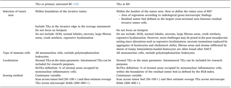
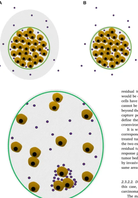
As illustration, two examples showing the patterns of response to NACT are provided inFigs. 1 and 2.
2.3.2.1. Concentric shrinkage, high cellularity (Fig. 1). In this case, the response consists in concentric tumor shrinkage, with high cellularity maintained.
As shown inFig. 1A, the area of regression usually extends beyond the residual tumor bed. Analyzing only the residual tumor bed area has two advantages: i) it complies with the definition of the Residual Cancer Burden[23,34]and ii) it is less affected by the micro- and macroscopic
modifications of the BC tissue after NACT, so its delineation is likely more reproducible compared to the delineation of the regression area. Furthermore, the use of a larger area as the denominator might some-times lead to a reduced TIL score. In addition, the adoption of the
residual tumor bed area as the area for post-NACT TIL assessment would be consistent with the assumption that TILs located near cancer cells have a higher biological and clinical value. On the other hand, it cannot be excluded that the assessment of TILs dispersed in the stroma beyond the residual tumor area, but within the area of regression, might capture potentially relevant information. More research is needed to define the importance of spatial heterogeneity of tumor immune mi-croenvironment in the post-neoadjuvant treatment setting.
It is worth noting that the area of regression does not necessarily corresponds to or is identified within the extent of the primary un-treated tumor but more often covers an intermediate surface between the two extremes, the extension of the primary untreated tumor and the residual tumor bed. Finally, in some cases of the concentric shrinkage response pattern, the area of regression may overlap with the residual tumor bed, with no signs of regression found beyond the area delimited by invasive cells. In such condition, both approaches would identify the same area for TILs evaluation (Fig. 1B).
2.3.2.2. Decrease in tumor cellularity, no concentric shrinkage (Fig. 2). In this case, pattern of response consists of residual scattered foci of carcinoma dispersed in a large stroma area.
The main controversy here is not the delineation of the area to be assessed for TILs, since the area of regression and the residual tumor bed may overlap (Fig. 2), but whether to consider the whole eligible stromal surface (within the selected area) as the denominator for TIL evaluation.
As already mentioned, TILs found immediately adjacent to the tumor cells/nests may carry more biologically relevant information than scattered TILs within the stroma far from the tumor foci [35]. However, definition of the extent of the area surrounding residual tumor cells/nests to be included in TIL analysis may be challenging. In addition, the amount of TILs surrounding residual scattered foci may be heterogeneous, which is another limitation of this approach. Using the average TIL score, determined on the whole eligible stromal surface, including TILs at distance from tumor foci, may be one solution. However, this does carry a risk of TIL score reduction.
The two paradigmatic patterns of response presented here represent the two extremes of a wide spectrum of possible mixed responses[23]. Moreover, a much-debated issue is whether TIL evaluation can be performed in case of pCR. Although challenging, the evaluation of the immune infiltrate in pCR cases might be technically feasible. In this condition, the sole definition of the area for TIL assessment that would make sense is the area of regression. However, the main uncertainty is the biological and clinical relevance of such analysis. To date, available evidence is inadequate and we encourage clinical research groups to score TILs even in pCR in order to acquire evidence.
Fig. 1. Concentric tumor shrinkage as response to NACT. Gray-sha-dowed surface: area of regression; green line: residual tumor bed; purple spots: mononuclear inflammatory cells. 1A: area of regression extending beyond the residual tumor bed; 1B: area of regression overlapping and confined to the residual tumor bed. (For interpreta-tion of the references to colour in thisfigure legend, the reader is referred to the web version of this article.)
Fig. 2. Reduced tumor cellularity as response to NACT, without concentric shrinkage. Gray-shadowed surface: area of regression; green line: residual tumor bed; purple spots: mononuclear inflammatory cells. (For interpretation of the references to colour in this figure legend, the reader is referred to the web version of this article.)
M.V. Dieci et al. Seminars in Cancer Biology 52 (2018) 16–25
2.3.3. Recommendations for a scoring method for TILs in the post-NACT residual disease setting
Careful discussion of lessons learned from the initial concordance study has led the members of the Working Group to propose a reference method for TIL assessment after neoadjuvant treatment.
The proposed method is based on the methodology used in the published studies [2,3,5,6,9,10], combined with adjustments derived from the experience of the RING study. A summary of the guidance in the form of a tutorial including pictures is available inTable 2and as Supplementary File 3.
We propose the residual tumor bed area as the area for TIL assess-ment in the post-NACT setting, in concordance with the Residual Cancer Burden definition[34]since this variable is widely used in daily practice and clinical studies. Clear instructions on how to measure the RCB are available and its prognostic significance has recently been established[36]. The clinical utility of TILs as an immune biomarker in RD may help discriminate poor or intermediate prognosis patients who would be suitable for clinical trials of post-neoadjuvant systemic treatments from the patients potentially cured by NACT. In this context, adding the residual tumor TILs to other factors, such as RCB, may further improve the performance of prognostic models. Indeed, RCB has been validated as a strong prognostic parameter capable of overcoming limitations of a simplistic dichotomization of response in pCR or non-pCR[34,36]. In order to enable such integration of TIL scores into post-NACT RD evaluation in breast cancer, the most coherent and pragmatic approach is to adopt the criteria used for delimitation of the tumor bed for the RCB index[34].
Related to the second main area of controversy, this referral method proposes to include the whole stroma surface of the identified residual tumor bed as the denominator for TIL assessment. This will avoid the introduction of additional subjective elements to the scoring method that would increase variability in interpretation, like the definition of the extent of the area around scattered tumor foci. However, if those scattered tumor foci are separated widely from each other, then the area around each foci should be considered for TIL assessment and then averaged in a similar manner as the TIL assessment in DCIS re-commends.
Furthermore, several ongoing studies are scoring TILs in the neoadjuvant setting on core biopsies taken after a short period of treatment (targeted therapy or chemotherapy) but prior to surgery. The current recommendations also apply to this setting. This means that only cores with tumor should be assessed. Cores with scarred and in-flammatory stroma but with no tumor cells should not be assessed. This is in line with the current recommendations on assessing only TILs surrounding tumor cells. Also, the rules proposed here for delineation of residual tumor area of course do not apply to core biopsies since such delineation is impossible on that type of tissue sample.
This scoring method is intended to provide guidance for the ongoing or future studies assessing the clinical relevance of TILs in post-neoadjuvant RD. It clearly does not claim to be perfect and fully ac-knowledges its limitations. However, putting forward a simple tool for TIL assessment would help homogenizing data from different studies and facilitate interpretation of results in a consistent fashion. The adoption of a unique scoring method in clinical research would, through acquisition of comparable results, allow a faster increase in scientific knowledge and the comprehension of the clinical utility of TILs in RD, besides avoiding waste of efforts and resources, as the ex-perience with Ki67 has shown.
3. TIL assessment in the ductal carcinoma in situ (DCIS) setting 3.1. Available evidence
There are only few studies which evaluated the prevalence and prognostic relevance of TILs in DCIS. TILs are present in most DCIS at varying levels (Fig. 3), however only a minority of DCIS show > 50%
stromal TILs, which represented only 6.5% of cases in the largest study
[11]. The most frequent TILs are CD3+ cells (CD4+ being more fre-quent than CD8 + ), followed by CD20+ cells and FOXP3+ T-reg-ulatory cells (Tregs)[37]. High TILs in DCIS have consistently been associated with adverse histopathologic features including high nuclear grade, comedo necrosis, high Ki67, high VNPI (Van Nuys Prognostic Index), as well as triple negative and HER2+ subtype [11,37,38]. Significantly higher levels of TILs have been found in cases of DCIS with micro-invasion[37,39,40].
Research to date has not demonstrated an association between TILs and recurrence risk in DCIS [11,39,40]. The largest study, which evaluated 1488 cases of DCIS with a median follow-up of 8.2 years, found no significant association between ipsilateral breast event and
Table 2
Recommendations for assessing tumor-infiltrating lymphocytes (TILs) in solid tumors in the residual disease setting.
1. TIL-assessment in the residual disease setting should be done within the borders of the residual tumor bed, as defined by the presence of the residual tumor cells, in analogy with the definition of the residual tumor bed of the Residual Cancer Burden (RCB)-index[21].
2. The entire largest cross-sectional area of the residual tumor bed should be used for histologic TIL-assessment. One section (4–5 μm) per patient can be considered to be sufficient for practical purposes.
3. However, if the residual tumor bed is large than 2 cm more slides need to be assessed, with one slide for each cm of tumor bed as a minimum. For example, if the largest diameter is > 5 cm, then at least 5 representative slides from the largest cross-sectional area should be considered. If the residual tumor bed is thus only 2 cm one slide is considered enough. Assessing numerous slides for each case should thus be possible mentioning the number of assessed slides specifically in the study protocol.
4. To assess TIL-cellularity it is helpful to scan across the sections of tumor bed (50–100× magnification) and then estimate the average TIL-cellularity from the different microscopic fields and slides (200–400× magnification).
5. Areas of tumor necrosis may be replaced by aggregates of histiocytes with cholesterol clefts. TILs associated with these necrotic areas should be excluded. 6. Exclude TILs in tumor zones with crush artefacts
7. Exclude TILs closely related to remaining foci of carcinoma in situ or normal lobules within the residual tumor bed.
8. TILs should be assessed when tumor cells are embedded within aggregates of histiocytes.
9. TILs can be assessed on core biopsies after a short period of treatment (targeted or chemotherapy), prior to surgery, only when they contain tumor cells. Cores with scarred and inflammatory stroma but with no tumor cells should not be assessed. 10. TILs should be reported separately for the stromal compartment (% stromal TILs) and the tumor cell compartment (% intra-epithelial tumoral TILs). The reasons are that in many tumors the TIL density in the two compartments is different. The denominator used to determine the% stromal TILs is the area of stromal tissue (i.e. area occupied by mononuclear inflammatory cells over total stromal area), not the number of stromal cells (i.e. fraction of total stromal nuclei that represent mononuclear inflammatory cell nuclei in the stroma). Similarly, for intra-epithelial tumoral TILs the tumor cell area is the denominator, not the stromal area.
11. Do not focus on hotspots.
12. All mononuclear cells (including lymphocytes and plasma cells) should be scored, but polymorphonuclear leukocytes are excluded.
13. TILs may provide more biological relevant information when scored as a continuous variable, since this will allow more accurate statistical analyses, which can later be categorized around different thresholds. However, in daily practice most pathologists will rarely report for example 13.5% and will round up to the nearest 5–10%, in this example thus 15%. Pathologist should report their scores in as much detail as he/she feels comfortable with.
14. TILs should be assessed as a continuous parameter. The percentage of stromal or intra-tumoral TILs is a semi-quantitative parameter for this assessment, for example, 80% stromal TILs means that 80% of the stromal area shows a dense mononuclear infiltrate. For assessment of percentage values, the dissociated growth pattern of lymphocytes needs to be taken into account. Lymphocytes typically do not form solid cellular aggregates, therefore the designation“100% stromal TILs” would still allow some empty tissue space between the individual lymphocytes.
15. Do not include: DCIS, normal lobules, necrosis, largefibrous areas, crush artefacts, regressive hyalinization.
16. In case of complete pathological regression, TILs may, for specific research purposes be assessed in the area of regression as defined by imaging, macroscopic and microscopic features.
TILs, using either continuous or categorical cut-points for TILs [11]. However, studies investigating the leukocyte subpopulations in DCIS have found some associations with the recurrence risk. In a study by Thompson et al. high numbers of FOXP3+ regulatory T-cells in DCIS were associated with increased recurrence risk [37]. In one study, which focused on 16 immune cell subpopulations in DCIS, high num-bers of activated CD8 + HLADR + cytotoxic T-cells, low CD8 + HLADR- T-cells and low CD115+ macrophages were associated with low recurrence risk[38]. Knopfelmacher and colleagues found an association between dense chronic inflammation around DCIS and high Oncotype DCIS score[41].
Evidence of an active adaptive immune response in DCIS suggests that immune-based strategies might be effective for treatment and secondary breast cancer prevention. Expression of the immune check-point ligand PD-L1 in 81% of TILs in DCIS cases, along with the sig-nificant association of PD-L1 expression and high TILs, as demonstrated in one study, suggests that PD-L1 blockade might be a valuable ap-proach[37]. Further evidence supporting an active adaptive immune response in DCIS is seen in the phenomenon known as “healing”. “Healing” is a regressive change in DCIS resulting in prominent peri-ductal fibrosis which often obliterates the DCIS. Healing in DCIS is significantly associated with high TILs, particularly with CD8+ TILs, and likely to represent a lymphocyte-mediated response to DCIS[42]. Healing is noted predominately in HER2+ DCIS. Pruneri and collea-gues also found highest TILs in the HER2+ DCIS subtype suggesting this is the most immunogenic DCIS [11]. Thus, although available evidence so far, based on a retrospective series, suggest that total TILs does not predict recurrence risk, preliminary studies support an active adaptive immune response in DCIS which could be targeted by im-mune-based therapies.
3.2. Methodological considerations of TIL assessment in ductal carcinoma in situ
3.2.1. TIL assessment on H & E
Based on published evidence [11] and discussions within the Working Group (SABCS 2016) on the methodology for TIL assessment in this particular setting, we propose recommendations for harmonizing TIL evaluation in carcinoma in situ of the breast (CIS). Although evi-dence is currently limited to DCIS, these recommendations are also applicable to TIL assessment in lobular carcinoma in situ. This method has been found to be very reproducible across 4 pathologists in the
study of Pruneri et al.[11]. A reference scoring sheet and a tutorial are included as Supplementary Files 4–5.
As in invasive breast cancer, TILs in breast CIS arefirst evaluated on H & E-stained sections of formalin-fixed, paraffin-embedded tissues. Three principal scenarios can be encountered: 1) pure CIS, 2) CIS with a micro-invasive carcinoma[43]and 3) CIS with an invasive carcinoma. The same methodology should be used in all situations, where thefirst rule is not to evaluate TILs in the areas with invasion, whatever the size of invasion is. The areas of microinvasion are frequently found very close to the CIS areas, which can render their distinction difficult from the in situ lesion. Data on TILs in microinvasive breast carcinomas are still limited. Higher TIL density in the areas with micro-invasion has been reported [40,44], however this is not encountered in all cases. More studies are needed to better understand the role of TILs in the microinvasive breast carcinoma, however, we recommend doc-umenting TIL counts in this setting in order to acquire insights into the global immunogenicity of the invasive cells. Similarly, we recommend indicating how the amount/density of the TILs surrounding the micro-invasive component compares to the rest of the lesion.
Current evidence supports the assessment of stromal (or“per-iductal”) TILs in CIS of the breast. Stromal TILs in CIS are the TILs located in the specialized breast stroma around ducts or acinifilled with CIS. As in invasive breast cancer[7], the% stromal TILs in breast CIS is calculated as the% of the specialized stromal area surface occupied by TILs. In cases where the specialized periductal stroma is not clearly recognizable, the area considered as the specialized stroma in breast CIS is defined, by some authors, as the area adjacent to the CIS islands (the“periductal” area) which may extend over 2 High-Power Fields, HPFs (x40, with thefield diameter of 0.5-0.7 mm in most modern mi-croscopes), whereas others have evaluated TILs within the area that extends over to 2 HPFs. Future studies comparing TIL density will en-hance precision in the issue of surface delineation for TIL density as-sessment in breast CIS. For the current guidelines, we suggest using the 2 HPFs rule until more evidence offers another approach.
CIS of the breast often spreads throughout the breast, not forming a solid tumor mass, compared to the invasive cancer. At present, the significance of TIL heterogeneity in breast CIS and the methods of its measurement are unclear, therefore we recommend using the average% of stromal TILs in all areas of CIS that may be encountered on different slides of the same case.
Intratumoral (or“intraepithelial”) TILs in CIS are much less present than stromal TILs; furthermore, their assessment is also less Fig. 3. TILs in DCIS. Pictures showing 4 different cases (A–D) with varying levels of TILs.
M.V. Dieci et al. Seminars in Cancer Biology 52 (2018) 16–25
reproducible on plain H & E sections. Therefore, at present, we do not recommend the use of intra-tumoral TILs in CIS of the breast and en-courage further methodological research enabling the more accurate characterization of this variable.
The existing evidence does not allow formal recommendation for a clinically relevant breast CIS TIL threshold(s). In order to retain in-formation and allow the most accurate statistical analyses, we suggest assessing TILs as a continuous parameter (0–100%). Pathologists should report their scores in as much detail as they feel comfortable with. Our current recommendations for TIL evaluation on H & E-stained breast CIS sections are summarized inTable 3. For general remarks, such as magnification, slide thickness, cell types to be included, the exclusion of TILs in and around normal lobules and in areas of necrosis or hyalini-zation or crush artefacts, we refer to the earlier recommendations on TIL assessment in invasive breast cancer[7].
3.2.2. Subtyping and functional assessment of TILs
Immunohistochemistry (chromogenic orfluorescent) can give in-formation on TIL composition and, partially, on their phenotype or/and functional status. As for TIL assessment on H & E-stained sections, it is highly recommended to use the full-face sections for IHC analyses. However, IHC staining of all the sections of a breast CIS lesion can be costly and the interpretation time consuming.
Tissue microarrays (TMAs) can be helpful in the evaluation of TIL phenotype by IHC. Studies using TMAs in TIL assessment in breast CIS are still very rare, so no recommendations for TMA construction aiming at TIL in situ phenotyping in breast CIS can be made at this point.
Beguinot-Cornillon and colleagues have evaluated TIL im-munophenotype of 131 breast DCIS using TMAs [40]. TMAs were constructed by sampling 3 cores of 0.6 mm diameter from each CIS. Cores were taken from areas with the densest TIL infiltration. In cases of micro-invasive carcinoma, one cylinder had to contain the micro-vasive component. By this approach TIL composition of richly in-filtrated pure DCIS significantly differs from the TIL composition of micro-invasive carcinoma, despite no difference in the level of lym-phocytic infiltration.
However, using TMAs for phenotyping TIL subpopulations does have limitations (risk of under-estimation of intra-tumor heterogeneity, material loss during processing, etc.), but has well known advantages for screening a high number of cases. In TIL phenotyping, TMAs are well suited to analyze ratios between TIL subpopulations (T-cells to B-cells, or CD8+ cells to FOXP3+ cells etc.) as the subpopulations can be counted on the same surface of the TMA spot. The most important difficulty lies in determining the area within which TILs will be counted, as the surface of stromal area is not identical in all TMA spots. This can be overcome by visual or machine-assisted estimation of the size of the area in which the counting was done or by use of ocular grids which highlight the area to evaluate.
Recently developed IHC methods of multiplex staining, which result in 2–5 color signals on the same slide, allows for co-localization of different TIL subpopulations also in relation to tumor cells. This ap-proach might be very useful in the analysis of development of anti- vs pro-CIS immune-response over time, as these changes may provide important clues of breast CIS progression[45].
4. Conclusion
Process for biomarker development is long, complex and requires efforts from the scientific community to proceed through the key steps of analytic validity, clinical validity and clinical utility[46,47].
TILs in the post-neoadjuvant residual disease setting are gaining increased importance as stratifying markers in clinical trials, con-sidering the growing interest on immunotherapeutic strategies after the 1st-line chemotherapy (neoadjuvant). Translational research performed in post-neoadjuvant treatment trials, for example in TNBC patients with RD after NACT (NCT02926196, NCT02954874) can help clarifying the
added value of TILs in this setting, as summarized in Savas et al.[48]. Three possible scenarios in case of RD after NACT may be outlined: (i) intermediate to high TILs in RD, (ii) low TILs in RD and (iii) change in TILs from before to after NACT. The first case represents the ideal candidate for immune checkpoint monotherapy/combination since it is conceivable that the presence of TILs reflects existing but partially ex-hausted anti-tumor immunity, which only needs to be maintained and/ or boosted. In the second scenario, the absence of an anti-tumor im-mune response would virtually undermine the effect of immunotherapy alone. The identification of genomic alterations associated with im-mune escape could enable the investigation of novel targeted agents, to be potentially associated with immunotherapy. The potential of the third scenario should be interpreted within the context of thefirst and the second scenario.
Similarly, the evaluation of breast CIS richness in TILs may give insights into the immunogenicity level of the lesion. Globally, lesions with no or very low TILs may be considered non-immunogenic, whereas intermediate to high TIL scores indicate the existence of some immune response to CIS.
In this review, the Working Group proposes recommendations to assess TILs in both the post-neoadjuvant residual disease and the CIS setting, acknowledging the limits of each method. In a similar mind Table 3
Recommendations for assessing tumor-infiltrating lymphocytes (TILs) in breast carcinoma in situ.
1. TILs can be evaluated for each type of CIS, whether it is ductal or lobular. This should be specified in the report and in advance. The same methodology applies to whether it concerns lobular or ductal breast carcinoma in situ.
2. TILs can be evaluated in CIS as part of an invasive cancer or in CIS with no invasive component. The same methodology applies. TILs should not be evaluated in the areas of invasive cancer.
3. Thefinal score of TILs in CIS score should not contain the score of TILs in the area containing invasive cells.
4. Evaluation of all sections containing CIS of one patient is recommended. Full-face sections (4–5 μm) are preferred over biopsies whenever possible. Cores or vacuum-assisted biopsies can be used in the neoadjuvant setting. No validated methodology has been developed to score TILs in CIS after neoadjuvant treatment.
5. These recommendations are for evaluation of TILs on hematoxylin & eosin-stained sections of formalin-fixed/paraffin-embedded tissues.
6. Only stromal TILs should be reported. TILs located within CIS-lesions (between the in situ tumor cells), i.e. intra-epithelial tumoral TILs, should not be assessed.
•
We recommend defining the stromal compartment as the area of the specialized stroma surrounding the CIS-lesions (the“periductal” area). If a DCIS is surrounded by afibrous rim with TIL immediately adjacent to it, these should be assessed as well. If this area of specialized stroma is not readily distinguishable, one can use a more arbitrary stromal area which extends over 2 HPF (x40).•
Any type of circumferential TIL infiltration should be taken into account (minimal, partial, subtotal or total).•
If many CIS lesions are encountered, TIL evaluation in all lesions should be done and the mean value should be derived. Do not focus on hot spots.•
TILs that are in continuity between the invasive lesions and the in situ lesions, without clear distinction whether they are associated to the invasive or to the in situ lesion, should be assessed within the 2HPF area for DCIS-TILs while the remaining TILs are part of the invasive component.•
No distinction to be made between TILs surrounding CIS lesions of different differentiation grades.7. TILs around normal lobules, wherever they are found, should not be assessed. 8. TILs in tumor zones with crush artefacts, necrosis, regressive hyalinization as well
as in the previous core biopsy site should not be assessed.
9. The result of TIL assessment should be reported as% of stromal area surface occupied by TILs. The authors recommend to report the result as a continuous parameter (0–100%). TIL evaluators should report their results in as much detail they feel comfortable with. In determining the percentage of stromal area surface occupied by TILs, the dissociated growth pattern of lymphocytes needs to be taken into account. Lymphocytes typically do not form solid cellular aggregates, therefore the designation“100% stromal TILs” would still allow some empty tissue space between the individual lymphocytes.
10. No formal recommendation for a clinically relevant TIL threshold(s) can be given at this stage. From our point of view, a valid methodology is currently more important than issues of thresholds for clinical use, which will be determined once a solid methodology is in place.
frame, TILs should be considered as just one piece of the immunogenic puzzle of the tumor and thus their assessment complementary to other approaches in clinical research. More clinical studies will determine to what extent TILs solely or combined with other morphological (for example, subtyping) or genomics-based variables (for example, gene expression profiles) will prove to be sufficiently robust, in terms of clinical utility, to be implemented in a daily practice setting. As an example, the Working Group has activated an international effort to include TILs in the RCB index in order to gain a more precise risk stratification in breast cancer after NACT.
These recommendations represent a consensus guidance for pa-thologists, aimed to achieve the highest possible consistency between studies. For this, a comparison of data from different studies is needed, using a similar methodology. Conceptually, the basics presented here may also be applied to other tumor types such as, for example, post-neoadjuvant ovarian, esophagus, head-and-neck and rectal cancer. Future updates on the proposed methodologies will be performed as more evidence becomes available and thefield progresses.
Conflict of interest
David Rimm has served as consultant or advisor for: Astra Zeneca, Agendia, Bethyl Labs, Biocept, Bristol-Myers Squibb, Cell Signaling Technology, Merck, OptraScan, Perkin Elmer and Ultivue. David Rimm receives funding from Astra Zeneca, Cepheid, Navigate/Novartis, Gilead Sciences, Pierre Fabre, and Perkin Elmer for research in his la-boratory.
Baljit Singh has served as consultant for Genomic Health. Torsten Nielsen declares the following conflicts of interest: Bioclassifier LLC (patent, ownership interest) and Nanostring Technologies (licensing agreements, consultancy). Torsten Nielsen declares fundings from the Canadian Cancer Society (not related to the present manuscript). Sherene Loi declares contracted research funding directly to her Institute from: Novartis, Pfizer, Merck, Genentech/Roche, Puma Biotechnology and Bristol-Myers Squibb. Stephen Hewitt is an em-ployee of the US federal Government. The other authors declare no conflict of interest.
Acknowledgements
Roberto Salgado was supported by a grant from the Breast Cancer Research Foundation (BCRF). Federico Rojo was supported by CIBERONCCB/16/12/00481 and PI15/00934.
Appendix A. Supplementary data
Supplementary data associated with this article can be found, in the online version, athttp://dx.doi.org/10.1016/j.semcancer.2017.10.003. References
[1] S.E. Stanton, S. Adams, M.L. Disis, Variation in the incidence and magnitude of tumor-infiltrating lymphocytes in breast cancer subtypes: a systematic review, JAMA Oncol. 2 (10) (2016) 1354–1360.
[2] C. Denkert, S. Loibl, A. Noske, M. Roller, B.M. Muller, M. Komor, et al., Tumor-associated lymphocytes as an independent predictor of response to neoadjuvant chemotherapy in breast cancer, J. Clin. Oncol. 28 (1) (2010) 105–113. [3] C. Denkert, G. von Minckwitz, J.C. Brase, B.V. Sinn, S. Gade, R. Kronenwett, et al.,
Tumor-infiltrating lymphocytes and response to neoadjuvant chemotherapy with or without Carboplatin in human epidermal growth factor receptor 2-positive and triple-negative primary breast cancers, J. Clin. Oncol. 33 (9) (2015) 983–991. [4] H.J. Lee, J.J. Lee, I.H. Song, I.A. Park, J. Kang, J.H. Yu, et al., Prognostic and predictive value of nanostring-based immune-related gene signatures in a neoad-juvant setting of triple-negative breast cancer: relationship to tumor-infiltrating lymphocytes, Breast Cancer Res. Treat. 151 (3) (2015) 619–627.
[5] M.V. Dieci, C. Criscitiello, A. Goubar, G. Viale, P. Conte, V. Guarneri, et al., Prognostic value of tumor-infiltrating lymphocytes on residual disease after primary chemotherapy for triple-negative breast cancer: a retrospective multicenter study, Ann. Oncol. 25 (3) (2014) 611–618.
[6] S. Loi, S. Dushyanthen, P.A. Beavis, R. Salgado, C. Denkert, P. Savas, et al., RAS/
MAPK activation is associated with reduced tumor-Infiltrating lymphocytes in triple-negative breast cancer: therapeutic cooperation between MEK and PD-1/PD-L1 immune checkpoint inhibitors, Clin. Cancer Res. 22 (6) (2016) 1499–1509. [7] R. Salgado, C. Denkert, S. Demaria, N. Sirtaine, F. Klauschen, G. Pruneri, et al., The
evaluation of tumor-infiltrating lymphocytes (TILs) in breast cancer: re-commendations by an international TILs working group 2014, Ann. Oncol. 26 (2) (2015) 259–271.
[8] S. Hendry, R. Salgado, T. Gevaert, P.A. Russell, T. John, B. Thapa, et al., Assessing tumor-infiltrating lymphocytes in solid tumors: a practical review for pathologists and proposal for a standardized method from the international immuno-oncology biomarkers working group: part 2: TILs in melanoma, gastrointestinal tract carci-nomas, non-small cell lung carcinoma and mesothelioma, endometrial and ovarian carcinomas, squamous cell carcinoma of the head and neck, genitourinary carci-nomas, and primary brain tumors, Adv. Anat. Pathol. (2017) [Epub ahead of print]. [9] S. Loi, N. Sirtaine, F. Piette, R. Salgado, G. Viale, F. Van Eenoo, et al., Prognostic
and predictive value of tumor-infiltrating lymphocytes in a phase III randomized adjuvant breast cancer trial in node-positive breast cancer comparing the addition of docetaxel to doxorubicin with doxorubicin-based chemotherapy: BIG 02-98, J. Clin. Oncol. 31 (7) (2013) 860–867.
[10] S. Loi, S. Michiels, R. Salgado, N. Sirtaine, V. Jose, D. Fumagalli, et al., Tumor infiltrating lymphocytes are prognostic in triple negative breast cancer and pre-dictive for trastuzumab benefit in early breast cancer: results from the FinHER trial, Ann. Oncol. 25 (8) (2014) 1544–1550.
[11] G. Pruneri, M. Lazzeroni, V. Bagnardi, G.B. Tiburzio, N. Rotmensz, A. DeCensi, et al., The prevalence and clinical relevance of tumor-infiltrating lymphocytes (TILs) in ductal carcinoma in situ of the breast, Ann. Oncol. 28 (2) (2017) 321–328. [12] T.M. Prowell, R. Pazdur, Pathological complete response and accelerated drug
ap-proval in early breast cancer, N. Engl. J. Med. 366 (26) (2012) 2438–2441. [13] P. Cortazar, L. Zhang, M. Untch, K. Mehta, J.P. Costantino, N. Wolmark, et al.,
Pathological complete response and long-term clinical benefit in breast cancer: the CTNeoBC pooled analysis, Lancet 384 (9938) (2014) 164–172.
[14] G. von Minckwitz, M. Untch, J.U. Blohmer, S.D. Costa, H. Eidtmann, P.A. Fasching, et al., Definition and impact of pathologic complete response on prognosis after neoadjuvant chemotherapy in various intrinsic breast cancer subtypes, J. Clin. Oncol. 30 (15) (2012) 1796–1804.
[15] Y. Asano, S. Kashiwagi, W. Goto, M. Takada, T. Takashima, T. Morisaki, et al., Prediction of survival after neoadjuvant chemotherapy for breast cancer by eva-luation of tumor-infiltrating lymphocytes (TILs) and residual cancer burden (RCB). [abstract], Proceedings of the 2016 San Antonio Breast Cancer Symposium, 2016 Dec 6–10; San Antonio, TX Philadelphia (PA): AACR, 2016 Cancer Research.;77(4 Suppl):Abstract nr P2-05-.
[16] W.H. Fridman, F. Pages, C. Sautes-Fridman, J. Galon, The immune contexture in human tumours: impact on clinical outcome, Nat. Rev. Cancer 12 (4) (2012) 298–306.
[17] E. Garcia-Martinez, G.L. Gil, A.C. Benito, E. Gonzalez-Billalabeitia, M.A. Conesa, T. Garcia, et al., Tumor-infiltrating immune cell profiles and their change after neoadjuvant chemotherapy predict response and prognosis of breast cancer, Breast Cancer Res. 16 (6) (2014) 488.
[18] M. Miyashita, H. Sasano, K. Tamaki, H. Hirakawa, Y. Takahashi, S. Nakagawa, et al., Prognostic significance of tumor-infiltrating CD8+ and FOXP3+ lympho-cytes in residual tumors and alterations in these parameters after neoadjuvant chemotherapy in triple-negative breast cancer: a retrospective multicenter study, Breast Cancer Res. 17 (2015) 124.
[19] C. Liedtke, C. Mazouni, K.R. Hess, F. Andre, A. Tordai, J.A. Mejia, et al., Response to neoadjuvant therapy and long-term survival in patients with triple-negative breast cancer, J. Clin. Oncol. 26 (8) (2008) 1275–1281.
[20] Breast Cancer NCCN Guidelines Version 2. (2016) Available from:https://www. nccn.org/professionals/physician_gls/pdf/breast.pdf.
[21] N. Masuda, S.J. Lee, S. Ohtani, Y.H. Im, E.S. Lee, I. Yokota, et al., Adjuvant cape-citabine for breast cancer after preoperative chemotherapy, N. Engl. J. Med. 376 (22) (2017) 2147–2159.
[22] C. Denkert, S. Wienert, A. Poterie, S. Loibl, J. Budczies, S. Badve, et al., Standardized evaluation of tumor-infiltrating lymphocytes in breast cancer: results of the ring studies of the international immuno-oncology biomarker working group, Mod. Pathol. 29 (10) (2016) 1155–1164.
[23] E. Provenzano, V. Bossuyt, G. Viale, D. Cameron, S. Badve, C. Denkert, et al., Standardization of pathologic evaluation and reporting of postneoadjuvant speci-mens in clinical trials of breast cancer: recommendations from an international working group, Mod. Pathol. 28 (9) (2015) 1185–1201.
[24] R. Salgado, C. Denkert, C. Campbell, P. Savas, P. Nuciforo, C. Aura, et al., Tumor-infiltrating lymphocytes and associations with pathological complete response and event-free survival in HER2-positive early-stage breast cancer treated with lapatinib and trastuzumab: a secondary analysis of the NeoALTTO trial, JAMA Oncol. 1 (4) (2015) 448–454.
[25] C. Denkert, G. von Minckwitz, S. Darb-Esfahani, B. Ingold Heppner, F. Klauschen, J. Furlanetto, et al., Evaluation of tumor-infiltrating lymphocytes (TILs) as pre-dictive and prognostic biomarker in different subtypes of breast cancer treated with neoadjuvant therapy—a metaanalysis of 3771 patients [abstract], Proceedings of the 2016 San Antonio Breast Cancer Symposium, 2016 Dec 6–10; San Antonio, TX Philadelphia (PA): AACR, 2017 Cancer Research.;77(4 Suppl):Abstract nr S1-09. [26] F. Andre, M.V. Dieci, P. Dubsky, C. Sotiriou, G. Curigliano, C. Denkert, et al.,
Molecular pathways: involvement of immune pathways in the therapeutic response and outcome in breast cancer, Clin. Cancer Res. 19 (1) (2013) 28–33.
[27] L. Galluzzi, A. Buque, O. Kepp, L. Zitvogel, G. Kroemer, Immunological effects of conventional chemotherapy and targeted anticancer agents, Cancer Cell 28 (6) (2015) 690–714.
M.V. Dieci et al. Seminars in Cancer Biology 52 (2018) 16–25
[28] S. Demaria, M.D. Volm, R.L. Shapiro, H.T. Yee, R. Oratz, S.C. Formenti, et al., Development of tumor-infiltrating lymphocytes in breast cancer after neoadjuvant paclitaxel chemotherapy, Clin. Cancer Res. 7 (10) (2001) 3025–3030.
[29] R. Gennari, S. Menard, F. Fagnoni, L. Ponchio, M. Scelsi, E. Tagliabue, et al., Pilot study of the mechanism of action of preoperative trastuzumab in patients with primary operable breast tumors overexpressing HER2, Clin. Cancer Res. 10 (17) (2004) 5650–5655.
[30] G. Bianchini, L. Gianni, The immune system and response to HER2-targeted treat-ment in breast cancer, Lancet Oncol. 15 (2) (2014) e58–68.
[31] S. Loibl, L. de la Pena, V. Nekljudova, D. Zardavas, S. Michiels, C. Denkert, et al., Phase II, randomized, parallel-cohort study of neoadjuvant buparlisib (BKM120) in combination with trastuzumab and paclitaxel in women with HER2-positive, PIK3CA mutant and PIK3CA wild-type primary breast cancer—NeoPHOEBE. [ab-stract], Proceedings of the Thirty-Eighth Annual CTRC-AACR San Antonio Breast Cancer Symposium, 2015 Dec 8–12; San Antonio, TX Philadelphia (PA): AACR, 2016 Cancer Res.;76(4 Suppl):Abstract nr P1-14-01.
[32] S.C.Y. Leung, T.O. Nielsen, L. Zabaglo, I. Arun, S.S. Badve, A.L. Bane, et al., Analytical validation of a standardized scoring protocol for Ki67: phase 3 of an international multicenter collaboration, NPJ Breast Cancer 2 (2016) 16014. [33] G. von Minckwitz, A. Schneeweiss, S. Loibl, C. Salat, C. Denkert, M. Rezai, et al.,
Neoadjuvant carboplatin in patients with triple-negative and HER2-positive early breast cancer (GeparSixto; GBG 66): a randomised phase 2 trial, Lancet Oncol. 15 (7) (2014) 747–756.
[34] W.F. Symmans, F. Peintinger, C. Hatzis, R. Rajan, H. Kuerer, V. Valero, et al., Measurement of residual breast cancer burden to predict survival after neoadjuvant chemotherapy, J. Clin. Oncol. 25 (28) (2007) 4414–4422.
[35] S. Nawaz, A. Heindl, K. Koelble, Y. Yuan, Beyond immune density: critical role of spatial heterogeneity in estrogen receptor-negative breast cancer, Mod. Pathol. 28 (12) (2015) 1621.
[36] W.F. Symmans, C. Wei, R. Gould, X. Yu, Y. Zhang, M. Liu, et al., Long-term prog-nostic risk after neoadjuvant chemotherapy associated with residual cancer burden and breast cancer subtype, J. Clin. Oncol. 35 (10) (2017) 1049–1060.
[37] E. Thompson, J.M. Taube, H. Elwood, R. Sharma, A. Meeker, H.N. Warzecha, et al., The immune microenvironment of breast ductal carcinoma in situ, Mod. Pathol. 29 (3) (2016) 249–258.
[38] M.J. Campbell, F. Baehner, T. O'Meara, E. Ojukwu, B. Han, R. Mukhtar, et al., Characterizing the immune microenvironment in high-risk ductal carcinoma in situ of the breast, Breast Cancer Res. Treat. (2016).
[39] A. Kim, S.H. Heo, Y.A. Kim, G. Gong, H. Jin Lee, An examination of the local cellular
immune response to examples of both ductal carcinoma In situ (DCIS) of the breast and DCIS with microinvasion, with emphasis on tertiary lymphoid structures and tumor infiltrating lymphoctytes, Am. J. Clin. Pathol. 146 (1) (2016) 137–144. [40] M. Beguinot-Cornillon, M.-M. Dauplat, F. Kwiatkowski, G. Lebouedec, L. Tixier,
C. Pomel, et al., Analysis of tumor-infiltrating lymphocytes (TILs) reveals biologi-cally different subgroups of breast ductal carcinoma in situ, Proceedings of the 107th Annual Meeting of the American Association for Cancer Research, 2016 Apr 16–20; New Orleans, LA Philadelphia (PA): AACR, 2016 Cancer Res.;76(Suppl. 14):Abstract nr 1465.
[41] A. Knopfelmacher, J. Fox, Y. Lo, N. Shapiro, S. Fineberg, Correlation of histo-pathologic features of ductal carcinoma in situ of the breast with the oncotype DX DCIS score, Mod. Pathol. 28 (9) (2015) 1167–1173.
[42] M. Morita, R. Yamaguchi, M. Tanaka, G.M. Tse, M. Yamaguchi, N. Kanomata, et al., CD8(+) tumor-infiltrating lymphocytes contribute to spontaneous healing in HER2-positive ductal carcinoma in situ, Cancer Med. 5 (7) (2016) 1607–1618. [43] S.R. Lakhani, I.O. Ellis, S.J. Schnitt, P.H. Tan, M.J. van de Vijver, WHO
Classification of Tumours of the Breast, 4th ed., IARC Press, Lyon, 2012. [44] M. Morita, R. Yamaguchi, M. Tanaka, G.M. Tse, M. Yamaguchi, H. Otsuka, et al.,
Two progressive pathways of microinvasive carcinoma: low-grade luminal pathway and high-grade HER2 pathway based on high tumour-infiltrating lymphocytes, J. Clin. Pathol. (2016).
[45] C.R. Gil Del Alcazar, S.J. Huh, M.B. Ekram, A. Trinh, L.L. Liu, F. Beca, et al., Immune escape in Breast cancer during In situ to invasive carcinoma transition, Cancer Discov. 7 (10) (2017) 1098–1115.
[46] L.N. Harris, N. Ismaila, L.M. McShane, F. Andre, D.E. Collyar, A.M. Gonzalez-Angulo, et al., Use of biomarkers to guide decisions on adjuvant systemic therapy for women with early-stage invasive breast cancer: american society of clinical oncology clinical practice guideline, J. Clin. Oncol. 34 (10) (2016) 1134–1150. [47] D.F. Hayes, J. Allen, C. Compton, G. Gustavsen, D.G. Leonard, R. McCormack, et al.,
Breaking a vicious cycle, Sci. Transl. Med. 5 (196) (2013) 196cm6.
[48] P. Savas, R. Salgado, C. Denkert, C. Sotiriou, P.K. Darcy, M.J. Smyth, et al., Clinical relevance of host immunity in breast cancer: from TILs to the clinic, Nat. Rev. Clin. Oncol. 13 (4) (2016) 228–241.
[49] A.S. Hamy, J.Y. Pierga, A. Sabaila, E. Laas, H. Bonsang-Kitzis, C. Laurent, A. Vincent-Salomon, P. Cottu, F. Lerebours, R. Rouzier, M. Lae, F. Reyal, Stromal lymphocyte infiltration after neoadjuvant chemotherapy is associated with ag-gressive residual disease and lower disease-free survival in HER2-positive breast cancer, Ann. Oncol. 28 (Sep (9)) (2017) 2233–2240.