HAL Id: hal-03020846
https://hal.archives-ouvertes.fr/hal-03020846
Submitted on 24 Nov 2020
HAL is a multi-disciplinary open access
archive for the deposit and dissemination of sci-entific research documents, whether they are pub-lished or not. The documents may come from teaching and research institutions in France or abroad, or from public or private research centers.
L’archive ouverte pluridisciplinaire HAL, est destinée au dépôt et à la diffusion de documents scientifiques de niveau recherche, publiés ou non, émanant des établissements d’enseignement et de recherche français ou étrangers, des laboratoires publics ou privés.
Bioactive Isocedrenes from Perezia multiflora
Sandra Bourgeade-Delmas, Christiane André-Barrès, Jeanne Lucas, Manon
Trinel, Denis Castillo Pareja, Valérie Jullian
To cite this version:
Sandra Bourgeade-Delmas, Christiane André-Barrès, Jeanne Lucas, Manon Trinel, Denis Castillo Pareja, et al.. Bioactive Isocedrenes from Perezia multiflora. Planta Medica, Georg Thieme Verlag, 2020, �10.1055/a-1298-4706�. �hal-03020846�
1
Bioactive isocedrenes from Perezia multiflora
Bourgeade-Delmas S.1, Andre-Barres C.2, Lucas J.1, Trinel M.1, Castillo Pareja D.H.3, Jullian
V.1,3
Affiliation
1 UMR 152 Pharmadev, Université de Toulouse, IRD, UPS, France
2 UMR 5068 LSPCMIB, Université de Toulouse, CNRS, UPS, France
3 Laboratorios de Investigación y Desarrollo, Facultad de Ciencias y Filosofía, Universidad
Peruana Cayetano Heredia, Lima 15102, Peru
Correspondence
Dr. Valérie Jullian, UMR 152 Pharmadev, Université de Toulouse, IRD, Faculté des sciences
pharmaceutiques, 35 chemin des maraichers, 31069 Toulouse Cedex France. E-mail :
[email protected] Phone : +33 5 62 25 68 86
Abstract
Four isocedrenes were isolated from an ethanolic extract of Perezia multiflora by
bioguided fractionation. One compound is new and three of them were already described in the
literature. For two of them, we proposed a revision of their 3D structures bases on molecular
modeling studies using DFT-NMR calculations. Antiparasitic activity of the four compounds
was evaluated using in vitro culture of Plasmodium falciparum and axenic amastigotes of
Leishmania amazonensis. Toxicity against mouses macrophages J774A.1 or THP-1 cells was
2
Key words
Perezia multiflora, isocedrene, sesquiterpenes, Plasmodium, Leishmania
Introduction
Perezia multiflora (Humb. & Bonpl.) Less. (Asteraceae) is a small herb growing in the
highlands of andean mountains (3800-4500m) in Peru, Bolivia, Chile, Argentina, Ecuador,
Colombia. In Peru, the plant is sold as fresh plant on andean markets and is very popular against
cough, bronchitis, fever.
Isocedrenes are sesquiterpenes first described in P. multiflora and Proustia pyrifolia [1]
by Bohlman, and then also in Trixis wrightii and T. inula [2]. Further studies have confirmed
the occurrence of this squeleton in various genera, like Jungia [3], Moscharia [4], Pleocarphus
[5], Acourtia [6], all belonging to the tribe Nassauviineae, subfamily Mutisioidaea in the
Asteraceae family [7]. The name isocedrene was first proposed by Bohlman because this
squeleton was similar to this of α-cedrene, with a methyl shifted from C-3 to C-5 [1]. However,
a biogenetical relationship between α-cedrene and isocedrene is unlikely, and isocedrene is
more probably formed from a cyperene squeleton [7]. Therefore, some authors proposed to use
the name trixane instead of isocedrene [8-11].
In our hands, ethanol extract from aerial part of P. multiflora displayed interesting
activity against in vitro culture of the parasite Plasmodium falciparum. In this work, we
describe the bioguided isolation of 4 isocedrenes sesquiterpenes (1-4), and their evaluation
against P. falciparum, Leishmania infantum axenic amastigotes and macrophages. One
isocedrene is new, and we propose a revision of the previously published structures of two of
them, based on careful examination of their NMR data and their comparison with computed
3
Results and discussion
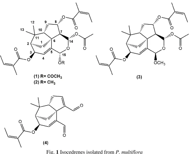
Fig. 1 Isocedrenes isolated from P. multiflora
4
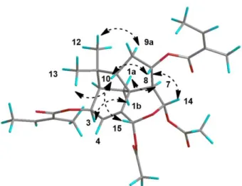
Fig. 3 Key NOESY correlations for compound 1
Compounds 1-4 were isolated from the ethanol extract of the aerial parts of P. multiflora
by bioguided fractionation.
Compound 1 was isolated as colorless oil. Its molecular formula was established as
C29H38O9 by positive HRESIMS (m/z 548.2856, [M + NH4]+) and showed 11 indices of
hydrogen deficiency. Careful examination of 2D-NMR spectra allowed us to determine the
planar structure, and to attribute all 1H and 13C NMR signals. The two methyl singulets at δH
0.74 and 1.19 present HMBC correlations with one another and carbon signals at δC 43.3 (C),
52.8 (CH), 60.5 (CH) which are typical of a gem-dimethyl group and allow to attribute C-11,
C-2 and C-10. 1H-1H COSY correlation between H-2 (δ
H 2.03, m) and the signal at δH 5.96
allows to attribute H-3. The position of the olefinic methine proton at δH 5.49 (q, J = 2.0 Hz,
H-4) was deduced by vicinal 1H-1H COSY correlation with H-3, and long-range 1H-1H COSY
correlation of H-3 and H-4 with the signal at δH 6.92 (dd, J = 2.8, 2.0 Hz) allowed us to attribute
H-15. This was confirmed by HMBC correlations between H-15, C-4, and C-5. Substitution of
C-15 (CH, δC 87.5) by an acetoxy group is confirmed by the HMBC correlation between H-15
and a carbonyl at δC 169.5, and the HMBC correlation of this latter signal with a methyl
singulet at δH 1.59. Starting from H-10 (1.72, m), successive 1H-1H COSY correlations
5
attribution as H-9a, H-9b and H-8 respectively. HMBC correlation of H-9b with a methine signal at δC 49.5 positioned C-7, and 1H-1H COSY correlation between H-7 (δH 2.09) and a
signal at δH 6.45 (d, J = 1.4 Hz) identified H-14. HMBC correlation of H-14 with a carbonyl at
δC 168.5, and the HMBC correlation of this carbonyl with a methyl singulet at δH 1.70
positioned an acetoxy substituent on C-14. The ethoxy link between C-14 and C-15 is
suggested by the chemical shifts of H-14 and H-15, typical of acetalic protons, and by HMBC
correlation between H-15 and C-14. HMBC correlation between a quaternary carbon at δC 47.5
and H-4 (5.49, q, J = 2.0 Hz) and H-7 or H-9 (δH 2.09) indicates the central position of C-6 (δC
47.5), and the isocedrene structure is closed by the attribution of H-1a (δH 1.81) thanks to its
COSY correlation with H-2 (δH 2.03), and HMBC correlation between C-1 (δC 44.5) and H-7
(δC 2.09). Signals typical of two angelate moieties are also present in the 1D and 2D spectra.
HMBC correlations between 2 carbonyl signals (δC 166.9 and 167.0) belonging to these
angelate with H-3 and H-8 positioned the angelate substituents on C-3 and C-8. This planar
structure, as well as the 1H NMR spectra were identical to these already described by Bohlman
for 3-angeloyl-8-angeloyloxyproustianol [1], however, the published 3D structure was not
coherent with our NMR data (coupling constants and NOESY), and prompted us to propose a
revised 3D structure. Bohlman’s structure was not coherent with the NOE effects observed
between H-8 and H-14, and between H-8 and H-10. Only a 8-angeloyl and a 14-acetoxy both in α position could explain these NOE effects. A β configuration for the 3-angeoyl moiety is
confirmed by the observed NOE effect between H-3 and H-1b, and this allowed us to
discriminate between H-1a and H-1b. The NOE effects between H-15 and H-10 allowed us to
determine the C-15 configuration. Furthermore, the NOE effects observed between H3-12 and
H-9a and H3-13 and H-10 allowed us to discriminate between H3-12 and H3-13, 9a and
6
The absolute configuration proposed (Fig. 1) was coherent with a biosynthetic origin
from a (-)-cyperene squeleton. It is also coherent with the absolute configuration of natural
trixanolides determined by Mosher’s method [11,12], and by ECD analysis supported by
theoretical calculation [10]. Furthermore, the ECD spectra of compound 1 showed the same
shape as these of previously reported trixanes [10]. Therefore compound 1 could be identified
as (2R,3R,6S,7S,8S,10S,14S,15R) 14,15-diacetoxy 3,8-diangeloyloxy,
14-15-epoxy-4-isocedrene.
Compound 2 was isolated as a colorless oil. Its molecular formula was established as
C28H38O8 by positive HRESIMS (m/z 525.2457, [M + Na]+) and showed 10 indices of
hydrogen deficiency.The 1H and 13C NMR data of 2, as well as the 2D NMR spectra, were
similar to those of 1, except for the disappearance of the signals of an acetoxy moiety, and the
appearance of signals of a methoxy group (δH 3.27, s, δC 55.5, CH3). The latter was positioned
on C-15, based on the the HMBC correlation of the methoxy protons with the carbon at δC
96.5. This latter signal could be attributed to C-15 because of the 1H-1H COSY correlation between the proton at δH 4.96 (dd, J = 2.7, 1.9 Hz, H-15) and the proton at δH 5.99 (dt, J = 4.8,
2.5 Hz, H-4). NOESY correlations and coupling constants were similar to those of 1, as well
as ECD spectra. Therefore compound 2 could be identified as (2R,3R,6S,7S,8S,10S,14S,15S)
14-acetoxy, 3,8-diangeloyloxy, 14-15-epoxy, 15-methoxy-4-isocedrene. This is the first
occurrence of this compound in the literature.
Compound 4 was isolated as a colorless oil. Its molecular formula was established as
C20H24O4 by positive HRAPCIMS (m/z 329.1742, [M + H]+) and showed 9 indices of hydrogen
deficiency. Analysis of 1H and 13C NMR data, as well as the 2D NMR spectra allowed us to
establish the structure of 4. This compound was first described by Bohlman as the product of
7
data matched with published ones. This compound may therefore be an artefact and the result
of the hydrolysis of 1, 2 or 3 during the purification on silica gel.
Compound 3 was isolated as a colorless oil. Its molecular formula was established as
C28H38O8 by positive HRESIMS (m/z 525.2458, [M + Na]+) and showed 10 indices of
hydrogen deficiency. 1H NMR data matched with published one for an isocedrene isolated from
P. runcinata but again, the published structure was not consistent with our data, especially with
the NOESY correlations [13].
Analysis of 1H and 13C NMR data, as well as the 2D NMR spectra indicates that
compound 3 was a diastereomer of compound 2. The most striking differences on the 1H spectra
were the chemical shifts of the H-1a, H-1b, and H-10 protons and the coupling constants of the
H-14 and H-15 protons. The strong difference between the coupling constant between H-14
and H-7 (1.7 Hz for compound 2 and 7.0 Hz for compound 3) prompted us to describe 3 as an
epimer of 2 with opposite configuration in C-14. However, this structure was not consistent
with the strong NOE effect observed between H-14 and H-8. The isocedrene structure could
present different conformations due to the conformational flexibility of the tetrahydropyrane
ring, which influences the geometry around C-14 and C-15. For compound 1 and 2 the data
matched with a conformation were H-14 and H-15 were both pseudo-equatorials. For
compound 3, NMR data could match with the structure of the epimer of 2 in C-15, but with a
different conformation of the tetrahydropyrane ring, were 14 would be pseudoaxial and
H-15 pseudoequatorial. Therefore, it seemed to us necessary to determine the major conformers
contributing to the structure of 1, 2, and 3 more accurately. Then calculation of the NMR data
corresponding to these conformers, and comparison with experimental spectra would permit to
evaluate the relevance of the structures we proposed for the three compounds.
For each compound, the conformers were modeled by DFT, using Gaussian 09 at the
8
optimized geometries at 298K, showing all positive frequencies and allowing evaluation of the
enthalpies of the minima.
13C NMR and 1H chemical shift calculations were then performed using the GIAO
NMR method with B3LYP/6-31+(d,p) and using the chloroform polarizable continuum model
(PCM) on optimized geometries at the B3LYP/6-31+G(d,p) level in the gas phase. Isotropic shielding constants (σ) for the 13C nucleus and 1H were transformed in chemical shifts (δ) using
linear regression procedure proposed by Tantillo [14]. The contribution of each conformer in
the constitution of the overall spectrum was based on Boltzmann conformational analysis.
Optimized geometries and calculated values on major conformers of compounds 1 and
2 match with their experimental data, which were also recorded in CDCl3. Detailed calculations
and 1H NMR simulated spectra of compounds 1 and 2 are consigned in tables SI.1-SI.6 and
figures SI.1-SI.2 of the supporting informations section.
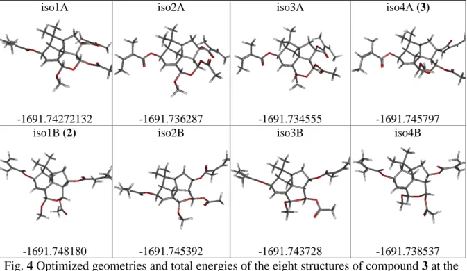
Concerning the compound 3 the four isomers corresponding to the two possible configurations
of H-14 and H-15 were considered, and for each structure, the two conformations (A and B) of
9
iso1A iso2A iso3A iso4A (3)
-1691.74272132 -1691.736287 -1691.734555 -1691.745797
iso1B (2) iso2B iso3B iso4B
-1691.748180 -1691.745392 -1691.743728 -1691.738537
Fig. 4 Optimized geometries and total energies of the eight structures of compound 3 at the B3LYP/6-31+G(d,p) in the gas phase.
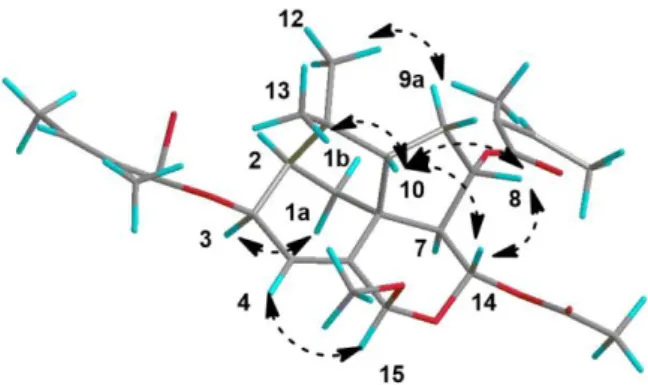
The strong NOE effect observed between H-14 and H-8 allows us to retain only isomers
where distance between H-14 and H-8 is less than 3Å. The experimental coupling constant
between H-14 and H-7 is around 7Hz; the calculated values which are the most approaching
this experimental one are respectively 8.4Hz for isomer 1A, 5.9Hz for isomer 2A and 8.1Hz
for isomer 4A, the other values being quite smaller. Furthermore, the root mean square
deviations (RMSD) on the calculated 13C chemical shifts has to be less than 2.94 with the
chosen method [13]; calculations of the RMSD of the eight isomers provide without any
ambiguity the matching of compound 3 with isomer 4A. (Table 3). For this structure, where
H-7 and H-14 are both pseudo axial (Fig. 5), the NOE effect observed between them is puzzling.
However, tables SI.4, SI.8 and SI.20 showed that in our conditions, NOE effects could be
observed between protons more than 3Å apart, and calculated distance between H-7 and H-14
10
Table 3. Calculated distances between H-14 and H-8, coupling constants between H-14 and
H-7, and RMSD obtained on 13C chemical shifts obtained for the eight structures modeled.
iso1A iso1B (2) iso2A iso2B iso3A iso3B iso4A (3) iso4B
d(H14-H8)/Å 2.64 2.74 2.70 3.72 2.64 3.70 2.38 2.89
J(H14-H7)/ Hz 8.4 1.7 5.9 3.8 5.1 4.1 8.1 1.8
RMSD/ppm 5,70 3,25 5,98 3,16 5,92 3,10 2,61 3,78
The summary of these DFT-NMR calculations and the comparison with the experimental data
are presented in the supplementary information.
Therefore, the structures of 1, 2 and 3 proposed (Fig. 1) are confirmed.
11
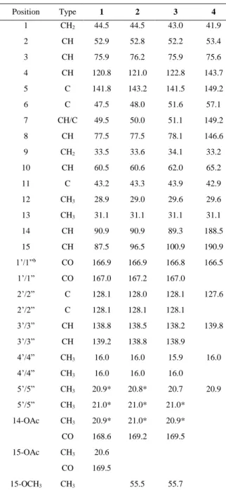
Table 1 13C NMR Spectroscopic Data (125 MHz, C6D6)for Compounds 1-4 (δ in ppm)
Position Type 1 2 3 4 1 CH2 44.5 44.5 43.0 41.9 2 CH 52.9 52.8 52.2 53.4 3 CH 75.9 76.2 75.9 75.6 4 CH 120.8 121.0 122.8 143.7 5 C 141.8 143.2 141.5 149.2 6 C 47.5 48.0 51.6 57.1 7 CH/C 49.5 50.0 51.1 149.2 8 CH 77.5 77.5 78.1 146.6 9 CH2 33.5 33.6 34.1 33.2 10 CH 60.5 60.6 62.0 65.2 11 C 43.2 43.3 43.9 42.9 12 CH3 28.9 29.0 29.6 29.6 13 CH3 31.1 31.1 31.1 31.1 14 CH 90.9 90.9 89.3 188.5 15 CH 87.5 96.5 100.9 190.9 1’/1”b CO 166.9 166.9 166.8 166.5 1’/1” CO 167.0 167.2 167.0 2’/2” C 128.1 128.0 128.1 127.6 2’/2” C 128.1 128.1 128.1 3’/3” CH 138.8 138.5 138.2 139.8 3’/3” CH 139.2 138.8 138.9 4’/4” CH3 16.0 16.0 15.9 16.0 4’/4” CH3 16.0 16.0 16.0 5’/5” CH3 20.9* 20.8* 20.7 20.9 5’/5” CH3 21.0* 21.0* 21.0* 14-OAc CH3 20.9* 21.0* 20.9* CO 168.6 169.2 169.5 15-OAc CH3 20.6 CO 169.5 15-OCH3 CH3 55.5 55.7 a : Signals overlap
12
Table 2 1H NMR Spectroscopic Data (500 MHz, C6D6) for Compounds 1-4 (δ in ppm)
Position 1 2 3 4 1 1.81a 1.84a 1.66 m 1.41 dd (12.2, 5.7) 1.91 m 1.95a 2.09a 2.13 dd (12.1, 2.0) 2 2.03 m 2.11a 2.08a 2.04a 3 5.96, dt (4.9, 2.5) 5.99 dt (4.8, 2.5) 5.92, ddd (4.3, 2.2, 0.8) 5.94 dd (4.6, 2.4) 4 5.49 q (2.0) 5.68a 5.37 se 5.84 dd (2.4, 1.7) 7 2.10 dd (8.8, 1.5) 2.11a 2.30 dd (7.0, 4.2) 8 5.20 ddd (10.5,8.9,5.9) 5.31 ddd (10.5, 8.8, 5.9) 5.50 ddd (7.1, 6.1, 4.2) 5.86 t (2.6) 9 1.32 m 1.40 m 1.58 ddd (13.9, 9.9, 6.2) 1.92 dd (9.1, 2.5) 2.07 2.13a 2.06a 1.96 dd (9.4, 2.7) 10 1.72 m 1.89 m 2.62 td (9.6, 1.9) 2.34 td (9.3, 1.9) 12 0.74 s 0.78 s 0.79 s 0.67 s 13 1.19 s 1.27 s 1.33 s 1.10 s 14 6.45 d (1.4) 6.41 d (1.7) 6.43 d (7.0) 9.62 s 15 6.92 dd (2.8, 2.0) 4.96 dd (2.7, 1.9) 4.91 se 9.09 s 3’/3” 5.71 m 5.70 m 5.70 m 5.77 qq (7.3, 1.5) 3’/3” 5.71 m 5.70 m 5.70 m 4’/4” 1.99 dq (7.3, 1.5) 2.00 dq (7.2, 1.5) 2.03 dq (7.2, 1.6) 2.04 dq (7.2, 1.5) 4’/4” 1.96 dq (7.2, 1.5) 1.95 dq (7.2, 1.6) 1.97 dq (7.2, 1.5) 5’/5” 1.82a 1.82a 1.82 m 1.82 p (1.5) 5’/5” 1.82a 1.82a 1.82 m 14-OAc 1.70 s 1.74 s 1.71 s 15-OAc 1.59 s 15-OMe 3.27 s 3.36 s aSignals overlapped
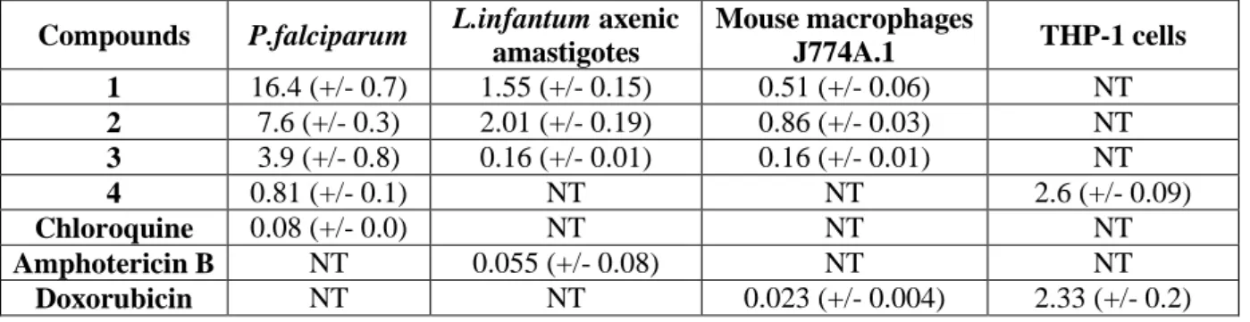
Antiparasitic activity of compounds 1, 2, 3 and 4 was evaluated on P. falciparum and
Leishmania infantum axenic amastigotes following already published protocols. To assess the
selectivity of their antiparasitic activity, their toxicity on J774A.1 macrophages or THP-1 cells
was evaluated. Results are reported in Table 4. The bioguided fractionation process which
allows the isolation of 1, 2, 3 and 4 was based on the activity of the ethanolic extract of P.
multiflora on in vitro culture of P. falciparum (IC50 = 4 µg/mL). The ethyl acetate fraction,
obtained after partition of ethanolic extract between ethyl acetate and water was further
13
concentrate the biological activity (IC50 = 0.8 µg/mL). Only compound 4 displayed a slightly
better antiplasmodial activity than the methanol fraction (IC50 = 0.8 µM or 0.26 µg/mL)
suggesting that some others compounds contribute to the activity of the extract. Compound 4
was the only one to present small selectivity against mammalian with a SI of 5. Compounds
showed only moderate activity against P. falciparum, and displayed no selectivity. This work
is the first report of antiplasmodial activity and cytotoxic activity of isocedrenes or trixanes.
These compounds displayed better activity on L. infantum axenic amastigotes, but in the same
range than their toxicity on J774A.1 macrophages. Antileishmanial activity of one trixane
isolated from Trixis antimenorrhoea has already been reported on promastigotes of L. infantum
and L. amazonensis [12].
Table 4 Biological activity of compounds 1-4 (IC50 values in µM)
Compounds P.falciparum L.infantum axenic
amastigotes Mouse macrophages J774A.1 THP-1 cells 1 16.4 (+/- 0.7) 1.55 (+/- 0.15) 0.51 (+/- 0.06) NT 2 7.6 (+/- 0.3) 2.01 (+/- 0.19) 0.86 (+/- 0.03) NT 3 3.9 (+/- 0.8) 0.16 (+/- 0.01) 0.16 (+/- 0.01) NT 4 0.81 (+/- 0.1) NT NT 2.6 (+/- 0.09) Chloroquine 0.08 (+/- 0.0) NT NT NT Amphotericin B NT 0.055 (+/- 0.08) NT NT Doxorubicin NT NT 0.023 (+/- 0.004) 2.33 (+/- 0.2) Conclusion
In this work, we describe four isocedrenes isolated from P. multiflora, a highly valued
herbal remedy in the andean mountains. One of them, compound 2 is new. Compound 1 was
already isolated from P. multiflora and compound 3 from P. runcinata, but carefull
examination of their NMR data, together with calculation of stability of their conformers and
prediction of their NMR spectra allowed us to propose revised 3D structure for both of them.
This work highlight the influence of the substitution of the tetrahydropyranose ring of
14
first report of biological activity of isocedrene sesquiterpenes. Their antiparasitic activity is not
selective enough, but it could be interesting to evaluate their cytotoxicity on cancerous cells
lines. As isocedrenes appeared here as potentially toxic molecules, it would be interesting to
evaluate their concentration in traditional preparations made with P. multiflora, to assess the
safety of use of this herbal remedy.
Materials and methods Chemical and reagents
Ethanol (96%) was from Laboratoire ALKOFARMA E.I.R.L, (Lima, Pérou).
Petroleum ether, cyclohexane, dichloromethane, ethyl acetate, methanol, were of analytical or
HPLC grade and bought from Fisher Scientific SAS. Merck Silica gel 60 (15–40 µm) was used
for medium pressure column chromatography (MPLC). Analytical thin-layer chromatography
was achieved on precoated silica gel plates (Merck, Kieselgel 60 F254, 0.25 mm) using UV 254
nm and a 1% vanillin/10% H2SO4 reagent in ethanol for visualization.
General procedures
MPLC column were run with Büchi medium pressure glass columns of various sizes,
with a Büchi pump manager C-615 connected to a Büchi pump module C-601. Optical rotations
were determined with a JASCO P2000 digital polarimeter. CD spectra were recorded on a
JASCO J-815, at 20°C, with a measurement range of 190-280 nm, a data interval of 0.2 nm, a
bandwidth of 1 nm and a scanning speed of 20nm/min, at a concentration of 7.75 ppm. The
NMR spectra were recorded on a Bruker Avance 500 MHz instrument with samples diluted in
C6D6 (δH 7.16 and δC 128.06) or on a Bruker Avance 300 MHz instrument with samples diluted
in CDCl3 (δH 7.28 and δC 77.1). UHPLC/MS analysis was performed using a UHPLC Ultimate
3000 system (Dionex) controlled by Chromeleon Xpress 6.8 software (Dionex), coupled with
an LTQ Orbitrap XL mass spectrometer (Thermo Fisher Scientific). All spectra were acquired
15
Plant material
Perezia multiflora (Bonpl.) Less. was purchased as a whole fresh plant in the Challwa
market (town of Huaraz, Peru) in august 2017. A voucher specimen was deposited at the
national herbarium of the museum of natural history (Universidad Mayor Nacional San
Marcos, Lima, Peru, deposit N° 026-2018-USM-MHN), where it was identified by specialists.
Whole plant was dried at ambient temperature in a ventilated place away from the sun. Aerial
parts and roots were separated, and aerial parts were grinded.
Extraction and isolation of pure compounds
Grinded aerial plant (375 g) was extracted successively three times in 3.3 L of EtOH
96% at ambient temperature during 12 h. Filtrates were pooled together and solvent was
evaporated under reduced pressure to give 53 g of extract. This extract was partitioned between
1 L of ethyl acetate and 1 L of water. A white precipitate was removed and ethyl acetate phase
was separated. The solvent was removed under reduced pressure to give 20.4 g of ethyl acetate
extract. This extract was further partitioned between 400 mL of petroleum ether and 400 mL
of methanol containing 10% of water. After removal of solvents under reduced pressure, 7.9 g
of petroleum ether extract and 10.3 g of methanol extract were obtained.
Purification of compounds 1-3: Methanol extract (5 g) was submitted to MPLC (diameter of
column: 4 cm, packed with 65 g of silica). Column was eluted with dichloromethane containing
increasing amount of methanol (500 mL 0%, 500 mL 1%, 500 mL 2%, 500 mL 5%, 500 mL
10% and 300 mL 100%) and 25 mL fractions were collected. Based on TLC analysis, the eluted
fractions were pooled into six fractions (F1 - F6) of which each was tested in the P. falciparum
assay. Fraction 2 (546 mg), displayed the best activity against P. falciparum (IC50 = 0.4
µg/mL). Fraction 2 (520 mg) was submitted to MPLC (diameter of column: 3 cm, packed with
16
acetate (500 mL 10%, 600 mL 15%, 500 mL 25%) and 20 mL fractions were collected.
Fractions were pooled according to their TLC profiles and 8 fractions were obtained (F2-1 –
F2-8). F2-4 (110 mg) was submitted to size exclusion chromatography (10 g of Sephadex®
LH-20 conditioned with methanol in a 1 cm diameter glass column). Column was eluted with
methanol, 2 mL fractions were collected, and pooled according to their TLC profile to give
compound 1 (89 mg). F2-1 (18.5 mg) was treated with the same protocol to give compound 3
(10.7 mg). F2-2 (37.8 mg) was submitted to size exclusion chromatography (1 g of Sephadex®
LH-20 conditioned with methanol in a 1 cm diameter column). Column was eluted with
methanol, 0.5 mL fractions were collected, and pooled according to their TLC profile to give
compound 2 (26.3 mg). F2-3 (35 mg) was treated with the same protocol to give 2 fractions
(F2-3-1, and F2-3-2).
Purification of compound 4: Methanol extract (5 g) was submitted to the same column
chromatography as described above. Fractions pooling was slightly different and gave 8 fractions (F1’ - F8’). Fractions F2’ (281 mg) and F3’ (302 mg) displayed the best activity
against P. falciparum (IC50 = 0.23 µg/mL and 0.7 µg/mL respectively). F2’ was submitted to
MPLC (diameter of column: 2.5 cm, packed with 35 g of silica). Column was eluted with
cyclohexane containing increasing amount of ethyl acetate (500 mL 10%, 600 mL 15%, 500
mL 25%, 350 mL 50%) and 20 mL fractions were collected. Fractions were pooled according
to their TLC profiles and 5 fractions were obtained (F2-1’ – F2-5’). F2-2’ (76.3 mg) was further
purified with size exclusion chromatography (40 g of Sephadex® LH-20 conditioned with
methanol in a 2 cm diameter glass column). Column was eluted with methanol, 2 mL fractions were collected, and pooled according to their TLC profile to give compound 4 (23.7 mg).
Compound 1: colorless oil, Rf 0.40, silica gel 60 F254, Petroleum ether/AcOEt (80:20); [α]𝐷20
17
1, 2 for data in C6D6 and table SI.21 for data in CDCl3. HR ESIMS (pos. ion mode) m/z
548.2856, [M + NH4]+ (calcd for C29H42NO9 548.2854)
Compound 2: colorless oil, Rf 0.60, silica gel 60 F254, Petroleum ether/AcOEt (80:20); [α]𝐷20
+3.0 (c 0.43, CH2Cl2), CD (CH3CN) Δε206.2 +25.5, Δε227.4 -7.6; 1H and 13C NMR : see tables 1,
2 for data in C6D6 and table SI.21 for data in CDCl3. HR ESIMS (pos. ion mode) m/z 525.2457,
[M + Na]+ (calcd for C28H38O8Na 525.2459)
Compound 3: colorless oil, Rf 0.65, silica gel 60 F254, Petroleum ether/AcOEt (80:20); [α]𝐷20
-38.2 (c 0.75, CH2Cl2), CD (CH3CN) Δε204.4 +25.3, Δε224.8 -7.9; 1H and 13C NMR : see tables
1, 2 for data in C6D6 and table SI.21 for data in CDCl3. HR ESIMS (pos. ion mode) m/z
525.2458, [M + Na]+ (calcd for C28H38O8Na 525.2459)
Compound 4: colorless oil, Rf 0.36, silica gel 60 F254, Petroleum ether/AcOEt (80:20); 1H
and 13C NMR : see tables 1, 2 for data in C6D6. HR APCIMS (pos. ion mode) m/z 329.1742,
[M + H]+ (calcd for C
20H25O4 329.1747)
Biological assays
Protocols for antiplasmodial and antileishmanial activity assays were already described [15].
Cytotoxicity evaluation on J774A.1
The evaluation of the tested molecules cytotoxicity by MTT assay was done on the J774A.1 cell
line (mouse macrophage cell line, Sigma-Aldrich). Briefly, cells (5.104 cells/mL) in 100 µL of
complete medium, [DMEM High glucose supplemented with 10% fetal calf serum (FCS), 2 mM
L-glutamine and antibiotics (100U/mL penicillin and 100 µg/mL streptomycin)] were seeded into
each well of 96-well plates and incubated at 37 °C in a humidified 5% CO2 with 95% air
atmosphere. After a 24 h incubation, 100 µL of medium with various product or extracts
18
°C. Each plate-well was then microscope-examined for detecting possible precipitate formation
before the medium was aspirated from the wells. 100 µL of MTT solution (0.5 mg/mL in RPMI)
were then added to each well. Cells were incubated for 2 h at 37 °C. After this time, the MTT
solution was removed and DMSO (100 µL) was added to dissolve the resulting formazan crystals.
Plates were shaken vigorously (300 rpm) for 5 min. The absorbance was measured at 570 nm with
a microplate spectrophotometer. DMSO or MeOH was used as blank and doxorubicin (purchased
from Sigma Aldrich) as positive control. CC50 were calculated by non-linear regression analysis
processed on dose–response curves, using TableCurve 2D V5 software.
Cytotoxicity evaluation on THP-1
The evaluation of the tested molecules cytotoxicity by MTT assay was done on the THP-1 cell line
(human monocyte cell line). Briefly, cells (0.77.105 cells/mL) in 200 µL of complete medium
[RPMI 1640 supplemented with 10% fetal calf serum (FCS), 2 mM L-glutamine and antibiotics
(100U/mL penicillin and 100 µg/mL streptomycin)] + PMA (50 ng/ml) were seeded into each well
of 96-well plates and incubated at 37 °C in a humidified 5% CO2 with 95% air atmosphere. After
a 96h incubation, Plates were rinse 3 times with medium and 100µl of medium were added. 100
µL of medium with various product or extracts concentrations and appropriate controls were added
and the plates were incubated for 72 h at 37 °C. Further treatment of the plates was as described
above for J774A.1 macrophages.
Supporting information
Detailed results of DFT calculations and NMR modelisations for compounds 1, 2, 3: geometries,
total energies, enthalpies and Boltzmann distribution ; experimental (CDCl3) and calculated 13C
chemical shifts in the chloroform PCM continuum model (SMD), and RMSD value ; calculated
1H chemical shifts and coupling constants for the major conformers ; experimental NOE effects
19
MestReNova software compared to experimental one in CDCl3 ; Table of 1H and 13C NMR data
in CDCl3 for compounds 1, 2, 3 ; 1D and 2D spectra of compounds 1, 2, 3 in C6D6.
Acknowledgements
The authors thank the logistic support of the LMI-LaVi (UPCH-IRD, Lima, Peru) and their
directors : Pr. Rosario Rojas (UPCH) and Dr. Michel Sauvain (IRD). The authors are grateful to
Pierre Lavedan, Marc Vedrenne and Caroline Toppan (NMR platform of the Institut de Chimie de
Toulouse, ICT) for NMR analyses and to Dr Stéphane Massou (NMR platform of the Institut de
Chimie de Toulouse, ICT) for fruitful discussions and help with the redaction of the SI section. The
authors thank Dr Charles-Louis Serpentini (IMRCP, UMR 5623 CNRS-Université Toulouse 3) for
the recording of CD spectra. The authors are indebted to the personnel of Universidad Nacional
Mayor San Marcos, Lima, Peru, (UNMSM) herbarium for plant determination and the Servicio
Nacional Forestal y de Fauna Silvestre (SERFOR) for issuing the P. multiflora collecting and
research permits (No 406-2016-SERFOR/DGGSPFFS and No 80-2017-SERFOR/DGGSPFFS).
Conflict of Interest
The authors declare no conflict of interest
References
1 Bohlmann F, Zdero C. Neue sesquiterpene mit anomalem Kohlenstoffgerust aus der
tribus Mutisieae. Chemische berichte 1979; 112: 427-434
2 Bohlmann F, Zdero C. Uber eine neue gruppe von sesquiterpenlactonene aus der
Gattung Trixis. Chemische berichte 1979; 112: 435-444
3 Bohlmann F, Zdero C, King RM, Robinson H. A tetracyclic sesquiterpene, further
isocedrene, and guaiene derivatives from Jungia stuebelii. Phytochemistry 1983; 22:
1201-1206
4 Singh P, Jakupovic J, Bohlmann F. Isocedrene derivatives and other sesquiterpenes
20
5 Zdero C, Bohlmann F, Niemeyer HM. Isocedrene and guaiane derivatives from
Pleocarphus revolutus. Journal of natural products 1988; 51: 509-512
6 Zdero C, Bohlmann F, Sanchez H, Dominguez XA. Isocedrene derivatives and other
constituents from Acourtia nana. Phytochemistry 1991; 30: 2695-2697
7 Zdero C, Bohlmann F, King RM, Robinson H. α-Isocedrene derivatives, 5-methyl
coumarins and other constituents from the subtribe Nassauviinae of the Compositae.
Phytochemistry 1986; 25: 2873-2882
8 De Riscala EC, Catalan CAN, Sosa VE, Gutierrez AB, Herz W. Trixane derivatives
from Trixis praestans. Phytochemistry 1988; 27: 2343-2346
9 Ybarra MI, Catalan CAN, Diaz JG, Herz W. A cyperane and trixanes from Jungia
polita. Phytochemistry 1992; 31: 3627-3629
10 Azevedo L, Faqueti L, Kritsanida M, Efstathiou A, Smirlis D, Franchi GCJ,
Genta-Jouve G, Michel S, Sandjo LP, Grougnet R, Biavatti MW. Three new trixane glycosides
obtained from the leaves of Jungia sellowii less. using centrifugal partition
chromatography. Beilstein Journal of Organic chemistry 2016; 12: 674-683
11 Kotowicz C, Hernandez LR, Cerda-Garcia-Rojas CM, Villecco MB, Catalan CAN,
Joseph-Nathan P. Absolute configuration of trixanolides from Trixis pallida. Journal of
Natural Products 2001; 64: 1326-1331
12 Maldonadoa EM, Salamanca E, Gimenez A, Saavedra G, Sterner O. Antileishmanial
metabolites from Trixis antimenorrhoea. Phytochemistry Letters 2014; 10: 281-286
13 Zdero C, Bohlmann F, Solomon J, Dominguez XA. Further isocedrene derivatives and
other constituents from Perezia species. Phytochemistry 1988; 27: 849-853
14 Lodewyk MW, Siebert MR, Tantillo DJ. Computational Prediction of 1H and 13C
Chemical Shifts: A Useful Tool for Natural Product, Mechanistic, and Synthetic
21
15 Castro I, Fabre N, Bourgeade-Delmas S, Saffon N, Gandini C, Sauvain M, Castillo D,
Bourdy G, Jullian V. Structural Characterization and Anti-infective Activity of
9,10-Seco-29-norcycloartane Glycosides Isolated from the Flowers of the Peruvian