S. Schmidt P. Chevallier B. Bessoud J.-Y. Meuwly C. Felley R. Meuli P. Schnyder A. Denys Received: 6 September 2006 Revised: 27 February 2007 Accepted: 19 April 2007 Published online: 10 May 2007
# Springer-Verlag 2007
Diagnostic performance of MRI for detection
of intestinal fistulas in patients
with complicated inflammatory
bowel conditions
Abstract The diagnostic perfor-mance of magnetic resonance imaging (MRI) for detection of intestinal fistulas, other than perianal, in patients with known complicated inflammato-ry bowel conditions (CIBC) was investigated. Our study group con-sisted of 20 patients (12 women, mean age 43 years) with CIBC, including Crohn’s disease (n=13), colonic di-verticulitis (n=3), colitis after radio-therapy (n=3) and of postoperative origin (n=1). Eleven surgically proven enteral fistulas were known in ten (50%) of these patients, being of enterovesical (n=3), enterocolic (n=2), enteroenteral (n=2), rectovaginal (n=2), rectovaginovesical (n=1) and of entercutaneous (n=1) localisation. The other ten patients (50%), used as the control group, showed MR features of CIBC, although without any fistulous tract. Multiplanar T1- and
T2-weighted sequences had been per-formed, including gadolinium-en-hanced acquisition with fat saturation (1.5 T). MR findings were
indepen-dently blindly and retrospectively reviewed by three radiologists for the presence and etiology of any fistula, as well as visualization and characteri-zation of the fistulous tract. Results were compared with surgical findings (n=16) and clinical evolution (n=4). Interobserver agreement was calcu-lated. Interobserver agreement kappa for fistula detection was 0.71. Overall sensitivity, specificity and accuracy for fistula detection were 78.6%, 75% and 77.2%, respectively. Sensitivity for fistula characterization was 80.6%, with visualization of the fistulous tract in all cases, whereby T1-weighted gadolinium-enhanced fat-saturated images were considered the most useful sequences. Gadolinium-en-hanced MRI is a reliable and repro-ducible tool for detection of enteral fistulas secondary to inflammatory conditions.
Keywords Intestinal fistula . Magnetic resonance imaging . Crohn’s disease
Introduction
Gastrointestinal fistulas are abnormal duct-like commu-nications between the gut and another epithelial-lined surface, such as an organ system, the skin surface, or another segment of the GI tract itself [1].
The major cause of acquired intestinal fistulas is Crohn’s disease. Formation of fistulas to adjacent organs occurs in 20–40% of these patients, with a cumulative risk of 33% after 10 years and 50% after 20 years of disease [2, 3].
Other complicated inflammatory bowel conditions (CIBC) result from prior radiotherapy combined with surgery for pelvic malignancies. Colonic diverticulitis may also cause similar fistulous complications.
Imaging plays a pivotal role for the detection and management of acquired gastrointestinal fistulas [1]. Their origin and anatomic course may initially be evaluated by fluoroscopic contrast medium-enhanced studies, such as direct fistulography, in the case of a cutaneous orifice, and oral or rectal opacification. However, neither the accurate S. Schmidt (*) . J.-Y. Meuwly .
R. Meuli . P. Schnyder . A. Denys Service de radiodiagnostic et radiologie interventionnelle, Centre Hospitalier, Universitaire Vaudois—CHUV, Rue du Bugnon, 1011 Lausanne, Switzerland e-mail: [email protected] Tel.: +41-21-3144607 Fax: +41-21-3144443 P. Chevallier
Imagerie Médicale, Hôpital Archet II, Nice, France B. Bessoud Radiologie Genérale, Hôpital Kremlin-Bicêtre, Kremlin-Bicêtre, France C. Felley Service de Gastroentérologie, University Hospital, CHUV, Lausanne, Switzerland
anatomic relationship of the fistula with the surrounding structures nor any possible inflammatory involvement of adjacent organs can be depicted by fluoroscopy alone [1,2, 4]. Cross-sectional modalities, such as computed tomogra-phy (CT) and magnetic resonance imaging (MRI) are able to demonstrate extraintestinal disease adjacent to the fistula. CT is rapid, generally available and less costly than MRI. It has been known for inherent high spatial resolution, especially with multidetector-row technique. Nevertheless, contrast resolution of CT still remains less than optimal for the depiction of intestinal fistulae. Based on indirect signs, CT may only suggest the presence of a fistulous tract; however, often without its direct delineation [1,5–8].
The decisive advantage of MRI is excellent intrinsic soft-tissue resolution together with primary multiplanar imaging capability [9]. During the last decade, MRI has evolved into a current standard of practice for perianal fistulas [10–13]. Intestinal fistulas of more proximal origin, however, have only recently been studied by MRI [14–18], all secondary to Crohn’s disease. However, to our knowl-edge, there has not yet been any study evaluating interobserver agreement in this regard. The aim of our study was, therefore, to determine if MRI allows the reliable and reproductible detection of intestinal fistulas, other than perianal, in patients known for CIBC of various origins.
Materials and methods
Patients
This retrospective study had met with written approval by our ethics committee. Informed oral consent had been obtained from each patient.
In our database of patients investigated by abdominal MRI within a defined study period of 14 months, we retrospectively found ten patients with CIBC and 11 surgically proven intestinal fistula. From the same database, we selected another ten patients, constituting our control group, who were equally known for CIBC, but without intestinal fistula. They had also undergone MRI during the same period for defining the extension of CIBC; namely, exclusion of intestinal fistula.
Our study population was, therefore, composed of 20 patients [12 women and eight men (age,22–71 years; mean age, 43.2 years)] They had been addressed to our department for MR investigation of CIBC due to underlying Crohn’s disease (n=13), secondary to prior radiotherapy (n=3), colonic diverticulitis (n=3), and post-operative inflammatory involvement of the colon second-ary to necrotizing pancreatitis (n=1). Only the two radiologists (S.S., A.D.) responsible for the study design, were aware of these clinical data.
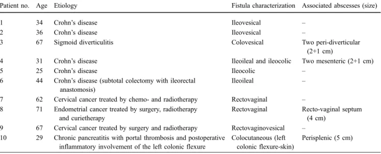
The detailed description of the ten patients with 11 intestinal fistulas, including their localization and asso-ciated abscesses can be seen in Table1.
The CIBC of our control group (n=10), without fistula, were characterized as follows. Eight patients were known for Crohn’s disease, among them four of the fibrostenosing category. Their corresponding MRI features were one (n=1) or two (n=3) substantially narrowed bowel segments. Three other Crohn’s patients belonged to the fistulizing/ perforating subtype, since mural inflammation was com-plicated by one (n=1) or two (n=2) associated abscesses, however without intestinal fistula confirmed by surgery. The eighth Crohn’s patient showed the active inflammatory type, characterized on MRI as important mural thickening of the terminal ileum. The two remaining patients of our control group had sigmoid diverticulitis, with one (n=1) or two (n=1) abscesses, respectively.
Table 1 Ten patients with 11 intestinal fistulas, including their etiology
Patient no. Age Etiology Fistula characterization Associated abscesses (size)
1 34 Crohn’s disease Ileovesical –
2 36 Crohn’s disease Ileovesical –
3 67 Sigmoid diverticulitis Colovesical Two peri-diverticular
(2+1 cm)
4 31 Crohn’s disease Ileoileal and ileocolic Two mesenteric (2+1 cm)
5 25 Crohn’s disease Ileocolic –
6 44 Crohn’s disease (subtotal colectomy with ileorectal anastomosis)
Ileoileal –
7 62 Cervical cancer treated by chemo- and radiotherapy Rectovaginal – 8 71 Endometrial cancer treated by surgery, radiotherapy
and curietherapy
Rectovaginal Recto-vaginal septum (4 cm)
9 67 Cervical cancer treated by surgery and radiotherapy Rectovaginovesical – 10 29 Chronic pancreatitis with portal thrombosis and postoperative
inflammatory involvement of the left colonic flexure
Colocutaneous (left colonic flexure-skin)
All these 11 intestinal fistulas were surgically treated at a mean delay of 21.4 days after MRI (range 14–40 days). Absence of fistula in the control group (n=10) was confirmed by surgery (n=6), performed for CIBC at an average of 31 days after MRI (range, 2–48 days). For each patient with surgical treatment (n=16), our reference standard was the histopathological result. In the remaining four patients without fistula, the negative MR findings were compared with follow-up (average, 40 months; range, 36–47 months). It was uneventful.
MR imaging
All MR studies were performed on a 1.5-T unit Symphony (phased array surface coil, Siemens Medical Systems, Erlangen, Germany). Patients were examined in the supine position.
The patients known to have Crohn’s disease fasted for 8 h prior to MRI. They had to drink 1–2 l of 5% methyl-cellulose for about 40–45 min before data acquisition in order to achieve good luminal small bowel distension, thus improving the visualization of any possible fistulous tract extending from bowel loops.
Immediately before MRI, a watery rectal enema was performed in all patients but four, who refused. Each of the 20 patients received 20 mg of scopolaminbutylbromid (Buscopan; Boehringer Ingelheim, Basel, Switzerland) i.v. or, if contraindicated, 1 mg of glucagon (GlucaGen; Novo Nordisk, Bagsvaerd, Denmark) i.v., to reduce potential image degradation resulting from bowel peristalsis.
We performed T1-weighted (T1WI) and T2-weighted (T2WI) MR sequences depending on the underlying disease and on the anatomical site of the suspected fistula. T1WI-sequences were always acquired with fat saturation (FS) before and 70 s after i.v. gadolinium (Gd) adminis-tration (DTPA-BMA, 0.1 mmol/kg of body weight: Omniscan, Nycomed Imaging, Oslo, Norway).
In patients with Crohn’s disease, we covered the whole region of the small and large intestine using axial and coronal T2-weighted (T2WI) half-Fourier single-shot turbo spin-echo sequences [HASTE: repetition time (TR) infinite, effective echo time (TE)eff 120 ms, echo train
136, echo spacing 4.3 ms, 1 excitation (NEX), matrix size 256×148]. They were followed by axial and coronal T1WI gradient-echo (GE) images (VIBE, TR 3.69, TE 1.59–1.64, flip angle 15°, 1 NEX, matrix size 256×176 in axial and 256×240 in coronal plane). Furthermore, axial True FISP sequences (true fast imaging with steady-state precession, TR 4.4, TE 2.2, 1 NEX, matrix size 256×148) were obtained [16,17].
Whenever the MRI was centered on the pelvis, axial and coronal T2WI turbo spin echo MR images without and with FS (TSE, TR 3,052–4,520, TE 86–121) were performed. They were followed by T1WI FS spin echo images (SE, TR 500–820, TE 12), acquired in axial, coronal and sagittal
plane. One excitation was used with a matrix size of 512×288 (axial), 512×307 (sagittal) and 512×384 (coronal).
We constantly used slices of 5-mm thickness in the axial plane and slices of 4-mm thickness in the coronal and sagittal plane (interslice gap 10%). The field of view (FOV) was 28–36×21–28 cm in the axial, 33–40×26–40 cm in the coronal and 26–28×26–28 cm in the sagittal plane.
If deemed necessary, the data acquisition was completed by high-resolution images of 3-mm thickness, performed as T2WI FS spin echo sequence [TSE, TR 3,052–4,520, TE 86–121, 2 NEX, matrix size of 512×245 (axial) or 512×205 (coronal)], centred on regions of interest.
The total examination time, including rectal enema, ranged from 30–40 min.
Image analysis
Three radiologists with 8 (observer 1), 10 (observer 2) and 6 (observer 3) years of experience in abdominal MRI were blinded to each patient’s clinical history and surgical findings. They independently read each MR examination, recording presence and etiology of any fistula as well as visualization and characterization of the fistulous tract. Before starting image analysis, the three observers only got to know the diagnosis“CIBC” in all patients with possible associated fistulous complications, but without other detail. A pre-reading session was organised and MR features of intestinal fistulas were defined in consensus. Each radiol-ogist had then to fill in one pre-established grid per patient during soft copy reading [Advantage Windows 7.4 GE work station, (General Electric Health Care, Milwaukee, USA)].
Inflammatory fistulas typically display a linear high signal intensity on T2WI MR sequences with a surround-ing zone of lower signal intensity representsurround-ing fibrosis or granulation tissue [7,13]. After i.v. Gd injection, a fistulous tract mostly exhibits longitudinal central low signal intensity surrounded by a thin contrast-enhanced rim, its inflammatory wall [13,19]. Very small fistulae measuring < 3 mm may not clearly demonstrate the central T1WI low signal intense fistulous tract. In these cases, the fistula merely appears as contrast-enhanced linear high or inter-mediate signal structure, seen on Gd-T1WI or T2WI sequence [13].
By comparing the different applied MR sequences with each other, the three observers separately analyzed the diagnostic quality of each MR sequence concerning the conspicuity of the fistulous tract and scored them as 3 (excellent), 2 (adequate) or 1 (poor). However, the aim was to establish the correct diagnosis for each patient using all the provided MR sequences together.
Finally, each of the three observers had to attribute an etiology to each fistula: underlying Crohn’s disease, colonic diverticulitis, after radiotherapy, of postoperative or unknown origin.
Statistical analysis
Statistical analysis was performed with a JMP 5.1 statistical package (SAS Institute, Cary, N.C.), including the whole study population of 20 patients.
Sensitivity, specificity, negative (NPV) and positive (PPV) predictive value, as well as accuracy for fistulae detection for each observer separately and altogether, were calculated.
Interobserver agreement for fistula detection was determined, according to the common kappa rating defined by Fleiss [20]: kappa <0.4=poor, 0.4–0.75=good, and > 0.75=excellent agreement.
The percentage of fistulae that each radiologist correctly characterized, was calculated. Characterization meant accu-rate anatomical localization and visualization of the fistulous tract. The percentage of fistulas identified by all three observers together was calculated, as well as the number of cases in which the etiology of the fistulae was found.
Results
The individual reading performance for fistula detection was as follows: sensitivity, specificity, NPV, PPV and
accu-racy were 81.8, 87.5, 90, 77.8 and 84.2% for observer one, 72.3, 50, 66.7, 57.1 and 63.2% for observer two, and 81.8, 87.5, 90, 77.8 and 84.2% for observer three, respectively. The overall diagnostic values were 78.6, 75, 82.2, 70.9 and 77.2%, respectively (Figs.1, 2). Interobserver agreement kappa between the three radiologists for fistula detection was 0.71; therefore, good [20]. The individual ability for correct characterization of the fistulas was 88.9, 75 and 77.8% for observer 1, 2 and 3, respectively. Correct localization of their extension was associated with visual-ization of the fistulous tract in all cases; altogether it was 80.6%. In general, intestinal fistulas communicating with a different organ, such as the bladder or the vagina, were more easily detected than fistulas extending between two adjacent bowel loops, especially in case of associated postinflam-matory or postoperative changes.
Observer 1 correctly identified the underlying disease in seven out of ten fistulae (70%), observer 2 in each of the 11 identified fistulas (100%) and observer 3 in eight out of ten fistulae (80%). Altogether, the correct etiology was attributed to each fistula in 83.3% of cases.
All observers considered T1WI FS Gd-enhanced images (Figs.1,2) as the most useful sequences for fistula detection and characterization (range 2.6–2.9, average 2.8), followed by HASTE and TSE T2WI, respectively (range 1.5–2.9, Fig. 1a–d A 31-year-old man
with acute exacerbation of long-standing Crohn’s disease (the patient refused the rectal administration of water before MRI). Coronal and axial Gd-enhanced T1-weighted images with fat suppression (TR/TE 3.69/1.64) show two intestinal fistulas. a, b On coronal images an ileoileal fistula is character-ized by a small fistulous tract (arrowhead), involving several inflamed distal ileal loops (thin arrows), with an associated small abscess (asterisk). c, d Secondly, axial images show an ileosigmoid fistula ex-tending as an irregular fistulous tract (arrowhead) between the equally inflamed and narrowed sigmoid colon (thick arrows) and terminal ileum (thin arrows)
average 2.3), then TSE T2WI FS (range 1.6–2.2, average 1.9), True-FISP (range 1–1.6, average 1.3) and pre-contrast T1WI FS MR sequences (range 1–1.1, average 1.1).
In order to avoid magnetic susceptibility artefacts resulting from metallic hip prostheses, which occurred in two patients of our study population, the T2WI HASTE MR sequence was found the most adequate technique.
In addition to the routine acquisition of axial slices, which the three radiologists considered the most useful ones, the sagittal plane was found particularly effective for the detection of intestinal pelvic fistulas communicating with the bladder or the genital organs. The coronal plane was especially helpful for demonstration of intestinal fistulas involving the small and large bowel (Figs.1,2).
Discussion
MRI has revealed excellent accuracy for perianal fistulas [11, 12], but more proximal intestinal fistulae are not routinely evaluated. In contrast, the contemporary imaging approach often remains characterized by conventional fluoroscopic studies, often combined with CT. They require the administration of an enteral contrast agent for
visualization of fistulous tracts. However, the tendency of hydrosoluble contrast agents to dilute with the intestinal luminal fluid and to become less opaque, limits the diagnostic value of these studies. Furthermore, they are negative in case of edematous occlusion of the fistulous tract preventing its opacification.
MRI does not require direct opacification of the fistulous tract. The excellent MR contrast resolution and the presence of fluid inside the active fistulous tract, working as natural luminal contrast agent, is sufficient for its delineation.
We chose the additional oral/rectal administration of methylcellulose/water to improve luminal bowel distension with consecutive better visualization of fistulas extending between different intestinal loops. The fistulous tract might then slightly enlarge due to the increased luminal pressure by this fluid challenge, and fistulas become more conspicuous. Our three observers considered the Gd-T1WI FS sequences as the most useful ones (mean score: 2.8) for fistula detection and characterization, similar to previous studies [13,21]. On Gd-T1WI FS sequences, the orifice of an intestinal fistula is characterized as mural point-shaped low signal intensity, surrounded by a thin contrast-enhanced rim. This feature seems to be more easily depicted than the focal discontinuity in the low signal muscularis of the wall Fig. 2a–d A 67-year-old
woman with subacute sigmoid diverticulitis complicated by urine tract infection. Coronal and axial Gd-enhanced T1WI images with fat suppression (TR/TE 3.69/1.64) demonstrate a sigmoidovesical fistula. a, b Axial images show that the fistula first extends within the thickenend and pathologically contrast-enhanced left anterolat-eral bladder wall (arrowhead), and then runs through an asso-ciated abscess located just above the bladder dome (asterisk). c, d Coronal images demon-strate the extension of the fis-tuluous tract (thin arrows) through the abscess (asterisk) and finally reaching the en-flamed sigmoid colon (thick arrows)
you have to rely on, when only T2WI sequences are performed [9]. Furthermore, possible associated abscess formations and the degree of intestinal wall inflammation, reflecting the risk of fistula formation, can simultaneously be exactly evaluated, especially important in underlying Crohn’s disease [21]. In our study, we detected adjacent abscesses in four out of ten patients with intestinal fistulas (40%).
Finally, by using water/methylcellulose, known as biphas-ic enteral contrast agents, we created an excellent contrast between the distended bowel lumen, exhibiting a low signal on T1WI sequences, and the inflamed, thus intensely contrast-enhanced, surrounding structures. On T2WI images, the fluid-filled fistuluos tract may be simulated by the water-filled high-signal-intensity bowel lumen.
Our study demonstrated a good interobserver agreement (kappa=0.71) for fistula detection, proving good reproduc-ibility for MRI in this regard. Our aim was to stress the objective nature of image interpretation obtained by three independent readings. To our knowledge, there has not been any other study determining interobserver agreement for intestinal fistula detection by MRI.
We deliberately chose a patient population uniformly known for CIBC, even the control group. Under these conditions, we consider the task to correctly select the positive patients as more difficult than if our study design had consecutively included all the patients investigated by intestinal MRI within this period. We, hereby, wanted to stress the additional diagnostic difficulty our readers had to overcome by detecting intestinal fistulas among patients showing each important MR features of bowel inflamma-tion anyway, mostly with adjacent abscesses or bowel stenoses.
There is no doubt that the sensibility of intestinal fistula detection primarily depends on the size of the fistulous tract, whatever technique may be used. In general, pelvic fistulas, of rectovaginal, uterine or vesical localization, are of larger diameter than the fistulous tract extending between bowel loops. Furthermore, the indirect sign of these pelvic fistulas—that is, air bubbles in the vagina, uterus or bladder in absence of previous catheterization—is a pathognomonic feature. It may also quite easily be depicted by other cross-sectional techniques. The detection of intestinal fistulae extending between bowel loops is
considered more difficult, because of mostly smaller size and the irrelevance of air bubbles, confirmed by our study. There are limitations to our study. The luminal intestinal fluid challenge we obtained by oral administration of methylcellulosis might have been more effective using the technique of enteroclysis. Patients, however, seldom readily accept the need for nasojejunal intubation. That is why we did without, while relying on the relaxing effect of the i.v. injected antiperistaltic agent, as reported previously [22,23]. Our study design is retrospective, including a quite small number of patients. However, to our knowledge, there is no previous study focusing on the MR detection of intestinal fistulas others than of perianal among a population of patients with CIBC of various origins.
Four patients out of our control group had no surgical correlation, but they had been followed-up closely for 40 months. This long uneventful delay, together with the excellent negative predictive value (91.7–100%) and the high specificity (96.7–100%) for MRI of intestinal fistulas revealed by other working-groups [16–18], reliably con-firms our negative results.
Furthermore, our chosen study group shows a high prevalence of intestinal fistulas (50%), which one may never come across in real clinical settings. We deliberately selected these patients; among them, each demonstrated inflammatory features of difficult interpretation, challeng-ing whether MRI could serve as an appropriate problem-solving imaging modality in this regard. This high concentration of complicated cases may—at least par-tially—compensate for our relatively small number of patients.
Finally, MRI remains a cross-sectional technique, gen-erally not permitting dynamic imaging. Therefore, we could not obtain information about fistula activity. MR fluoroscopy has been attempted for small bowel disease [24, 25], but mainly to monitor the luminal filling and degree of distension during enteroclysis [16,17].
In conclusion, our results show that MRI is a reliable and reproducible tool for the detection of inflammatory enteral fistulas of others than perianal location. We emphasize the particular diagnostic value of MRI in selecting patients with intestinal fistulas among a study population with CIBC of various origins.
References
1. Pickhardt PJ, Bhalla S, Balfe DM (2002) Acquired gastrointestinal fistu-las: classification, etiologies, and im-aging evaluation. Radiology 224:9–23
2. Gore RM, Balthazar EJ, Ghahremani GG, Miller FH (1996) CT features of ulcerative colitis and Crohn’s disease. AJR Am J Roentgenol 167:3–15 3. Schwartz DA, Loftus EV, Tremaine WJ
et al (2002) The natural history of fistulizing Crohn’s disease in Olmsted County, Minnesota. Gastroenterology 122:875–880
4. Maglinte DDT, Gourtsoyiannis N, Rex D, Howard TH, Kelvin FM (2003) Classification of small bowel Crohn’s subtypes based on multimodality im-aging. Radiol Clin N Am 41:285–303
5. Kuhlman JE, Fishman EK (1990) CT Evaluation of enterovaginal and vesi-covaginal fistulas. J Comput Assist Tomogr 14:390–394
6. Goldman SM, Fishman EK, Gatewood IMB, Jones B, Siegelman SS (1985) CT in the diagnosis of enterovesical fistulae. AJR Am J Roentgenol 129:1229–1233
7. Hession P, Mannion RAJ, Finan P, Chalmers AG (1997) Imaging appear-ances of ileouterine fistula complicating recurrent adenocarcinoma of the rec-tum. Br J Radiol 70:415–417
8. Murphy JM, Lee G, Sharma SC, Doble A, Lomas DJ (1999) Vesicouterine fistula: MRI diagnosis. Eur Radiol 9:1876–1878
9. Outwater E, Schiebler ML (1993) Pel-vic fistulas: findings on MR images. AJR Am J Roentgenol 160:327–330 10. Bartram C, Buchanan G (2003)
Imag-ing anal fistula. Radiol Clin N Am 41:443–457
11. Morris J, Spencer JA, Ambrose NS (2000) MR imaging classification of perianal fistulas and its implications for patient management. Radiographics 20:623–635
12. Spencer JA, Chapple K, Wilson D, Ward J, Windsor ACI, Ambrose NS (1998) Outcome after surgery for peri-anal fistula: predictive value of MR imaging. AJR Am J Roentgenol 171:403–406
13. Semelka RE, Hricak H, Kim B et al (1997) Pelvic fistulas: appearances on MR images. Abdom Imaging 22:91–95
14. Albert JG, Martiny F, Krummenerl A et al (2005) Diagnosis of small bowel Crohn’s disease: a prospective com-parison of capsule endoscopy with magnetic resonance imaging and fluoroscopic enteroclysis. Gut 54:1721–1727
15. Pilleul F, Godefroy C, Yzebe-Beziat D et al (2005) Magnetic resonance imag-ing in Crohn’s disease. Gatroenterol Clin Biol 29:803–808
16. Gourtsoyiannis NC, Grammatikakis J, Papamastorakis G et al (2006) Imaging of small intestinal Crohn’s disease: comparison between MR enteroclysis and conventional enteroclysis. Eur Radiol 16:1915–1925
17. Masselli G, Casciani E, Polettini E et al (2006) Assessment of Crohn’s disease in the small bowel: prospective com-parison of magnetic resonance entero-clysis with conventional enteroentero-clysis. Eur Radiol 16:2817
18. Herrmann KA, Michaely HJ, Seiderer J et al (2006) The“star-sign” in mag-netic resonance enteroclysis: a charac-teristic finding of internal fistulae in Crohn’s disease. Scand J Gastroenterol 41:239–241
19. Healey JC, Philips RR, Reznek RH, Crawford RA, Armstrong P, Shepherd JH (1996) The MR appearance of vaginal fistulas. AJR Am J Roentgenol 167:1487–1489
20. Fleiss JL (1985) Statistical methods for rates and proportions, 2nd edn. Wiley, New York, pp 211–236
21. Gourtsoyiannis N, Papanikolaou N, Grammatikakis J, Prassopoulos P (2002) MR enteroclysis: technical con-siderations and clinical applications. Eur Radiol 12:2651–2658
22. Borthne AS, Abdelnoor M, Hellund JC et al (2005) MR imaging of the small bowel with increasing concentrations of an oral osmotic agent. Eur Radiol 15:666–671 23. Ajaj W, Goyen M, Schneemann H et al (2005) Oral contrast agents for small bowel distension in MRI: influence of the osmolarity for small bowel disten-tion. Eur Radiol 15:1400–1406 24. Prassopoulos P, Papanikolaou N,
Grammatikakis J, Rousomoustakakt M, Maris T, Gourtsoyiannis N (2001) MR enteroclysis: imaging of Crohn disease. Radiographics 21:S161–S172
25. Umschaden HW, Szolar D, Gaser J, Umschaden M, Haselbach H (2000) Small bowel disease: comparison of MR enteroclysis images with conven-tional enteroclysis and surgical find-ings. Radiology 215:717–725