HAL Id: hal-03196133
https://hal.archives-ouvertes.fr/hal-03196133
Submitted on 12 Apr 2021HAL is a multi-disciplinary open access
archive for the deposit and dissemination of sci-entific research documents, whether they are pub-lished or not. The documents may come from teaching and research institutions in France or abroad, or from public or private research centers.
L’archive ouverte pluridisciplinaire HAL, est destinée au dépôt et à la diffusion de documents scientifiques de niveau recherche, publiés ou non, émanant des établissements d’enseignement et de recherche français ou étrangers, des laboratoires publics ou privés.
Reduction of [Cp*2Mo2O5] in Aqueous Medium:
Structure and Properties of a Triangular Mixed
Oxo-Hydroxo-Bridged Product,
[Cp*3Mo3(µ-O)2(µ-OH)4](X)2 (X = CF3CO2 or
CF3SO3)
Funda Demirhan, Bahar Çağatay, Deniz Demir, Miguel Baya, Jean-Claude
Daran, Rinaldo Poli
To cite this version:
Funda Demirhan, Bahar Çağatay, Deniz Demir, Miguel Baya, Jean-Claude Daran, et al.. Reduction of [Cp*2Mo2O5] in Aqueous Medium: Structure and Properties of a Triangular Mixed Oxo-Hydroxo-Bridged Product, [Cp*3Mo3(µ-O)2(µ-OH)4](X)2 (X = CF3CO2 or CF3SO3). European Journal of Inorganic Chemistry, Wiley-VCH Verlag, 2006, 2006 (4), pp.757-764. �10.1002/ejic.200500665�. �hal-03196133�
Reduction of Cp*
2Mo
2O
5in an aqueous medium. Structure and properties
of a triangular mixed oxo-hydroxo-bridged product, [Cp*
3Mo
3(-O)
2(-OH)
4](X)
2, with X = CF
3CO
2or CF
3SO
3Funda Demirhan*,a Bahar Çağatay,a Deniz Demir,a Miguel Baya,b Jean-Claude Daranb and
Rinaldo Poli*b
aCelal Bayar University, Faculty of Sciences & Liberal Arts, Department of Chemistry,
45030, Muradiye-Manisa, Turkey
b Laboratoire de Chimie de Coordination, UPR CNRS 8241, 205 Route de Narbonne, 31077
Toulouse Cedex, France
Proofs to: Prof. Rinaldo Poli Tel +33 561333195
Fax +33 561553003
E-mail: poli@lcc-toulouse.fr
Keywords: Molybdenum, aqueous organometallic chemistry, metal-metal bonds, oxo ligands, hydroxo ligands, hydrogen bonding.
Abstract
The reduction of Cp*2Mo2O5 with Zn in a MeOH-H2O solution that is acidified with either
CF3COOH or CF3SO3H leads to the formation of [Cp*3Mo3(-O)2(-OH)4]2+ ion as a
trifluoroacetate or trifluoromethylsulfonate salt. The structure of the compound is confirmed by X-ray analyses. The anions establish hydrogen-bonding interactions with all four bridging OH groups. DFT calculations afford bonding parameters in close agreement with the observed structure and indicate that the cluster is best described as a valence-delocalized Mo313+ species. The 5 metal electrons are distributed among an a-type (z2) orbital, which
insure most of the metal-metal attraction, and two essentially metal-metal nonbonding e-type (xy) orbitals with a slight Mo-(-O) *-type contribution. Because of the C2 symmetry, the
latter orbitals are not degenerate. The calculations show that the unpaired electron is located in a MO with equal contribution from two Mo atoms, in agreement with the experimental observation of coupling of the unpaired electron to two Mo atoms in the isotropic EPR spectrum.
1. Introduction
We have recently initiated a research program aimed at developing the chemistry of high oxidation state organometallic compounds in water, in the spirit of “green” chemistry[1-3]
and with the long term goal of exploring the catalytic and electrocatalytic potential of organometallic compounds in water.[4, 5] Unlike the low-valent organometallic systems,
which are generally supported by hydrosoluble modified phosphine or cyclopentadienyl ligands,[6-8] the high-oxidation state complexes are supported by oxo ligands, which make
them hydrosoluble by virtue of hydrogen bonding and protonation equilibria, yielding charged hydroxo and aqua complexes. Although oxo-supported high oxidation organometallics are now well established and have interesting catalytic properties,[9, 10] their
systematic investigation in water has received little attention. We further argue that the greater metal electronegativity in the higher oxidation states confers a higher degree of covalency to the metal carbon bonds with odd-electron carbon ligands (i.e. alkyls, allyls, cyclopentadienyls, etc.), which consequently may become quite resistant toward hydrolytic decomposition. For a redox-active metal, reduction of a high oxidation state oxo complex should favour the generation of aqua ligands, opening the way to the generation of open coordination sites for substrate activation, catalytic and electrocatalytic applications.[11]
We have so far focused our attention on the speciation[12] and electrochemical
behaviour[13, 14] of compound Cp*2Mo2O5. The Cp*-Mo bond is quite hydrolytically stable,
being split only quite slowly at very low pH.[15] By chemical reduction with zinc in an acidic
nature of which seems to delicately depend on the nature of the acid used. Namely, when using acetic acid, we isolated the dinuclear MoIV complex Cp*2Mo2(-O)2(-O2CCH3)2, I,[16]
whereas the use of CF3COOH under the same conditions (mixed MeOH/H2O solvent, Zn as
reducing agent) led to the crystallization of the MoV trinuclear complex [Cp*
3Mo3(
-O)(-O)3(-O2CCF3)3]+, II, as a salt of the Zn2(O2CCF3)62- dianion.[17] The formation of these
compounds demonstrates the removal of water ligands (from the expected protonation of oxo ligands, following the reduction process), generating vacant positions that are filled by the anions of the acids used. In previous contributions,[11, 18] we have mentioned that another
product may also be crystallized from the reaction mixture obtained by reduction in the presence of CF3COOH, depending on the solvent and conditions used for the crystallization.
This is another triangular cluster to which the formula [Cp*3Mo3(-OH)x(-O)6-x]2+ was
assigned on the basis of an X-ray structural analysis. However, this product could not be properly characterized because of severe disorder problems in the cation as well as in the CF3COO- anion. This situation could not even allow the unambiguous identification of the x
value (number of hydrogen atoms on the bridging oxygen atoms), and thus of the number of electrons in the cluster. We have now repeated the same reaction with a related strong acid, CF3SO3H, and crystallized the corresponding product. We report here the structure of these
compounds and a description of their properties.
2. Results
The general procedure for the reduction of Cp*2Mo2O5, i.e. zing reduction in a
water-methanol solution acidified by CF3COOH, has been described previously.[19] Numerous
subsequent syntheses and crystallization of the resulting product consistently gave the same kind of crystals, always affected by the initially observed severe disorder problem (see Introduction).[20] Unfortunately, the same type of crystals, showing the same kind of disorder,
were consistently obtained. Interestingly, the same product (as revealed by the EPR study, vide infra) was also obtained in the presence of other weaker acids, in an attempt to obtain other kinds of carboxylato derivatives analogous to the previously reported Cp*2Mo2
(-O)2(-O2CCH3)2, when working at the same low pH.[16] This observation attests to the
thermodynamic stability of this product.
Although badly disordered, the structure of the trifluoroacetate salt serves to identify the structural motif and to set the stage for the discussion of this compound. A view of the central Mo3O6 core, including the interaction with the two CF3CO2 anions, is shown in Figure
1a. The three Mo atoms identify a nearly ideal equilateral triangle (vide infra), each edge of which is symmetrically bridged by two oxygen atoms, one above and one below the Mo3
plane. Thus, the three O atoms above the metal plane, and the three below, define two additional approximately equilateral triangles that are parallel to and staggered with respect to the Mo3 triangle. The two anions are located above and below the Mo3 plane and establish
hydrogen bonding interactions with the bridging O atoms, which desymmetrize the triangle. Both bridging O atoms of one particular Mo3 edge (namely Mo1-Mo2 in Figure 1a) are
second O atom of each CF3COO- anion interacts with a bridging O atom of a different edge
(O4, bridging Mo1-Mo3, and O6, bridging Mo2-Mo3). The four O···O distances, in the range 2.59-2.64 Å, unambiguously show the presence of 4 OH groups. However, the quality of the structure is not sufficient to establish whether the two remaining bridging O atoms carry additional H atoms or not. Although of low quality, the structure of the trifluoroacetate salt indicates that the unique Mo1-Mo2 distance is slightly longer (2.797 Å) than the other two (Mo1-Mo3, 2.786 Å; Mo2-Mo3, 2.785 Å). It is important to stress that the spectroscopic and magnetic properties are insufficient to make an unambiguous choice, because both a (OH)4(O)2 and a (OH)6 structure correspond to an odd number of electrons (paramagnetic).
Thus, they would not be easily distinguishable by EPR spectroscopy. They are likewise difficult to distinguish by infrared spectroscopy.
< Figure 1>
(b) Structure of the trifluoromethylsulfonate salt
The reduction process described in the previous section was repeated in the presence of triflic acid, with two objectives in mind. First, the different anion may allow a less problematic crystal structure determination. Second, and most important, the triflate anion has three equivalent oxygen atoms available for hydrogen bonding. Therefore, should the two additional bridging functions that are not involved in hydrogen bonding with the
trifluoroacetate anions (i.e. O3 and O5 in Figure 1a) also bear hydrogen atoms, the triflate ions would be expected to establish an interaction with them. Conversely, the lack of such interactions may be taken as strong indication for the absence of H atoms in these positions.
Complex [Cp*3Mo3(-O)2(-OH)4](CF3SO3)2 crystallizes under the same experimental
conditions that lead to the crystallization of its trifluoroacetate congener. The compound dissolves readily in chlorinated solvents (dichloromethane, chloroform) giving identical EPR properties with those of the triflouoroacetate salt (vide infra). It important to note that the crude product also gave NMR resonances that could be attributed to the Cp* protons of one or more diamagnetic products. However, a sample of carefully cleaned single crystals from the batch used for the X-ray structural analysis yielded NMR-silent CDCl3 solutions.
The single crystal X-ray analysis gave an orthorhombic unit cell and the structure was solved in the Pbca space group. A peculiar twinning problem prevented refinement of the data to very low residuals (see Experimental section). However, the structure was apparently not complicated by interstitial solvent, nor by disorder. The strongest residual peak and hole in the last difference Fourier map were reasonably low and without any chemical significance. A view of the [Cp*3Mo3(-O)2(-OH)4]2+ cation is shown in Figure 2.
<Figure 2>
The relative arrangement of the ions in the structure is shown in Figure 1(b). Like in the related trifluoroacetate salt, only two of the three bridging groups on each triangular face establish hydrogen bonds with the oxygen atoms of the CF3SO3 anions. The third O atom of
each trifluoromethylsulfonate anion does not point toward the third bridging O atom. Specifically, the CF3SO3 anion located on the same side occupied by the bridging O1, O2 and
O3 atoms places the third SO3 oxygen atom away from (exo) and the CF3 group on the same
side as (endo) the triangle of the bridging O atoms. One CF3 fluorine atom is in proximity of
atom O1, but the F···O distance (3.22 Å) is too long to envisage a hydrogen bond (sum of van der Wall radii of F and O = 2.75 Å). The second CF3SO3 anion, which is located on the same
side as O4, O5 and O5 from the Mo3 triangle, places the third O atom endo and the CF3 group
exo, but the anion is tilted in such a way as to place the third O atom at 4.02 Å from O5, clearly indicating a repulsive interaction (sum of van der Wall radii of the two O atoms = 2.80 Å). This evidence allows us to propose the molecular formula of the dicationic trimetallic cluster as having four bridging hydroxo and two bridging oxo groups. Although the structure of the trifluoroacetate salt (vide supra) is much less precisely determined than that of the trifluoromethylsulfonate salt, a comparison of the hydrogen bonded O···O contacts shows that these are tighter with the former anion and looser with the latter one, in agreement with their relative basicity scale.
Although the structure has no crystallographically imposed symmetry, the trimetallic dication possesses an approximate C2 local symmetry, the pseudo-symmetry axis passing
through the Mo2 atom and the middle of the Mo1-Mo3 bond. We shall refer to all averaged bonding parameters by using the symbol Mo for the unique atom Mo2 and the symbol Mo’ for the two pseudo-symmetry related Mo1 and Mo3 atoms. The unique Mo’-Mo’ bond is marginally but significantly longer than the two Mo-Mo’ bonds, possibly as a consequence of the different nature of the bridging groups. Indeed, the bridging Mo-(-O) and Mo’-(-O)
bonds are significantly shorter than the corresponding Mo-(-OH) and Mo’-(-OH) bonds. The alternative interpretation as assigning a greater oxidation state (V) to atom Mo and a lower one (IV) to atoms Mo’ seems excluded by the DFT calculations (vide infra). There is no significant difference between the Mo-(-O) and Mo’-(-O) bonds on one side [average 1.95(1) Å], and between the Mo-(-OH) and Mo’-(-OH) bonds on the other side [average 2.05(1) Å], in agreement with an insignificant difference in metal ionic radius between Mo and Mo’. This analysis further serves to confirm the interpretation of atoms O1 and O5 as (-O) rather than (-OH) groups. The Mo-Mo distances are slightly longer than those found in related triangular Cp*3Mo3(-O)6-type clusters, e.g. 2.730(13) Å in Cp*4Mo5O11 (where the
central [Cp*3Mo3(-O)6]- unit is capped by a [Cp*MoVIO2(-O)MoVIO2]+ fragment),[21] and
2.744(9) Å in Cp*6Mo8O16 (where two equivalent [Cp*3Mo3(-O)6]- units are bridged by a
[MoV
2O2(-O)2]2+ fragment).[22] They are identical, on the other hand, to the average
distance reported for compound [Cp*3Mo3(-OH)n(-O)6-n]Cl2 (namely 2.78(3) Å). The
latter structure, however, was poorly defined because of solvent disorder.[21]
(c) Mass spectrometric characterization
Both salts (trifluoroacetate and triflate) have been investigated by electrospray mass spectrometry (positive mode) in mixed H2O-MeOH solvents. In both cases, the spectrum
shows the presence of trinuclear species and the absence of mono- and dinuclear species. For the trifluoroacetate salt, the major envelope is satisfactorily simulated on the basis of the
formula [Cp*3Mo3(-O)5(-OMe)]+, see Figure 3. This shows that, under the conditions of
the electrospray MS experiment, the dication readily loses one proton and exchanges the residual bridging OH group with the methanol solvent. The shape of the simulated envelope matches very closely the experimental one, but the major peak m/z value leaves a doubt as to the exact number of protons. This question is resolved by the additional experiments that will be presented in later sections. The minor envelope (Figure 3c) corresponds to the formula ([Cp*3Mo3(-O)5(-OMe)]++CF3COOH+MeOH) and therefore gives direct evidence
for the presence of the trifluoroacetate anion. This envelope overlaps with a weaker peak distribution at higher m/z values, which could correspond to further addition of a water molecule. A parallel FAB (NBA matrix) spectrometric analysis shows the absence of solvent association phenomena. However, the spectrum is more complex, because of reduction and fragmentation of the trinuclear unit. The highest peak distributions observed correspond to Cp*3Mo3O4(OH)2 (massif centered around m/z = 791) and Cp*3Mo3O4(OH) (massif centered
around m/z = 774).
The spectrum of the triflate salt is closely related to that of the trifluoroacetate analogue. The major envelope, centered around m/z = 804, is identical to that observed for the trifluoroacetate analogue (the major peak is found at m/z = 804.25 in this case), whereas the smaller envelope at higher m/z values is centered at 1036.35, corresponding to ([Cp*3Mo3(-O)5(-OMe)]++CF3SO3H+2MeOH+H2O). Thus, this MS also gives direct
evidence for the presence of the triflate counterion.
(d) EPR spectroscopic properties
Solutions of the complex [Cp*3Mo3(-O)2(-OH)4]2+ (either salt) in dichloromethane
exhibit a relatively sharp EPR spectrum at room temperature, see Figure 4, whereas the compounds are NMR silent in CDCl3. The absence of an NMR spectrum is consistent with
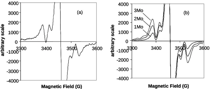
the odd electron nature of the cluster. The observation of an isotropic EPR spectrum at relatively high temperature, on the other hand, clearly indicates that the system must have a single unpaired electron (S = ½) in a non degenerate orbital. An accurate analysis of the Mo hyperfine coupling pattern illuminates us on the localization of the unpaired electron. As Figure 4 shows, simulations were carried out using hyperfine coupling constant, shape and line broadening parameters from the experimental spectrum, but assuming coupling to either one, two, or all three cluster Mo atoms. The height and shape of the satellite peaks, relative to the central resonance and especially relative to each other, unambiguously identify the spectrum as involving coupling to two equivalent Mo nuclei. Unfortunately, the relative broadness of the spectrum conceals the outer peaks of the 1:2:3:4:5:6:5:4:3:2:1 envelope (6.25% overall intensity), which is generated by the isotopomers having two I = 5/2 Mo nuclei. The spectrum was also recorded at temperatures down to 210 K in an attempt to ameliorate the spectral resolution, but no significant change was noted.
(e) Magnetic susceptibility
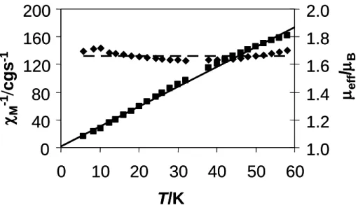
The compound exhibits a simple Curie behaviour, consistent with the present of a single unpaired electron. Measurements carried out in the 6-60 K range show a linear -1 vs.
T plot, with a negligible value for the Curie temperature (-0.38 K), see Figure 1. The corresponding effective magnetic moment, which is approximately temperature independent, averages 1.66B after correction for the ligands diamagnetism. This is rather close to the
expected spin-only value for one unpaired electron (1.73 B). This result is consistent with
the EPR spectrum and confirms that the latter is representative of the bulk sample.
<Figure 5>
(f) DFT Calculation
In order to further confirm the assignment of the chemical composition and electronic structure, we have carried out a theoretical study. Before describing the computational results, it is useful to qualitatively analyze the electronic structure on the basis of first principles and relevant previous knowledge. [21, 23, 24] The cluster may be viewed as resulting
from the assembling of three “four-legged piano stools” of the Cp*MoO4 type, whose
electronic structure is well known,[25, 26] as shown in Scheme 1. Taking a D3h-symmetrical
structure with six identical bridging ligands as a first approximation, it is possible to envisage that the interaction of the z2-type orbitals will lead to one bonding (of type a) and two
degenerate antibonding (of type e) combinations, and that the interaction of the xy-type orbitals will equally lead to one a-type bonding and two degenerate antibonding combinations of e-type. The overlap between the xy orbitals being much weaker, the energy of the MOs resulting from these orbitals will be less perturbed from that of the starting fragment orbitals. However, as well documented for other bridging systems, * mixing with the bridging ligands lone pairs should destabilize the xy orbital combinations.[27] It is easily predictable
that this destabilization should be strongest for the symmetric (a-type) orbital, as shown in Scheme 1. The asymmetric nature of the bridging ligands (four OH and two O), however, is expected to alter this scheme, particularly by removing the degeneracy of the e-type combination.
<Scheme 1>
The DFT optimized geometry agrees quite well with the experimental geometry, see Table 1. The minor variations could partly be attributed to the different environment (hydrogen-bonded solid state structure with the CF3SO3 anion for the experimental geometry,
free “gas phase” dication for the DFT optimized one), and partly to the well known overestimation of bond distances by DFT. In particular, it is comforting to see the same trend of Mo-O bond distances [Mo-(-O) < Mo-(-OH)] in the optimized and experimental parameters, once again confirming the structural assignment.
<Table 1>
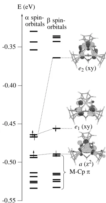
The calculated electronic structure, see Figure 6, confirms the qualitative expectations. The bonding a(z2) orbital is located just above the group of the six MO’s describing the three
double-sided Mo-Cp* interactions. Above it, we find the pair of e(xy) orbitals, which contains three electrons. The calculation converges with two electrons in the e1-type orbital
and one in the e2-type orbital, whose degeneracy (in the ideal C3 symmetry) is substantially
split by the different nature of the bridging groups (O vs. OH). It is rewarding to see that the unpaired electron, according to the calculations, chooses to reside in the orbital from the e set that has the nodal plane parallel to the C2 symmetry axis (e2), rather than in the orbital with
the nodal plane perpendicular to it (e1). The half-occupied orbital has contributions only from
the two Mo’ atoms and a symmetry imposed zero contribution from the Mo atom, in perfect agreement with the result of the EPR analysis above. Furthermore, the DFT calculation suggests that the cluster is completely valence-delocalized. Indeed, the computed natural charge[28] is very similar on atoms Mo (+1.075) and Mo’ (+1.100 and 1.101), and so is the
computed Mulliken spin density (-0.820, 0.849 and 0.847, respectively). We note that the mean value of S2 from the spin-unrestricted calculation is high (see Computational Details),
suggesting mixing of an excited [a(z2)]2[e1(xy)]1[e2(xy)]1[a(xy)]1 configuration into the
ground state. However, according to the result of the EPR experiment, this contribution must be negligible for the real system, because the isotropic resonance is relatively sharp at room temperature and unaffected by cooling.
< Figure 6>
A somewhat related triangular Mo3 cluster, [Mo3CuS4(H2O)10]4+, which was described
as having a CuI ion and a Mo311+ core,[29] has an electronic structure similar to that described
here, although there are two additional electrons, thereby also involving occupation of the a-type orbital. In that case, the experimental observation of electronic coupling to a single Mo nucleus in the EPR spectrum was interpreted as resulting from a valence-trapped MoIVMoIVMoIII structure, the unpaired electron being mostly localized on the MoIII center,
although the calculations (at the EHMO level) indicated a delocalized shape for the MOs, similar to that reported here.
3. Discussion
It is useful to compare the salts reported here with a closely related one reported earlier by Bottomley et al., namely [Cp*3Mo3(-O)6-n(-OH)n]2+(Cl-)2.[21] This compound was
obtained by a synthetic strategy closely related to ours (zinc reduction of Cp*MoO2Cl in
CHCl3 in the presence of concentrated HCl). The X-ray structure presented for this
compound was affected by severe disorder, preventing the unambiguous determination of the number of bridging OH groups, like for the trifluoroacetate complex reported here. We have already pointed out above that the reported Mo-Mo distances for the dichloride salt are very similar to those reported here for the [Cp*3Mo3(-O)2(-OH)4]2+(CF3SO3-)2 salt. For
value, but also suggested the presence of a redox disproportionation equilibrium in solution, which involves other species characterized by n = 4 and n = 6. This suggestion was based on magnetic, EPR, and NMR observations. In particular, the EPR spectrum was assigned to the n = 6 species, whereas two NMR resonances, a sharp one and a broad one, where assigned to the diamagnetic n = 5 species and to the paramagnetic n = 4 species, respectively. The room temperature isotropic EPR spectrum reported for solutions of the dichloride salt seems very similar to that of the trifluoromethylsulfonate and trifluoroacetate salts reported here, but it was interpreted in that case as arising from coupling to three equivalent Mo nuclei. An argument given in favor of the above scheme was the fact that the n = 4 species could not yield an observable isotropic EPR spectrum, given the expected (e)3 electronic
configuration.[21] Our DFT calculation, however, shows that the asymmetry associated to the
distribution of the two -O and four -OH groups significantly splits this orbital degeneracy. The trifluoromethylsulfonate and trifluoroacetate salts reported here (at least batches of clean single crystals) did not show any sharp or even broadened NMR signal that could be assigned to the Cp* ligand in a diamagnetic or paramagnetic compound. All evidence, therefore, points to the fact that the trifluoromethylsulfonate salt reported here maintains its identity as a single compound in solution.
Another useful comparison involves compounds Cp*4Mo5O11[21] and Cp*6Mo8O16,[22]
both being described as containing the central [Cp*3Mo3(-O)6]- unit (vide supra). In this
unit, there are only 4 cluster electrons (Mo3IV,V,V) and the structures are stabilized by capping
salts of [Cp*3Mo3(-O)2(-OH)4]2+ exhibit interesting electrochemical properties, which may
dependent on pH and/or on the presence of other metal ions.
4. Conclusion
We have shown in this paper that the zinc reduction of Cp*2Mo2O5 in an acidic
aqueous environment leads to the triangular [Cp*3Mo3(-O)2(-OH)4]2+ cluster, containing a
paramagnetic (5-electron) trimetallic core. The spectroscopic evidence points to a solution structure identical to the solid state structure, with the unpaired electron located in a delocalized molecular orbital that has atomic orbital participation from only two Mo atoms. A study of the redox properties and acid/base properties of this system, followed by an investigation of its chemical reactivity, are now in progress and will be reported in due course.
3. Experimental Section
3.1 General Methods
All preparations and manipulations were carried out with Schlenk techniques under an oxygen-free argon atmosphere. All glassware was oven-dried at 120 C. Solvents were dried by standard procedures and distilled under dinitrogen prior to use. EPR spectra were measured on a Elexsys E500 BRUKER spectrometer (X-band) equipped with both a frequencemeter and gaussmeter. Mass spectra were recorded with a NERMAG R10-10
instrument. The starting compound, Cp*2Mo2O5, was prepared as described in the
literature.[19]
3.2. Synthesis and crystallization of compound [Cp*3Mo3(-O)2(-OH)4](CF3SO3)2 To a solution of [Cp*2Mo2O5] (0.05 g; 0.092 mmol) in MeOH-H2O (1:1, 6 mL), after
acidification with concentrated CF3SO3H (10 drops), was added metallic zinc (0.48 g; 7.38
mmol). The mixture was stirred under argon at room temperature for two-days during which time it changed from a clear yellow solution to a green suspension. The mixture was filtered and the solid was dried in vacuo. The solid was extracted with THF and, after filtration, addition of n-hexane yielded a green precipitate (0.021 g; 31.3%). A single crystal for the X-ray analysis was obtained by diffusion of a n-hexane layer into a green CH2Cl2 solution at
room temperature. Elemental analysis. Calcd. for C32H49F6Mo3O12S2.2CH2Cl2 (MW =
1261.53): C, 32.37; H, 4.23. Obtained: C, 32.92; H, 3.65. EPR (CH2Cl2): g = 1.962, aMo =
26.2 G. Average eff in the 6-60 K range: 1.66 B (diamagnetic correction: -7.91·10-4 cgs).
3.3. X-ray analyses
Single crystal of compounds [Cp*3Mo3(-O)2(-OH)4](X)2 (X = CF3CO2, CF3SO3)
were mounted under inert perfluoropolyether at the tip of glass fibre and cooled in the cryostream of the Oxford-Diffraction XCALIBUR CCD diffractometer. Data were collected using the monochromatic MoK radiation (= 0.71073). The structure was solved by direct methods (SIR97[30]) and refined by least-squares procedures on F2 using SHELXL-97.[31]
Although the model could be easily defined by direct methods, its refinement appeared to be very poor with large elongated ellipsoids for the Cp* and rather high R and wR2 values. The elongated ellipsoids suggest a free rotation of the Cp ring around the Mo-Cp* centroid axis. However, all attempts to refine a disordered Cp* model failed. The occurrence of a twinned structure by pseudo merohedry, considering a monoclinic cell, was also attempted but again without improvement of the refinement. Trying to refine using a non centrosymmetric space group did not improve the result either. Careful examination of the reciprocal space revealed that the crystal used is certainly not single but an attempt to integrate using different domains failed. There is, however, no doubt about the reality of the model as confirmed by different analytical methods. All H atoms attached to carbon were introduced in the calculation in idealised positions and treated as riding models. Owing to the poor quality of the refinement, the H atom attached to the hydroxyl groups could not be located. The drawings were obtained with the help of ORTEP32.[32] The structures was solved by direct methods (SIR97)[30] and
refined by least-squares procedures on F2 using SHELXL-97.[31] All H atoms attached to
carbon were introduced in calculation in idealised positions and treated as riding models. The drawing of the molecules was realised with the help of ORTEP32.[32] The statistically
distributed disulfide and dichloromethane molecules were freely refined. The crystal data and refinement parameters are shown in Table 2 and selected geometric parameters are collected in Table 1.
3.4. Computational details
The DFT calculation was carried out on a model system where the Cp* ligands were replaced on the simpler Cp rings. The starting geometry was based on the crystallographically determined structure of the triflate salt and no symmetry restrictions were imposed. The geometry was fully optimized and the resulting minimum of the potential energy surface (PES) was verified by the positive value of all second derivatives of the energy. The calculation, which used the spin-unrestricted formulation, was performed using the B3LYP three-parameter hybrid density functional method of Becke,[33] as implemented in
the Gaussian03 suite of programs.[34] The basis sets used for the geometry optimizations are
the standard 6-31G** for C, H and P atoms, and the standard LANL2DZ basis set, which included the Hay and Wadt effective core potentials (ECP),[35] for the metal atoms. The
value of <S2> resulting from the calculation is 1.39 (0.81 after annihilation). This shows that
there is significant spin contamination, indicating mixing with low-lying excited states. The correspondence between the resulting Kohn-Sham spin-orbitals was verified by displaying contour plots with the aid of the program MOLDEN.[36]
Acknowledgements
This research was supported by the CNRS in France and by TUBITAK in Turkey. Supplemental travel support by a bilateral CNRS-TUBITAK (TBAG-U/62-102T211) program is also gratefully acknowledged. In addition, RP acknowledges support from the European Commission through the Research Training Network “AQUACHEM” (contract
No. MRTN-CT-2003-503864) and the COST D29 program (working group 0009-03). FD thanks the University of Celal Bayar (FEF 2002/101) for a research grant.
References
[1] T. Chan, L. Li, Y. Yang, W. Lu, ACS Symp. Ser. 2002, 819, 166-177. [2] F. Joó, Acc. Chem. Res. 2002, 35, 738-745.
[3] I. T. Horvath, Acc. Chem. Res. 2002, 35, 685. [4] D. Sinou, Topics Curr. Chem. 1999, 206, 41-59. [5] D. Sinou, Adv. Synth. Catal. 2002, 344, 221-237.
[6] P. Kalck, F. Monteil, Adv. Organometal. Chem. 1992, 34, 219-284. [7] B. E. Hanson, Coord. Chem. Rev. 1999, 186, 795-807.
[8] C. Muller, D. Vos, P. Jutzi, J. Organometal. Chem. 2000, 600, 127-143. [9] W. A. Herrmann, E. Herdtweck, M. Flöel, J. Kulpe, U. Küsthardt, J. Okuda,
Polyhedron 1987, 6, 1165-1182.
[10] W. A. Herrmann, Comments Inorg. Chem. 1988, 7, 73-107. [11] R. Poli, Chem. Eur. J. 2004, 10, 332-341.
[12] E. Collange, J. Garcia, R. Poli, New J. Chem. 2002, 26, 1249-1256.
[13] J. Gun, A. Modestov, O. Lev, D. Saurenz, M. A. Vorotyntsev, R. Poli, Eur. J. Inorg. Chem. 2003, 482-492.
[14] J. Gun, A. Modestov, O. Lev, R. Poli, Eur. J. Inorg. Chem. 2003, 2264-2272. [15] E. Collange, L. Metteau, P. Richard, R. Poli, Polyhedron 2004, 23, 2605-2610. [16] F. Demirhan, P. Richard, R. Poli, Inorg. Chim. Acta 2003, 347, 61-66.
[17] F. Demirhan, J. Gun, O. Lev, A. Modestov, R. Poli, P. Richard, J. Chem. Soc, Dalton Trans. 2002, 2109-2111.
[18] E. Collange, F. Demirhan, J. Gun, O. Lev, A. Modestov, R. Poli, P. Richard, D. Saurenz, in Perspectives in Organometallic Chemistry, Vol. 287 (Eds.: C. G. Screttas, B. R. Steele), Royal Society of Chemistry, London, 2003, pp. 167-182.
[19] D. Saurenz, F. Demirhan, P. Richard, R. Poli, H. Sitzmann, Eur. J. Inorg. Chem.
2002, 1415-1424.
[20] Footnote1.
[21] F. Bottomley, J. Chen, K. F. Preston, R. C. Thompson, J. Am. Chem. Soc. 1994, 116, 7989-7995.
[22] J. R. Harper, A. L. Rheingold, J. Am. Chem. Soc. 1990, 112, 4037-4038. [23] P. Hofmann, N. Roesch, H. R. Schmidt, Inorg. Chem. 1986, 25, 4470-4478. [24] F. Bottomley, S. Karslioglu, Organometallics 1992, 11, 326-337.
[26] T. A. Albright, J. K. Burdett, M. H. Whangbo, Orbital Interactions in Chemistry, J. Wiley & Sons, New York, 1985.
[27] F. Abugideiri, J. C. Fettinger, R. Poli, Inorg. Chim. Acta 1995, 229, 445-454. [28] A. E. Reed, L. A. Curtiss, F. Weinhold, Chem. Rev. 1988, 88, 899-926.
[29] R. Miyamoto, S. Kawata, M. Iwaizumi, H. Akashi, T. Shibahara, Inorg. Chem. 1997, 36, 542-546.
[30] A. Altomare, M. Burla, M. Camalli, G. Cascarano, C. Giacovazzo, A. Guagliardi, A. Moliterni, G. Polidori, R. Spagna, J. Appl. Cryst. 1999, 32, 115-119.
[31] G. M. Sheldrick, SHELXL97. Program for Crystal Structure refinement, University of Göttingen, Göttingen, Germany, 1997.
[32] L. J. Farrugia, J. Appl. Crystallogr. 1997, 32, 565. [33] A. D. Becke, J. Chem. Phys. 1993, 98, 5648-5652.
[34] M. J. Frisch, G. W. Trucks, H. B. Schlegel, G. E. Scuseria, M. A. Robb, J. R. Cheeseman, J. Montgomery, J. A., T. Vreven, K. N. Kudin, J. C. Burant, J. M.
Millam, S. S. Iyengar, J. Tomasi, V. Barone, B. Mennucci, M. Cossi, G. Scalmani, N. Rega, G. A. Petersson, H. Nakatsuji, M. Hada, M. Ehara, K. Toyota, R. Fukuda, J. Hasegawa, M. Ishida, T. Nakajima, Y. Honda, O. Kitao, H. Nakai, M. Klene, X. Li, J. E. Knox, H. P. Hratchian, J. B. Cross, C. Adamo, J. Jaramillo, R. Gomperts, R. E. Stratmann, O. Yazyev, A. J. Austin, R. Cammi, C. Pomelli, J. W. Ochterski, P. Y. Ayala, K. Morokuma, G. A. Voth, P. Salvador, J. J. Dannenberg, V. G. Zakrzewski, S. Dapprich, A. D. Daniels, M. C. Strain, O. Farkas, D. K. Malick, A. D. Rabuck, K. Raghavachari, J. B. Foresman, J. V. Ortiz, Q. Cui, A. G. Baboul, S. Clifford, J. Cioslowski, B. B. Stefanov, G. Liu, A. Liashenko, P. Piskorz, I. Komaromi, R. L. Martin, D. J. Fox, T. Keith, M. A. Al-Laham, C. Y. Peng, A. Nanayakkara, M. Challacombe, P. M. W. Gill, B. Johnson, W. Chen, M. W. Wong, C. Gonzalez, J. A. Pople, Gaussian 03, Revision B.04, Gaussian, Inc., Pittsburgh PA, 2003.
[35] P. J. Hay, W. R. Wadt, J. Chem. Phys. 1985, 82, 270-283.
[36] G. Schaftenaar, Molden v3.2, CAOS/CAMM Center Nijmegen, Toernooiveld, Nijmegen, The Netherlands, 1991.
Captions for Figures
Figure 1. An ORTEP view of the central Mo3O6 core of the [Cp*3Mo3(-O)2(-OH)4]2+ in
the acetate salt (left, the disordered F atoms are not shown) and
trifluoromethylsulfonate salt (right), showing the hydrogen bonding interactions with the two anions.
Figure 2. An ORTEP view of the [Cp*3Mo3(-O)2(-OH)4]2+ ion from the structure of the
trifluoromethylsulfonate salt. For clarity, the hydrogen atoms of the Cp* ligands are not shown and the ellipsoids of the Cp* C atoms are replaced by spheres of arbitrary radius. The ellipsoids of the Mo and O atoms are shown at the 30% probability level.
Figure 3. (a) Electrospray mass spectrum (positive mode) of compound [Cp*3Mo3(-O)4
(-OH)2](O2CF3)2 in MeOH. Excerpts of this spectrum are shown in (b) and (c), and
a simulation of the envelope corresponding to the [Cp*3Mo3(-O)5(-OMe)]+ ion
is given in (d).
Figure 4. EPR spectrum of [Cp*3Mo3(-O)2(-OH)4](CF3SO3)2 in CH2Cl2 solution. (a)
Experimental spectrum. (b) Simulated spectra with coupling to one, two, and three equivalent Mo nuclei (simulation parameters: Gaussian line shape, linewidth = 18 G). All spectra are scaled to the same peak-to-peak height for the central resonance.
Figure 5. Magnetic properties (molar magnetic susceptibility, M, and effective magnetic
moment, eff) of compound [Cp*3Mo3(-O)2(-OH)4](CF3SO3)2.
Figure 6. Electronic structure of the [Cp*3Mo3(-O)2(-OH)4]2+ cluster as resulting from a
gas-phase DFT calculation. The shape of the spin-orbitals connected by a dashed line is essentially identical (only one for each pair is shown).
Figure 1
2.64 Å
2.64 Å
2.59 Å
2.71 Å
2.76 Å
2.65 Å
2.76 Å
2.62 Å
2.64 Å
2.64 Å
2.59 Å
2.71 Å
2.76 Å
2.65 Å
2.76 Å
2.62 Å
Figure 2
Mo1
Mo2
Mo3
O4
O5
O6
O1
O2
O3
Mo1
Mo2
Mo3
O4
O5
O6
O1
O2
O3
Figure 3
(b)
(c)
x4 800 810 790 820 790 820 790 790 820820 804.06(a)
800 810 790 820 804.80 970 960 950 940 930 949.60(d)
1000 900 800 700 500 600(b)
(c)
x4 800 810 790 820 790 820 790 790 820820 804.06(a)
800 810 790 820 804.80 970 960 950 940 930 949.60(d)
1000 900 800 700 500 600Figure 4 -4000 -3000 -2000 -1000 0 1000 2000 3000 4000 3300 3400 3500 3600 Magnetic Field (G) ar b itr ar y scale (a) -4000 -3000 -2000 -1000 0 1000 2000 3000 4000 3300 3400 3500 3600 Magnetic Field (G) ar b itr ar y scale 1Mo 2Mo 3Mo (b) -4000 -3000 -2000 -1000 0 1000 2000 3000 4000 3300 3400 3500 3600 Magnetic Field (G) ar b itr ar y scale (a) -4000 -3000 -2000 -1000 0 1000 2000 3000 4000 3300 3400 3500 3600 Magnetic Field (G) ar b itr ar y scale (a) -4000 -3000 -2000 -1000 0 1000 2000 3000 4000 3300 3400 3500 3600 Magnetic Field (G) ar b itr ar y scale 1Mo 2Mo 3Mo (b) -4000 -3000 -2000 -1000 0 1000 2000 3000 4000 3300 3400 3500 3600 Magnetic Field (G) ar b itr ar y scale 1Mo 2Mo 3Mo (b)
Figure 5
0
40
80
120
160
200
0
10
20
30
40
50
60
T/K
M -1cgs
-11.0
1.2
1.4
1.6
1.8
2.0
eff/
B0
40
80
120
160
200
0
10
20
30
40
50
60
T/K
M -1cgs
-11.0
1.2
1.4
1.6
1.8
2.0
eff/
BFigure 6
-0.40
-0.45
-0.50
-0.35
-0.55
M-Cp
e
1(xy)
e
2(xy)
E (eV)
a (z
2)
Scheme 1 a (z2) e1 (z2) e2 (z2) a (xy) e1 (xy) e2 (xy) z2 xy
Table 1. Selected bond distances (Å) and angles (°) for complex [Cp*3Mo3(-O)2
(-OH)4]2+: observed (from the X-ray structure of the CF3SO3 salt) and DFT
optimized (gas-phase).
Distancea,b Experimental DFT optimized
Mo-Mo’ 2.7854(13), 2.7820(13) 2.859, 2.876 Mo’-Mo’ 2.8244(12) 2.946 Mo-(-O) 1.946(8), 1.959(8) 1.979, 1.987 Mo-(-OH) 2.041(8), 2.061(7) 2.108, 2.110 Mo’-(-O) 1.949(8), 1.956(8) 1.938, 1.943 Mo’-(-OH) 2.048(8), 2.053(7), 2.055(8), 2.049(8), 2.028(8), 2.053(8) 2.090, 2.094, 2.097, 2.099, 2.106, 2.108 Mo-CNT 2.0145(9) 2.082 Mo’-CNT 2.0058(9), 2.0168(9) 2.079, 2.080 Anglea,b Experimental DFT optimized
Mo’-Mo-Mo’ 60.97(3) 61.92 Mo-Mo’-Mo’ 59.46(3), 59.57(3) 58.91, 59.16 Mo-(-O)-Mo’ 91.0(3), 90.9(3) 93.61, 93.83 Mo-(-OH)-Mo’ 85.1(3), 85.9(3) 85.96, 85.97 Mo’-(-OH)-Mo’ 87.5(3), 87.0(3) 88.93, 88.94 CNT-Mo-(-O) 112.7 (2), 114.7(2) 114.04, 116.94 CNT-Mo-(-OH) 112.2(2), 112.7(2) 113.22, 113.92 CNT-Mo’-(-O) 113.4(2), 113.9(2) 113.01, 113.92 CNT-Mo’-(-OH) 110.8(2), 113.4(2), 115.1(2) 111.7(2), 114.8(2), 115.0(2) 112.94, 115.54, 117.05, 112.99, 115.30, 117.88
aMo indicates the unique atom, with the Cp*Mo(O)
2(OH)2 coordination environment. Mo’
indicates the two symmetry related atoms, with the Cp*Mo(O)(OH)3 coordination
Table 2. Crystal data and refinement parameters for [Cp*3Mo3(-O)2(-OH)4](CF3SO3)2
Empirical formula C32 H35 F6 Mo3 O12 S2
Formula weight 1087.62
Temperature 180(2) K
Wavelength 0.71073 Å
Crystal system Orthorhombic
Space group Pbca
Unit cell dimensions a = 15.5156(6) Å = 90° b = 16.8899(7) Å = 90° c = 31.0271(12) Å = 90° Volume 8130.9(6) Å3 Z 8 Density (calculated) 1.777 Mg/m3 Absorption coefficient 1.098 mm-1 F(000) 4360 Crystal size 0.24 x 0.12 x 0.077 mm3 Theta range for data collection 2.82 to 26.37°
Index ranges -19<=h<=19, -18<=k<=21, -38<=l<=38 Reflections collected 59030
Independent reflections 8302 [R(int) = 0.0997] Completeness to theta = 26.37° 99.8 %
Absorption correction Semi-empirical from equivalents Max. and min. transmission 0.9623 and 0.7409
Refinement method Full-matrix least-squares on F2 Data / restraints / parameters 8302 / 0 / 487
Goodness-of-fit on F2 1.088
Final R indices [I>2sigma(I)] R1 = 0.1145, wR2 = 0.2423 R indices (all data) R1 = 0.1279, wR2 = 0.2495 Largest diff. peak and hole 2.867 and -3.776 e.Å-3
Graphical abstract 2.64 Å 2.64 Å 2.59 Å 2.71 Å 2.76 Å 2.65 Å 2.76 Å 2.62 Å 2.64 Å 2.64 Å 2.59 Å 2.71 Å 2.76 Å 2.65 Å 2.76 Å 2.62 Å