HAL Id: inserm-00278426
https://www.hal.inserm.fr/inserm-00278426
Submitted on 13 May 2008
HAL is a multi-disciplinary open access
archive for the deposit and dissemination of sci-entific research documents, whether they are pub-lished or not. The documents may come from teaching and research institutions in France or abroad, or from public or private research centers.
L’archive ouverte pluridisciplinaire HAL, est destinée au dépôt et à la diffusion de documents scientifiques de niveau recherche, publiés ou non, émanant des établissements d’enseignement et de recherche français ou étrangers, des laboratoires publics ou privés.
Effects of the selective neurotensin antagonist SR
142948A on
3,4-methylenedioxymethamphetamine-induced
behaviours in mice.
Cynthia Marie-Claire, Stefano Palminteri, Patrizia Romualdi, Florence Noble
To cite this version:
Cynthia Marie-Claire, Stefano Palminteri, Patrizia Romualdi, Florence Noble. Effects of the selective neurotensin antagonist SR 142948A on 3,4-methylenedioxymethamphetamine-induced behaviours in mice.. Neuropharmacology, Elsevier, 2008, 54 (7), pp.1107-11. �10.1016/j.neuropharm.2008.03.001�. �inserm-00278426�
Effects of the selective neurotensin antagonist SR 142948A on
3,4-methylenedioxymethamphetamine-induced behaviours in mice
Running title : NT1 receptor blockade decreases MDMA effects
Cynthia Marie-Claire a,b,c,1, Stefano Palminteri d,1 , Patrizia Romualdi d, Florence Noble a,b,c,*
a Université Paris Descartes, Faculté de Pharmacie, Neuropsychopharmacologie
des addictions, et Université Paris7, Paris F-75010, France
b CNRS, UMR7157, Paris F-75006, France c INSERM, U705, Paris F-75006, France
d Dept. Pharmacology, University of Bologna, Irnerio 48 – 40126 Bologna, Italy
1 Equivalent contribution
* Corresponding author Tel : 33-1-53-73-95-61 Fax : 33-1-53-73-97-19
florence.noble@univ-paris5.fr
HAL author manuscript inserm-00278426, version 1
HAL author manuscript
ABSTRACT
Neurotensin is one of the genes previously found up-regulated in mice striatum after acute injection of MDMA (9mg/kg). In order to examine the pharmacological significance of this effect the involvement of the neurotensinergic system in MDMA-induced behaviors was explored in mice using the neurotensin receptor antagonist SR142948A (1mg/kg). We found that acute administration of the antagonist inhibited the MDMA-elicited locomotor activity. SR142948A pre-treatment had no effect on the acquisition of conditioned place preference (CPP) to MDMA but abolished the expression of this behavior. We also studied the effects of acute and repeated exposure to MDMA on the mRNA level of neurotensin in mice striatum. Kinetic analysis of the regulation 1, 2, 6 and 12 hours after acute injection of MDMA showed that the drug transiently up-regulate neurotensin mRNA in this structure. The time course of the modulation suggests that the effects observed with SR142948A are attributable to the release of a preexisting endogenous pool rather than the newly synthesized peptide. Repeated exposure to MDMA following the same injection pattern used in the CPP paradigm revealed an increase in mRNA level of neurotensin in mice striatum. These results indicate that endogenous neurotensin play a role in both the acute locomotor activity and the expression of CPP induced by MDMA.
Keywords : neurotensin, MDMA, gene expression, behavior, mice
2
INTRODUCTION
MDMA (3,4-methylenedioxymethamphetamine) is the psychoactive compound of the widely abused drug “ecstasy”. This compound is structurally related to both amphetamine and hallucinogens. In rodents, MDMA has been shown to have psychostimulant and rewarding effects, similar to those observed by administration of amphetamine and cocaine (Salzmann et al., 2003; Trigo et al., 2006). The mechanism of action of MDMA is complex and not yet completely known. At the neurochemical level MDMA produces the release of both dopamine (DA) and serotonin (5-HT) from nerve terminals (for review, see: (Colado et al., 2004). By interacting with their respective receptors, these neurotransmitters are responsible for the acute and long lasting effects of MDMA. Moreover, MDMA displays a moderate affinity also to other receptors, whose activation could be at the origin of some of its effects (Battaglia et al., 1988). At the cellular level MDMA administration in mice causes a variety of transcriptional events, probably responsible for the long term effects and the addictive properties of this substance, in several brain structures (Salzmann et al., 2006; Salzmann et al., 2003). Among these, up-regulation of the gene coding for the precursor of the peptide neurotensin was recently observed in our laboratory in mice striatum (Salzmann et
al., 2006). It is well known that this tridecapeptide is closely related with dopamine transmission in
the central nervous system. The biological actions of neurotensin are initiated by binding to three different receptor subtypes NTS1, NTS2 and NTS3. However it clearly appears that the majority of the central effects of neurotensin appear to be exerted through the high affinity NTS1. Thus it has been shown that endogenous neurotensin, through activation of NTS1 receptors, mediates some behavioural effects of psychostimulants. Pre-treatment with an antagonist of the NTS1 receptor, SR48692, reduced the number of rearing induced by acute cocaine injection (Betancur et al., 1998) and delayed the development of cocaine sensitization (Horger et al., 1994 ). Furthermore, the locomotor sensitization to amphetamine could be blocked when the drug was co-administered with SR48692 (Panayi et al., 2005; Panayi et al., 2002; Rompre and Perron, 2000).
The aim of this study was to explore the role of endogenous neurotensin on behavioral effects of MDMA, including hyperlocomotion and the acquisition and expression of conditioned place preference. The potent and selective NTS1 receptor antagonist SR142948A was used. In addition, we further explored with real time quantitative PCR the gene expression of neurotensin precursor following acute and chronic MDMA administration in the striatum of mice.
4
MATERIALS AND METHODS
Animals and drugs : Male CD-1 mice (Charles River, L’arbresle, France), weighing 22-24g at the beginning of the experiments, were housed in a temperature controlled environment (21±2°) under a 12 h light/dark cycle (lights on 7:00-19:00) with food and water provided ad libitum. All experiments were performed in strict accordance with the guidelines for the use and care of experimental animals promulgated by the European Community (EEC No. 86/609). All efforts were made to minimize animal suffering and to use only the number of animals necessary to produce reliable scientific data. D,L-MDMA (Lipomed, Arlesheim, Switzerland) and the neurotensin receptor antagonist SR142948A (generous gift from Sanofi-Aventis) were dissolved in saline solution (0.9 % NaCl). All drugs were injected intraperitoneally (i.p.) in a volume of 0.1 ml per 10 g of body weight. The doses of MDMA and SR148942A were chosen based on published data (Salzmann et al, 2003; Azmi et al, 2006; Blinder et al, 2002).The antagonist was injected 20 minutes before MDMA.
Conditioned Place Preference (CPP) paradigm: As previously described an unbiased place preference conditioning procedure was used (Salzmann et al., 2003). The place preference apparatus consisted of two conditioning compartments (15x15x15 cm) separated by a neutral area. The conditioning compartments were differentiated by a distinctive sensory cue: the wall coloring pattern (black or strips). Movements and location of mice were recorded by computerized monitoring software (Videotrack, Viewpoint, Lyon, France). The protocol consists of three phases. Preconditioning phase: drug-naïve mice had free access to all compartments for 20 min, and the time spent in each compartment is recorded. Conditioning phase: this phase consisted in 6 days where each conditioning chamber was closed. On the first conditioning day, mice were treated with drugs or saline according to their group and were placed after the injection in one of the conditioning environments individually for 20 min. The following conditioning day, all mice were given saline in the opposite compartment, and this sequence was repeated during the next 4 days. The designation of drug-paired chamber was random and resulted in an approximately equal
representation of the two conditioning chambers as the drug-paired chamber across groups for all experiments. Post-conditioning phase: this phase took place 24 h after the final conditioning session (day 7). This phase was carried out exactly as the preconditioning phase. Results are expressed in scores (mean ± s.e.m.) calculated as the difference between the time spent in the drug-paired compartment during the post-conditioning phase minus the time spent in the same compartment in the preconditioning phase. The total number of visits and the distance traveled (arbitrary unit) were also recorded during the test day.
Experiment I: effect of SR142948A on the acquisition of MDMA –induced CPP.
On conditioning days 1, 3 and 5, animals received SR142948A (1 mg kg–1; i.p.) or saline 20 min before MDMA (9 mg kg–1; i.p) or saline injection, and then were placed in the drug-paired compartment for 20 min.
Experiment II: effect of SR142948A on the expression of MDMA –induced CPP
During the test phase (day 7) animals received SR142948A (1 mg kg–1; i.p.) or saline 20 min before being placed in the apparatus for final recording.
Locomotor Activity : Locomotor activity was recorded in transparent activity boxes (10x18x14 cm; Imetronic, Bordeaux, France). Displacements were measured by photocell beams located across the long axis, 20 mm above the floor (horizontal activity). Naïve mice, not prehabituated to the activity boxes, received MDMA (9 mg kg–1; i.p.) or saline injection 20 minutes after SR142948A (0.25 or 1 mg kg–1; i.p.) or saline, and their locomotor activity was immediately recorded for 60 minutes. Locomotor activity was expressed in scores (means ± s.e.m.) as the number of interruption of the photocell beams, recorded every 10 minutes.
Drugs treatments and brain dissection for quantitative real-time PCR: For the study of neurotensin gene (Nts) expression following acute drug administration mice, previously injected with MDMA (9 mg kg–1; i.p) or saline, were killed by cervical dislocation 1, 2, 6, 12 or 24 hours after a single injection. To study the effect of chronic MDMA administration on gene expression, two groups of mice were treated with the same administration schedule used in CPP general
6
protocol. Animals were chronically treated with MDMA (9 mg kg–1; i.p) or saline according to their group on days 1, 3 and 5, and with saline on days 2, 4 and 6, as previously described. These mice were then killed by cervical dislocation on day seven, 24 hours after the last injection. After cervical dislocation the brain was quickly removed, frozen in isopentane at –50 °C, and placed in an acrylic matrix (David Kopf Instruments, Tujunga, CA, USA) allowing the reproducible slicing of 1 mm coronal sections. A 2 mm-section was cut, corresponding approximately to bregma +0.26 mm to – 0.46 mm according to the mouse brain atlas (Paxinos and Franklin, 2000). Striatum was then dissected free–hand in ice within the slice, and stored at –80 °C until processing.
RNA isolation and reverse transcription for quantitative real-time PCR: Total RNA used for quantitative PCR experiments was extracted from individual dorsal striata by a modified acid-phenol guanidinium method, following the manufacturer’s protocol (RNAble®, Eurobio, Courtaboeuf, France). The quality of the RNA samples was determined by electrophoresis through agarose gels and staining with ethidium bromide. Quantification of total RNA was assessed using a NanoDrop® ND-1000 spectrophotometer (NanoDrop® Technologies, USA). Reverse transcription of RNA was performed in a final volume of 20 μl containing 1× first strand buffer (Invitrogen, France), 500 μM each dNTP, 20 U of Rnasin ribonuclease inhibitor (Promega, France), 10 mM dithiothreitol, 100 U of Superscript II Rnase H− reverse transcriptase (Invitrogen, France), 1.5 μM random hexanucleotide primers (Amersham Biosciences, France) and 1 μg of total RNA as previously described (Salzmann et al, 2003).
Real time quantitative RT-PCR: Fluorescent PCR reactions were performed on a Light-Cycler® instrument (Roche Diagnostics, Meylan, France) using the LC-FastStart DNA Master SYBR Green I kit (Roche Diagnostics). The cDNAs were diluted 500-fold and 5 μl were added to the PCR reaction mix to yield a total volume of 10 μl. The reaction buffer contained 4 mM MgCl2 and 0.5
μM of each primer. The PCR reactions were performed with 10-12 samples/drug treatment, each sample being prepared with bilateral dorsal striata from one mouse. Quantification was made on the basis of a calibration curve using cDNA from an untreated mouse brain as previously described
(Salzmann et al., 2006). PCR primers were chosen with the assistance of Oligo 6.42 software (MedProbe, Norway). In addition to the transcript of neurotensine (Nts), the hypoxanthine-guanine-phosphoribosyl-transferase transcript (Hprt) was also quantified and each sample was normalized on the basis of its Hprt content. The nucleotide sequences of the primers used for Nts and Hprt quantification have been previously described (Salzmann et al., 2006). Results are expressed as Nts transcript/Hprt transcript.
Statistical analysis: All series of data were analyzed with Statview 5.0 software. For conditioned place preference and real time quantitative RT-PCR results, data were analyzed using one-way ANOVA between subjects, followed by a Fisher-PLSD test for post hoc comparisons. For locomotor activity times x treatments interactions were analyzed by two-way ANOVA, for repeated measures followed by a one-way ANOVA and a Fisher-PLSD test for post hoc comparisons. The level of significance was set at P <0.05.
8
RESULTS
Effect of SR142948A on MDMA-induced hyperlocomotion: The effects of two doses of SR148942A on MDMA-induced locomotor activity were tested (Figure 1A and 1B). In both cases (0.25 or 1mg kg-1 of SR142948A) two-way ANOVA for repeated measures showed a significant
treatment effect, time effect and time x treatment interaction.
As shown in Figure 1A, administration of MDMA increased locomotor activity as compared to saline-treated animals throughout the 60 min period of testing (saline/MDMA vs saline/saline, 10 min.: P<0.0001, 20 min.: P<0.0001, 30 min.: P=0.0003, 40 min.: P=0.0010, 50 min.: P<0.0001 and 60 min: P<0.0001). This hyperactivity was antagonized by pre-injection of SR142948A (0.25 mg kg–1; i.p) 20 min before MDMA treatment (saline/MDMA vs SR142948A/MDMA, 10 min.: P=0.0065, 20 min.: P=0.0025, 30 min.: P=0.0003, 40 min.: P=0.0026, 50 min.: P=0.0035 and 60 min: P=0.0045). We also observed a significant difference in locomotor activity between SR142948A/saline and SR142948A/MDMA group, while no significant difference was found between saline/saline and SR142948A/saline treated animals (Figure 1A).
As shown in Figure 1B, administration of MDMA increased locomotor activity as compared to saline-treated animals throughout the 60 min. period of testing (saline/MDMA vs saline/saline, 10 min.: P=0.0002, 20 min.: P=0.0091, 30 min.: P=0.0003, 40 min.: P<0.0001, 50 min.: P<0.0001 and 60 min: P<0.0001). This hyperactivity was antagonized by pre-injection of SR142948A (1 mg kg–1; i.p) 20 min before MDMA treatment, as shown on figure 1B (saline/MDMA vs SR142948A/MDMA, 10 min.: P=0.0174, 20 min.: P=0.0204, 30 min.: P=0.0009, 40 min.: P=0.0002, 50 min.: P<0.0001 and 60 min: P<0.0001). We also observed a significant difference in locomotor activity between SR142948A/saline and SR142948A/MDMA group, while no significant difference was found between saline/saline and SR142948A/saline treated animals (Figure 1B).
As no effect was observed following administration of SR142948A (1 mg kg-1) alone, and that this
dose was able to totally blocked the hyperlocomotion induced by MDMA, 1 mg.kg-1 was chosen for the rest of the studies.
Effect of SR142948A on the acquisition of MDMA-induced CPP: One-way ANOVA (F3,64=3.843;
P=0.0136) revealed a significant treatment effect between the four groups of animals. Post hoc
comparisons showed a significant effect of MDMA as compared to control group (saline/MDMA vs saline/saline: P=0.0125) and no significant effect of SR142948A pretreatment on the acquisition of MDMA-induced conditioned place preference (saline/MDMA vs SR142948A/MDMA: P=0.5671) (Figure 2A).
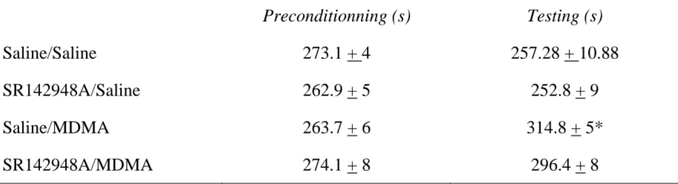
Effect of SR142948A on the expression of MDMA-induced CPP: Comparison of preconditioning times spent in the drug-paired compartment did not show any significant difference between the groups, indicating the unbiased characteristics of the experimental design (Table 1). One-way ANOVA (F3,67=6.974; P=0.0004) revealed a significant treatment effect between the four groups of
animals. Post hoc comparisons showed a significant effect of MDMA compared to control group (MDMA/saline vs saline/saline: P=0.0002). Interestingly the administration of SR142948A 20 min before the postconditioning test was able to antagonize the expression of this behavioral effect of MDMA (MDMA/saline vs MDMA/SR142948A: P=0.0233) (Figure 2B). Furthermore, no differences were found in the total number of entries in the two conditioning compartments for the four groups of animals, saline/saline, SR142948A/saline, Saline/MDMA and SR142948A/MDMA (number of entries = 125.3 ± 9.6, 111.7 ± 8.3, 127.5 ± 4.8 and 115.9 ± 7.6, respectively) (p = 0.435). The total distances covered by the animals during the 18 minutes of the test in the four groups showed no differences for saline/saline, SR142948A/saline, Saline/MDMA and SR142948A/MDMA (total distance = 4524.94 ± 175.63, 4451.72 ± 285.05, 4913.65 ± 152.77 and 4845.75 ± 259.18, respectively) (p = 0.397). These results indicate that the mice were not hypoactive with this dose of antagonist on the day of the test and that the observed effect is indeed due to a blockade of the expression of the MDMA-induced CPP.
10
Effect of acute MDMA administration on Nts gene expression: Real time quantitative PCR was used to study the kinetics of Nts gene expression induced by acute injection MDMA in mouse striatum. Nts expression levels were analyzed at 1h, 2h, 6h 12h and 24h after acute MDMA injection. At 1h the expression level of Nts was similar in the control and MDMA-treated animals while one-way ANOVA showed a significant increase of Nts transcription (1.7 and 2.3 fold change) at 2h (F1,20=5.618; P=0.0279) and 6h (F1,22=18,246; P=0,0003) respectively. 12h and 24h after the
MDMA injection the Nts expression level returned to basal (Figure 3).
Effect of chronic MDMA administration on Nts gene expression: The effect of the MDMA administration schedule used in the CPP procedure was measured at the time of the post-conditioning test (24h after the last injection) on Nts gene expression. One-way ANOVA (F1,21=5.695; P=0.0265) showed a significant effect of MDMA chronic treatment on Nts
transcription. A 1.3-fold increase of the Nts transcript was found in MDMA-treated animals as compared to controls (Figure 4).
DISCUSSION
Neurotensin was one of the genes found up-regulated by a single MDMA injection in mice striatum in a previous microarray study (Salzmann et al., 2006). Neurotensin is a 13 amino acids peptide, widely distributed in the brain, involved in several behavioral functions including locomotion, reward, stress and pain modulation (Geisler et al., 2006). In order to better understand the role of this peptide in the behavioral responses induced by MDMA, we studied the effects of a neurotensin receptor antagonist on MDMA-induced hyperlocomotion and CPP. We also measured the effects of acute and chronic MDMA on the expression level of neurotensin in mice striatum.
Acute administration of MDMA in mice induced a locomotor hyperactivity, in good agreement with previous studies (Salzmann et al., 2003). This effect was blocked by the NTS1 receptor antagonist SR142948A. Interestingly, while SR142948A inhibited the MDMA-induced hyperlocomotion immediately after the injection, at this time point no modulation of the Nts mRNA level could be detected in the first hour. However acute MDMA injection induced a significant increase in Nts mRNA in the striatum at 2 and 6 hours after the injection. As neurotensin is stored in dense core vesicles and released in a calcium-dependent manner, our results suggest that this pre-existing endogenous neurotensin pool is involved in the MDMA-induced hyperlocomotor effect and not newly synthesized neuropeptide as a delay in the increase of mRNA level was observed. Moreover, it could be speculated that the mRNA increase observed might correspond to a cell response to restore rapidly the neurotransmitter pool. The increased release of endogenous neurotensin following systemic administration of MDMA could play a direct or indirect role in the hyperlocomotor effects induced by MDMA. In the striatum, it has been shown that neurotensin increases dopaminergic signalling mainly via the activation of a relatively high density of NTS1 receptors located on striatal dopamine terminals (Ferraro et al., 1997).
High densities of neurotensin immunoreactive nerve terminals nearby dopaminergic cell bodies have been observed within the ventral tegmental area where the peptide is also co-localized within a distinct group of dopaminergic neurons projecting to the cerebral cortex (Berger et al., 1992;
12
Hokfelt et al., 1984). These neuro-anatomic distributions suggest that neurotensin transmission is also relevant in regulating dopaminergic neuronal pathways involved in reward at the level of the midbrain dopamine cell bodies well known to play a key role in the effects induced by drugs of abuse. Studies have provided evidences of the rewarding effects of MDMA in different animal models, such as self-administration and conditioned place preference (Salzmann et al., 2003; Trigo
et al., 2006). Thus it was also interesting to evaluate whether the neurotensin receptor antagonist
was able to modify the rewarding effects of MDMA evaluated in the CPP. This model is based on the fact that the pairing of neutral distinctive environmental stimuli with a drug (primary reward) results in an acquired preference for those specific stimuli (secondary or conditioned reward). The CPP consists of an acquisition phase, and an expression phase in which drug-free animals are tested for their preference for the environment previously paired with the drug. Different neurochemical mechanisms appear to mediate the acquisition and expression of this incentive learning, and it has been shown in the case of amphetamine and morphine that the neurons involved in the expression and acquisition of CPP are anatomically distinct at least within the nucleus accumbens (Fenu et al., 2006; Sellings and Clarke, 2003). Thus it was interesting to investigate the effect of SR142948A on both these phases using MDMA as primary reward. The NTS1 receptor antagonist had no effect on the acquisition but blocked the expression of MDMA CPP.
This could suggest that NTS1 receptors are not involved in mediating the rewarding effects of MDMA, while they play a key role in the behaviour elicited by conditioned reward to MDMA-paired stimuli. In line with this we found that, at the time of the test (expression), Nts mRNA level was up-regulated in the striatum of the mice. It is unlikely that the NTS1 antagonist blocked the expression of MDMA CPP because of aversive properties, as repeated administration of SR142948A did not induce significant effects.
These results suggest that neurotensin may be a neural substrate for reward expectation. A signal may be delivered when the animal is placed in the drug-paired compartment, increasing the release of neurotensin, which through activation of NTS1 receptors may influence the processing of
predictions and the choice of reward-maximizing action. This process may directly or indirectly involved other neurotransmitter systems, as it has been shown for instance that glutamate transmission play a key role in expression of morphine or cocaine CPP (Cervo and Samanin, 1995; Harris et al., 2004), as well as dopamine (Garris et al., 1999; Schultz et al., 1997) or enkephalins (Mas Nieto et al., 2001). It is possible that activation of NTS1 receptors is effective only at a critical period in which animals must remember the place and cues associated with drug discrimination. In conclusion, our results show that endogenous neurotensin is involved in both MDMA-induced hyperlocomotion and CPP. Although acute injection of MDMA up-regulates transiently Nts mRNA levels in mice striatum, it is the endogenous pool of the peptide that is involved in the locomotor activity induced after acute MDMA. Moreover, neurotensin receptors activation is important for the expression but not the acquisition of MDMA-induced CPP. Repeated MDMA injections following the CPP pattern induced an up-regulation of Nts mRNA in the striatum. In order to determine whether this modulation is specific to the striatum, studies of Nts mRNA levels in several structures of the mice brain are currently under investigation in our laboratory.
14
Acknowledgements: S. Palminteri was supported by a fellowship for foreign thesis of the
University of Bologna, Faculty of Pharmacy, Biotechnology Course. The authors thank Arberie Peci for preliminary studies.
REFERENCES
Adams, D. H., Hanson, G. R., Keefe, K. A., 2001. Differential effects of cocaine and methamphetamine on neurotensin/neuromedin N and preprotachykinin messenger RNA expression in unique regions of the striatum. Neuroscience 102, 843-851.
Adams, D. H., Hanson, G. R., Keefe, K. A., 2005. 3,4-Methylenedioxymethamphetamine increases neuropeptide messenger RNA expression in rat striatum. Molecular Brain Research 133, 131-142. Battaglia, G., Brooks, B. P., Kulsakdinun, C., De Souza, E. B., 1988. Pharmacologic profile of MDMA
(3,4-methylenedioxymethamphetamine) at various brain recognition sites. Eur J Pharmacol 149, 159-163.
Berger, B., Gaspar, P., Verney, C., 1992. Colocalization of neurotensin in the mesocortical dopaminergic system. Restricted regional and laminar distribution in rat, lack of colocalization in human. Annals of the New York Academy of Sciences 668, 307-310.
Betancur, C., Cabrera, R., de Kloet, E. R., Pelaprat, D., Rostene, W., 1998. Role of Endogenous Neurotensin in the Behavioral and Neuroendocrine Effects of Cocaine. Neuropsychopharmacology 19, 322-332.
Cervo, L., Samanin, R., 1995. Effects of dopaminergic and glutamatergic receptor antagonists on the acquisition and expression of cocaine conditioning place preference. Brain Research 673, 242-250. Colado, M. I., O'Shea, E., Green, A. R., 2004. Acute and long-term effects of MDMA on cerebral
dopamine biochemistry and function. Psychopharmacology (Berl) 173, 249-263.
Costa, F. G., Frussa-Filho, R., Felicio, L. F., 2001. The neurotensin receptor antagonist, SR48692, attenuates the expression of amphetamine-induced behavioural sensitisation in mice. European Journal of Pharmacology 428, 97-103.
Fenu, S., Spina, L., Rivas, E., Longoni, R., Di Chiara, G., 2006. Morphine-conditioned single-trial place preference: role of nucleus accumbens shell dopamine receptors in acquisition, but not expression. Psychopharmacology 187, 143-153.
Ferraro, L., WT, O. C., T, A., K, F., S, T., 1997. Differential effects of intrastriatal neurotensin(1-13)
16
and neurotensin(8-13) on striatal dopamine and pallidal GABA release. A dual-probe microdialysis study in the awake rat. European Journal of Neuroscience 9, 1838-1846.
Garris, P. A., Kilpatrick, M., Bunin, M. A., Michael, D., Walker, Q. D., Wightman, R. M., 1999. Dissociation of dopamine release in the nucleus accumbens from intracranial self-stimulation. Nature 398, 67-69.
Geisler, S., Berod, A., Zahm, D. S., Rostene, W., 2006. Brain neurotensin, psychostimulants, and stress - emphasis on neuroanatomical substrates. Peptides 27, 2364-2384.
Harris, G. C., Wimmer, M., Byrne, R., Aston-Jones, G., 2004. Glutamate-associated plasticity in the ventral tegmental area is necessary for conditioning environmental stimuli with morphine. Neuroscience 129, 841-847.
Hokfelt, T., Everitt, B., Theodorsson-Norheim, E., Goldstein, M., 1984. Occurrence of neurotensinlike immunoreactivity in subpopulations of hypothalamic, mesencephalic, and medullary catecholamine neurons. The Journal of comparative neurology 222, 543-559.
Horger, B., Taylor, J., Elsworth, J., Roth, R., 1994 Preexposure to, but not cotreatment with, the neurotensin antagonist SR 48692 delays the development of cocaine sensitization. Neuropsychopharmacology 11, 215-222.
Mas Nieto, M., Wilson, J., Walker, J., Benavides, J., Fournie-Zaluski, M.-C., Roques, B. P., Noble, F., 2001. Facilitation of enkephalins catabolism inhibitor-induced antinociception by drugs classically used in pain management. Neuropharmacology 41, 496-506.
Panayi, F., Colussi-Mas, J., Lambás-Señas, L., Renaud, B., Scarna, H., Berod, A., 2005. Endogenous neurotensin in the ventral tegmental area contributes to amphetamine behavioral sensitization. Neuropsychopharmacology 30, 871-879.
Panayi, F., Dorso, E., Lambás-Señas, L., Renaud, B., Scarna, H., Berod, A., 2002. Chronic blockade of neurotensin receptors strongly reduces sensitized, but not acute, behavioral response to D-amphetamine. Neuropsychopharmacology 26, 64-74.
Paxinos, G., Franklin, K., 2000. The Mouse Brain in Stereotaxic Coordinates. Academic Press.
Rompre, P.-P., Perron, S., 2000. Evidence for a role of endogenous neurotensin in the initiation of amphetamine sensitization. Neuropharmacology 39, 1880-1892.
Salzmann, J., Canestrelli, C., Noble, F., Marie-Claire, C., 2006. Analysis of transcriptional responses in the mouse dorsal striatum following acute 3,4-methylenedioxymethamphetamine (ecstasy): Identification of extracellular signal-regulated kinase-controlled genes. Neuroscience 137, 473-482. Salzmann, J., Marie-Claire, C., Le Guen, S., Roques, B. P., Noble, F., 2003. Importance of ERK
activation in behavioral and biochemical effects induced by MDMA in mice. Br J Pharmacol 140, 831-838.
Schultz, W., Dayan, P., Montague, P. R., 1997. A Neural Substrate of Prediction and Reward. Science 275, 1593-1599.
Sellings, L. H. L., Clarke, P. B. S., 2003. Segregation of Amphetamine Reward and Locomotor Stimulation between Nucleus Accumbens Medial Shell and Core. J. Neurosci. 23, 6295-6303.
Trigo, J., Panayi, F., Soria, G., Maldonado, R., Robledo, P., 2006. A reliable model of intravenous MDMA self-administration in naïve mice. Psychopharmacology 184, 212-220.
18
FIGURES LEGENDS
Figure 1. Effect of 2 doses of SR142948A on MDMA-induced hyperlocomotion. A) SR142948A
(0.25 mg kg-1, i.p) and B) SR142948A (1 mg kg-1; i.p.). Mice were injected with SR142948A or saline followed 20 min later by MDMA (9 mg kg-1; i.p.) or saline (n=10-12/group). The motor activity was monitored immediately after MDMA administration each 10 min for 60 min. Data represent means ± s.e.m. of photobeam disruptions (n=12/goup). **P<0.01, ***P<0.001 vs SR142948A/MDMA group, #P<0.05, ##P<0.01, ###P<0.001vs MDMA/saline group (Fisher-PLSD).
Figure 2.A) Effect of SR142948A on the acquisition of MDMA-induced CPP. Mice
(n=17-19/group) received SR142948A (1 mg kg-1; i.p.) or saline 20 min before MDMA (9 mg kg-1; i.p.) or saline during the conditioning phase. B) Effect of SR142948A on the expression MDMA-induced CPP. Mice (n=17-19/group) received SR142948A (1 mg kg-1; i.p.) or saline 20 min before being placed in the experimental apparatus during the postconditioning phase. Data are expressed in scores calculated as the difference between preconditioning and postconditioning time spent in drug-paired compartment (means ± s.e.m.). *P<0.05, ***P<0.001 vs saline/saline group; #P<0.05
vs MDMA/saline group (Fisher-PLSD).
Figure 3. Kinetics of the effect of acute MDMA injection (9 mgkg-1; i.p.) on Nts gene transcription.
Mice were killed 1, 2, 6, 12 and 24 hours after MDMA injection. The mRNA levels were measured as fluorescent intensities using quantitative real-time PCR and normalized to Hprt mRNA levels. Values represent fold change in Nts gene expression, calculated as the ratio + SEM of the means values of MDMA-treated animals versus saline-treated at each time point (n=12/group). *P<0.05 and ***P<0.001 MDMA vs saline group (Fisher-PLSD).
Figure 4. Effect of chronic MDMA administration on Nts gene expression in striatum. Mice
(n=11-12/group) were treated with MDMA (9 mg kg -1) or saline following the chronic administration schedule used to induce CPP (see methods for details). Mice were killed and brains were removed
and dissected 24 hours after the last injection. Data represent means ± SEM. of Nts/Hprt ratio. *P<0.05 vs saline group (Fisher-PLSD).
20
0 10 20 30 40 50 60 70 0 100 200 300 400 Saline/Saline SR142948A 1 mg kg-1/Saline saline/MDMA *** ** *** *** *** *** ### # # ### ### ### SR142948A 1 mg kg-1/MDMA B Time (min.) L o c o m o to r a c tiv it y 0 10 20 30 40 50 60 70 0 100 200 300 400 *** *** *** *** ** *** ## ## ## ### ## ## Saline/Saline saline/MDMA SR142948A 0.25 mg kg-1/MDMA SR142948A 0.25 mg kg-1/Saline A Time (min.) Loc om ot or a c ti v it y
-50 0 50 100
A
***
***
-- -1 9 1 9 MDMA (mg kg-1) SR148942A (mg kg-1) Sc o re( s ) -50 0 50 100B
***
#*
-- -1 9 1 9 MDMA (mg kg-1) SR148942A (mg kg-1) Sc o re (s )0 4 8 12 16 20 24 0 1 2 3
***
*
Time (h) Fol d c h angeSaline MDMA 0 50 100 150
*
Nt s /H p rtB
Saline MDMA 0 50 100 150 Nt s /Hp rtA
Table I. Time spent in drug-associated compartment during the preconditioning and the testing phase Preconditionning (s) Testing (s) Saline/Saline 273.1 + 4 257.28 + 10.88 SR142948A/Saline 262.9 + 5 252.8 + 9 Saline/MDMA 263.7 + 6 314.8 + 5* SR142948A/MDMA 274.1 + 8 296.4 + 8
Effect of SR142948A pre-treatment of the expression of MDMA-induced place preference. Values are the mean+s.e.m. from 17-19 mice per group. *P<0.05 vs preconditioning phase (Fisher-PLSD).