HAL Id: hal-02557467
https://hal-amu.archives-ouvertes.fr/hal-02557467
Submitted on 3 Jun 2020
HAL is a multi-disciplinary open access archive for the deposit and dissemination of sci-entific research documents, whether they are pub-lished or not. The documents may come from teaching and research institutions in France or abroad, or from public or private research centers.
L’archive ouverte pluridisciplinaire HAL, est destinée au dépôt et à la diffusion de documents scientifiques de niveau recherche, publiés ou non, émanant des établissements d’enseignement et de recherche français ou étrangers, des laboratoires publics ou privés.
outcome in three hospital centres in Senegal from 2008
to 2018
Doudou Sow, Maodo Ndiaye, Lamine Sarr, Mamadou Kanté, Fatoumata Ly,
Pauline Dioussé, Abdou Magip Gaye, Cheikh Sokhna, Stephane Ranque,
Babacar Faye
To cite this version:
Doudou Sow, Maodo Ndiaye, Lamine Sarr, Mamadou Kanté, Fatoumata Ly, et al.. Mycetoma epidemi-ology, diagnosis management, and outcome in three hospital centres in Senegal from 2008 to 2018. PLoS ONE, Public Library of Science, 2020, 15 (4), pp.e0231871. �10.1371/journal.pone.0231871�. �hal-02557467�
RESEARCH ARTICLE
Mycetoma epidemiology, diagnosis
management, and outcome in three hospital
centres in Senegal from 2008 to 2018
Doudou SowID1,2,3*, Maodo Ndiaye4, Lamine Sarr5, Mamadou D. Kante´4, Fatoumata Ly6, Pauline Diousse´7, Babacar T. Faye2, Abdou Magip Gaye8, Cheikh Sokhna3,
Ste´phane Ranque9, Babacar Faye2
1 Service de Parasitologie-Mycologie, UFR Sciences de la Sante´, Universite´ Gasbon Berger de Saint Louis, Saint Louis, Se´ne´gal, 2 Service de Parasitologie-Mycologie, Faculte´ de me´decine, Universite´ Cheikh Anta Diop de Dakar, Dakar, Se´ne´gal, 3 UMR VITROME, Campus International IRD-UCAD de l’IRD, Dakar, Se´ne´gal, 4 Service de Dermatologie, Hoˆpital Aristide Le Dantec, Dakar, Se´ne´gal, 5 Service d’Orthope´ die, Hoˆpital Aristide Le Dantec, Dakar, Se´ne´gal, 6 Service de Dermatologie, Institut d’Hygiène Sociale, Dakar, Se´ne´gal, 7 Service de Dermatologie, Centre Hospitalier Re´gional de Thiès, Thiès, Se´ne´gal, 8 Service d’anatomie pathologie, Hoˆ pital Aristide Le Dantec, Dakar, Se´ne´gal, 9 Aix Marseille Univ, IRD, APHM, SSA, VITROME, IHU-Me´ diterrane´e Infection, Marseille, France
*doudou.sow@ugb.edu.sn
Abstract
Mycetoma is a neglected tropical disease caused by various actinomycetes or fungi. The disease is characterized by the formation of tumor like-swellings and grains. Senegal is an endemic country where mycetoma cases are under-or misdiagnosed due to the lack of capacities and knowledge among health workers and the community; and where the man-agement of eumycetoma, burdened by a high amputation rate, is currently inadequate. This study aimed to update data on the epidemiology of mycetoma cases diagnosed in three hos-pital centres in Senegal over a 10 years-period. A total of 193 patients, diagnosed from 2008 to 2018, were included in the study. The most frequent presentation was eumycetoma (47.2%); followed by actinomycetoma (36.8%); it remained undetermined in 16.1% of the patients. The mean age was 38.3 years (68.4% of the patients were between 15 and 45 years-old); the male: female ratio was a 2.94; and most were farmers. One hundred fifty-six (80.8%) patients had used phytotherapy before attending the hospital. Mycetoma was mainly located to the lower limbs (91.2%). Grains were observed in 85% of the patients; including white (25.6%) and yellow (4.3%) grains. The etiological diagnosis was complex, resulting in negative direct microscopy, culture and/or histopathology findings, which explains that 16.1% remained uncharacterized. In most of cases, actinomycetoma were treated with a combination of cotrimoxazole, amoxicillin/clavulanic acid, and streptomycin; whereas eumy-cetoma cases were treated with terbinafine. The surgery was done in 100 (51.8%) of the patients including 9 in actinomycetoma, 78 in eumycetoma and 13 in undetermined form. The high number of uncharacterized mycetoma in this study, the delay in attending a quali-fied health-care facility, and the lack of available adequate antifungal drug, point out the need to strengthen mycetoma management capacities in Senegal.
a1111111111 a1111111111 a1111111111 a1111111111 a1111111111 OPEN ACCESS
Citation: Sow D, Ndiaye M, Sarr L, Kante´ MD, Ly F,
Diousse´ P, et al. (2020) Mycetoma epidemiology, diagnosis management, and outcome in three hospital centres in Senegal from 2008 to 2018. PLoS ONE 15(4): e0231871.https://doi.org/ 10.1371/journal.pone.0231871
Editor: Abdallah M. Samy, Faculty of Science, Ain
Shams University (ASU), EGYPT
Received: March 5, 2020 Accepted: March 29, 2020 Published: April 24, 2020
Peer Review History: PLOS recognizes the
benefits of transparency in the peer review process; therefore, we enable the publication of all of the content of peer review and author responses alongside final, published articles. The editorial history of this article is available here:
https://doi.org/10.1371/journal.pone.0231871
Copyright:© 2020 Sow et al. This is an open access article distributed under the terms of the
Creative Commons Attribution License, which permits unrestricted use, distribution, and reproduction in any medium, provided the original author and source are credited.
Data Availability Statement: All relevant data are
within the paper.
Funding: The authors received no specific funding
Introduction
Mycetoma is a tropical chronic granulomatous disease, with the formation of tumor-like soft tissue swelling and the formation of grains [1]. This disease usually results from small trau-matic implantation of causative agent in subcutaneous tissue mainly located to the foot [2]. The disease is responsible of massive distortion, deformities and disabilities, and can be life-threatening if not adequately managed [3]. The involved infectious agents are either aerobic filamentous actinomycetes, causing actinomycetoma, or filamentous fungi, causing eumyce-toma. Actinomycetoma are commonly caused byStreptomyces somaliensis and Nocardia spp.
whileMadurella mycetomatis is the commonest agent in eumycetoma [4–6].
The treatment of mycetoma cases depends on whether the causative agent is an actinomy-cete or a fungus. Whereas actinomycetoma are usually adequately treated with antibacterials, eumycetoma show usually a relatively poor response to available antifungal drugs. Therefore, surgery is recommended as the best treatment option of eumycetoma. The surgical treatments ranges from broad surgical excision to limb amputation [7,8].
Mycetoma was recently added to the WHO list of Neglected Tropical Diseases (NTD) fol-lowing the Geneva meeting in May 2016. This recognition has brought much more attention to this disease. However, the global burden of mycetoma is still unknown due to the absence of case reporting system [1,9]. A recent meta-analysis has estimated a mycetoma prevalence up to 1.81 and 3.49 cases per 100,000 habitants in Sudan and Mauritania, respectively [2]. Senegal, a country located within the « mycetoma belt » area is burdened with a relatively high mycetoma prevalence. Some studies have described the profile of actinomycetoma and eumycetoma cases diagnosed in the country through cases series a decade ago [10–12]. The most recent papers have mainly focused on case reports and have described the epidemiology in a single heath care centre [13,14]. This study aims to provide updated data on the epidemiology, clinical pre-sentation, laboratory diagnosis, and treatment of mycetoma, based on the patients who had consulted in three health care facilities located in two regions of Senegal.
Materials and methods
This descriptive, retrospective case series study was carried out between January 2008 and December 2018, in three hospital centres, including Le Dantec university hospital and the Institut d’Hygiene Sociale hospital, which are both located in the capital city Dakar, and the regional hospital of Thies located 70 km East from Dakar. The clinical records of 193 patients with mycetoma who visited the Dermatology wards of these hospitals and/or the orthopaedic ward of Le Dantec hospital, during the study period were thoroughly reviewed.
Mycetoma diagnosis criteria were compatible clinical examination findings (tumor like-swellings, presence of sinus tracts, discharge of pus and/or grains) and, when available, a labo-ratory diagnosis confirmation.
Clinical diagnosis was mainly based on the colour of the grain and the evolution of the lesion. The microbiological aspects included: 1) the direct microscopic examination of the specimens or the grains, in saline or cotton blue solution after adding 30% potassium hydrox-ide (KOH), and 2) the culture on both Sabouraud dextrose agar plus chloramphenicol and Lowenstein- Jensen agar. Biopsies have been made in each case for histological examination after Periodic acid–Schiff (PAS), Grocott’s methenamine silver (GMS) and hematoxylin and eosin (H&E) staining. X-rays, ultrasound, and CT scan examinations have been performed to investigate severe lesions with bone involvement. At the time of this study, molecular diagnosis was not available at any of the centres. Therefore, species identification could not be confirmed molecularly.
Competing interests: The authors have declared
Medical treatment consisted of trimethoprim-sulfamethoxazole, or a combination of tri-methoprim-sulfamethoxazole, streptomycin, and amoxicillin/clavulanic acid for actinomyce-toma and terbinafine or itraconazole for eumyceactinomyce-toma. In case it was not known if the lesion was an actino- or an eumycetoma, patients were treated with a combination of trimethoprim-sulfamethoxazole, streptomycin, amoxicillin/clavulanic acid and/or itraconazole.
Eumycetoma were also treated surgically. The surgical treatment consisted either in a cura-tive surgical resection of the lesion or a limb amputation in case of bone involvement. Lymph-adenopathy surgery consisted of lymph node dissection followed by histopathological
examination.
The patients’ demographic, place of residency, clinical and biological data were entered into excel table and statistical analysis was performed using TM R2.15.0 software (R Founda-tion for Statistical Computing, Vienna, Austria). Categorical variables were described as per-centages while mean and standard error were used for continuous variables.
Ethical statement
This study has been approved by the Ethics Committee of the Cheikh Anta Diop University (Ref number: 0237/2017/CER/UCAD). All the data collected from the study participants have been de-identified. Informed consent was not required as the study consisted in retrospective review of the files of patients who had been diagnosed and treated for mycetoma.
Results
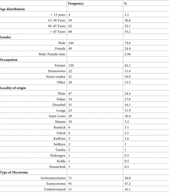
A total of 193 patients diagnosed with mycetoma have been included in this study. The major-ity of these patients were recruited in Dakar, 108 (55.9%) at Le Dantec hospital, 53 (27.5%) at the Institut d’Hygiène Sociale hospital, and 32 (16.6%) at the Thies regional hospital. The mean age of the patients was 38.3±16.4 years (range: 8–84 years). Most of the patients were 30 years old or more as presented inTable 1; 74.6% of them were male, with a 2.94 male/female ratio (Table 1). Regarding their occupation, 120 (62.1%) were farmers; 22 (11.4%) housewives; 21(10.9%) shopkeeper; and 30 (15.5%) had another occupation, including 7 students and 10 unemployed persons.
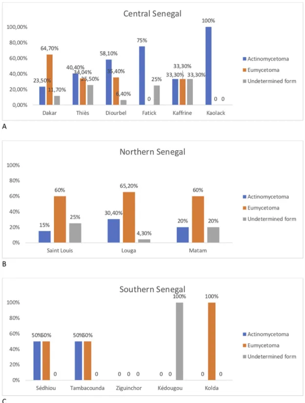
The geographic distribution across Senegal of the mycetoma cases herein analysed is illus-trated inTable 1. Most of the patients originated from the central and northern regions of the country including 47 cases from Thies (24.4%), 31 from Diourbel (16.1%), 23 from Louga (11.9%) and 20 from St Louis (10.4%). Patients originating from the capital city Dakar accounted for 34 cases (17.6%); the remaining patients were distributed in the East and South-ern part of Senegal (Fig 1). The mycetoma cases were classified into three groups according to the type of grains and laboratory criteria: there were 71 (36.8%) actinomycetoma, 91 (47.2%) eumycetoma, and 31 (16.1%) undetermined form (Table 1). Noteworthy, there were more eumycetoma cases in the northern regions than in the other part of the country (Fig 1).
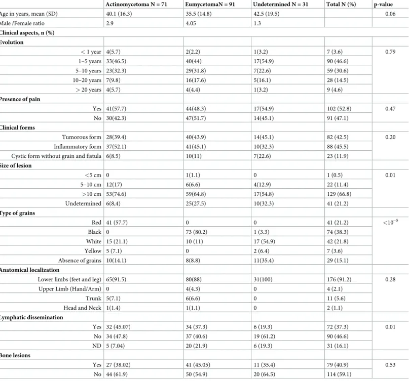
At presentation, the duration of the lesions varied according to cases. The majority of patients 90 (46.6%) had mycetoma for 1 to 5 years while 37 (19.1%) had the infection for more than 10 years (Table 2). In 102 cases (52.8%) the patients experienced pain. In the majority of these patients a painkiller was added to their treatment. The clinical form, the size of the lesion, the type of grains, the anatomical localization (Figs2and3), the lymphatic dissemination and the bone lesion are presented inTable 2.
Diagnosis of mycetoma was confirmed in 97 cases (50.3%) using microbiological examina-tions and/or histology. The culture has isolated only 56 species (29%).Actinomadura pelletieri
was the most isolated species (24) followed byMadurella mycetomatis (21), Falciformispora senegalensis (5), Streptomyces somaliensis (2), Scedosporum apiospermum (1), Penicillium sp.
(1),Fusarium solani (1), and Pseudallescheria boydii (anamorphic: Scedosporium boydii/Scedos-porium boydii) (1).
X-Ray examination was done in all the patients and was normal in the majority of cases 114 (59.1%). Bone lesions were identified in 79 patients (40.9%).
At interview, 156 (80.8%) patients have reported history of phytotherapy including 60 cases who have used oral plus local medicinal plants (Table 3), however the nature of the plants used was not mentioned. Mycetoma, clinical cases were treated regarding the type of mycetoma and the availability of the drug. Most of actinomycetoma cases were treated with trimetho-prim-sulfamethoxazole, or a combination of trimethotrimetho-prim-sulfamethoxazole, streptomycin, and amoxicillin/clavulanic acid (Table 3). In eumycetoma cases, itraconazole and terbinafine were used as antifungal agent combined in most cases with surgical treatment as shown in
Table 3. Surgical treatment consisted of wide local excision and amputation (Table 3).
Table 1. Demographic data of the study population.
Frequency % Age distribution < 15 years 4 2.1 15–30 Years 59 30.6 30–45 Years 62 32.1 > 45 Years 68 35.2 Gender Male 144 74.6 Female 49 24.4
Male: Female ratio 2.94
Occupation Farmer 120 62.1 Housewives 22 11.4 Street vendor 21 10.9 Other 30 15.5 Locality of origin Thiès 47 24.4 Dakar 34 17.6 Diourbel 31 16.1 Louga 23 11.9 Saint-Louis 20 10.4 Matam 10 5.2 Kaolack 6 3.1 Fatick 4 2.1 Kaffrine 3 1.6 Se´dhiou 2 1 Tamba 2 1 Ke´dougou 1 0.5 Kolda 1 0.5 Nouatchok 1 0.5 Type of Mycetoma Actinomycetoma 71 36.8 Eumycetoma 91 47.2 Undetermined 31 16.1 https://doi.org/10.1371/journal.pone.0231871.t001
Fig 1. Distribution of mycetoma types across Senegal.
Out of the 33 red-grain actinomycetoma cases treated with trimethoprim-sulfamethoxazole alone, 20 recovered (60.6% cure rate), 4 was lost to follow-up and 9 were still on treatment. Outcome was worse if these red-grain actinomycetoma cases were treated with trimethoprim-sulfamethoxazole + streptomycin + amoxicillin/clavulanic acid as out of the 38 patients, 10 recovered (26.3% cure rate), 2 had a recurrence, 4 was lost to follow-up and 22 were still on treatment.
Table 2. Demographic and clinical findings according to the type of mycetoma.
Actinomycetoma N = 71 EumycetomaN = 91 Undetermined N = 31 Total N (%) p-value
Age in years, mean (SD) 40.1 (16.3) 35.5 (14.8) 42.5 (19.5) 0.06
Male /Female ratio 2.9 4.05 1.3
Clinical aspects, n (%) Evolution < 1 year 4(5.7) 2(2.2) 1(3.2) 7 (3.6) 0.79 1–5 years 33(46.5) 40(44) 17(54.9) 90 (46.6) 5–10 years 23(32.3) 29(31.8) 7(22.6) 59 (30.6) 10–20 years 7(9.8) 16(17.6) 5(16.1) 28 (14.5) > 20 years 4(5.7) 4(4.4) 1(3.2) 9 (4.6) Presence of pain Yes 41(57.7) 44(48.3) 17(54.9) 102 (52.8) 0.47 No 30(42.3) 47(51.7) 14(45.1) 91 (47.1) Clinical forms Tumorous form 28(39.4) 40(43.9) 14(45.1) 82 (42.5) 0.20 Inflammatory form 37(52.1) 41(45.1) 10(32.3) 88 (45.5)
Cystic form without grain and fistula 6(8.5) 10(11) 7(22.6) 23 (11.9)
Size of lesion <5 cm 0 1(1.1) 0 1 (0.5) 0.01 5–10 cm 12(17) 6(6.6) 4(12.9) 22 (11.4) >10 cm 53(74.6) 59(64.8) 17(54.8) 129 (66.8) Undetermined 6(8,4) 25(27.5) 10(32.3) 41 (21.2) Type of grains Red 41 (57.7) 0 0 41 (21.2) <10−5 Black 0 73 (80.2) 1 (3.3) 74 (38.3) White 15 (21.1) 10 (11) 17 (54.9) 42 (21.8) Yellow 5 (7.1) 0 2 (6.4) 7 (3.6) Absence of grains 10(14.1) 8(8.8) 11(35.4) 29 (15.1) Anatomical localization
Lower limbs (feet and leg) 65(91.5) 80(88) 31(100) 176 (91.2) 0.28
Upper Limb (Hand/Arm) 0 4(4.3) 0 4 (2.1)
Trunk 5(7.1) 6(6.6) 0 11 (5.6)
Head and Neck 1(1.4) 1(1.1) 0 2 (1.1)
Lymphatic dissemination Yes 32 (45.07) 34 (37.3) 6 (19.3) 72 (37.3) 0.01 No 34 (47.8) 37 (40.6) 19 (61.2) 90 (46.6) ND 5 (7.04) 20 (21.9) 6 (19.3) 31 (16.1) Bone lesions Yes 27 (38.02) 41 (45.05) 11 (35.4) 79 (40.9) 0.53 No 44 (61.9) 50 (54.9) 20 (64.5) 114 (59.1) https://doi.org/10.1371/journal.pone.0231871.t002
Fig 2. A dorsolumbar tumoral actinomycetoma cases due toActinomadura pelletieri.
https://doi.org/10.1371/journal.pone.0231871.g002
Fig 3. “Madura foot” caused byActinomadura pelletieri.
Out of the white or yellow grain mycetoma cases, treatment with trimethoprim-sulfameth-oxazole alone (12 patients) yielded 4 recovery (33.3% cure rate), 1 recurrence, 3 patients lost to follow-up and 4 cases still on treatment. Out of the 4 patients treated with the combination tri-methoprim-sulfamethoxazole + streptomycin + amoxicillin/clavulanic acid, 2 was lost to fol-low-up and 2 were still on treatment. The 15 patients treated with Itraconazole+ antibiotics have presented 2 recovery (13% cure rate), 2 recurrence, 7 lost to follow-up and 4 cases still on treatment.
Out of the 68 black-grain eumycetoma cases treated with terbinafine and surgery, 20 recov-ered (29.4% cure rate), 2 had a recurrence, 14 were lost to follow-up and 32 were still on t-reatment. Outcome was better if these black-grain eumycetoma cases were treated with Itraconazole, trimethoprim-sulfamethoxazole + streptomycin + amoxicillin/clavulanic acid and surgery as all the 23 patients recovered (100% cure rate). The patients with eumycetoma cases were treated with terbinafine 500 mg twice daily for 24–48 weeks. In the 20 patients treated with terbinafine who recovered, one patient had a small lesion (less than 5 cm), 4 patients had lesions between 5–10 cm, and 10 patients had lesions larger than 10 cm.
Terbinafine treatment was combined with surgery in 49 of the 68 patients. Among these patients, 31 were treated with surgical removal while 18 were amputated. The 20 patients who recovered had been treated with terbinafine combined with surgery. Ten of them were treated with surgical removal and 10 were amputated. No patient has been cured with terbi-nafine only.
Table 3. Treatment and outcome according to the type of mycetoma.
Actinomycetoma N = 71 Eumycetoma N = 91 Undetermined N = 31 Total N (%) p-value Traditional phytotherapy Yes 61(86) 76(83.5) 19(61.3) 156 (80.8) 0.009 No 10(14) 15(16.5) 12(38.7) 37 (19.2) Local+Oral 21(29.5) 31(34.1) 8(25.8) 60 (31.1) 0.91 Local 14(19.7) 14(15.3) 3(9.7) 31 (16.1) Oral 26(36.6) 31(34.1 8(25.8) 65 (28.5) Medical treatment Trimethoprim-sulfamethoxazole 33(46.5) 0 12(38.7) 45(23.3) <10−5
trimethoprim-sulfamethoxazole + Amox/Ac Clavulanique + Streptomycine
38(53.5) 0 4(12.9) 42(21.8)
trimethoprim-sulfamethoxazole + Amox/Ac Clavulanique + Itraconazole 0 23(25.3) 15(48.4) 38(19.7) Terbinafine 0 68(74.7) 0 68(35.2) Surgery Yes 9(12.7) 78(85.7) 13(41.9) 100(51.8) <10−5 No 62(87.3) 13(14.3) 18(58.1) 93(48.2) Type of surgery Resection 5(7.1) 43(47.2) 6(19.3) 54(27.9) 0.02 Amputation 3(4.2) 35(38.5) 7(22.6) 45(23.3)
Lymph node dissection 1(1.4) 0 0 1(0.5)
Outcome Full recovery 30 (42.3) 43 (47.3) 6 (19.4) 79(40.9) <10−5 Recurrence 2 (2.8) 2 (2.2) 3 (9.6) 7(3.7) Lost to follow-up 8 (11.3) 14 (15.4) 12 (38.7) 34(17.6) Ongoing treatment 31 (43.6) 32 (35.1) 10 (32.3) 73(37.8) https://doi.org/10.1371/journal.pone.0231871.t003
At the time of survey, 79 (40.9%) patients has presented a full recovery. Only 7(3.7%) patients has presented recurrence (Table 3).
Discussion
With the view of strengthening mycetoma control, it is important to provide accurate and updated epidemiological information in endemic regions. This study provides, for the first time in Senegal, mycetoma cases series diagnosed in three hospital centres located in two dif-ferent regions and allows an overview of the distribution of clinical cases. The mycetoma prev-alence within the study period is underestimated because we have selected only well
documented patients to strengthen the disease description.
The patients with mycetoma described in this series were young adults in general with an mean age of 38 years, which is in accordance with previous reports in Senegal and many other countries that have shown the higher frequency of mycetoma between 20 and 40 years of age [11,12,15,16]. However, 35.2% of our patients were more than 45 years old. This category of patients was also the most infected group in a series reported in Brazil with an mean age of 48 years [17]. The excess of male patients found in this study is in agree with the literature [1,3,5,11]. Some authors have suggested the role of hormonal and genetics factors in this ele-vated male: female ratio [18,19].
Patients were more frequently diagnosed with eumycetoma (47.2%) than actinomycetoma (37.8%) in our study. In contrast, previous studies in Senegal have reported a relatively higher prevalence of actinomycetoma in their series [11,16]. This discrepancy might be explained by a recruitment bias because most of these data originated from the Le Dantec hospital derma-tology ward where actinomycetoma form are usually managed. However, the predominance of eumycetoma cases in our series is in agree with the findings of Ndiayeet al., who have
reported 70% eumycetomaversus 30% actinomycetoma in Senegal [12]. Other reports confirm the relatively high prevalence of eumycetoma in the West-African region [20,21]. Most of cases in our study were diagnosed based on clinical aspects and grain color. Only half of the cases were confirmed by mycological techniques and histopathology. Indeed, the diagnosis and the confirmation of mycetoma cases is challenging in our resources limited settings. In Senegal, culture and histopathology are the gold standard methods in hospital settings. How-ever, both techniques are operator dependent and need experience which could have its reflec-tions on the accuracy. Many difficulties have been noted in our laboratory settings, including negative culture, the misidentifications of the causative fungal agents and the inability of the technicians to correctly describe the histopathological appearance of mycetoma causative agents. This situation emphasized the need for capacity building of our laboratory technicians on mycetoma diagnosis.
Despite the use of diverse laboratory techniques coupled with clinical findings, the etiologi-cal agent remained undetermined in 16% of the patients in our study. This is quite similar to Fahalet al., where the etiological agent was undetermined in 13% of their patients [3]. The dif-ficulty of identifying the etiological agents can be explained by the lack of adequate diagnostic tools in most of our endemic countries and the impossibility in certain cases to differentiate the causative agents using the available mycological and histological techniques [22,23]. For example, it is well known that histological features cannot differentiateAcremonium spp from Fusarium spp, which both produce yellow to white grains with heterogeneous histological
aspects [2]. The direct nucleotide sequencing in biopsy samples of the 16S rRNA gene, to doc-ument actinomycetoma, and the rRNA gene internal transcribed spacer regions, to docdoc-ument eumycetoma, have been proposed to enhance pathogen identification capacities [24–28].
The geographical distribution of the patients in our study revealed the predominance of eumycetoma in the Northern region of Senegal and of actinomycetoma in the Central and Southern part of the country is in agreement with previous reports in the country [10–12]. This spatial distribution is probably associated with eco-climatic factors, because the dry tropi-cal climate in the North contrasts to the higher annual rainfall in the South. The effect of envi-ronmental factors and climate on the distribution of mycetoma cases have been demonstrated in many other countries within the African continent and elsewhere [3,15,29–31]. It is impor-tant to note the relatively high number of patients originating from the capital-city Dakar in our study, which is considered as a non-endemic area. One explanation might be the usually protracted incubation lag of the disease; the patients had probably been infected before immi-grating to the capital-city. Another likely explanation is that specialized health-care facilities that are capable to manage mycetoma are mainly available in the capital-city.
Patients with mycetoma usually attend the clinic at late stage of the disease [32]. Accord-ingly, most of the times the lag between the disease onset and hospital attendance exceeded 10 years in many of the patients in our study. Most of the patients seen in our study presented with local pain, which is probably associated with an advanced stage of the disease. This late presentation at the hospital can be explained by the lack of capacity among health workers, particularly in rural area, and consequently the absence of health education among patients and [33,34]. Most of the patients in this study had used of traditional medicine before attend-ing specialized clinics. This is in line with previous reports [35].
The clinical presentation was quite similar in both actinomycetoma and eumycetoma; how-ever, bone lesions appeared at an earlier stage in actinomycetoma than in eumycetoma. In this study, most of the lesions was tumor-like, irrespective of the mycetoma type. The lower limbs (feet and leg) were the most affected sites as described in many previous studies [1,3,11,12]. However, it is important to note that 37.3% of lesions have spread to the lymph nodes and 40.9% have reached the bones. The frequency of lymphadenopathy is relatively high in this series compared to reports from Sudan (10.3%) or Mexico (1.65%) [1,3]. It is well known that mycetoma may spread to the lymph nodes particularly in case of multiple inadequate surgery [33,36,37]. Some authors have also demonstrated the possibility of blood borne spread in cer-tain cases. This situation emphasized the need to train surgeons on the management of myce-toma and to highlight that local anaesthesia is contraindicated in mycemyce-toma [7].
Most of the mycetoma treatment used in this study is in line with previous published reports. For actinomycetoma, trimethoprim-sulfamethoxazole administrated alone gave better results (60.6% cure rate) compared to the combination of trimethoprim-sulfamethoxazole and the other antibiotics. However, it was difficult to give any conclusion in this last group as most of the patients was still on treatment (trimethoprim-sulfamethoxazole + streptomycin + amoxi-cillin/clavulanic acid) at the time of this study. Indeed, several studies have shown the excellent clinical response of the combination Co-trimoxazole and Amikacin Sulfate achieving in some studies a cure rate of about 90% [38,39]. So, this combination remains the recommended treat-ment for actinomycetoma cases.
For black grain eumycetoma cases, terbinafine was used in the majority of the patients in this series with a low cure rate (29.3%) compared to the excellent response obtained with the combination itraconazole and antibiotics. These results are in line with previous reports show-ing the low response of eumycetoma to terbinafine. For example, Ndiaye et al have reported 25% cure rate in 23 patients treated with terbinafine [40]. However, our study has some limita-tions as several patients did not completed their follow-up. So, there is a need for further stud-ies to compare the efficacy of these two drugs.
Another big challenge in the control of mycetoma is the management cost particularly in eumycetoma. Treatments are expensive and several patients do not continue their follow up.
Therefore, there is a need to find innovative and cost-effective strategies to manage mycetoma cases in our endemic settings.
In conclusion, this study has highlighted the limitations of mycetoma diagnosis in Senegal, the delayed access to healthcare and treatment, and the lack of adequate therapeutic resources. The limitations on fungal species identification is also a challenge as the management and out-come of cases depends on the causative agent. The baseline data provided will contribute to the establishment of a surveillance system for better control and advocate for more resources to fight this devastating disease.
Author Contributions
Conceptualization: Doudou Sow, Maodo Ndiaye, Babacar Faye. Data curation: Doudou Sow, Babacar T. Faye.
Formal analysis: Ste´phane Ranque.
Investigation: Doudou Sow, Maodo Ndiaye, Lamine Sarr, Mamadou D. Kante´, Fatoumata Ly, Pauline Diousse´, Abdou Magip Gaye, Cheikh Sokhna.
Software: Babacar T. Faye.
Supervision: Doudou Sow, Mamadou D. Kante´, Babacar Faye. Validation: Babacar T. Faye, Ste´phane Ranque.
Writing – original draft: Doudou Sow, Ste´phane Ranque.
Writing – review & editing: Doudou Sow, Maodo Ndiaye, Lamine Sarr, Mamadou D. Kante´, Fatoumata Ly, Pauline Diousse´, Babacar T. Faye, Abdou Magip Gaye, Cheikh Sokhna, Ste´-phane Ranque, Babacar Faye.
References
1. Bonifaz A, Tirado-Sa´nchez A, Caldero´n L, Sau´l A, Araiza J, Herna´ndez M, et al. Mycetoma: Experience of 482 Cases in a Single Center in Mexico. PLoS Negl Trop Dis. 2014; 8(8):e3102.https://doi.org/10. 1371/journal.pntd.0003102PMID:25144462
2. van de Sande WWJ. Global Burden of Human Mycetoma: A Systematic Review and Meta-analysis. PLoS Negl Trop Dis. 2013; 7(11):e2550.https://doi.org/10.1371/journal.pntd.0002550PMID:
24244780
3. Fahal A, Mahgoub ES, Hassan AME, Abdel-Rahman ME. Mycetoma in the Sudan: An Update from the Mycetoma Research Centre, University of Khartoum, Sudan. PLoS Negl Trop Dis. 2015; 9(3): e0003679.https://doi.org/10.1371/journal.pntd.0003679PMID:25816316
4. Estrada R, Cha´vez-Lo´pez G, Estrada-Cha´vez G, Lo´pez-Martı´nez R, Welsh O. Eumycetoma. Clin Der-matol. 2012; 30(4):389–96.https://doi.org/10.1016/j.clindermatol.2011.09.009PMID:22682186
5. Fahal A, Mahgoub ES, EL Hassan AM, Jacoub AO, Hassan D. Head and Neck Mycetoma: The Myce-toma Research Centre Experience. PLoS Negl Trop Dis. 2015 13; 9(3):e0003587.https://doi.org/10. 1371/journal.pntd.0003587PMID:25768090
6. de Hoog GS, van Diepeningen AD, Mahgoub E-S, van de Sande WWJ. New species of Madurella, causative agents of black-grain mycetoma. J Clin Microbiol. 2012; 50(3):988–94.https://doi.org/10. 1128/JCM.05477-11PMID:22205798
7. Suleiman SH, Wadaella ES, Fahal AH. The Surgical Treatment of Mycetoma. PLoS Negl Trop Dis. 2016; 10(6):e0004690.https://doi.org/10.1371/journal.pntd.0004690PMID:27336736
8. Gismalla MDA, Ahmed GMA, MohamedAli MM, Taha SM, Mohamed TA, Ahmed AE, et al. Surgical management of eumycetoma: experience from Gezira Mycetoma Center, Sudan. Trop Med Health. 2019; 47(1).https://doi.org/10.1186/s41182-018-0129-2PMID:30675125
9. van de Sande WWJ, Maghoub ES, Fahal AH, Goodfellow M, Welsh O, Zijlstra E. The Mycetoma Knowl-edge Gap: Identification of Research Priorities. PLoS Negl Trop Dis. 2014; 8(3):e2667.https://doi.org/ 10.1371/journal.pntd.0002667PMID:24675533
10. Dieng MT, Niang SO, Diop B, Ndiaye B. [Actinomycetomas in Senegal: study of 90 cases]. Bull Soc Pathol Exot. 1990; 98(1):18–20. PMID:15915967
11. Dieng M-T, Sy M-H, Diop B-M, Niang S-O, Ndiaye B. [Mycetoma: 130 cases]. Ann Dermatol Venereol. 2003; 130(1 Pt 1):16–9. PMID:12605151
12. Ndiaye D, Ndiaye M, Sène PD, Diouf MN, Diallo M, Faye B, et al. Myce´tomes diagnostique´s au Se´ne´gal de 2008à2010. J Mycol Me´dicale. 2011; 21(3):173–81.https://doi.org/10.1016/j.mycmed.2011.07. 003PMID:24451559
13. Ndiaye M, Diatta BA, Sow D, Diallo M, Diop A, Diadie S, et al. [A dorsolumbar tumoral actinomycosic mycetoma, an unusual mycetoma presentation]. J Mycol Medicale. 2014; 24(1):44–7.https://doi.org/ 10.1016/j.mycmed.2013.10.002PMID:24387810
14. Diongue K, Boye A, Bre´chard L, Diallo MA, Dione H, Ndoye NW, et al. Dermatophytic mycetoma of the scalp due to an atypical strain of Microsporum audouinii identified by MALDI-TOF MS and ITS sequenc-ing. J Mycol Medicale. 2019; 29(2):185–8.https://doi.org/10.1016/j.mycmed.2019.03.001PMID:
30956063
15. Lo´pez-Martı´nez R, Me´ndez-Tovar LJ, Bonifaz A, Arenas R, Mayorga J, Welsh O, et al. [Update on the epidemiology of mycetoma in Mexico. A review of 3933 cases]. Gac Med Mex. 2013; 149(5):586–92. PMID:24108347
16. Develoux M, Dieng MT, Kane A, Ndiaye B. [Management of mycetoma in West-Africa]. Bull Soc Pathol Exot. 2003; 96(5):376–82. PMID:15015843
17. Castro LGM, Piquero-Casals J. Clinical and mycologic findings and therapeutic outcome of 27 myce-toma patients from São Paulo, Brazil. Int J Dermatol. 2008; 47(2):160–3.https://doi.org/10.1111/j. 1365-4632.2008.03447.xPMID:18211487
18. van de Sande WWJ, Fahal A, Tavakol M, van Belkum A. Polymorphisms in catechol-O-methyltransfer-ase and cytochrome p450 subfamily 19 genes predispose towards Madurella mycetomatis -induced mycetoma susceptibility. Med Mycol. 2010; 48(7):959–68.https://doi.org/10.3109/
13693781003636680PMID:20184498
19. Vera-Cabrera L, Salinas-Carmona MC, Waksman N, Messeguer-Pe´rez J, Ocampo-Candiani J, Welsh O. Host defenses in subcutaneous mycoses. Clin Dermatol. 2012; 30(4):382–8.https://doi.org/10. 1016/j.clindermatol.2011.09.008PMID:22682185
20. Adoubryn KD, Koffi KE, Troh E, Doukoure B, Kouadio-Yapo CG, Ouhon J, et al. Les myce´tomes auto-chtones de Coˆte d’Ivoire: caractères e´pide´miologiques et e´tiologiques des cas confirme´ s. J Mycol Me´di-cale. 2009; 19(2):71–6.https://doi.org/10.1016/j.mycmed.2008.11.004
21. Adoubryn KD, Koffi KE, Doukoure B, Troh E, Kouadio-Yapo CG, Ouhon J, et al. Myce´tomes des Ouest-Africains non re´sidants en Coˆte d’Ivoire. J Mycol Me´dicale. 2010; 20(1):26–30.https://doi.org/10.1016/ j.mycmed.2009.11.007
22. Verghese A, Klokke AH. Histologic diagnosis of species of fungus causing mycetoma. Indian J Med Res. 1966; 54(6):524–30. PMID:5947017
23. Ahmed SA, van de Sande WWJ, Stevens DA, Fahal A, van Diepeningen AD, Menken SBJ, et al. Revi-sion of agents of black-grain eumycetoma in the order Pleosporales. Persoonia—Mol Phylogeny Evol Fungi. 2014; 33(1):141–54.https://doi.org/10.3767/003158514X684744PMID:25737597
24. Zhi X-Y, Li W-J, Stackebrandt E. An update of the structure and 16S rRNA gene sequence-based defini-tion of higher ranks of the class Actinobacteria, with the proposal of two new suborders and four new families and emended descriptions of the existing higher taxa. Int J Syst Evol Microbiol. 2009; 59 (3):589–608.https://doi.org/10.1099/ijs.0.65780-0
25. Steingrube VA, Brown BA, Gibson JL, Wilson RW, Brown J, Blacklock Z, et al. DNA amplification and restriction endonuclease analysis for differentiation of 12 species and taxa of Nocardia, including recog-nition of four new taxa within the Nocardia asteroides complex. J Clin Microbiol. 1995; 33(12):3096– 101. PMID:8586680
26. Ahmed AO, Mukhtar MM, Kools-Sijmons M, Fahal AH, de Hoog S, van den Ende BG, et al. Develop-ment of a species-specific PCR-restriction fragDevelop-ment length polymorphism analysis procedure for identi-fication of Madurella mycetomatis. J Clin Microbiol. 1999; 37(10):3175–8. PMID:10488173
27. Mhmoud NA, Ahmed SA, Fahal AH, de Hoog GS, Gerrits van den Ende AHG, van de Sande WWJ. Pleurostomophora ochracea, a Novel Agent of Human Eumycetoma with Yellow Grains. J Clin Micro-biol. 2012; 50(9):2987–94.https://doi.org/10.1128/JCM.01470-12PMID:22760037
28. de Hoog GS, van Diepeningen AD, Mahgoub E-S, van de Sande WWJ. New Species of Madurella, Causative Agents of Black-Grain Mycetoma. J Clin Microbiol. 2012; 50(3):988–94.https://doi.org/10. 1128/JCM.05477-11PMID:22205798
29. Mathur, Vyas MC. Mycetoma—epidemiological and pathologic study of 133 cases. Indian J Pathol Microbiol. 1989; 32(1):75. PMID:2592035
30. Sharma NL, Mahajan VK, Agarwal S, Katoch VM, Das R, Kashyap M, et al. Nocardial mycetoma: diverse clinical presentations. Indian J Dermatol Venereol Leprol. 2008; 74(6):635–40.https://doi.org/ 10.4103/0378-6323.45110PMID:19171991
31. Mathur D, Bakshi R. Incidence and changing pattern of mycetoma in western Rajasthan. Indian J Pathol Microbiol. 2008; 51(1):154.https://doi.org/10.4103/0377-4929.40433PMID:18417892
32. Fahal A, Mahgoub ES, EL Hassan AM, Abdel-Rahman ME, Alshambaty Y, Hashim A, et al. A New Model for Management of Mycetoma in the Sudan. PLoS Negl Trop Dis. 2014; 8(10):e3271.https://doi. org/10.1371/journal.pntd.0003271PMID:25356640
33. Ahmed AA, van de Sande WW, Fahal A, Bakker-Woudenberg I, Verbrugh H, van Belkum A. Manage-ment of mycetoma: major challenge in tropical mycoses with limited international recognition: Curr Opin Infect Dis. 2007; 20(2):146–51.https://doi.org/10.1097/QCO.0b013e32803d38fePMID:17496572
34. Poncio Mendes R, Negroni R, Bonifaz A, Pappagianis D. New aspects of some endemic mycoses. Med Mycol. 2000; 38 Suppl 1:237–41. PMID:11204151
35. Ezaldeen EA, Fahal AH, Osman A. Mycetoma Herbal Treatment: The Mycetoma Research Centre, Sudan Experience. PLoS Negl Trop Dis. 2013; 7(8):e2400.https://doi.org/10.1371/journal.pntd. 0002400PMID:23991244
36. El Hassan AM, Mahgoub ES. Lymph node involvement in mycetoma. Trans R Soc Trop Med Hyg. 1972; 66(1):165–9.https://doi.org/10.1016/0035-9203(72)90065-xPMID:5065467
37. Fahal A. Mycetoma: a thorn in the flesh. Trans R Soc Trop Med Hyg. 2004; 98(1):3–11.https://doi.org/ 10.1016/s0035-9203(03)00009-9PMID:14702833
38. Welsh O, Al-Abdely HM, Salinas-Carmona MC, Fahal AH. Mycetoma medical therapy. PLoS Negl Trop Dis. 2014; 8(10):e3218.https://doi.org/10.1371/journal.pntd.0003218PMID:25330342
39. Fahal Ahmed Hassan. Mycetoma. Khartoum Medical Journal. 2011; 4 (01): 514–23
40. N’Diaye B, Dieng MT, Perez A, Stockmeyer M, Bakshi R. Clinical efficacy and safety of oral terbinafine in fungal mycetoma. Int J Dermatol. 2006; 45: 154–7.https://doi.org/10.1111/j.1365-4632.2004.02392. xPMID:16445509