Original Article
Management and outcome of Ebstein’s anomaly in children
Angela Oxenius,1,2 Christine H. Attenhofer Jost,3 Rene´ Preˆtre,2,4 Hitendu Dave,2,4 Urs Bauersfeld,1,2
Oliver Kretschmar,1,2 Burkhardt Seifert,5 Christian Balmer,1,2Emanuela R. Valsangiacomo Buechel1,2
1Division of Pediatric Cardiology, University Children’s Hospital; 2Pediatric Research Centre (PRC), University of
Zurich;3Cardiovascular Centre Zurich, Klinik im Park;4Cardiothoracic Surgery, University Children’s Hospital;
5
Division of Biostatistics, Institute of Social and Preventive Medicine, University of Zurich, Zurich, Switzerland Abstract Objectives: To assess clinical presentation, treatment, and outcome of children with Ebstein’s anomaly. Background: Data on long-term outcome of children with Ebstein’s anomaly are scarce. Methods: Retrospective analysis of all children with Ebstein’s anomaly treated between February, 1979 and January, 2009 in a single tertiary institution. Primary outcomes included patient survival and need for intervention, either cardiac surgery or catheter intervention. Results: A total of 42 patients were diagnosed with Ebstein’s anomaly at a median age of 5 days ranging from 1 day to 11.7 years. Symptoms included cyanosis, heart murmur, and/or dyspnoea. Associated cardiac anomalies occurred in 90% of the patients. Average follow-up was 9.5 plus or minus 7.0 years. The overall mortality rate was 14%. Of the six patients, three died postnatally before treatment. Cardiac surgery and/or catheter-guided interventions were required in 33 patients (79%). Cardiac surgery was performed in 21 (50%) patients at a median age of 9.1 years (range 0.1–16.5 years), including biventricular repair in 13 (62%), one-and-a-half chamber repair in seven (33%), and a staged single-ventricle repair in one. Peri-operative mortality was 4%. Catheter-guided interventions consisted of device closure of an atrial septal defect in three cases and radiofrequency ablation of accessory pathways in nine patients. The estimated 10-year survival was 85.3 plus or minus 5.6%. Conclusion: In children, Ebstein’s anomaly is usually diagnosed in the first year of age. Even though children with Ebstein’s anomaly often require an intervention, their peri-operative mortality is low and long-term survival is good. Symptomatic newborns requiring an intervention may have a worse outcome.
Keywords: Congenital heart defect; tricuspid valve; intervention
Received: 28 June 2011; Accepted: 3 February 2012; First published online: 15 March 2012
E
BSTEIN’S ANOMALY IS A RARE CONGENITAL HEARTdefect occurring in 1–5 of 200,000 live births, accounting for less than 1% of all congenital
heart defects.1 It encompasses a wide anatomic
spectrum of abnormalities of the tricuspid valve and of the right ventricle including atrialisation of parts of the right ventricle due to apical displacement of the septal and posterior tricuspid valve leaflets. Additional cardiac anomalies are common and include atrial septal defect in up to 90%, accessory pathways
leading to ventricular pre-excitation in
approxi-mately 15% of cases,2 and more rarely
noncompac-tion of the left ventricular myocardium.3 Ebstein’s
anomaly may manifest at any age, ranging from the prenatal stage to older age.4 Clinical presentation is
usually age dependent:5 neonates may present with
cyanosis, congestive heart failure, and marked cardi-omegaly; older children may have signs of progressive right ventricular failure; adolescents and adults often present with palpitations due to arrhythmias or mild symptoms such as exertional dyspnoea.1
The techniques of tricuspid valve reconstruction have evolved during the last decades, and a biventricular repair with a competent tricuspid valve can usually
be achieved with good results.6–8
Correspondence to: Dr A. Oxenius, MD, Department of Pediatric Cardiology, University Children’s Hospital, Steinwiesstrasse 75, 8032 Zurich, Switzerland. Tel: 141 44 266 70 22; Fax: 141 44 266 79 81; E-mail: angela.oxenius@kispi. uzh.ch
Ebstein’s anomaly is being increasingly diagnosed early in life. Previous studies identified cyanosis and need for early intervention as risk factors for poor
outcome in neonates and young children.9–13
Thus, management of symptomatic children with Ebstein’s anomaly remains challenging and addi-tional interventions such as systemic-to-pulmonary artery shunts, cavopulmonary shunts, and radio-frequency ablations of supraventricular arrhythmias due to pre-excitation and/or transcatheter device closure of interatrial shunts may be necessary before or after tricuspid valve surgery.14,15In the most severe cases, the Fontan procedure with right ventricular exclusion may be the surgical treatment of choice.12,16 We sought to assess clinical presentation, treat-ment strategies, and outcome in a cohort of neonates and children with Ebstein’s anomaly.
Methods
Medical and surgical reports, echocardiographic find-ings, and 12-lead electrocardiograms of all consecutive liveborns diagnosed with Ebstein’s anomaly at our institution between 1979 and 2009 were retrospectively reviewed. Particular focus was given to symptoms, treatment strategies including surgical and catheter-guided interventions, as well as to follow-up. Follow-up was performed either by arranging a clinical visit to our centre (33 patients) or by contacting the treating physician (three patients).
Primary end-points included patient survival and need for intervention, either cardiac surgery or catheter-guided interventions including radiofrequency ablation and/or interventional device closure of secundum atrial septal defect.
The study was approved by the local ethics committee.
Surgical strategy and operative technique
Indications for surgical intervention in the neonatal period included ventilator dependency, prostaglandin-dependent circulation, and/or severe cardiac failure. A symptomatic newborn was allowed time for transition from the foetal circulation, particularly for regression of neonatal pulmonary hypertension. Usually, a systemic-to-pulmonary artery shunt was chosen as the primary modality to relieve cyanosis.
Intracardiac corrective surgery was performed in older children presenting with one or a combination of the following: congestive heart failure, New York Heart Association functional class III, cyanosis with saturations below 90%, and arrhythmias refractory to medical treatment. Although difficult to determine in retrospect, the most common indication was presence of right-sided heart congestion resulting
from moderate or severe tricuspid regurgitation. Biventricular correction involving tricuspid valve repair with plication of the atrialised right ventricle free wall and a pulmonary commissurotomy or either transannular monocusp patch or right ventricle to pulmonary artery valved conduit was the preferred technique. Tricuspid valve repair involved detachment of the displaced septal, posterior, and even anterior leaflet from their abnormal attachment to the free wall of the right ventricle, re-attachment of the leaflets to the virtual neo-annulus, and then ultimately reducing the neo-annulus with an annuloplasty. The valve was then bestowed upon with a parachute subvalvar apparatus, by creating multiple artificial neo-chordae
(Goretexs suture, W.L. Gore & Associate, Inc.,
Flagstaff, Arizona, United States of America). When tricuspid valve competence could not be achieved, the valve was replaced with a biological valve. We have used an age-adjusted Contegras(Contegra, Medtronic, Inc., Minneapolis, Minnesota, United States of America) xenograft valve indigenously housed in a
Goretexs tube to replace the tricuspid valve in one
patient. In cases of significant displacement of the tricuspid valve leaflets, with borderline functional right ventricle, a bidirectional Glenn anastomosis was performed to create a one-and-a-half ventricle repair. Most severe forms of Ebstein’s anomaly with severely impaired right ventricular volume and function underwent a staged univentricular repair. The surgical technique chosen was determined by the severity of tricuspid valve dysplasia and of impairment of the right ventricular size and function, as well as presence of functional pulmonary atresia.
Catheter intervention
Indication for transcatheter device closure of a secundum atrial septal defect was presence of a haemodynamic relevant shunt if oxygen saturation was normal at rest and during exercise testing, or presence of cyanosis or desaturation below 90%, at rest or during exercise.
Catheter ablation
Invasive electrophysiology examination with radio-frequency ablation was performed in all patients with obvious accessory pathway on electrocardiogram and/or clinical symptomatic tachyarrhythmias. If radio-frequency ablation was inefficient in treating recurrent tachyarrhythmias, surgical cryoablation was planned during surgical repair.
Statistical methods
Continuous data are expressed as median and range, nominal data as frequencies. Actuarial survival was analysed using the Kaplan–Meier method.
Peri-operative mortality was defined as mortality within 30 days after operation.
Results
General findings
A total of 42 patients with Ebstein’s anomaly, 52% boys, were included in the study. Diagnosis was made at a median age of 5 days – range from 1 day to 11.7 years, and in 86% of the patients during the first year of life (Fig 1). In 9 cases, Ebstein’s anomaly was diagnosed prenatally: four of them died, two received tricuspid valve repair at a later age, one had a one-and-a-half ventricle repair, and one patient never underwent any intervention. A syndromal disorder occurred in three patients, including Down’s syndrome, VACTERL-association, and un-classified dysmorphic features in one each. Clinical symptoms at the time of presentation of the 33 patients who were not diagnosed prenatally are shown in Table 1. Additional cardiac anomalies and morbities occurred in 90% of all patients (Table 2). Left ventricular ejection fraction was reduced in two of five patients with a left ventricular noncompac-tion. Of the 11 patients with Wolff–Parkinson– White syndrome, two presented with tachycardia postnatally, seven later in life, and the other two remained asymptomatic.
Diagnosis was primarily made by transthoracic echocardiography in all patients; cardiac magnetic
resonance imaging was additionally performed for right ventricular evaluation in 11.
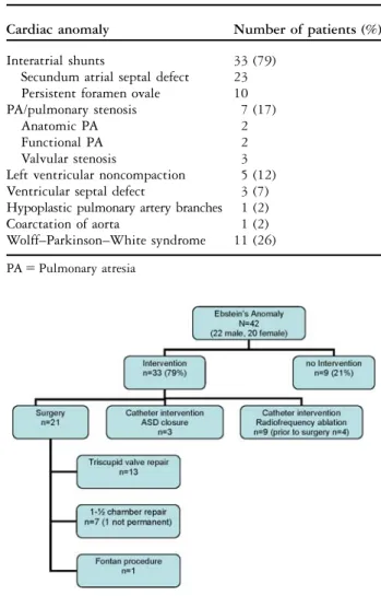
Invasive treatment was required in 33 of 42 patients (79%; Fig 2).
At least one surgical intervention was necessary in 21 patients (50%) at a median age of 9.1 years (0.1–16.5).
Biventricular repair
Biventricular repair was achieved in 13 patients (62%), and consisted of tricuspid valve reconstruction, annuloplasty (DeVega), and closure of secundum atrial septal defect if required. Figures 3 and 4 show a 12-year-old patient with moderate Ebstein’s anomaly and severe, respectively, mild tricuspid regurgitation before and after surgery. In two patients, a redo tricuspid valve repair was necessary because of residual severe regurgitation 2 weeks and 9 months, respec-tively, after first repair. In two patients with atrial reentry tachycardia, cryoablation was performed in the right atrium during surgery. Pacemaker implantation
Figure 1.
Age distribution at the time of diagnosis of Ebstein’s anomaly in children.
Table 1. Clinical presentation.
Symptoms Number of patients (%)
Heart murmur 14 (33)
Cyanosis 12 (29)
Arrhythmias 2 (5)
Congestive heart failure 2 (5)
Syndromal disorder 3 (7) Figure 2.
Summary of interventions. ASD 5 atrial septal defect.
Table 2. Cardiac anomalies and morbidities.
Cardiac anomaly Number of patients (%)
Interatrial shunts 33 (79)
Secundum atrial septal defect 23 Persistent foramen ovale 10
PA/pulmonary stenosis 7 (17)
Anatomic PA 2
Functional PA 2
Valvular stenosis 3
Left ventricular noncompaction 5 (12) Ventricular septal defect 3 (7) Hypoplastic pulmonary artery branches 1 (2)
Coarctation of aorta 1 (2)
Wolff–Parkinson–White syndrome 11 (26)
was necessary in one patient because of sick sinus syndrome. Pre-operative radiofrequency ablation was performed in four patients with a Wolff–Parkinson– White syndrome.
One-and-a-half ventricle repair
Approximately 33%, that is, seven, children were palliated with a cavopulmonary anastomosis result-ing in a one-and-a-half ventricle repair. In these
patients, a modified Blalock–Taussig Shunt was first performed in the neonatal period. The bidirectional cavopulmonary anastomosis was then performed at a median age of 7.4 years (0.4–12.8 years). Of these patients, four additionally had an anatomical or functional pulmonary atresia with insufficient pulmonary blood flow. In one case, cavopulmonary anastomosis was taken down and physiology returned successfully into a biventricular repair after 2.8 years.
Figure 3.
Transthoracic echocardiography of a 12-year-old patient with moderate form of Ebstein anomaly and severe tricuspid regurgitation. Apical 4 chamber view. ARV 5atrialised right ventricle; LV 5 left ventricle; RA 5 right atrium; RV 5 right ventricle; arrow points to apical displacement of the septal tricuspid valve leaflet.
Figure 4.
Transthoracic echocardiography of the same patient as in Figure 3 after surgical repair. Apical 4 chamber view. LV 5 left ventricle; RA 5 right atrium; RV 5 right ventricle; arrow points to reconstructed tricuspid valve.
This patient had previously obtained tricuspid valve
replacement by using insertion of a Contegras
xenograft housed in a Goretexs tube in tricuspid
position at the age of 6 months. The xenograft required repeated replacement at the age of 1.5 and 2.5 years because of inadequate size during somatic growth. At the end of follow-up, this patient was 5 years old and presented in functional class II receiving diuretics, betablocker, and aspirin medi-cation. There was one patient who underwent radiofrequency ablation before cavopulmonary ana-stomosis and another in whom one right-sided cryoablation was performed during surgery, because of recurrent intraatrial tachycardias.
Single-ventricle palliation
There was one patient (5%) with Ebstein’s anomaly and severe right ventricular hypoplasia who was treated with a staged univentricular repair (Fontan), consisting of Blalock–Taussig Shunt during the neonatal period, a bidirectional cavopulmonary anastomosis at the age of 2.1 years, and an extracardiac conduit and obliteration of the right ventricle at the age of 4.5 years.
Catheter interventions
A transcatheter device closure of a secundum atrial
septal defect using an Amplatzers (AGA Medical
Corporation, Golden Valley, Minnesota, United States of America) Septal Occluder was performed successfully in three patients (7%). None of these patients had preceding additional surgery. Indication for interventional closure was ventricular volume overload without cyanosis in one child and mild
cyanosis (O2 saturation 90%) due to bidirectional
shunting across the atrial septum, as well as New York Heart Association functional class II in the other two. The median age at device closure was 13.9 years (1.2–16.4 years). In all patients, oxygen saturation improved to over 95% and no right ventricular failure or increase in tricuspid regurgitation occurred during follow-up.
Catheter ablation of the accessory pathway was performed in 11 patients with Wolff–Parkinson– White Syndrome, at a median age of 9.3 years (6.1–14.5 years), and successfully completed with-out any arrhythmia recurrence in 78% of the cases. In two patients, ablation was not effective; in one of these patients, a second catheter ablation was success-ful, whereas in the other patient surgical cryoablation was performed at time of surgical repair.
Follow-up
Median follow-up was 9.2 years (1 month to 29.4 years). All patients after tricuspid valve reconstruction were
in functional classes I–II without any medication. The regurgitation on echocardiography ranged from mild to moderate. Patients with cavopulmonary anastomosis usually were in functional class II, with 50% of them receiving diuretics.
In all, six patients died. Peri-operative mortality was 4%, as one neonate died early post-operatively. Approximately 7%, that is, three, patients died postnatally because of congestive heart failure before they could undergo surgical repair. Of the patients, two were born prematurely at 31 and 36 gestational weeks, respectively, and one presented with hydrops fetalis. In both, the diagnosis Ebstein’s anomaly was known prenatally. There were two patients who had a sudden cardiac death; one child under betablocker medication and without any previous intervention died at the age of 3.4 years because of cardiovascular collapse caused by intraatrial reentry tachycardia. The other patient, who had been previously palliated with a modified Blalock– Taussig Shunt, presented with a severe viral infection and in dismal conditions at the age of 6 months.
The 10-year survival estimate was 85.3 plus or minus 5.6% (Fig 5).
Discussion
This paper reports the clinical presentation and our treatment algorithms, as well as outcome of children diagnosed with Ebstein’s anomaly. Despite a severe form of Ebstein’s anomaly in most of these children, outcome in this series was quite good; however, this required frequent interventions. Contrarily to adults, these interventions rarely include tricuspid valve replacement, but tricuspid valve repair, besides closure of interatrial communications and arrhythmia surgery.
Figure 5.
Diagnosis and presentation of Ebstein’s anomaly In patients with Ebstein’s anomaly, varying degrees of right ventricular dysfunction and tricuspid regurgita-tion, presence of an interatrial shunting, and/or right ventricular outflow tract obstruction yield to a wide spectrum of presentation. In addition, arrhythmias may originate from reentry pathways and from right atrial dilatation because of volume overload. The clinical symptoms of the disease are mainly correlated to the age of the patient. Thus, newborns may present severely symptomatic with cyanosis and congestive heart failure.10 In contrast, adult patients usually present with exercise intolerance, tachyarrhythmias, or signs of progressive congestive heart failure.17
The very young age at diagnosis observed in our cohort reflects the fact that clinical conditions are worse in younger patients.9,11Prenatal diagnosis was made in 21% of our patient group reflecting severe Ebstein’s anomaly, as 56% of them died during the first 3.4 years of life. Survival of young children with severe Ebstein’s anomaly is mainly warranted by early intervention, as it was the case in 79% of our patients. Diagnostic evaluation
Echocardiography is the first-line modality for assessing Ebstein’s anomaly. As children present a good imaging window, usually transthoracic echo-cardiography enables accurate evaluation of the tricuspid valve and the size and function of both ventricles. Echocardiographic assessment should include evaluation of abnormal trabeculations in the left ventricle, as left myocardial noncompaction in Ebstein’s anomaly has been described,3and impaired left ventricular function may worsen prognosis. In our cohort, left ventricular noncompaction was diagnosed in five patients by echocardiography. None of these patients showed signs of left heart failure.
Nowadays, cardiac magnetic resonance is of additional value for exact quantitative right ven-tricular assessment (Fig 6), and demonstration of the displacement of the tricuspid valve leaflets in all three dimensions (Supplementary Movie S1), which can be useful when planning surgical repair during follow-up, if cardiac failure occurs. Cardiac mag-netic resonance is the ideal modality for serial quantification of right ventricular function.18
Surgical interventions in young children with Ebstein’s anomaly
Management of neonates with Ebstein’s anomaly is challenging and needs to be individualised for each particular patient. Biventricular repair is usually preferable if the right ventricular size and function are adequate. Surgical repair with tricuspid valve reconstruction can be performed in young children
with low mortality and good long-term durability of the valve, provided that tricuspid valve
compe-tence is achieved.19,20 Whenever feasible, in
children tricuspid valve reconstruction is preferable to valve replacement. In addition to well-known complications such as thromboembolism, endocar-ditis, and valvular degeneration, in neonates and small children valve prosthesis may be too large at the time of insertion, with the risk of disruption of the geometry of the adjacent structures. Later during somatic growth the valve may result too small. In our series, biventricular repair with tricuspid valve reconstruction was achieved in 62% of the patients undergoing surgery. Never-theless, a redo rate of 15% for tricuspid valve reconstruction reflects the challenges of this valve reconstruction during infancy.
In neonates requiring initial palliation with a
systemic-to-pulmonary shunt, a one-and-a-half
ventricle repair with a cavopulmonary anastomosis should be considered after 2–3 months if their functional capacity of the right ventricle remains marginal or insufficient. A well-functioning one-and-a-half ventricle repair with an unloaded right ventricle may be preferable to a compromised
biventricular repair.21,22 The prevalence of 38% of
children who underwent one-and-a-half ventricle repair observed in our cohort is similar to the one reported by the European Congenital Heart
Sur-geons Association.22 However, the same authors
report significantly higher mortality in young infants and higher rate of tricuspid valve replace-ment even in older patients, than we found in our
Figure 6.
Steady-state free precession cardiovascular magnetic resonance image in an axial plane showing severe displacement of the septal leaflet (black arrow) and a coaptation deficit of the tricuspid valve (white arrow). ARV 5atrialised right ventricle; RA 5 right atrium; RV 5 right ventricle.
study. Thus, considering that symptomatic neonates with Ebstein’s anomaly tend to have unfavourable anatomic and pathophysiologic conditions with potentially a worse prognosis, our data may suggest that an accurate planning and an individual-ised treatment strategy enable successful surgical approach towards biventricular repair and preservation of the tricuspid valve.19
Treatment strategies for tachyarrhythmias
Radiofrequency ablation is safe and effective for treatment of various types of tachycardias occurring
in Ebstein’s anomaly.23 Nevertheless, in patients
with Ebstein’s anomaly, catheter ablation presents lower success rate with some arrhythmia recurrence in comparison with procedures performed in
structurally normal hearts.24 In contrast, surgical
cryoablation for accessory pathway-mediated tachy-cardia provides excellent results with freedom from
arrhythmia recurrence.25,26 In our patients, the
success rate of radiofrequency ablation was 78%. Owing to the fact that arrhythmias are one of the leading symptoms in older children and adults with Ebstein’s anomaly, an accurate electrophysiological investigation is indicated already during childhood and radiofrequency ablation of accessory pathways should be performed when appropriate. In case of arrhythmias recurrence, surgical cryoablation can be planned at the time of surgical repair.
Transcatheter device closure of interatrial shunts Transcatheter device closure of a secundum atrial septal defect can represent the only required interven-tion in selected patients, without significant tricuspid valve regurgitation and with good right ventricular function. Atiq et al14report about six children with Ebstein’s anomaly and without prior procedures, who received successful device closure of a secundum atrial septal defect. Similarly, in our three cases transcatheter device closure led to a normalisation of oxygen saturation and improvement of clinical symptoms. Thus, transcatheter device closure of a secundum atrial septal defect in patients with Ebstein’s anomaly, who do not require tricuspid valve repair, seems to be a safe and efficient way of improving oxygen saturation under rest and exercise. Moreover, it may help in
preventing paradoxical thromboembolisms.15
Never-theless, when considering transcatheter device closure, the degree of tricuspid valve regurgitation needs to be previously carefully assessed, as interatrial shunt closure alone may worsen right ventricular function.27
Limitations
This is a retrospective descriptive study. Even though not exceedingly small for such a rare disease
like Ebstein’s anomaly, the size of our patient group, and the few patients with adverse outcome did not allow a statistical analysis for risk factors for poor outcome. However, our findings reflect the observa-tions reported by others, that neonates with cyanosis and/or congestive heart failure are the group at risk for mortality. In fact, five of the six deaths in our patient group consisted of neonates with a severe form of Ebstein’s anomaly.
Conclusion
Ebstein’s anomaly is a complex congenital structural heart defect with a broad anatomic and clinical spectrum. In particular, the management of newborn patients, usually affected by the most severe form of the disease, is challenging. An individualised treat-ment strategy considering the clinical symptoms of the patient, the anatomic and haemodynamic conditions and the associated abnormalities, and the choice of the most appropriate interventions, as surgery, catheter-guided interventions, radiofrequency ablations, may provide excellent results and good outcome.
Supplementary materials
For supplementary materials referred to in this article, please visit http://dx.doi.org/doi:10.1017/ S1047951112000224
References
1. Attenhofer Jost CH, Connolly HM, Dearani JA, Edwards WD, Danielson GK. Ebstein’s anomaly. Circulation 2007; 115: 277–285. 2. Brown ML, Dearani JA, Danielson GK, et al. Functional status after operation for Ebstein anomaly: the Mayo Clinic experience. JACC 2008; 52: 460–466.
3. Attenhofer Jost CH, Connolly HM, Warnes CA, et al. Noncompacted myocardium in Ebstein’s anomaly: initial description in three patients. J Am Soc Echocardiogr 2004; 17: 677–680. 4. Celermajer DS, Bull C, Till JA, et al. Ebstein’s anomaly:
presentation and outcome from fetus to adult. JACC 1994; 23: 170–176.
5. Attenhofer Jost CH, Connolly HM, Edwards WD, Hayes D, Warnes CA, Danielson GK. Ebstein’s anomaly – review of a multifaceted congenital cardiac condition. Swiss Med Wkly 2005; 135: 269–281.
6. Danielson GK, Maloney JD, Devloo RA. Surgical repair of Ebstein’s anomaly. Mayo Clin Proc 1979; 54: 185–192. 7. Carpentier A, Chauvaud S, Mace L, et al. A new reconstructive
operation for Ebstein’s anomaly of the tricuspid valve. J Thorac Cardiovasc Surg 1988; 96: 92–101.
8. da Silva JP, Baumgratz JF, da Fonseca L, et al. The cone reconstruction of the tricuspid valve in Ebstein’s anomaly. The operation: early and midterm results. J Thorac Cardiovasc Surg 2007; 133: 215–223.
9. Kapusta L, Eveleigh RM, Poulino SE, et al. Ebstein’s anomaly: factors associated with death in childhood and adolescence: a multi-centre, long-term study. Eur Heart J 2007; 28: 2661–2666. 10. Celermajer DS, Cullen S, Sullivan ID, Spiegelhalter DJ, Wyse RK, Deanfield JE. Outcome in neonates with Ebstein’s anomaly. JACC 1992; 19: 1041–1046.
11. Yetman AT, Freedom RM, McCrindle BW. Outcome in cyanotic neonates with Ebstein’s anomaly. Am J Cardiol 1998; 81: 749–754.
12. Shinkawa T, Polimenakos AC, Gomez-Fifer CA, et al. Manage-ment and long-term outcome of neonatal Ebstein anomaly. J Thorac Cardiovasc Surg 2010; 139: 354–358.
13. Bove EL, Hirsch JC, Ohye RG, Devaney EJ. How I manage neonatal Ebstein’s anomaly. Semin Thorac Cardiovasc Surg Pediatr Card Surg Annu 2009; 12: 63–65.
14. Atiq M, Lai L, Lee KJ, Benson LN. Transcatheter closure of atrial septal defects in children with a hypoplastic right ventricle. Catheter Cardiovasc Interv 2005; 64: 112–116.
15. Munayer Calderon JE, Maza Juarez G, Carpio JC, et al. Percutaneous closure of patent foramen ovale with amplatzer device. Report of two cases. Arch Cardiol Mex 2005; 75: 306–309.
16. Badiu CC, Schreiber C, Horer J, et al. Early timing of surgical intervention in patients with Ebstein’s anomaly predicts superior long-term outcome. Eur J Cardiothorac Surg 2010; 37: 186–192. 17. Paranon S, Acar P. Ebstein’s anomaly of the tricuspid valve: from fetus to adult: congenital heart disease. Heart 2008; 94: 237–243. 18. Cantinotti M, Bell A, Razavi R. Role of magnetic resonance imaging in different ways of presentation of Ebstein’s anomaly. J Cardiovasc Med (Hagerstown) 2008; 9: 628–630.
19. Boston US, Dearani JA, O’Leary PW, Driscoll DJ, Danielson GK. Tricuspid valve repair for Ebstein’s anomaly in young children: a 30-year experience. Ann Thorac Surg 2006; 81: 690–695; discussion 695–696.
20. Wu Q, Huang Z, Pan G, Wang L, Li L, Xue H. Early and midterm results in anatomic repair of Ebstein anomaly. J Thorac Cardiovasc Surg 2007; 134: 1438–1440; discussion 1440–1432. 21. Knott-Craig CJ, Goldberg SP. Management of neonatal Ebstein’s anomaly. Semin Thorac Cardiovasc Surg Pediatr Card Surg Annu 2007; 10: 112–116.
22. Sarris GE, Giannopoulos NM, Tsoutsinos AJ, et al. Results of surgery for Ebstein anomaly: a multicenter study from the European Congenital Heart Surgeons Association. J Thorac Cardiovasc Surg 2006; 132: 50–57.
23. Hebe J. Ebstein’s anomaly in adults. Arrhythmias: diagnosis and therapeutic approach. Thorac Cardiovasc Surg 2000; 48: 214–219.
24. Cappato R, Schluter M, Weiss C, et al. Radiofrequency current catheter ablation of accessory atrioventricular pathways in Ebstein’s anomaly. Circulation 1996; 94: 376–383.
25. Khositseth A, Danielson GK, Dearani JA, Munger TM, Porter CJ. Supraventricular tachyarrhythmias in Ebstein anomaly: management and outcome. J Thorac Cardiovasc Surg 2004; 128: 826–833.
26. Greason KL, Dearani JA, Theodoro DA, Porter CB, Warnes CA, Danielson GK. Surgical management of atrial tachyarrhythmias associated with congenital cardiac anomalies: Mayo Clinic experience. Semin Thorac Cardiovasc Surg Pediatr Card Surg Annu 2003; 6: 59–71.
27. Taguchi K, Matsumura M, Ishikawa M, et al. Deterioration of Ebstein disease after closure of atrial septal defect. Jpn J Surg 1982; 12: 126–129.