HAL Id: tel-03183825
https://tel.archives-ouvertes.fr/tel-03183825
Submitted on 29 Mar 2021
HAL is a multi-disciplinary open access archive for the deposit and dissemination of sci-entific research documents, whether they are pub-lished or not. The documents may come from teaching and research institutions in France or abroad, or from public or private research centers.
L’archive ouverte pluridisciplinaire HAL, est destinée au dépôt et à la diffusion de documents scientifiques de niveau recherche, publiés ou non, émanant des établissements d’enseignement et de recherche français ou étrangers, des laboratoires publics ou privés.
Investigating the molecular pathways driving the
sumoylation/desumoylation balance in rat hippocampal
synapses
Lenka Schorova
To cite this version:
Lenka Schorova. Investigating the molecular pathways driving the sumoylation/desumoylation balance in rat hippocampal synapses. Molecular biology. COMUE Université Côte d’Azur (2015 - 2019), 2018. English. �NNT : 2018AZUR4021�. �tel-03183825�
Etude des mécanismes de régulation
synaptique de la balance
sumoylation/désumoylation
Lenka SCHOROVA
Equipe : Implication physiologique et pathophysiologique de la sumoylation
neuronale
Présentée en vue de l’obtention du grade de docteur en Interactions Moléculaires et Cellulaire
d’Université Côte d’Azur Dirigée par : Stéphane Martin Soutenue le : 28 Mars 2018
Devant le jury, composé de : Guillaume Bossis, Dr, Rapporteur Jeremy Henley, Prof, Rapporteur Jacques Noel, Prof, Présidant du Jury
Etude des mécanismes de régulation
synaptique de la balance
sumoylation/désumoylation
Jury :
Président du jury
Jacques Noël, Prof, UCA Rapporteurs
Jeremy Henley, Prof, Bristol UK
Titre : Etude des mécanismes de régulation synaptique de la balance sumoylation/désumoylation
Résumé
La SUMOylation est une modification post-traductionnelle essentielle pour toutes les cellules eucaryotes. C’est un processus enzymatique qui permet la liaison covalente du polypeptide SUMO sur des résidus de lysine de protéines cibles. Les SUMO protéases (SENP)
déconjuguent SUMO des protéines SUMOylées et sont donc critiques pour maintenir
l’équilibre physiologique entre la forme modifiée et non modifiée d’un substrat. Les synapses se composent de deux compartiments : l’élément présynaptique ou terminaison axonale et le compartiment postsynaptique également appelé épine dendritique. Les synapses sont des structures très riches en protéines qui sont centrales pour la transmission et la plasticité synaptique. Il existe de nombreux éléments impliquant la SUMOylation au niveau des synapses où elle régule la fonction de multiples protéines. La dérégulation de la balance SUMOylation / déSUMOylation a notamment été mise en évidence dans plusieurs pathologies cérébrales présentant un dysfonctionnement de la fonction synaptique. Pour envisager le développement de nouvelles stratégies thérapeutiques de ces maladies, il est indispensable de mieux comprendre les mécanismes moléculaires régissant cet équilibre. Mon laboratoire de thèse a préalablement montré que l'activation des récepteurs
métabotropiques du glutamate (mGluR) augmente de façon transitoire le temps de résidence post-synaptique de l'enzyme de conjugaison de la SUMOylation Ubc9. Cette rétention transitoire est dépendante de la cascade d’activation PLC/PKC et qui conduit à l’augmentation des niveaux de sumoylation synaptique et à la régulation de la
communication neuronale. Cependant, aucune donnée n’est aujourd’hui disponible dans la littérature concernant la régulation de la désumoylation synaptique. Au cours de ma thèse, j'ai combiné l’utilisation de l'imagerie en temps réel sur cellules vivantes avec des approches de biochimie et d’agents pharmacologiques spécifiques pour identifier les mécanismes de régulation du transport de la déSUMOylase SENP1. J'ai ainsi démontré que l'activation neuronale augmente les niveaux synaptiques de SENP1. Cette augmentation synaptique de SENP1 résulte de la modification de la vitesse de diffusion de l’enzyme entre les dendrites et les synapses d’une part, et d’autre part, de l’augmentation drastique du temps de rétention synaptique de l’enzyme. Je rapporte également que ce mécanisme de régulation dynamique de SENP1 implique l'activation directe des récepteurs mGlu du groupe I.
Pour résumé, mon travail révèle que l'équilibre SUMOylation / déSUMOylation repose sur une régulation spatio-temporelle distincte des deux enzymes Ubc9 et SENP1. De plus, je suggère la participation d'autres acteurs de la signalisation (comprenant la PKC) dans la régulation du transport synapto-dendritique de SENP1. Mon travail met ainsi en lumière de nouveaux mécanismes de régulation du processus de SUMOylation synaptique qui sont importants pour la communication cérébrale.
Title: Investigating the molecular pathways driving the sumoylation/desumoylation balance in rat hippocampal synapses
Abstract
Sumoylation is a vital posttranslational protein modification that takes place in all eukaryotic cells. Sumoylation occurs as an enzymatic process that conjugates SUMO peptides to target proteins. SUMO proteases (SENP) deconjugate SUMO from modified proteins and thus are critical for maintaining the balanced levels of SUMOylated and un-SUMOylated substrates required for normal physiology. Neuronal synapses consist of two compartments:
presynaptic - the axon terminals and postsynaptic - dendritic spines. Synapses are protein-rich structures that are essential to synaptic transmission and plasticity. There is a strong evidence that sumoylation occurs in synapses and regulates the function of synaptic
proteins. Indeed, distortion of the SUMO balance has been linked to several pathologies with dysfunctional synaptic function. Gaining a deeper understanding into the molecular
mechanisms regulating the SUMO balance is a prerequisite to envisaging the development of novel therapeutic strategies.
My PhD host laboratory has previously shown that the activation of mGlu5 receptors transiently increases the postsynaptic residency time of the SUMO-conjugating enzyme Ubc9 in a PLC/PKC-dependent manner increasing synaptic sumoylation levels and regulating neuronal communication. However, to date there have been no reports on the regulation of desumoylation at synapses. During my PhD thesis, I used a combination of real-time live-cell confocal imaging, biochemistry and pharmacological approaches to identify SENP1 (SENtrin specific Protease 1) regulatory mechanisms at synapses. I provided
evidence that synaptic activation increases SENP1 protein levels at synapses at a time-scale that is distinct from the Ubc9 enzyme. I showed that the increase in synaptic SENP1 upon synaptic activation is a result of two processes: Although (a) fewer SENP1 proteins enter into spines at low diffusion speed (b) a significant proportion of SENP1 becomes immobile and is retained in spines. I demonstrate that the regulatory mechanism of this SENP1 dynamics involves direct activation of Group I mGlu receptors.
Altogether, I propose that the SUMO balance is achieved via a distinct spatio-temporal regulation of Ubc9 and SENP1 enzymes at synapses. Moreover, I suggest a participation of additional signalling players (incl. PKC) in SENP1 regulation at synapses. These findings reveal novel mechanisms and add to the understanding of the SUMO balance in neuronal communication.
mé rodin
ě,
to my family,
á ma famille,
ACKNOWLEDGEMENTS
If the only prayer you say in your entire life is Thank you, it will e e ough. – Meister Eckhart
Bar s ur t dow / Now I a see the oo . – Mizuta Masahide
First, I wish to thank the members of my dissertation committee: Prof Jeremy Henley, Dr Guillaume Bossis, Prof Jacques Noël and Dr Stéphane Martin for generously offering their expertise and time to review this document.
Mé největší díky patří mé rodině, která po celou dobu mých studií stála při mně a podporovala mě. Prošli jsme si časy v odloučení a stesku ale v lásce a vzpomínkách. Bez vás, maminko, tatínku,
sestřicko, Františku, Nicolko, Martínku a Matine bych to nezvládla. Upřímně a z celého sdrce vám
děkuji.
My next heartfelt thank you belongs to my darling Anouar. Your enthusiasm, honesty, humbleness and love made me a better person. You encouraged me and scientifically advised which helped me immensely. Thank you. Je voudrais également exprimer ma gratitude à la fa ille d’Anouar. Je re ercie Aicha, Fredj, Tarek, Sami, Rakia, Hatem, et tous les petits ui ’ont toujours donné le sourire. Je vous remercie de votre soutien et de la délicieuse nourriture tunisienne.
It is a genuine pleasure to express my gratitude to my supervisor Dr Stéphane Martin. I sincerely appreciate your guidance, expertise, patience, support and help that you devoted to me throughout the years. Your dedication to research will always serve me as an example. Thank you.
This thesis is the result of a team effort and therefore I would like to thank all my colleagues
from the SM tea : G énola Poupon, Carole Gwizdek, Marta Prieto García, Alessandra Folci
and Marie Pronot. Gwen, thank you for your help. More than that however, I feel thankful to have met such an honest and perceptive person like you. Thank you for being a friend. Carole, thank you for your advice and support during my PhD. I admire your pedagogical attitude and passion for science, both of which I believe you have passed on to me. Thank you for that.
Cocolinas (Marta and Ale), thank you for your participation in this project and your lovely friendship. A special thanks goes to Marta for an incredible batch of V3. Marie, you helped me greatly and I very much appreciate your effort. We all had great fun in and outside the lab which kept me going and I will never forget these times. Thank you guys.
A big thank you goes to my other dear friends and colleagues: Jana, Denisa, Liudmyla, Magda, Sandrine, Nedra, Amine and Aisling. Thank you for making me feel like I never left home.
I also thank to all the e ers of the IPMC and Signalife co unities. A very big thank
you to Frédèric Brau and Sophie Abelanet for their imaging advice and help. Here comes the time for the spinning-disc microscope to be used by someone else than me. Tell them to be nice with it.
Last, but not least, I would like to thank the PhD program Labex Signalife for funding my PhD project and Dr Beck for her administrative support.
SUMMARY
Sumoylation is a vital posttranslational protein modification that takes place in all eukaryotic cells. Sumoylation occurs as an enzymatic process that conjugates SUMO peptides to target proteins. SUMO proteases (SENP) deconjugate SUMO from modified proteins and thus are critical for maintaining the balanced levels of SUMOylated and un-SUMOylated substrates required for normal physiology. Neuronal synapses consist of two compartments: presynaptic - the axon terminals and postsynaptic - dendritic spines. Synapses are protein-rich structures that are essential to synaptic transmission and plasticity. There is a strong evidence that sumoylation occurs in synapses and regulates the function of synaptic proteins. Indeed, distortion of the
SUMO balance has been linked to several pathologies with dysfunctional synaptic function.
Gaining a deeper understanding into the molecular mechanisms regulating the SUMO balance is a prerequisite to envisaging the development of novel therapeutic strategies.
My PhD host laboratory has previously shown that the activation of mGlu5 receptors transiently increases the postsynaptic residency time of the SUMO-conjugating enzyme Ubc9 in a PLC/PKC-dependent manner increasing synaptic sumoylation levels and regulating neuronal communication. However, to date there have been no reports on the regulation of desumoylation at synapses. During my PhD thesis, I used a combination of real-time live-cell confocal imaging, biochemistry and pharmacological approaches to identify SENP1 (SENtrin specific Protease 1) regulatory mechanisms at synapses. I provided evidence that synaptic activation increases SENP1 protein levels at synapses at a time-scale that is distinct from the Ubc9 enzyme. I showed that the increase in synaptic SENP1 upon synaptic activation is a result of two processes: Although (a) fewer SENP1 proteins enter into spines at low diffusion speed (b) a significant proportion of SENP1 becomes immobile and is retained in spines. I demonstrate that the regulatory mechanism of this SENP1 dynamics involves direct activation of Group I mGlu receptors.
Altogether, I propose that the SUMO balance is achieved via a distinct spatio-temporal regulation of Ubc9 and SENP1 enzymes at synapses. Moreover, I suggest a participation of additional signalling players (incl. PKC and CaMKII) in SENP1 regulation at synapses. These
findings reveal novel mechanisms and add to the understanding of the SUMO balance in neuronal communication.
LIST OF CONTENTS
ACKNOWLEDGEMENTS SUMMARY LIST OF FIGURES LIST OF TABLES1. INTRODUCTION ... 1
1.1 The hippocampus... 1 Neuroanatomy of the hippocampal formation ... 4
A. Dentate gyrus ...5 B. Hippocampus proper ...7 C. Subicular complex ...11 D. Entorhinal complex ...11 1.2 Neuronal synapse ... 13 Chemical synapses ... 14 A. The presynapse ...15 a) Presynaptic trafficking ...15
b) Structure and composition of presynaptic termini ...18
Active Zone ...19
o v-SNAREs ...22
o calcium sensors associated with synaptic vesicles ...22
o t-SNAREs...23
o actin cytoskeleton ...24
o mitochondria ...25
o synaptic vesicle recycling machinery ...25
B. The postsynapse ...25
a) Morphology of dendritic spines ...26
b) Dendritic and spinal cytoskeleton ...27
Actin cytoskeleton in dendritic arborisation and spines ...29
Microtubules in dendritic arborisation and spines ...31
c) Components of the postsynaptic density (PSD) ...33
1.3 The mechanisms of glutamatergic neurotransmission ... 37
Glutamate receptors ... 38
A. Ionotropic glutamate receptors ...39
a) NMDARs ...40
b) AMPARs ...43
c) Kainate receptors ...45
B. Metabotropic glutamate receptors ...46
1.4 Posttranslational modification implicated in synaptic function ... 50 A. Phosphorylation ...50 a) Presynaptic phosphorylation ...51 b) Postsynaptic phosphorylation ...53 B. Palmitoylation ...56 C. Ubiquitination ...58 D. Sumoylation ...61 a) Presynaptic sumoylation ...65 La protein ...65 Synapsin Ia ...66 Syntaxin-1A ...68 Synaptotagmin ...68 RIM α ...69 CRMP2 ...70 Kv channels ...71
Metabotropic glutamate receptors ...72
b) Postsynaptic sumoylation ...73
Regulatory mechanisms of sumoylation at the postsynapse ...74
FMRP ...75
Kainate receptors ...78
Arc ...78
NOTE: Misleading data on synaptic sumoylation ...79
1.5 Subject of thesis study:Investigating the molecular pathways driving the sumoylation/desumoylation balance in rat hippocampal synapses ... 82
SENP proteases ... 82
SENP1 ...83
Working hypothesis... 87
Experimental approaches ... 89
1. Live-cell imaging to dissect the dynamic properties of SENP1 spino-dendritic diffusion ...89
a) Long-duration time-lapse imaging ...89
b) Synaptic entry vs exit of SENP1 ...90
2. Investigation into endogenous synaptic SENP1 ...91
3. Pharmacological interventions to target SENP1 upstream regulators ...92
2. RESULTS and DISCUSSION ... 93
I. Is SENP1 spino-dendritic diffusion regulated by synaptic activity? ... 94
a) Validation of experimental tools ...94
Is GFP-SENP1 distributed as the endogenous SENP1 in cultured rat
hippocampal neurons? ...95
b) Does an increase in synaptic activity alter the subcellular distribution of GFP-SENP1? ...98
c) What are the dynamic properties of WT GFP-SENP1 spino-dendritic exchange upon synaptic activation? ...103
Investigating SENP1 dynamics of synaptic entry ...103
Investigating SENP1 synaptic exit ...106
Does synaptic localisation of endogenous SENP1 increase upon sustained synaptic activity? ...108
II. Activation of which glutamatergic receptors is responsible for the regulation of SENP1 spino-dendritic diffusion? ... 113
a) Are NMDA receptors involved in SENP1 spino-dendritic redistribution? ...113
b) Are Group I mGlu receptors involved in SENP1 spino-dendritic redistribution? ...115
III. Is SENP1 trafficking dependent upon microtubules? ... 123
IV. Does SENP1 accumulation in spines affect SUMO1/2/3-ylation levels? ... 126
V. Is catalytic activity of SENP1 important for its spino-dendritic redistribution? .... 129
3.
PERSPECTIVES ... 132
4. CONCLUSION ... 138
5.
ANNEX ... 139
Annexed Article 1 ... 139 Annexed Article 2 ... 164 Annexed Article 3 ... 2316. REFERENCES ... 268
LIST OF FIGURES
1. INTRODUCTIONFigure 1. Historical reminder of hippocampal terminology ...1
Figure 2. Discovery of hippocampal structure ...2
Figure 3. Section of the rabbit hippocampus stained with the original Golgi method ...3
Figure 4. The hippocampal formation ...4
Figure 5. The tri-neuronal circuit between principal cells of the hippocampal formation ...7
Figure 6. Nissl-stained section and line drawing illustrating the general organisation of the hippocampal formation in the rat ...8
Figure 7. Grid and place cells ...12
Figure 8. Synaptic complex in the CA1 region of the hippocampus ...14
Figure 9. Axonal boutons and dendritic spines ...18
Figure 10. 3D reconstruction of a mossy fibre bouton and CA3 thorny excrescences ...19
Figure 11. Dense projections of the active zone and heterogeneity of synaptic vesicle pool ...21
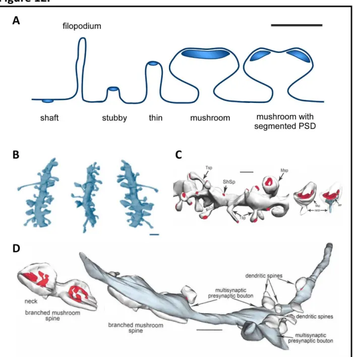
Figure 12. Dendritic spine morphology ...27
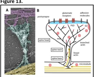
Figure 13. Cytoskeletal organisation of dendritic spines ...30
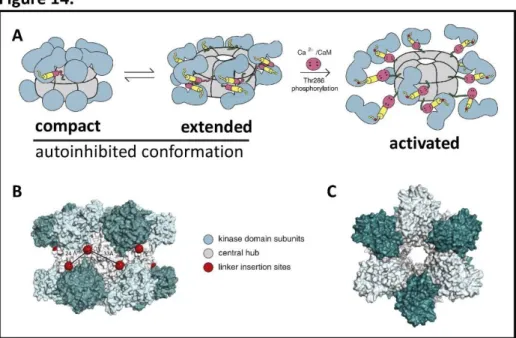
Figure 14. Structure of CaMKII ...35
Figure 15. Subunit composition of ionotropic glutamate receptors ...39
Figure 16. NMDAR subunit diversity and expression pattern ...41
Figure 17. Subunit composition and ion permeability of AMPAR ...43
Figure 18. Signal transduction of Group I mGluRs ...47
Figure 19. CaMKII phosphorylation of GluA1 subunit of AMPAR can mediate differential plasticity responses ...53
Figure 20. The SUMO enzymatic pathway ...63
Figure 22. In vitro FMRP sumoylation assay and FMRP mechanism of action in dendrites
...77
Figure 23. Structural, evolutionary and functional differences of SENP proteases ...82
Figure 24. Developmental regulation of SENP1 distribution in the rat brain ...84
Figure 25. Neuronal activity-dependent regulation of SENP1 redistribution at the synapse ...85
Figure 26. Schematic model of Ubc9 regulation at postsynaptic sites ...87
Figure 27. The principle of Fluorescence Recovery After Photobleaching (FRAP) ...90
Figure 28. The principle of photoconversion ...91
2. RESULTS AND DISCCUSSION Figure 29. Expression of WT GFP-SENP1 decreases SUMO1/2/3-modified protein levels in COS7 cells ...95
Figure 30. Distribution of SENP1 in dendrites and spines ...96
Figure 31. Nuclear localisation of SENP1 in neurons ...97
Figure 32. Activity-dependent redistribution of WT GFP-SENP1 into spines ...99
Figure 33. Synapto-dendritic redistribution of WT GFP-SENP1 under basal/control neuronal activity ...100
Figure 34. SENP1 postsynaptic entry is regulated by synaptic activity ...102
Figure 35. SENP1 synaptic entry is regulated by synaptic activity in a time-dependent manner ...104
Figure 36. SENP1 postsynaptic entry is regulated by synaptic activity in a time-dependent manner ...105
Figure 37. Synaptic exit of WT Dendra2-SENP1 ...106
Figure 38. Endogenous SENP1 localisation at synapses ...109
Figure 39. Synaptosomal preparation from cultured cortical neurons ...110
Figure 40. TIF preparation from primary cortical neurons ...111
Figure 41. SENP1 protein levels in TIF fraction ...112
Figure 43. Activation of mGluR1/5 regulates SENP1 postsynaptic entry ...116
Figure 44. Localisation of endogenous SENP1 at synapses ...118
Figure 45. Activation of Group I mGluRs increases endogenous SENP1 levels at PSD ...119
Figure 46. mGluR5 participates in the regulation of SENP1 synaptic diffusion...120
Figure 47. SENP1 synaptic diffusion is mGluR5-dependent ...121
Figure 48. Application of TTX reduces spontaneous neuronal activity ...122
Figure 49. FRAP measurements can determine the binding properties of studied proteins ...124
Figure 50. Microtubule stability is involved in spino-dendritic exchange of WT GFP-SENP1 ...125
Figure 51. Sustained synaptic activity alters SUMO1/2/3-ylation levels in TIF fraction ...128
Figure 52. Synaptic activation triggers accumulation of GFP-SENP1 C603S in spines ...129
Figure 53. Synaptic redistribution of GFP-SENP1 C603S is regulated by synaptic activity ....131
3. PERSPECTIVES Figure 54. Mass spectrometry to identify SENP1 interactome ...133
Figure 55. PKC may play a role in the regulation of SENP1 spino-dendritic exchange ...135
Figure 56. CaMKII may play a role in the regulation of SENP1 spino-dendritic diffusion ...136
Figure 57. Scheme of the newly identified and putative regulatory mechanisms of SENP1 spino-dendritic diffusion ...137
LIST OF TABLES
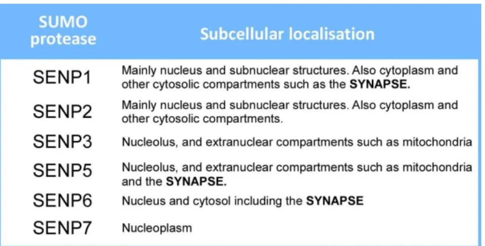
1. INTRODUCTIONTable 1. Implication of sumoylation in synaptopathies ...64 Table 2. Subcellular localisation of SENP proteases ...83
1
1. Introduction
This introduction is aimed to provide an overview of the anatomical and cellular features of the hippocampal formation. I further discuss the structure of a typical excitatory synapse, which is followed by a description of the mechanisms of glutamatergic transmission. In the last part, I focus on posttranslational modifications that play important roles at the synapse. Sumoylation is discussed in detail as it is the topic of my thesis. I believe that providing this information facilitates understanding into the thesis subject which concerns the investigation into regulatory mechanisms of postsynaptic desumoylation. I have to admit that the process of acquiring this background knowledge including a vast literature review and the write-up process was a very useful exercise.
1.1 The hippocampus
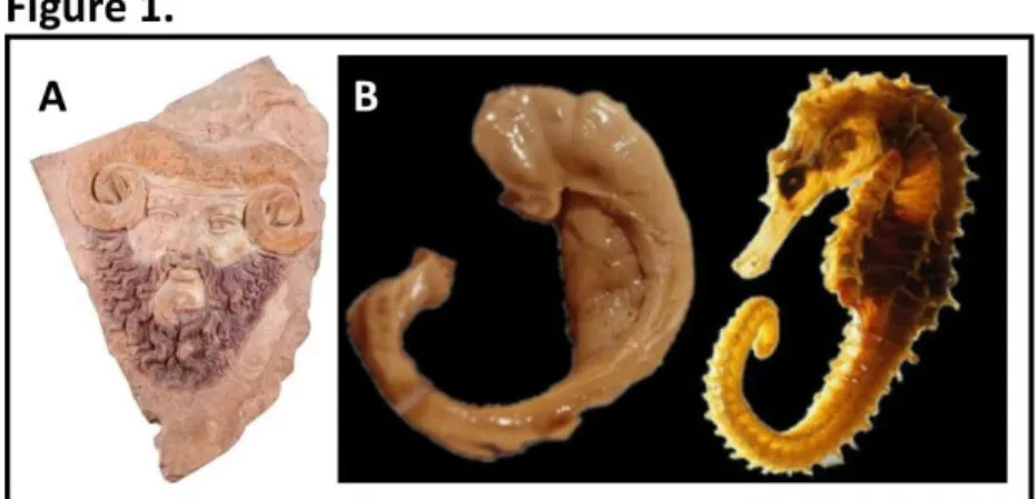
It is not a coincidence that the hippocampus is the most studied part of the brain serving as a model system for neurobiological studies. Its intrinsic structure has draught the attention of anatomists since ancient Egypt (~300 B.C.). Alexandrian scholars observed on the horizontal midsection of the hippocampus a curved structure resembling horns of the ram deity Ammon
(Fig. 1A), and therefore named it cornu ammonis in Latin. This terminology survived until now as the acronym (CA) for hippocampal subregions. The name hippocampus’ was first used by the
Figure 1. Historical reminder of hippocampal terminology. A. A terracotta cast of ram-horned Jupiter Ammon, 1st
century A.D. (Museo di Scultura Antica Giovanni Barracco, Rome, http://en.museobarracco.it/). B. Comparison of the dissected human hippocampus (left) with sea horse Hippocampus leria (right). Adapted from The Hippocampus Book (Andersen et al., 2007).
2
Bolognese anatomist Guilio Cesare Aranzi (circa 1564) undeniably because of its similarity with the sea horse (Fig. 1B), genus Hippocampus, where hippo means in ancient Greek horse’ and
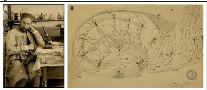
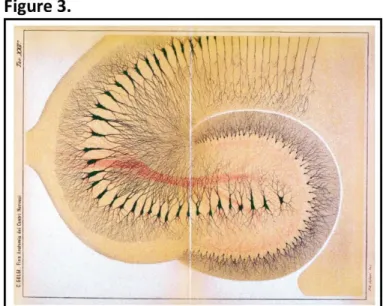
kampos sea monster’. However, it was not until the late 19th century that the Spanish physician Dr Santiago Ramón y Cajal (Fig. 2A)mostly with the use of the Golgi method (Fig. 3) depicted the cellular organisation of many tissues structures including the structure of nerve cells and neuromuscular junction. These findings were published in 1893 as Manual de Histología Normal
y Técnica Micrográfica. Later, Ramón y Cajal set himself for a thorough study of the nervous
system and published his observations along with a more detailed cellular organisation of the hippocampus (Fig. 2B) in Histologie du système nerveux de l'homme & des vertébrés in 1909.
Simultaneously, Karoly Schaffer, a Hungarian neurologist, was interested in hippocampal axonal fibres and their length, and found that they have short as well as long branches connecting with other areas of the cortex. He discovered the so-called collateral fi er s ste ’ that connects the CA3 to the CA1 region of the hippocampus, known today as Schaffer collaterals. Another pioneering neuroanatomist Rafael Lorente de Nó (1934) bolstered the work of Ramón y Cajal by adding to the study of the many hippocampal cell types and their arborisations, and distinguished the hippocampal subregions CA1, CA2 and CA3.
Until 1930s the hippocampus was thought to be part of the olfactory system, perhaps due to its size in macrosmatic animals (e.g. rodents and insectivores) as it is considerably large in the
Figure 2. Discovery of hippocampal structure. A. Santiago Ramón Cajal, the Father of odern Neuroscience’. B. Cellular organisation of the rat hippocampus by S. Ramón y Cajal. Both images were taken and modified from (Swanson et al., 2017).
3
context of the whole brain. This view was challenged throughout the years since the hippocampus was not found to be directly connected with the olfactory bulb (a review by (Brodal, 1947)). Another important influencer in the field of neuroanatomy was James Papez who proposed the existence of a circuit (known as Papez circuit, 1937) that interconnected cortical and subcortical structures including the hippocampus and supposedly mediated emotions. The most prominent functional importance of the hippocampus was uncovered about 60 years ago when patient H. M. suffered from amnesia due to surgical excision of the medial temporal lobe for epilepsy relief performed by Dr Scoville (1957). Since then, the hippocampus has been extensively studied for its involvement in memory. Early experiments on hippocampectomized primates and rodents, however, failed to bring a convincing proof of memory deficits. Nonetheless, some of the observations included defects in exploration, habituation to novelty and navigation, which prompted the idea of existence of more than one type of memory. An important milestone pro-hippocampus-mediated memory was made with the introduction of a more appropriate behavioural test – the object recognition task – by David Gaffan (1974) and its optimization for use in the monkey by Mortimer Mishkin and Jean Delacour (1975). Nowadays, the hippocampus is accepted to be part of the li ic s ste ’, a ter that as first used the French neurologist Pierre Paul Broca (1878), which mediates not only memory formation but also emotions, motivation, learning, spatial navigation and olfaction.
Figure 3: Section of the rabbit hippocampus stained with the original Golgi method (1886). Source: The Hippocampus Book (Andersen et al., 2007).
4 Neuroanatomy of the hippocampal formation
The hippocampal formation (Fig. 4 and 6) is widely accepted to refer to several closely related regions: the dentate gyrus, hippocampus proper (CA1, CA2 and CA3), subicular complex and
entorhinal cortex. Although the volume of the human hippocampus is about 100 times that of
the rat and 10 times that of the monkey, the basic hippocampal architecture, particular cellular organisation and sea horse’ shape is present throughout all mammals. The hippocampus is buried in the medial temporal lobe of the human brain, whereas in rat it is localised rather rosto-caudally. An intriguing feature of the hippocampal formation is that it is largely nonreciprocal, with unidirectional projections. This is different from what we see in neocortical areas where it is normal practice that region A projects into region B and region B projects back to region A, showing a large degree of reciprocity. Much of the neocortical input to the hippocampal formation is received through the entorhinal cortex. As depicted in Figure 4, the entorhinal cortex
Figure 4. The hippocampal formation. A. Neurons in layer II of the entorhinal cortex (EC) project to the dentate gyrus and the CA3 region via the perforant pathway. Neurons in layer III of EC project to CA1 and the subiculum (Sub) via the perforant and alvear pathways. The granule cells of the dentate gyrus (DG) project to CA3 via mossy fibre projections. CA3 pyramidal neurons project to CA1 via Schaffer collaterals. CA1 pyramidal neurons project to Sub. Both CA1 and Sub neurons project back to the deep layers of EC. B. Projections along the transverse axis of the hippocampal formation.
5
sends axons to the dentate gyrus, however, the dentate gyrus does not project to the entorhinal cortex. Granule cell axons of the dentate gyrus called mossy fibres project towards the pyramidal cells of the CA3 region and again this pathway is unidirectional; so are the projections from CA3 to CA1 called Schaffer collaterals, and CA1 to the subiculum.
It should be emphasized that no brain region functions in isolation. The regions of the hippocampal formation are innervated by and send projections to other brain nuclei, which is vital for their function. There are three major fibre bundles providing input innervation to the hippocampal formation. The first is the angular bundle containing fibres that originate in the entorhinal cortex and innervate the dentate gyrus, hippocampus and subiculum. The second pathway is the fimbria-fornix pathway that interconnects the hippocampal formation with the basal forebrain, hypothalamus and brain stem. The third major fibre system is called dorsal and
ventral hippocampal commissures and contain some 350,000 fibres. They connect one
hippocampal formation of one hemisphere with the hippocampal formation of the contralateral hemisphere.
To follow on the architectonic organisation of the hippocampal formation, the specific regions will be separately and briefly described below. For the sake of simplicity, this will be done taking the rodent hippocampal formation as a model system.
A. Dentate gyrus
The dentate gyrus (DG) has three distinct layers, from the superficial side: molecular layer,
granule cell layer and polymorphic cell layer (see scheme in Fig. 5). The principal cells, that is to
say those that project out of DG, are the granule cells whose cell bodies (10 µm x 18 µm) lay within the granule cell layer. There are about 1.2 x 10⁶ granule cells in one dentate gyrus. The
granule neuron has a very specific cone-shaped dendritic tree extending its branches in the molecular cell layer. Granule cells have spiny dendrites with estimates between 3600-5600 spines per cell depending on their particular localisation along the granule cell layer. Interestingly, the number of granule cells does not vary in adult animals, however, young animals that were exposed to enriched environments show bigger dentate gyri with more granule cells in adulthood. Except granule cells, the dentate gyrus contains local interneurons that unlike the granule cells do not project to other areas of the hippocampal formation. The pyramidal basket
6
cell is the most studied interneuron. Their cell bodies are localised at the edge of the granule cell layer. The name basket’ comes from the appearance of the cone-like axonal plexus that surrounds and connects with cell bodies of granule cells. The basket neuron has usually one principal apical aspiny dendrite extending to the molecular layer and several basal dendrites in the polymorphic cell layer. Basket cells are positive for the µ-aminobutyric acid (GABA) and thus provide an inhibitory input. The molecular cell layer is mostly occupied by dendrites and axons, however, it also contains cell bodies of interneurons named MOPP (molecular layer performant path-associated) and chandelier cells that are axo-axonic cells innervating the axon initial segment of granule cells. In regard to the polymorphic cell layer, that is also referred to as the hilus, the most common cell type is the mossy cell with large triangular or multipolar cell bodies (25-35 µm). Mossy cells are further characterized by large, complex and dense spines that are called thorny excrescences located on proximal dendrites. These spines form glutamatergic synapses with large boutons of mossy fibre axons of granule cells (as depicted in Fig. 5). Of note, the ord oss ’ is used in t o distinct cell t pes: oss fi res of granule cells and oss cells, which can be confusing but it is apt considering the mossy appearance of both.
Noteworthy, the dentate gyrus receives major excitatory innervations from the entorhinal cortex (Fig. 4) through so called perforant pathway for the fact that the fibres leaving the angular bundle perforate the subiculum. A population of hypothalamic cells sends mostly glutamatergic projections to granule cells; and noradrenergic and dopaminergic projections are received from the brain stem. The subcortical regions send only few inputs towards the dentate gyrus and the most prominent is the one from septal nuclei using acetylcholine or GABA as a neurotransmitter. Importantly, the dentate gyrus is a source of adult neural stem cells that reside in the
subgranular zone and give rise to new granule cells throughout the life. The dentate gyrus plays
a substantial role in cognition and emotions. The cognitive functions relate to spatial memory. In particular, DG has been involved in so called pattern separation, the ability to distinguish between similar experiences which is crucial for episodic memory, its storage and retrieval. The emotional function of DG involves the regulation of mood and anxiety, and it has also been associated with behaviours with a strong component of stress and fear (reviewed in (Scharfman, 2016)).
7
B. Hippocampus proper
Alike the dentate gyrus, the hippocampus proper, also referred to as A on’s horn, has a curved structure (Fig. 6). It is divided into three subregions: CA1, CA2 and CA3 (Fig. 4 and 6). These subregions have laminar architecture with five layers, superficially: stratum alveole (ALV),
stratum oriens (OR), stratum pyramidale (PYR), stratum radiatum (RAD) and stratum lacunosum moleculare (LAC-MOL; Fig. 5). The CA3 field contains an additional thin acellular layer the stratum lucidum occupied by mossy fibres. Stratum alveole presents a fibre-rich thin layer. Stratum oriens
contains basal dendrites of pyramidal cells and several types of interneurons. Moreover, some of the fibres connecting CA3 to CA3 and CA3 to CA1 (Schaffer collaterals) are also located in stratum oriens. The somata of hippocampal principal cells, called pyramidal cells, lay in the stratum pyramidale. They are tightly packed in CA1 and more loosely in CA2 and CA3. Owing to the U shape of the hippocampus, the CA1 pyramidal cells are upside down compared to CA3. Stratum radiatum consists of connecting fibres of CA3 to CA3 neurons, and Schaffer collaterals. The lacunosum moleculare layer is formed mostly by fibres from the entorhinal cortex and thalamus. Numerous interneurons are also scattered in both strata radiatum and lacunosum moleculare.
Figure 5. The tri-neural circuit between principal cells of the hippocampal formation. A scheme of axonal terminations of principal cells on target principal cells. Axons of the dentate gyrus (DG) granule cells, termed mossy fibres, innervate the giant excrescences of CA3 pyramidal cells. CA3 axons called Schaffer collaterals terminate on the CA1 dendrites. The names of individual layers are also indicated and further described within the text. The image was taken and adapted from (Hammond, 2001).
8
Pyramidal cells are the most numerous cell type found not only in the hippocampus but also
in the cerebral cortex and amygdala. Although the pyramidal neurons present a certain degree of variability, their cellular architecture is stereotypical. Cell bodies of pyramidal cells tend to have a teardrop/rounded pyramid shape. They have a longer apical dendritic tree that extends from the pointy end of the soma and is terminated by a dendritic turf. The basal portion of the pyramidal cell forms a basal cluster of shorter dendrites. Apical dendrites of hippocampal pyramidal neurons pass through the stratum radiatum and ramify in stratum lacunosum moleculare, whereas basal dendrites arborize in stratum oriens. The pyramidal cells of the CA3 region that are closest to the DG do not extend their dendrites to stratum lacunosum moleculare and therefore are not influenced by projections from the entorhinal cortex, but synapse with mossy fibres of DG. Dendrites of pyramidal cells have numerous spines. The most prominent are the thorny excrescences of CA3 cells forming synapses with mossy fibres. Axons of pyramidal cells run through stratu alveus e itting nu erous collaterals efore leaving the A on’s horn through the pre- and postcommissural fornixes (axon bundles; Fig 5).
There is a vast literature concerning studies performed on the CA1 pyramidal neurons, especially focused on synaptic transmission and plasticity. This is mostly attributed to their morphology, cell viability in culture and well trackable connections with CA3. The dendrites of
Figure 6. Nissl-stained section (left) and line drawing (right) illustrating the general organization of the hippocampal formation in the rat. Scale bar = 1 mm. Taken and modified from The Hippocampus Book(Andersen et al., 2007).
9
CA1 neurons are covered with about 30,000 spines that receive excitatory synaptic inputs (Megias et al., 2001). Thus, density and morphology of dendritic spines has been used as a functional measure for excitatory efficacy that is closely correlated with cognitive function including memory formation. Indeed, changes in spine density and morphology have been reported in many neurological disorders. For instance, a massive loss of synapses in CA1 region is associated with cognitive decline in murine models of Alzhei er’s disease (Perez-Cruz et al., 2011) (Merino-Serrais et al., 2011) (Lazcano et al., 2014). On the other hand, a neurodevelopmental disorder - the Fragile X syndrome is characterized by an increased number of immature dendritic spines in the CA1 region (reviewed in (He and Portera-Cailliau, 2013)). Under physiological conditions, the highest density of spines is found in strata radiatum and oriens, much lower in stratum lacunosum moleculare. Asymmetrical synapses, which are presumably excitatory, can be also formed on dendritic shafts usually in the apical dendritic tuft. It was estimated that about 1,700 inhibitory symmetrical synapses converge on a single CA1 pyramidal neuron targeting usually the soma, axon and spine-free proximal apical and proximal basal dendrites (Megias et al., 2001).
CA2 pyramidal neurons have been long overlooked probably due to the small size of the CA2 region when compared to CA1 or CA3. More recent studies have determined their particular synaptic properties and involvement in social and spatial memory and pathological conditions such as schizophrenia (reviewed in (Dudek et al., 2016) (Srinivas et al., 2017)). CA2 pyramidal neurons can be distinguished from the CA1 and CA3 pyramidal cells based on morphology, connectivity and molecular markers. According to Lorent de Nó (1934), CA2 pyramidal cell dendrites lack thorny excrescences that form synapses with mossy fibres from DG and are characteristic of the CA3 pyramidal neuron dendrites; although we now know that this is variable between species. More recent discoveries identified specific axonal projections from the thalamus that are indicative of the boundary between CA2 and CA3 fields. In addition, CA2 pyramidal neurons project mainly to stratum oriens of CA1, whereas CA3 pyramidal neurons project to stratum radiatum of CA1. Furthermore, there are more parvalbumin- and reelin-expressing interneurons in CA2 than in CA1 or CA3. Interestingly, the CA2 field is relatively
10
resistant to injury as well as to induction of plasticity processes such as long-term plasticity (LTP). Intriguing, however, is the finding that the CA2 pyramidal cells possess all proposed plasticity mediators characteristic of CA1 neurons. The research laboratory of Dr Serena M. Dudek has carried out extensive studies on the CA2 area concerning plasticity processes. In the past years, they showed that one possible cue for plasticity resistance could be through changes in calcium dynamics. Her team also proposed the RGS14 (Regulator of G-protein Signalling 14) scaffold protein to play a key role in suppression of LTP in CA2 pyramidal cells (Simons et al., 2009) (Lee et al., 2010) (Vellano et al., 2011). In addition, given the high resistance of the CA2 region to apoptosis, CA2 may prove a suitable model to study diseases with impairments in social and spatial memory processing.
The CA3 region receives three major excitatory inputs: from mossy fibres of DG, from EC and local from the CA3 neurons. This unique interconnectivity makes the CA3 network highly excitable. For this reason, the CA3 region has attracted increasing attention for its role in memory and susceptibility to seizures and degeneration. CA3 pyramidal neurons are morphologically very similar to CA1 neurons, however, the CA3 neurons are on average larger. The largest CA3 neurons are in the distal and smallest in the proximal portion of CA3 to DG. The dendritic ramification is characterised by a shorter basal dendritic tree within stratum oriens, a short apical dendritic trunk in stratum lucidum that arborizes into two or more long apical trunks. These long apical trunks further ramify into shorter dendritic branches in stratum radiatum and long dendritic trunks continue to stratum lacunosum moleculare. As mentioned above, CA3 neurons are studded with thousands of dendritic spines. The most apparent are the thorny excrescences that form about 40 clusters on each CA3 neuron. Generally, these branched dendritic spines synapse with a single mossy fibre bouton. A single CA3 neuron projects its axon to all CA3, CA2 and CA1 regions. These axons are myelinated with abundant boutons. The estimates show between 15,000 and 60,000 synapses that can be formed by a single CA3 axon. Some CA3 axon boutons also innervate interneurons which interestingly happens at a single release site. As in CA1 this single synapse is very powerful able to generate an action potential in the postsynaptic interneuron. In regard to the high excitability, the CA3 neurons show a typical bursting pattern
11
comprising of several action potentials that last 30 to 50 ms. Because the CA3 pyramidal neurons do not possess a large primary apical dendrite like the CA1 neurons, only a restricted number of studies have focused on the dendritic excitability and ion channels in CA3 (Andersen et al., 2007).
C. Subicular complex
The subicular complex (Fig. 4) including prosubiculum, subiculum, presubiculum, postsubiculum and parasubiculum forms a continuum of the CA1 as it begins where the Schaffer collaterals end (reviewed in (Andersen et al., 2007) (Ding, 2013) (O'Mara, 2005)). The pyramidal cells of the subicular complex are more disperse compared to the tightly packed layer of CA1 pyramidal cells. Despite the fact that the subicular complex constitutes the major output of the hippocampal formation it is a poorly investigated brain structure. Some of its roles were identified in the encoding and retrieval of memory, and in neurodegenerative disease and epilepsy. The subicular pyramidal neuron has a typical morphology with dendrites comprising spines. The subiculum receives input from CA1 as well as EC layer II and III. Importantly, the subicular output innervates local areas: EC layer V, presubiculum and parasubiculum and also more distant cortical structures such as the prefrontal cortex, olfactory nucleus, thalamus, amygdaloid complex and others. The particular subicular cortices can be identified based on the expression of specific genes and neurochemicals.
D. Entorhinal cortex
The na e ’entorhinal’’ is based on its position as it is partially enclosed by the olfactory – rhinal sulcus. Early studies of the entorhinal cortex by Ramón y Cajal and Lorente de Nó defined the cytoarchitectonic organisation which is today accepted with some minor changes: EC is divided into two main subregions, the medial and lateral EC, both of which have a 6-layer laminar structure with four cellular and two acellular layers. Much interest devoted to EC has begun in early 1990s with the discovery that this brain area was prone to early neurodegeneration in Alzhei er’s disease (Van Hoesen et al., 1991). The entorhinal cortex plays an indispensable role
12
in the feedforward and feedback flow of information bridging the hippocampus and the neocortex. Recent studies provide evidence that the medial EC processes spatial information, whereas the lateral EC governs pathways encoding object information. A famous trio of scientists
(John O’Keefe and Ed ard and Ma -Britt Moser) who were awarded the Nobel Prize in Physiology
or Medicine in 2014 made a breakthrough discovery of a GPS syste in the rain – the place
and grid cells (Fig. 7). Place cells are CA1/CA3 hippocampal pyramidal neurons that fire specifically based on spatial localisation. Even more interesting is the finding that the firing of place cells does not depend on the local CA3/dentate gyrus innervation but rather the spatial information is received from the medial EC. The medial EC neurons are highly responsive to change in position. These neurons show a firing field pattern with regularly shaped triangular or hexagonal grids, thence called grid cells (reviewed in (Moser et al., 2015)). The realisation about where we are in space provides one of the most fundamental information for survival. The crucial function of place and grid cells function is evident in Alzhei er’s disease where disorientation is a common early symptom.
Figure 7. Grid and place cells. A. An EC grid cell firing pattern. The black trace shows the trajectory of a foraging rat
in part of a 1.5-m-diameter-wide square field. Spike locations of the grid cell are in red. Each red dot corresponds to one spike. Blue equilateral triangles illustrate the regular hexagonal structure of the grid pattern. B. Grid cell (left) and place cell (right) firing and activity. The top part shows trajectories with spike locations. The bottom color-coded rate maps show high activity (red) and low activity (blue) firing. Grid cells are thought to provide much, but not all, of the entorhinal spatial input to place cells. Adapted from (Moser et al., 2015).
13
1.2 Neuronal synapse
Most studies investigating synapses have been carried out using the hippocampal circuitry as a model system. Therefore, the previous chapter aimed at introducing the hippocampal formation so it would set the niche for further characterization of synapses, to which the current chapter is devoted.
The notion that a contact between two neurons is the place where information transmission occurs was first suggested by Ramon y Cajal in 1888. Later, an English neurophysiologist Charles Scott Sherrington (1897) introduced the term synapse’ fro the Greek to clasp to give a name to these specialized zones of interneuronal communication. Currently, the synapse is no longer seen only as a static junction between neurons but a very dynamic organelle whose function is tightly regulated in time and space owing to the constituting molecular interactions (Choquet and Triller, 2013). Deciphering the structure and molecular organisation of synapses is an essential step toward understanding the molecular mechanisms that underlie synaptic transmission and plasticity - processes that are the foundation of physiological brain function. Noteworthy, there is a tendency to see cellular reactions as linear, but particularly in neurites, differential concentrations, position as well as the reactive state of soluble and bound synaptic proteins determine the regulatory cues in these highly dynamic and precise processes that mediate both presynaptic and postsynaptic portions of neurotransmission.
Primarily, neuronal synapses can be characterized based on the type of transmission – chemical and electrical. Electrical transmission is mediated via so called gap junctions, i.e. electrical synapses, through a direct cytoplasmic exchange of ions and small molecules between
neighbouring neurons.Importantly, the two types of neurotransmission coexist and interact in
both the developing and adult brain (Pereda, 2014) (Nagy et al., 2018). This introduction will describe and refer to the most abundant form of transmission at chemical synapses composed of axonal termini (boutons) and dendritic spines in the hippocampus.
14
Chemical synapses
A characteristic of chemical synapses is the presence of a synaptic cleft – a gap between the axonal terminus of a presynaptic neuron and the dendritic specialisation of a postsynaptic neuron (reviewed in (Harris and Weinberg, 2012) (Hammond, 2001) (Pickel and Segal, 2014) (Nicholls et al., 2012)). The process of transmission is mediated by a change in electrical potential in the presynaptic cell that consequently leads to the release of neurotransmitter molecules. Chemical synapses are either excitatory or inhibitory depending on the neuromodulatory effect the neurotransmitter has on the receiving postsynaptic neuron. A large body of literature describes the prototypical chemical neuron-to-neuron synapses that are indeed the most abundant and extensively studied synapses in the brain. The synaptic complex is a basic unit of each functional chemical synapse. It comprises of three components: the presynapse, cleft and postsynapse. The synaptic complex shows a particular asymmetric organisation. The most prominent asymmetric trait is the presence of synaptic vesicles (40-60 nm) exclusively in the presynapse and a submembraneous electron-dense zone in the postsynapse. Additionally, the synaptic complex is surrounded by modulatory astroglial processes. Thus the synaptic complex together with astroglia can be seen as a mesh-like structure on an electron microscopy section (Fig. 8). Excitatory synapses are formed mainly on dendritic spines, unlike the inhibitory synapses that preferably connect to the cell soma and axonal initial segments with only small percentage found
Figure 8. Synaptic complex in the CA1 region of the hippocampus. A. An electron microscopy section that was colour coded to show excitatory axon (green), spiny dendrites (yellow), an aspiny dendrite (dark red), sparse inhibitory axons (orange) and astroglial processes (light blue). B. Asymmetric synapses (green arrows), a non-spiny dendrite (ns) with a mitochondrion (mito), two dendritic spines (sp) of which one has a perforated PSD (red arrow). Adapted from (Harris and Weinberg, 2012).
15
along spiny and aspiny dendrites, hence the sparse distribution of inhibitory synapses that can be seen in the synaptic complex (Fig. 8).
This part of introduction will summarize the existing knowledge of the structure, types and composition of the synaptic complex.
A. The presynapse
The first piece of evidence pointing to the synapse as a dynamic organelle came with the finding that neurotransmission relies upon calcium-driven fusion of neurotransmitter-filled vesicles with presynaptic membrane. This notion was further reinforced by the discovery of endocytic pathway that dynamically recycles these vesicles (Heuser and Reese, 1973).
a) Presynaptic trafficking
During neuronal development, upon neuronal cell determination and morphogenesis, synapses are to be formed. The majority of synaptic material required for synaptic formation is synthesized in the cell body and transported over long distances to and from synaptic loci by microtubules-associated molecular motors. The molecular organization of axonal versus dendritic microtubules differs. According to in vivo studies, axonal microtubules (MTs) have their minus ends oriented exclusively toward the cell body, whereas dendritic microtubules show mixed orientation with more abundant distal plus-ends in vertebrates when compared to invertebrates (reviewed in (Chia et al., 2013); (Stone et al., 2008)). However, it is not only the polarity of MTs itself that is critical in determining whether molecular cargoes will be targeted toward the presynapse or postsynapse, the microtubule-associated proteins (MAPs) also play an important role. Upon genetic manipulations, presynaptic cargoes can be misplaced into dendrites, as shown in mutants for kinesin and other MT-binding proteins such as UNC-33 and UNC-44 in Ceanorhabditis elegans (Seeger and Rice, 2010) (Maniar et al., 2011).
Axonal trafficking of biomolecules can be classified based on the direction toward the cell body as retrograde, or toward the axonal terminus as anterograde. Anterograde trafficking is carried out by kinesins. These motor proteins are important for both the export of cargo molecules from the Golgi apparatus and their subsequent transport to destination sites. The exact mechanisms of cargo sorting and loading remain to be determined.
16
An average pyramidal neuron possesses thousands of synapses. What are the exact signalling cues that regulate how cargoes get distributed between synapses is yet to be elucidated. Most of the investigations into the molecular mechanisms of axonal trafficking have been performed in C. elegans and Drosophila melanogaster. The axonal anterograde transport includes kinesin-3 motor proteins KIF1A and KIF1Bβ that were shown to transport synaptic vesicle-associated proteins in the form of SV precursors to presynaptic sites (Sabo et al., 2006). Binding of motor molecules onto the MTs is followed by ATP hydrolysis that initiates the transport along MT tracks.
The lack of KIF1A and KIF1Bβ reduced the number of SVs as well as SV proteins in the presynapse
(Chia et al., 2013). The kinesin-1 motor complex transports presynaptic membrane proteins such as SNAP-25, Bassoon, Piccolo, RIM and syntaxin-1. Upon reaching the end of microtubules, molecular cargoes are unloaded for delivery to presynaptic sites presumably by additional local regulatory cues (Yagensky et al., 2016). For instance, one local regulatory mechanism involves the small GTPase Rab3. DENN/MADD, a Rab3 guanine nucleotide exchange factor, binds to the kinesin-3 complex and promotes anterograde transport of synaptic vesicles associated with Rab3 in the GTP-bound state (Niwa et al., 2008). Depleting DENN/MADD of its enzymatic activity or locking Rac3 in GTP-bound state impairs transport of these vesicles to distal presynaptic sites (Niwa et al., 2008). Phosphorylation presents another mechanism capable of controlling distal
axonal cargo targeting. Phosphorylation of kinesins by the GSK3β kinase leads to cargo release
(Morfini et al., 2004). GSK3β is selectively active in growth cones and thereby most likely participates in formation of synapses de novo (Morfini et al., 2004). A mechanism controlling cargo pausing and loading has also been documented along MTs en route. Loss-of-function mutations in arl-8 that encodes the small G-protein ARL-8 lead to proximal accumulation of presynaptic specializations and loss of synapses in distal axons, which results in defects in neurotransmission in C. elegans (Wu et al., 2013). Thereafter, ARL-8 and JNK were reported to act in an antagonistic way to balance cargo self-assembly and facilitate cargo trafficking en route (Klassen et al., 2010) (Wu et al., 2013). An interesting phenomenon has been described in the process of synaptic vesicle recycling. The recycled material shuttles between local as well as remote boutons involving both kinesin and dynein motors. Remarkably, the retrogradely
17
transported vesicles are likely to be captured by distal as opposed to proximal presynaptic sites (Maeder et al., 2014).
Sustained and optimal presynaptic function requires the molecular motor dynein, which mediates retrograde transport of biomolecules from the presynapse to the nucleus. In response to synaptic activity, retrograde movement of messenger molecules functions as a feedback signal that triggers changes in gene expression. In turn, specific products of gene expression can regulate the strength of synaptic transmission (Panayotis et al., 2015). For instance, calcium ion waves implement fast response synapse to soma communication and are most efficient for synapses localised closer to the soma. Slower and long distance synapse to soma communication involves extracellular signalling molecules, neurotrophins, such as BDNF (brain derived neurotrophic factor). Principally, neurotrophins bind to their receptors (Trk [tyrosine kinase] or p75NTR) at the presynaptic membrane which triggers receptor autophosphorylation and activation of downstream signalling cascades via MAPK, PLCγ and PI3K (Pazyra-Murphy et al., 2009).
In addition to changes in gene expression, retrograde transport is crucial for degradation and turnover of unwanted or damaged proteins and organelles. During axonal development, protein degradation at the axonal tip decreases with an enhanced retrograde transport of the ubiquitin-proteasome system (UPS) (Hsu et al., 2015). In regard to mature presynapses, previous studies demonstrated that the UPS functions rather locally within synaptic boutons to acutely control levels of presynaptic proteins and thereafter the efficacy of neurotransmission (Speese et al., 2003). Autophagy, however, is a degradation mechanism that depends on the retrograde
transport of presynaptic components. These components including synaptic vesicles and
α-synuclein are cleared via autophagosomes. This degradation pathway is quite challenging since autophagosomes must be transported across long distances to lysosomes that usually reside in the cell soma. A recent study implicated JIP1, a kinesin-1 activator that binds dynein, and the autophagosomal protein LC3 in the clearance of presynaptic proteins. Preventing JIP1 binding to
18
LC3 results in defects in retrograde trafficking of autophagosomes as well as impairment of autophagosomes fusion with lysosomes (Fu et al., 2014b).
Noteworthy, although kinesin and dynein motors mediate unidirectional traffic, they are known to bind synaptic cargoes simultaneously. Axonal microtubule tracks are not continuous. They can break, or encounter various obstacles and therefore the option of switching between the two directions is very convenient for bypassing such difficulties. Importantly, these opposite-polarity motors were found to activate one another and this way efficiently carry synaptic cargoes in either direction.
MT motors also respond to presynaptic plasticity processes. Repetitive stimulation of neurons in culture enhances the formation of new presynaptic boutons, a process that is dependent upon trafficking of presynaptic components by kinesin-1. Similarly, mice that were placed in an enriched environment expressed increased levels of kinesin-3 motor KIF1A which is directly correlated with increased trafficking of presynaptic cargoes (Kondo et al., 2012).
b) Structure and composition of presynaptic termini
The excitatory axospinous synapses in the stratum radiatum of the hippocampal CA1 are prototypic and highly abundant synapses formed predominantly by unmyelinated axons. Most of these axons are the Schaffer collaterals originating in the CA3 region. The axonal termini often form swellings referred to as boutons that are filled with many neurotransmitter-containing
Figure 9. Axonal boutons and dendritic spines. (Left and right), 3D reconstructions of axons and axonal boutons (light green) of Schaffer collaterals. Synaptic vesicles are visible within each bouton. Dendritic spines (grey) with PSD (red) converge onto synaptic boutons. In the right panel, a red disc represents a reconstructed PSD from the depicted dendritic spine. Abbreviations: dcv, dense core vesicles; MSB, multi-synaptic bouton; SSB, single synaptic bouton; NSB, non-synaptic bouton; mito, mitochondrion; mvb, multivesicular body. Taken from (Harris and Weinberg, 2012).
19
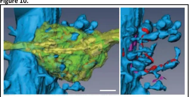
vesicles and can be clearly seen by electron microscopy (Fig. 9). The majority (~75%) of axonal boutons establish a single contact synapse, about 21% form multi-synapse contacts and ~4% lack their postsynaptic counterpart (Sorra et al., 2006). These pre-existing but unconnected boutons are an advantage when it comes to rapid synaptogenesis as there is no need for generation of presynaptic termini de novo (Petrak et al., 2005) (Harris and Weinberg, 2012). Another type of axonal bouton that is worth mentioning is the robust bouton of granule neurons of the dentate gyrus that converge onto multiple thorny excrescences of CA3 pyramidal neurons (Fig. 10). In the cerebellum, large specialized axonal termini termed synaptic glomeruli can be found. These originate from cerebellar granule cells and synapse with dendritic spines of Purkinje neurons.
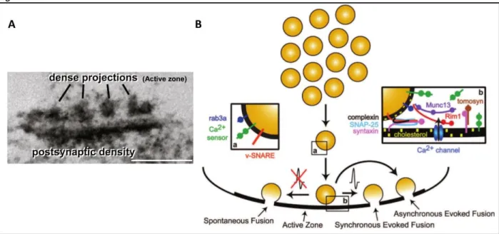
The active zone
At chemical synapses, action potentials trigger calcium influx into the presynaptic terminal, which typically leads to the fusion of SVs with the presynaptic active zone membrane and neurotransmitter release. The active zone (AZ) is a biomolecule-rich electron-dense region that can be found in the proximity to the presynaptic membrane opposed to the postsynaptic density.
Figure 10. 3D reconstruction of a mossy fibre bouton and CA3 thorny excrescences. Left, a mossy fibre bouton (yellow-green) converging onto thorny excrescences (blue spines). Right, CA3 thorny excrescences with reconstructed synaptic (red) and non-synaptic connections (magenta). Scale bar 1 µm. Taken from (Harris and Weinberg, 2012).
20
Typically, many synapticvesicles occupy this region ready to dock and release neurotransmitter
molecules. Intriguingly, AZ can be visualised as cytoplasmic dense projections that are organized into a presynaptic grid-like structure (Fig 11A). Why AZ adopts this particular shape is uncertain but it most likely has to do with SV mobilization and release as suggested by (Fernandez-Busnadiego et al., 2010). The excitatory SVs measure ~35 nm in diameter and are filled with glutamate molecules. Emerging evidence suggests that SVs are not a homogenous population of organelles and can be discriminated both structurally and molecularly. There exist at least three types of SV pools depending on SV availability for membrane mobilization and release: resting, recycling and readily releasable pools (Fig. 11B). The existence of different pools of SVs is obvious during a phenomenon called synaptic depression. Upon repetitive bursts of action potentials (APs) a reduction in postsynaptic response can be measured reflecting the fact that the RRP empties and another AP comes before this pools gets replenished. Oftentimes, a homeostatic lower steady level of transmission is established, in which the release is balanced by the slow refilling (Alabi and Tsien, 2012). In hippocampal synaptic boutons, only a few vesicles have the readily releasable pool status. After the readily releasable pool gets discharged (short 10-40 Hz
stimulation), neurotransmitter release occurs from the secondary glutamate depot – the
recycling pool. This total releasable pool of SVs (including both recycling and readily releasable pools) represents as little as ~100 vesicles. On the other hand, the resting pool is defined as a set of vesicles that are extremely reluctant to trafficking toward the AZ membrane and remain unreleased even after prolonged stimulation. Although the resting pool represent about 75% of total SV content in a presynaptic terminal, the physiological function of these vesicles remains unclear.
The release of SVs can be evoked in three distinct ways (Fig. 11B): a. synchronous vesicle release – electrical stimulation precedes synchronous currents that are triggered in the
postsynaptic cell; b. asynchronous – a delayed vesicle fusion upon a stimulus; c. spontaneous –
occurs in the absence of action potential, releases a very small portion of SVs and generates miniature postsynaptic currents (Crawford and Kavalali, 2015). The release of SV content is mediated by exocytosis, a well-orchestrated process that relies on spatial organisation and dynamics of fusion machinery components. Three-step SV release has been well documented:
21
SVs attachment to the plasma membrane (docking), fusion-preparatory phase (priming) and Ca2+
influx-dependent fusion (Milovanovic and Jahn, 2015). The SNARE (soluble N-ethylmaleimide-sensitive factor attachment protein receptor) family of proteins lies at the centre of the SV release process. v-SNARE (vesicular SNARE) proteins bind to target membrane SNARE (t-SNARE) proteins to form a complex that is essential for the fusion of vesicular and plasma membranes. Canonically, the component of v-SNARE VAMP2 (vesicle-associated membrane protein 2, also known as synaptobrevin-2) binds to the members of t-SNARE syntaxin 1 and SNAP-25 (synaptosomal-associated protein of 25 kDa) to bring the juxtaposed membranes together for fusion and neurotransmitter release. This process is catalysed by Ca2+ binding to the calcium
sensor synaptotagmin 1 (Crawford and Kavalali, 2015) (Sudhof, 2013). The SV membrane contains a range of proteins important for exocytosis. Evidence suggests that some v-SNAREs, calcium sensors and other vesicular proteins are involved in SV trafficking to segregate vesicle pools prior to the release. Thus, expression of these membrane proteins on SVs could function as a molecular code predictive of their function within the presynapse (Wilhelm et al., 2014).
Figure 11. Dense projections of the active zone and heterogeneity of the synaptic vesicle pool. A. Electron micrograph of a phosphotungstic acid stained synapse with pre- and postsynaptic specializations, scale bar = 200 nm (taken from (Sudhof, 2012). B. A scheme of synaptic vesicles that are shuttled to the active zone for fusion and neurotransmitter release: synchronously, in response to an action potential; asynchronously after an action potential; or spontaneously, in the absence of action potentials. Vesicular proteins (a) confer heterogeneity to synaptic vesicle populations while cytosolic and plasma membrane molecules (b) coordinate with vesicular proteins to determine the fusion process. Taken from (Crawford and Kavalali, 2015).