HAL Id: inserm-02388110
https://www.hal.inserm.fr/inserm-02388110
Submitted on 1 Dec 2019
HAL is a multi-disciplinary open access archive for the deposit and dissemination of sci-entific research documents, whether they are pub-lished or not. The documents may come from teaching and research institutions in France or abroad, or from public or private research centers.
L’archive ouverte pluridisciplinaire HAL, est destinée au dépôt et à la diffusion de documents scientifiques de niveau recherche, publiés ou non, émanant des établissements d’enseignement et de recherche français ou étrangers, des laboratoires publics ou privés.
epilepsy: is there a link?
Francesco Mattia Noè, Nicola Marchi
To cite this version:
Francesco Mattia Noè, Nicola Marchi. Central nervous system lymphatic unit, immunity and epilepsy: is there a link?. Epilepsia Open, The International League Against Epilepsy 2019, 4 (1), pp.30-39. �10.1002/epi4.12302�. �inserm-02388110�
30
|
wileyonlinelibrary.com/journal/epi4 Epilepsia Open. 2019;4:30–39.1
|
INTRODUCTION
The brain has been considered as an immune- privileged organ mainly due to the presence of brain barriers that restrict the re-location of immune cells and the uncertain existence, or rele-vance, of a central nervous system (CNS) lymphatic drainage.1,2
This notion was recently amended because of studies describ-ing the presence of functional lymphatic vessels in the menin-ges surrounding the brain and the spinal cord.3,4 Experimental
evidence has demonstrated that the meningeal lymphatic ves-sels (MLVs) play a role in the drainage of macromolecules in the brain parenchyma3,4, and were proposed as a route of com-munication between the CNS and the immune system.5
We here examine MLVs as a contributor of fluid drain-age in the CNS, integrating the interstitial fluid (ISF) and the cerebrospinal fluid (CSF) paths. We define this system as a CNS- lymphatic unit, discussing the potential association be-tween flawed MLVs, CSF- ISF drainage, and the generation
Received: 13 September 2018
|
Revised: 8 December 2018|
Accepted: 6 January 2019 DOI: 10.1002/epi4.12302C R I T I C A L R E V I E W
Central nervous system lymphatic unit, immunity, and epilepsy:
Is there a link?
Francesco M. Noé
1,2|
Nicola Marchi
3This is an open access article under the terms of the Creative Commons Attribution-NonCommercial-NoDerivs License, which permits use and distribution in any medium, provided the original work is properly cited, the use is non- commercial and no modifications or adaptations are made.
© 2019 The Authors. Epilepsia Open published by Wiley Periodicals Inc. on behalf of International League Against Epilepsy.
1Neuro-Lymphatic Group, A.I. Virtanen
Institute for Molecular Sciences, University of Eastern Finland, Kuopio, Finland
2Biology of Neuro-Immune
Interaction, HiLife-Neuroscience Center, Helsinki University, Helsinki, Finland
3Cerebrovascular Mechanisms of
Brain Disorders, Department of Neuroscience, Institute of Functional Genomics (UMR5203 CNRS – U1191 INSERM), University of Montpellier, Montpellier, France
Correspondence
Francesco M. Noé, HiLife-Neuroscience Center, Helsinki University, Helsinki, Finland.
and
Nicola Marchi, Institute of Functional Genomics, Montpellier, France. Emails: francesco.noe@helsinki.fi and nicola.marchi@igf.cnrs.fr
Funding information
Terveyden Tutkimuksen Toimikunta, Grant/ Award Number: #309479/2017; Citizens United for Research in Epilepsy; Agence Nationale de la Recherche, Grant/Award Number: 16-CE37-0013 and ANR-17-CE34-0005
Summary
The recent definition of a network of lymphatic vessels in the meninges surrounding the brain and the spinal cord has advanced our knowledge on the functional anatomy of fluid movement within the central nervous system (CNS). Meningeal lymphatic vessels along dural sinuses and main nerves contribute to cerebrospinal fluid (CSF) drainage, integrating the cerebrovascular and periventricular routes, and forming a circuit that we here define as the CNS- lymphatic unit. The latter unit is important for parenchymal waste clearance, brain homeostasis, and the regulation of immune or inflammatory processes within the brain. Disruption of fluid drain mechanisms may promote or sustain CNS disease, conceivably applicable to epilepsy where extracel-lular accumulation of macromolecules and metabolic by- products occur in the inter-stitial and perivascular spaces. Herein we address an emerging concept and propose a theoretical framework on: (a) how a defect of brain clearance of macromolecules could favor neuronal hyperexcitability and seizures, and (b) whether meningeal lym-phatic vessel dysfunction contributes to the neuroimmune cross- talk in epileptic pathophysiology. We propose possible molecular interventions targeting meningeal lymphatic dysfunctions, a potential target for immune- mediated epilepsy.
K E Y W O R D S
acquired epilepsy, central nervous system immune surveillance, immune epilepsy, meningeal lymphatic vessels, parenchymal clearance
of a pro- ictogenic brain environment. We examine the partic-ipation of MLVs in the neuroimmune interaction, in response to brain- derived antigens.
1.1
|
Lymphatic vessels: basic functions
New knowledge of MLVs is emerging,3,4,6–8 including the
an-atomic localization and the implication for draining of solutes and immune cells. However, the exact functions of MLVs in both healthy and pathological conditions remain to be char-acterized. Due to shared anatomical and functional aspects between meningeal and peripheral lymphatic vessels, we here refer to the latter to revise the lymphatic system fundamental aspects. In the periphery, lymphatic vessels develop in close association with veins in the subcutaneous tissues and along-side arteries in the viscera. Lymphatic capillaries are consti-tuted of a thin wall of endothelial cells, with smooth muscle cells and an adventitia layer present in larger vessels. The ex-istence of openings in the endothelium and specialized valves allows for the collection of interstitial fluid, molecules, and proteins that have leaked from adjacent blood vessels due to damage or pressure changes, cleaning the tissue from the accu-mulating by- products.9 Anatomic and functional defects of the
peripheral lymphatic system result in the disruption of drain-age and the development of lymphedema (accumulation of protein- rich fluid).10,11 Primary lymphedema is caused by
con-genital mutations in the genes involved in lymphatic vessel de-velopment (eg, Vascular endothelial growth factor receptor 3 [VEGFR- 3]). Secondary lymphedema is a consequence of in-creased tissue pressure following trauma or tumors compress-ing the vessels, surgeries (eg, the removal of lymph nodes), scar tissue, chronic venous insufficiency, obesity, and infec-tions (eg, filariasis, first cause of lymphedema in developing countries).11 Each of these conditions can result in the
over-load of lymphatic transport capacity due to the obstruction or interruption of lymphatic vessels, favoring edema formation.
The lymphatic system is also a key player in immune surveillance.12 Lymphatic vessels drain soluble and cell-
associated antigens from the tissues into regional lymph nodes, where they are presented to T and B lymphocytes via specialized antigen- presenting cells (APCs). The inter-action between APCs, lymphocytes, and the lymph node environment establishes whether naive lymphocytes will mount an effector response, become tolerant, or undergo apoptosis to avoid autoimmunity. Therefore, lymphatic vessels play a central role in immune- cell activation and differentiation.13,14
1.2
|
The meningeal lymphatic vessels
In the CNS, the lymphatic vessels are located in the dura mater facing the subarachnoid space, lining the dural sinuses (the sinuses on the calvarium and the pterygopalatine and
the middle meningeal arteries on the cranial base; Figure 1), or along the cranial nerves (trigeminal, optic, and spinal nerves).3,4,15 Experimental evidence suggests that MLVs are
important for the collection of the interstitial fluid solutes from the brain parenchyma, draining into lymph nodes lo-cated in the neck (deep and superficial cervical lymph nodes, dcLNs and scLNs, respectively), and participate to the transport of T cells, dendritic cells, and macrophages.3 The
dcLNs are the primary collectors of the MLVs constituting the draining lymph nodes of the CNS3,4, and are indicated as
the principal lymph nodes involved in the immune response to CNS- derived antigens16. MLVs are involved in: (a) CNS
fluid movement, (b) drainage of solutes from the brain pa-renchyma, and (c) modulation of the immune response to CNS- derived antigens. MLV dysfunction could participate in the pathogenesis of neurodegenerative diseases, where accumulation of macromolecules in brain parenchyma and a neuro- immune cross- talk occur.17,18
2
|
THE CNS- LYMPHATIC UNIT
AND PARENCHYMAL WASTE
ACCUMULATION: IMPLICATIONS FOR
SEIZURES AND EPILEPSY
2.1
|
Blood- brain barrier impairment,
macromolecule accumulation, and neuronal
hyperexcitability
The blood- brain barrier (BBB) is a functional- anatomic unit and a fundamental segment of the cerebrovascular tree. The BBB consists of a multicellular assembly of endothelial cells, astrocytes, and pericytes,30 with a main function of
separat-ing the circulatseparat-ing blood solutes and cells from the ISF and the brain parenchyma.31
BBB damage and dysfunction play an important role in gen-erating and sustaining ictal activity.32–34 Neuronal
hyperexcit-ability can be induced following BBB damage through different mechanisms, including: (a) rapid disequilibrium in parenchymal
Key Points
• Meningeal lymphatic vessels are functionally con-nected to CSF-ISF drainage pathways, constitut-ing the CNS-lymphatic unit
• The CNS-lymphatic unit contributes to brain in-terstitial clearance and impacts the neuroimmune interactions
• Functional alterations of the CNS-lymphatic unit may contribute to the pathogenesis of acquired and immune epilepsies
32
|
NOÉ aNd MaRCHIionic concentrations (eg, K+), impacting the initiation and
prop-agation of action potentials32; (b) perivascular and parenchymal accumulation of serum proteins,35–37 which can promote
neuro-nal damage and hyperexcitability38 (Box 2); (c) the setting up of a self- sustaining cycle between seizure activity and BBB permea-bility, driven by increased interstitial glutamate levels,39 metabolic mismatch (hypometabolism, hypoxia), and/or edema, all resulting in or perpetrating neuroinflammation.40–42 BBB abnormalities are associated with transient vasogenic or cytotoxic edema in cortical and subcortical ictal regions.32,43,44 Following BBB damage, in-terstitial protein accumulation promotes water entry into the brain
as well as changes in the lipophilicity of the perivascular space.45
It is therefore plausible to assume that BBB damage occurring during seizures will interfere with ISF formation and its move-ment along the arteriole- capillary routes (Box 1). Disrupted ISF circulation during seizures could, in turn, favor the interstitial ac-cumulation of waste products (eg, hyperphosphorylated tubulin- associated protein, pTau,46,47), sustaining astrocytes and microglia
activation, neuroinflammation and ictal activity.48
2.2
|
Macromolecule clearance and the
meningeal lymphatic vessels (MLVs)
The MLVs contribute to the clearance of solutes from the brain parenchyma.4 Clearance of cortically injected ovalbumin
(45 kDa) was significantly reduced in K14- VEGFR3- Ig mice (K14flt4- tg, a model of congenital lymphedema lacking a func-tional meningeal lymphatic drainage49) as compared to control.
By measuring the intensity of the fluorescent signal, Aspelund et al4 demonstrated that, in physiologic conditions, ovalbumin
is cleared from the brain and transported to the dcLNs, presum-ably through the MLVs located at the base of the skull. In the absence of a functional MLVs (K14flt4- tg mice) ovalbumin accumulates in the brain parenchyma. Similar results were ob-tained by Louveau et al8 (identifying 5 “hotspots” of lymphatic
drainage in the meninges), and using mice undergoing surgical ligation of the lymphatic vessels afferent to the dcLNs.3,4 These
data demonstrate that MLVs play an important role in the clear-ance of interstitial accumulating molecules, strengthening the notion of dcLNs as collectors of brain drainage pathways.
Plog et al50 demonstrated that ISF draining along the
peri-vascular space ends in the dcLNs. By impairing the CSF- ISF exchange (using pharmacologic, surgical, and physical manip-ulations24) the authors observed a reduced clearance of tracers
(including ovalbumin) from the brain and a defect in drainage toward the dcLNs. These results suggest that CSF, ISF, and the meningeal lymphatic flows are functionally connected and con-tribute as a whole to interstitial clearance. Building from this evidence, here we specify a CNS- lymphatic unit, constituted by the structures allowing ISF and CSF movement (ventricles, peri-vascular space, and basement membrane of capillaries) and the MLVs. Impaired clearance of toxic molecules (eg, amyloid beta or pTau) is a trait of neurodegenerative diseases contributing to neuronal hyperexcitability (Box 2). Therefore, a functional modification of the CNS- lymphatic unit could be pathologic.
3
|
ROLE OF MENINGEAL
LYMPHATIC VESSELS IN CNS
IMMUNE SURVEILLANCE
3.1
|
T cells in the CNS lymph nodes
The role of adaptive immunity in the pathophysiology of CNS diseases is emerging.71–73 Here we address the mechanisms
FIGURE 1 Schematic representation of lymphatic flow (light blue) located in the meninges, at the calvarium and cranial fossa (dorsal and ventral side of the cranium). A, In the calvarium, MLVs are located along the dural sinuses and the middle meningeal artery (red), as well as along the rostral rhinal vein and the tentorium around the pineal gland. B, C In the cranial fossa, MLVs are along the pterygopalatine artery (red), the optic and trigeminal nerves, the hypophysis, and the IX- XI cranial nerves. MLVs cover the spinal canal, exiting together with the cranial nerves toward the lymph nodes (C). FM, foramen magnum; MLVs, meningeal lymphatic vessels; MMA, middle meningeal artery; PPA, pterygopalatine artery; RRV, rostral rhinal vein; SSS, sinus sagittalis superior; ST, sinus transversus; TG, trigeminal nerve
of adaptive neuroimmunity, focusing on the link between MLVs, dcLNs, and T- cell activation. Available studies point to a pivotal role of dcLNs in CNS immune surveil-lance.16,74–76 As previously demonstrated,16,77 the immune
response to CNS- derived antigens is regional. Antigens drained from the CSF or present in the meninges trigger a T- cell response,77 whereas antigens expressed in the brain
pa-renchyma induce preferably a humoral immune reaction.16
CSF- ISF clearance (Box 1 and Figure 3) follows distinct pathways (ventricles, periventricular organs, subarachnoid and parenchyma space, or cortical and subcortical regions) determining specific antigen- draining routes toward second-ary lymphoid organs, perhaps influencing the immune re-sponse (Figure 4). ISF drains mainly to the dcLNs,4,27 while
solutes present in the CSF flow into both scLNs and dcLNs, as well as to lumbar LNs.19,78–80 In the dcLNs, brain- derived
antigens elicit a CNS- specific T- helper immune response (Section 3.2), whereas immune response triggered in the superficial or lumbar lymph nodes has been proposed to be skewed toward CD8+ T- cell activation.81
3.2
|
Role of deep cervical lymph nodes in
brain immune tolerance and response
By injecting immunogenic tumor- derived antigen directly into the brain parenchyma, Harling- Berg and colleagues dem-onstrated that, in the dcLNs, the evoked immune response is T- helper type 2 (Th2) and B- cell mediated, resulting in anti-body production.16 Injuries to the CNS (eg, optic nerve injury)
promote a similar immune response associated with the up-regulation increase of regulatory T cells (Treg, a cell
subpopu-lation pivotal in maintaining tolerance to self- antigens and in preventing autoimmune disease,82 Figure 4). Dissimilarly, in
the peripheral lymphatic organs, CNS- derived antigens elicit a cytotoxic immune response (CD8+ T- cell mediated), without activation of the Treg subpopulation.82,83 The source of the CNS-
derived antigens (parenchymal vs meningeal) may determine the lymph nodes to which the antigens drain to, eventually in-fluencing the immune response. This was proposed as a mecha-nism to provide brain protection from pathogen infection, at the same time preserving neurons from autoimmune attacks.84 Of
Box 1: CNS fluids
The cerebrospinal (CSF) and the interstitial fluid (ISF) are the principal fluid components of the CNS. The CSF is an ultrafil-trate of blood plasma, with a low protein content in the ventricles. At spinal cord level, protein concentration in the CSF is higher and includes a component of white blood cells. Its main functions are to protect the brain (buoyancy and shock absorp-tion), to maintain brain homeostasis, and to accumulate waste products, (eg, brain cell metabolites). The CSF is produced by the choroid plexus (up to 80%) filling the lateral, third, and fourth ventricles, while the remaining 20% may derive from the ependymal cells lining the ventricles and from the subarachnoid space.19,20 The CSF circulates through the ventricles, the
cisterns, and fills the subarachnoid space, and may re- enter the cortex via dispersion along large caliber arteries/arterioles.19,21
A component of the CSF flows along the Virchow- Robin space and in the perivascular space (pia and the glia limitans). The CSF is also assumed to enter the periventricular organs directly from the ventricles22 (Figure 2). The CSF has a pulsatile flow
(along the antero- posterior axis), as systolic and diastolic pressure changes impact CSF flow velocity and direction21. The
CSF exits the CNS via the arachnoid villi into the sinus sagittalis superior or flows to the nasal lymphatics through the cribri-form plate and along principal nerve routes (olfactory, optic, and spinal nerves, where MLVs are also located; Figure 3). The ISF fills the extracellular space within the brain parenchyma (15%- 20% of total brain volume).23 The ISF has a unique
composition of ions, proteins, peptides, and neurotransmitters, essential to maintain the isotonicity of the brain cellular microenvironment.20 The ISF derives at the BBB from secretion processes, where water follows ionic transport into the
brain and across the endothelium (reviewed in Brinker et al19). Starling's forces (oncotic vs hydrostatic pressure) control
the production of BBB exudate in disease conditions, when serum proteins can access the brain. Movement of ISF in the extracellular space may follow diffusion and convection mechanisms. However, the relative contribution of the two re-mains to be defined.21,22 The interchange and mixing between the CSF and ISF is difficult to estimate, as it may vary
de-pending on brain region (eg, depth of the cortical layers or proximity to ventricles where the CSF can diffuse).
The ISF drains along 3 potential pathways23: (a) the ventricle wall through ependymal cells, (b) the perivascular (and the
Virchow- Robin) space at the surface of the brain,24 and (c) the blood vessel wall (basement membrane)20,25,26 (Figure 2).
The first 2 pathways allow for ISF- CSF interchange, whereas the third one assumes a direct flow of ISF to the MLVs. Clearance of molecules from the CNS was proposed to be compartmentalized: solute drainage from the brain parenchyma occurs along the perivascular pathways into the dcLNs,27 whereas CSF from ventricles and subarachnoid spaces drain to
both scLNs and dcLNs27–29 (Figure 3). Modifications of CSF- ISF drainage due to congenital malformations or as result of
lymphatic vessel obstruction could generate proinflammatory conditions due to solute and cell accumulation, promoting neuroimmune reactions.
34
|
NOÉ aNd MaRCHIBox 2: pTau accumulation and neuronal
hyperexcitability
Deposits of hyperphosphorylated tubulin- associated pro-tein (pTau) are correlated with neurodegeneration and axonal injury in patients with epilepsy and in experimen-tal models (for a comprehensive review see Ali et al,51
Saletti et al,52 and Zheng et al53). Accumulation of pTau
was reported in brain specimens obtained from patients with focal cortical dysplasia or acquired epilepsy (eg, post- traumatic),47,54–56 as well as in temporal lobe
epi-lepsy patients with no history of TBI.57,58 Results were
corroborated by using experimental models of epilepsy59
or of TBI associated with the development of sei-zures.60,61 pTau has been implicated in the regulation of
neuronal network synchronization62,63 and in
neuroplas-ticity changes64,65 resulting in hyperexcitability.63 In a
murine model of Alzheimer disease, the reduction of pTau levels corresponded to decreased electroencepha-lographic seizures.62 From a pharmacologic point of
view,66,67 the administration of sodium selenate (a potent
activator of tau phosphatase PP2A) resulted in the de-crease of pTau and in the reduction of network hyperex-citability or seizure susceptibility,68 as well as in the
inhibition of epileptogenesis.69 These results support
pTau as a common component of neurodegenerative dis-eases, including acquired epilepsies.70 As accumulation
of pTau is associated with neuronal network excitability, favoring pTau clearance could result in an antiepileptic effect.
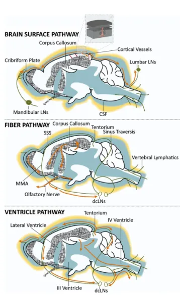
FIGURE 3 Schematic representation of solute drainage in the CNS. A, Cerebrospinal fluid (CSF) in the subarachnoid space is drained through the cribriform plate and collected by the lymphatic vessels present in the nasal cavity (afferent of the mandibular lymph nodes) or is reabsorbed into sinuses via the arachnoid villi. Alternatively, a part of the CSF recirculates from the subarachnoid space into the brain parenchyma along the perivascular spaces surrounding penetrating arteries (Box 1), and exchange with the interstitial fluid in the superficial layers of the neocortex. CSF flowing along the spinal canal is drained though the MLVs and allegedly transported to the lumbar lymph nodes. B, One main route for ISF and solute movement within the brain is along the white matter tracts (eg, corpus callosum, anterior commissures, and stria terminalis), and along the olfactory and optic nerve projections. Here solutes can be collected by the MLVs present in the dura mater running along the intracranial surface of the nerves and transported to the deep cervical lymph nodes (dcLNs). C, Alternatively, solutes can be transported to the ventricular system drained with the CSF. MLVs present in the tentorium and around sinus confluence, as well as the one along the rostral rhinal vein are putative collectors of the solutes drained through this pathway. CSF, cerebrospinal fluid; dcLNs, deep cervical lymph nodes; MLVs, meningeal lymphatic vessels; SSS, sinus sagittalis superior; MMA, middle meningeal artery
FIGURE 2 Schematic representation of cerebrospinal fluid (CSF) and interstitial fluid (ISF) production and circulation in the brain. CSF is mainly produced by the choroid plexus, whereas ISF derives from secretion at the level of the blood brain barrier (BBB). CSF and ISF interchange and mix at the level of the ventricles, and along the perivascular space or the capillary basement membrane. Arrows show direction and relative contribution of CSF and ISF to the net fluid circulation
interest, pharmacologic depletion of Treg in the dcLNs resulted
in neurodegeneration in a model of optic nerve lesion.82 The MLVs are afferent to the dcLNs.4 We speculate that
functional obstruction of MLVs could result in a detour of brain- derived antigens toward alternative secondary lym-phatic organs (eg, scLNs, lumbar lymph nodes, or spleen), circumventing the regulation of the neuroimmune response provided by the dcLNs. As a result, the antigens drained from the brain could promote a cytotoxic CD8- mediated auto immune reaction. Our preliminary data obtained using K14flt4- tg mice (lacking MLVs and dcLNs) support this hy-pothesis showing CD8+ T- cell immune response specifically in the cortical areas surrounding the lesion in a model of traumatic brain injury (TBI; controlled cortical injury [CCI] delivered unilaterally to the somatosensory cortex).
4
|
MENINGEAL LYMPHATIC
VESSELS AND THE
DEVELOPMENT OF AUTOIMMUNE
ENCEPHALITIDES
The International League Against Epilepsy (ILAE) has in-cluded autoimmunity among the etiologies of epilepsy: “im-mune epilepsy is the direct result of an im“im-mune disorder, in
which seizures are a core symptom, and the hallmark is the presence of autoimmune- mediated brain inflammation”.85 Autoimmune encephalitides are classified as follows: (a) en-cephalitides with pathogenic antibodies against cell surface proteins (eg, anti- NMDA [N- methyl- d- aspartate] receptor,
anti- LGI1, anti- VGKC complex); (b) T- cell diseases against intracellular antigens (eg, GAD65); and (c) encephalitides associated with other autoimmune disorders (eg, lupus cer-ebritis).86 Seizures and status epilepticus are common
symp-toms in autoimmune encephalitides,87 which can be resistant to antiepileptic drugs (AEDs) and respond better to immune therapies.88 Autoimmune encephalitides can relapse,86 sug-gesting the presence of a functional defect in the immune surveillance of the CNS.
4.1
|
CNS- lymphatic unit and Rasmussen
encephalitis pathophysiology: a proposed link
Here we focus on Rasmussen encephalitis (RE), described as focal seizures due to chronic localized encephalitis of probable viral origin.89 RE is a slow- progressing neurologic
disorder, characterized by unilateral brain atrophy and the presence of active microglia/macrophage nodules.90,91 RE is
associated with focal aware or focal impaired awareness sei-zures with motor onset, or with focal to bilateral tonic- clonic
FIGURE 4 Cartoon schematizing alternative immune responses toward brain- derived antigens. A, Soluble antigens from the brain parenchyma are transported along the interstitial fluid (ISF) route and the MLVs (blue- green) to the dcLNs. Here, depending on the inflammatory milieu, they can elicit immune tolerance mechanisms or a noncytotoxic immune reaction (Th2 mediated under Treg regulation), protecting neurons
and astrocytes from degeneration. B, A functional defect in one or more elements of the CNS lymphatic unit (eg, MLV congenital malformation or obstruction secondary to brain trauma) could result in drainage of brain- derived antigens to secondary lymphoid organs other thatn the dcLNs (eg, to the spleen via arachnoid villi and the venous system (blue) or to peripheral LNs via the cribriform plate), bypassing the specific neuroimmune response elicited in the dcLNs. Cytotoxic CD8+ T cells could be activated against neuronal or astrocytic self- antigens, homing to the brain, where kill targeted cells (ie, neurons and/or astrocytes). Ag, antigen; APC, antigen- presenting cell; dcLNs, deep cervical lymph nodes; MLVs, meningeal lymphatic vessels; Th0, naive T cell; Th2, type 2 CD4+ T helper; Treg, regulatory T cell
36
|
NOÉ aNd MaRCHI seizures, and poor response to AEDs.92 Studies performedusing brain specimens obtained from RE patients have in-dicated the presence of brain- infiltrating cytotoxic CD8+ T cells undergoing clonal local expansion.93–95 The infiltrating
CD8+ T cells are juxtaposed to neurons and astrocytes, with granzyme- B–containing granules polarized toward neuronal or astrocytic membranes.
In his original paper,89 Rasmussen proposed a brain viral
infection as the initiating event eliciting the CD8+ T- cell im-mune response. This would explain the clonal composition of the T- cell receptor repertoire found in the brain of RE pa-tients94 and the observed hemispheric distribution with
cen-trifugal expansion,96 suggestive of a focal infection. However,
no sign of viral infection has been found in brain specimens obtained from RE patients.96
Here we propose the hypothesis (Figure 4) that the CD8+ - mediated immune response observed in RE may be the re-sult of insufficient lymphatic drainage, either congenital (as in primary lymphedema) or consequent to the obstruction of the lymphatic flow. Under this condition, the control of the neuroimmune response provided by the MLVs- dcLNs may fail and brain- derived antigens could reach the peripheral lymph nodes, where a cytotoxic CD8+ T- cell mediated re-sponse occurs. Activated CD8+ T cells could home back to the brain and selectively target those cells (ie, neurons or glia) expressing the self- antigen. This could result in the specific neuronal and astrocytic cell loss observed in RE brains.97,98
A possible objection to our hypothesis is that autoimmune responses are usually not focal, whereas RE is. However, localized brain infiltration of activated CD8+ T cells may be facilitated in areas of BBB dysfunction and ongoing neuroinflammation. The latter could be the consequence of a cellular imprint of precedent insults and of a regional damage following head trauma or hypoxic events.99 Under
these conditions, proinflammatory cytokines can upregulate the expression of adhesion molecules (ICAM- 1, VCAM- 1, and E- selectin) on endothelial cells.100 These factors bind
to specific ligands expressed by the activated leukocytes al-lowing the adhesion, rolling, and migration of activated T cells across the brain endothelium79. In summary, RE could
therefore be the result of a double- hit, specifically a, reduced CNS- lymphatic unit efficiency (activating autoimmune T cells) and a brain insult, inducing regional neuroinflamma-tion and BBB dysfuncneuroinflamma-tion, that promotes focal lymphocyte CNS recruitment.
5
|
CNS- LYMPHATIC UNIT
IMPAIRMENT AND MODULATORY
APPROACHES
Strategies aimed at regenerating the lymphatic system may represent a supporting therapeutic intervention. It is known
that inflammation can directly promote lymphangiogenesis, an extensive and localized growth of lymphatic vessels.101
Tissue- infiltrating inflammatory cells (eg, CD11b+/Gr- 1+ macrophages) are capable of forming tube- like structures displaying lymphatic markers (ie, Lymphatic vessel endothe-lial hyaluronic acid receptor [Lyve- 1], Prospero homeobox protein 1 [Prox1], and podoplanin)102 and producing the
vas-cular endothelial growth factors VEGF- C and VEGF- D, pro-moting the genesis of new lymphatic vessels via VEGFR- 3 signaling.102 The newly- formed lymphatic vessels contribute
to restore the fluid drainage and counteract the inflammatory processes.102–105 It is therefore possible to exploit
lymphang-iogenic mechanisms to restore a compromised lymphatic system. For instance, the lymphangiogenesis- inducing fac-tor VEGF- C can be administered locally to recover lym-phatic drainage. The administration of the soluble form of the human recombinant (hr)VEGF- C106 or its localized viral
vectors- mediated over- expression107,108 resulted in growth of
functional and mature lymphatic vessels in animal models of peripheral lymphedema. Similarly, intracerebroventricular injections of adenoviral VEGF- C vector induced the growth of lymphatic capillaries in the meningeal compartment.109
However, the functionality of these newly generated MLVs is uncertain, and further studies are required to decipher the ability of the new lymphatic vessels to clear parenchymal sol-utes and to control neuroimmunity.
6
|
CONCLUDING REMARKS
Experimental evidence points to MLVs as a structural compo-nent of the CNS- lymphatic unit, impacting brain homeostasis, solute interstitial clearance, immune surveillance or inflam-mation. We have reviewed how alterations of the physiologic drainage of brain fluids could determine the accumulation of macromolecules within the brain parenchyma, resulting in the alteration of the extracellular ionic equilibrium, ulti-mately impacting neuronal excitability. The correct drainage of brain- derived antigens could be important for the allosta-sis of the neuroimmune cross- talk. We updated the hypoth-esis supporting the involvement of dcLNs in immune CNS surveillance and proposed that functional alterations of the MLVs (primary afferent vessels of the dcLNs) could result in autoimmune reactions. We suggested that a dysfunction of the CNS- lymphatic unit could be implicated in the pathophysiol-ogy of specific forms of epilepsy, as in situations where the primary cause is unknown (eg, Rasmussen encephalitis).
Moreover, functional of the CNS- lymphatic unit due to congenital defects or as a result of brain trauma, tumors, or infections could contribute to acquired or immune epilepsies. Addressing the dynamics of the CNS- lymphatic unit in the context of ictal activity could be important to disclose new therapeutic targets.
ACKNOWLEDGMENTS
FMN Funding: Academy of Finland research Fellowship (#309479/2017). NM Funding: ANR- Epicyte, ANR HepatoBrain, CURE Innovator Award. The authors thank Sara Wojciechowski for her contribution to revising the manuscript. We thank Muriel Asari (Institute of Functional Genomics, Montpellier, France) for the graphic design.
DISCLOSURE
None of the authors has any conflict of interest to disclose. The authors confirm that have read the Journal's position on issues involved in ethical publication and affirm that this re-port is consistent with those guidelines.
ORCID
Francesco M. Noé https://orcid.org/0000-0002-0019-1615
Nicola Marchi https://orcid.org/0000-0001-9124-0226
REFERENCES
1. Barker CF, Billingham RE. Immunologically privileged sites. Adv Immunol. 1977;25:1–54.
2. Shirai Y. Transplantation of the rat sarcoma in adult heteroge-neous animals. Japan Med World. 1921;1:14–5.
3. Louveau A, Smirnov I, Keyes TJ, et al. Structural and functional features of central nervous system lymphatic vessels. Nature. 2015;523:337–41.
4. Aspelund A, Antila S, Proulx ST, et al. A dural lymphatic vascular system that drains brain interstitial fluid and macromolecules. J Exp Med. 2015;212:991–9.
5. Louveau A. Cerebral lymphatic drainage: implication in the brain immune privilege. Med Sci (Paris). 2015;31:953–6.
6. Absinta M, Ha SK, Nair G, et al. Human and nonhuman primate meninges harbor lymphatic vessels that can be visualized nonin-vasively by MRI. Elife. 2017;6:e29738.
7. Kuo PH, Stuehm C, Squire S, et al. Meningeal lymphatic vessel flow runs countercurrent to venous flow in the superior sagittal sinus of the human brain. Tomography. 2018;4:99–104.
8. Louveau A, Herz J, Alme MN, et al. CNS lymphatic drainage and neuroinflammation are regulated by meningeal lymphatic vascu-lature. Nat Neurosci. 2018;21:1380–91.
9. Alitalo K. The lymphatic vasculature in disease. Nat Med. 2011;17:1371–80.
10. Brouillard P, Boon L, Vikkula M. Genetics of lymphatic anoma-lies. J Clin Invest. 2014;124:898–904.
11. Kerchner K, Fleischer A, Yosipovitch G. Lower extremity lymph-edema update: pathophysiology, diagnosis, and treatment guide-lines. J Am Acad Dermatol. 2008;59:324–31.
12. Betterman KL, Harvey NL. The lymphatic vasculature: development and role in shaping immunity. Immunol Rev. 2016;271:276–92. 13. Podgrabinska S, Kamalu O, Mayer L, et al. Inflamed lymphatic
endothelium suppresses dendritic cell maturation and func-tion via mac- 1/ICAM- 1- dependent mechanism. J Immunol. 2009;183:1767–79.
14. von Andrian UH, Mempel TR. Homing and cellular traffic in lymph nodes. Nat Rev Immunol. 2003;3:867–78.
15. Antila S, Karaman S, Nurmi H, et al. Development and plasticity of meningeal lymphatic vessels. J Exp Med. 2017;214:3645–67. 16. Harling-Berg CJ, Park TJ, Knopf PM. Role of the cervical
lym-phatics in the Th2- type hierarchy of CNS immune regulation. J Neuroimmunol. 1999;101:111–27.
17. Carare RO, Hawkes CA, Jeffrey M, et al. Review: cerebral amy-loid angiopathy, prion angiopathy, CADASIL and the spectrum of protein elimination failure angiopathies (PEFA) in neurode-generative disease with a focus on therapy. Neuropathol Appl Neurobiol. 2013;39:593–611.
18. Amor S, Puentes F, Baker D, et al. Inflammation in neurodegen-erative diseases. Immunology. 2010;129:154–69.
19. Brinker T, Stopa E, Morrison J, et al. A new look at cerebrospinal fluid circulation. Fluids Barriers CNS. 2014;11:10.
20. Abbott NJ. Evidence for bulk flow of brain interstitial fluid: significance for physiology and pathology. Neurochem Int. 2004;45:545–52.
21. Abbott NJ, Pizzo ME, Preston JE, et al. The role of brain barriers in fluid movement in the CNS: is there a ‘glymphatic’ system? Acta Neuropathol. 2018;135:387–407.
22. Hladky SB, Barrand MA. Mechanisms of fluid movement into, through and out of the brain: evaluation of the evidence. Fluids Barriers CNS. 2014;11:26.
23. Lei Y, Han H, Yuan F, et al. The brain interstitial system: anat-omy, modeling, in vivo measurement, and applications. Prog Neurobiol. 2017;157:230–46.
24. Iliff JJ, Wang M, Liao Y, et al. A paravascular pathway facili-tates CSF flow through the brain parenchyma and the clearance of interstitial solutes, including amyloid beta. Sci Transl Med. 2012;4:147ra111.
25. Carare RO, Bernardes-Silva M, Newman TA, et al. Solutes, but not cells, drain from the brain parenchyma along basement membranes of capillaries and arteries: significance for cerebral amyloid angiopathy and neuroimmunology. Neuropathol Appl Neurobiol. 2008;34:131–44.
26. Lonser RR, Sarntinoranont M, Morrison PF, et al. Convection- enhanced delivery to the central nervous system. J Neurosurg. 2015;122:697–706.
27. Szentistvanyi I, Patlak CS, Ellis RA, et al. Drainage of inter-stitial fluid from different regions of rat brain. Am J Physiol. 1984;246:F835–44.
28. Kida S, Pantazis A, Weller RO. CSF drains directly from the subarachnoid space into nasal lymphatics in the rat. anatomy, histology and immunological significance. Neuropathol Appl Neurobiol. 1993;19:480–8.
29. Hladky SB, Barrand MA. Elimination of substances from the brain parenchyma: efflux via perivascular pathways and via the blood- brain barrier. Fluids Barriers CNS. 2018;15:30.
30. Giannoni P, Badaut J, Dargazanli C, et al. The pericyte- glia inter-face at the blood- brain barrier. Clin Sci (Lond). 2018;132:361–74. 31. Daneman R, Prat A. The blood- brain barrier. Cold Spring Harb
Perspect Biol. 2015;7:a020412.
32. Marchi N, Granata T, Ghosh C, et al. Blood- brain barrier dys-function and epilepsy: pathophysiologic role and therapeutic ap-proaches. Epilepsia. 2012;53:1877–86.
33. Bar-Klein G, Lublinsky S, Kamintsky L, et al. Imaging blood- brain barrier dysfunction as a biomarker for epileptogenesis. Brain. 2017;140:1692–705.
38
|
NOÉ aNd MaRCHI34. Abbott NJ, Friedman A. Overview and introduction: the blood- brain barrier in health and disease. Epilepsia. 2012;53(Suppl 6):1–6. 35. Frigerio F, Frasca A, Weissberg I, et al. Long- lasting pro- ictogenic
effects induced in vivo by rat brain exposure to serum albumin in the absence of concomitant pathology. Epilepsia. 2012;53:1887–97. 36. Tomkins O, Shelef I, Kaizerman I, et al. Blood- brain barrier
dis-ruption in post- traumatic epilepsy. J Neurol Neurosurg Psychiatry. 2008;79:774–7.
37. Maggio N, Itsekson Z, Dominissini D, et al. Thrombin regulation of synaptic plasticity: implications for physiology and pathology. Exp Neurol. 2013;247:595–604.
38. Marchi N, Oby E, Batra A, et al. In vivo and in vitro effects of pi-locarpine: relevance to ictogenesis. Epilepsia. 2007;48:1934–46. 39. Vazana U, Veksler R, Pell GS, et al. Glutamate- mediated blood-
brain barrier opening: implications for neuroprotection and drug delivery. J Neurosci. 2016;36:7727–39.
40. Klement W, Garbelli R, Zub E, et al. Seizure progression and inflam-matory mediators promote pericytosis and pericyte- microglia clus-tering at the cerebrovasculature. Neurobiol Dis. 2018;113:70–81. 41. Vezzani A, Lang B, Aronica E. Immunity and inflammation in
epilepsy. Cold Spring Harb Perspect Med. 2015;6:a022699. 42. Marchi N, Granata T, Janigro D. Inflammatory pathways of
sei-zure disorders. Trends Neurosci. 2014;37:55–65.
43. Mendes A, Sampaio L. Brain magnetic resonance in status epilep-ticus: a focused review. Seizure. 2016;38:63–7.
44. Tomkins O, Feintuch A, Benifla M, et al. Blood- brain barrier break-down following traumatic brain injury: a possible role in posttrau-matic epilepsy. Cardiovasc Psychiatry Neurol. 2011;2011:765923. 45. Marchi N, Betto G, Fazio V, et al. Blood- brain barrier damage
and brain penetration of antiepileptic drugs: role of serum pro-teins and brain edema. Epilepsia. 2009;50:664–77.
46. Omalu BI, DeKosky ST, Hamilton RL, et al. Chronic traumatic encephalopathy in a national football league player: part II. Neurosurgery. 2006;59:1086–92; discussion 1092-3.
47. Omalu BI, DeKosky ST, Minster RL, et al. Chronic traumatic en-cephalopathy in a national football league player. Neurosurgery. 2005;57:128–34. discussion 128-34.
48. Marchi N, Banjara M, Janigro D. Blood- brain barrier, bulk flow, and interstitial clearance in epilepsy. J Neurosci Methods. 2016;260:118–24.
49. Makinen T, Jussila L, Veikkola T, et al. Inhibition of lymphangio-genesis with resulting lymphedema in transgenic mice expressing soluble VEGF receptor- 3. Nat Med. 2001;7:199–205.
50. Plog BA, Moll KM, Kang H, et al. A novel technique for mor-phometric quantification of subarachnoid hemorrhage- induced microglia activation. J Neurosci Methods. 2014;229:44–52. 51. Ali I, Silva JC, Liu S, et al. Targeting neurodegeneration to
pre-vent post- traumatic epilepsy. Neurobiol Dis. 2019;123:100–9. 52. Saletti PG, Ali I, Casillas-Espinosa PM, et al. In search of
antiepi-leptogenic treatments for post- traumatic epilepsy. Neurobiol Dis. 2019;123:86–99.
53. Zheng P, Shultz SR, Hovens CM, et al. Hyperphosphorylated tau is implicated in acquired epilepsy and neuropsychiatric comorbid-ities. Mol Neurobiol. 2014;49:1532–9.
54. Sarnat HB, Flores-Sarnat L. Infantile tauopathies: hemimegalen-cephaly; tuberous sclerosis complex; focal cortical dysplasia 2; ganglioglioma. Brain Dev. 2015;37:553–62.
55. Sen A, Thom M, Martinian L, et al. Pathological tau tangles localize to focal cortical dysplasia in older patients. Epilepsia. 2007;48:1447–54.
56. Thom M, Liu JY, Thompson P, et al. Neurofibrillary tangle pa-thology and braak staging in chronic epilepsy in relation to trau-matic brain injury and hippocampal sclerosis: a post- mortem study. Brain. 2011;134:2969–81.
57. Puvenna V, Engeler M, Banjara M, et al. Is phosphorylated tau unique to chronic traumatic encephalopathy? phosphorylated tau in epileptic brain and chronic traumatic encephalopathy. Brain Res. 2016;1630:225–40.
58. Monti G, Tondelli M, Giovannini G, et al. Cerebrospinal fluid tau proteins in status epilepticus. Epilepsy Behav. 2015;49:150–4. 59. Liang Z, Liu F, Iqbal K, et al. Dysregulation of tau
phosphoryla-tion in mouse brain during excitotoxic damage. J Alzheimers Dis. 2009;17:531–9.
60. Hawkins BE, Krishnamurthy S, Castillo-Carranza DL, et al. Rapid accumulation of endogenous tau oligomers in a rat model of trau-matic brain injury: possible link between trautrau-matic brain injury and sporadic tauopathies. J Biol Chem. 2013;288:17042–50. 61. Shultz SR, Wright DK, Zheng P, et al. Sodium selenate reduces
hyperphosphorylated tau and improves outcomes after traumatic brain injury. Brain. 2015;138:1297–313.
62. Roberson ED, Halabisky B, Yoo JW, et al. Amyloid- beta/fyn- induced synaptic, network, and cognitive impairments depend on tau levels in multiple mouse models of alzheimer's disease. J Neurosci. 2011;31:700–11.
63. Decker JM, Kruger L, Sydow A, et al. The tau/A152T mutation, a risk factor for frontotemporal- spectrum disorders, leads to NR2B receptor- mediated excitotoxicity. EMBO Rep. 2016;17:552–69. 64. Kandratavicius L, Monteiro MR, Hallak JE, et al. Microtubule-
associated proteins in mesial temporal lobe epilepsy with and without psychiatric comorbidities and their relation with granular cell layer dispersion. Biomed Res Int. 2013;2013:960126. 65. Sotiropoulos I, Galas MC, Silva JM, et al. Atypical, non- standard
functions of the microtubule associated tau protein. Acta Neuropathol Commun. 2017;5:91.
66. Engel T, Goni-Oliver P, Lucas JJ, et al. Chronic lithium admin-istration to FTDP- 17 tau and GSK- 3beta overexpressing mice prevents tau hyperphosphorylation and neurofibrillary tangle formation, but pre- formed neurofibrillary tangles do not revert. J Neurochem. 2006;99:1445–55.
67. Bhowmik M, Khanam R, Saini N, et al. Activation of AKT/ GSK3beta pathway by TDZD- 8 attenuates kainic acid induced neurodegeneration but not seizures in mice. Neurotoxicology. 2015;46:44–52.
68. Jones NC, Nguyen T, Corcoran NM, et al. Targeting hyperphos-phorylated tau with sodium selenate suppresses seizures in rodent models. Neurobiol Dis. 2012;45:897–901.
69. Liu SJ, Zheng P, Wright DK, et al. Sodium selenate retards epilepto-genesis in acquired epilepsy models reversing changes in protein phos-phatase 2A and hyperphosphorylated tau. Brain. 2016;139:1919–38. 70. Wilson L, Stewart W, Dams-O'Connor K, et al. The chronic and
evolving neurological consequences of traumatic brain injury. Lancet Neurol. 2017;16:813–25.
71. Molteni M, Rossetti C. Neurodegenerative diseases: the immuno-logical perspective. J Neuroimmunol. 2017;313:109–15. 72. Gendelman HE, Mosley RL. A perspective on roles played by
in-nate and adaptive immunity in the pathobiology of neurodegener-ative disorders. J Neuroimmune Pharmacol. 2015;10:645–50. 73. Bauer J, Becker AJ, Elyaman W, et al. Innate and adaptive
74. Walsh JT, Watson N, Kipnis J. T cells in the central nervous system: messengers of destruction or purveyors of protection? Immunology. 2014;141:340–4.
75. Urra X, Miro F, Chamorro A, et al. Antigen- specific immune re-actions to ischemic stroke. Front Cell Neurosci. 2014;8:278. 76. de Vos AF, van Meurs M, Brok HP, et al. Transfer of central
ner-vous system autoantigens and presentation in secondary lymphoid organs. J Immunol. 2002;169:5415–23.
77. Perry VH. A revised view of the central nervous system microen-vironment and major histocompatibility complex class II antigen presentation. J Neuroimmunol. 1998;90:113–21.
78. Weller RO, Galea I, Carare RO, et al. Pathophysiology of the lym-phatic drainage of the central nervous system: implications for pathogenesis and therapy of multiple sclerosis. Pathophysiology. 2010;17:295–306.
79. Engelhardt B, Carare RO, Bechmann I, et al. Vascular, glial, and lymphatic immune gateways of the central nervous system. Acta Neuropathol. 2016;132:317–38.
80. Cserr HF, Harling-Berg CJ, Knopf PM. Drainage of brain extra-cellular fluid into blood and deep cervical lymph and its immuno-logical significance. Brain Pathol. 1992;2:269–76.
81. Thomas DL, Kranz DM, Roy EJ. Experimental manipulations of afferent immune responses influence efferent immune responses to brain tumors. Cancer Immunol Immunother. 2008;57:1323–33. 82. Walsh JT, Zheng J, Smirnov I, et al. Regulatory T cells in cen-tral nervous system injury: a double- edged sword. J Immunol. 2014;193:5013–22.
83. Widner H, Moller G, Johansson BB. Immune response in deep cer-vical lymph nodes and spleen in the mouse after antigen deposition in different intracerebral sites. Scand J Immunol. 1988;28:563–71. 84. Galea I, Bechmann I, Perry VH. What is immune privilege (not)?
Trends Immunol. 2007;28:12–8.
85. Scheffer IE, Berkovic S, Capovilla G, et al. ILAE classification of the epilepsies: position paper of the ILAE commission for classi-fication and terminology. Epilepsia. 2017;58:512–21.
86. Lancaster E. The diagnosis and treatment of autoimmune enceph-alitis. J Clin Neurol. 2016;12:1–13.
87. Vincent A, Irani SR, Lang B. The growing recognition of immunotherapy- responsive seizure disorders with autoantibodies to specific neuronal proteins. Curr Opin Neurol. 2010;23:144–50. 88. Irani SR, Vincent A, Schott JM. Autoimmune encephalitis. BMJ.
2011;342:d1918.
89. Rasmussen T, Olszewski J, Lloydsmith D. Focal seizures due to chronic localized encephalitis. Neurology. 1958;8:435–45. 90. Pardo CA, Nabbout R, Galanopoulou AS. Mechanisms of
ep-ileptogenesis in pediatric epileptic syndromes: rasmussen en-cephalitis, infantile spasms, and febrile infection- related epilepsy syndrome (FIRES). Neurotherapeutics. 2014;11:297–310. 91. Robitaille Y. Neuropathologic aspects of chronic
encephali-tis. In: Andermann F (Ed). Chronic encephalitis and epilepsy. Rasmussen's syndrome Ed. Boston, MA: Butterworth‐Heinemann, 1991:p. 79–110.
92. Varadkar S, Bien CG, Kruse CA, et al. Rasmussen's encephalitis: clinical features, pathobiology, and treatment advances. Lancet Neurol. 2014;13:195–205.
93. Li Y, Uccelli A, Laxer KD, et al. Local- clonal expansion of in-filtrating T lymphocytes in chronic encephalitis of rasmussen. J Immunol. 1997;158:1428–37.
94. Schwab N, Bien CG, Waschbisch A, et al. CD8 + T- cell clones dominate brain infiltrates in rasmussen encephalitis and persist in the periphery. Brain. 2009;132:1236–46.
95. Al Nimer F, Jelcic I, Kempf C, et al. Phenotypic and functional complexity of brain- infiltrating T cells in rasmussen encephalitis. Neurol Neuroimmunol Neuroinflamm. 2017;5:e419.
96. Granata T, Andermann F. Rasmussen encephalitis. Handb Clin Neurol. 2013;111:511–9.
97. Bien CG, Bauer J, Deckwerth TL, et al. Destruction of neurons by cytotoxic T cells: a new pathogenic mechanism in rasmussen's encephalitis. Ann Neurol. 2002;51:311–8.
98. Bauer J, Vezzani A, Bien CG. Epileptic encephalitis: the role of the innate and adaptive immune system. Brain Pathol. 2012;22:412–21.
99. Stamatovic SM, Keep RF, Andjelkovic AV. Brain endothelial cell- cell junctions: how to “open” the blood brain barrier. Curr Neuropharmacol. 2008;6:179–92.
100. Haraldsen G, Kvale D, Lien B, et al. Cytokine- regulated expres-sion of E- selectin, intercellular adheexpres-sion molecule- 1 (ICAM- 1), and vascular cell adhesion molecule- 1 (VCAM- 1) in human mi-crovascular endothelial cells. J Immunol. 1996;156:2558–65. 101. Kim H, Kataru RP, Koh GY. Inflammation- associated
lymphangiogenesis: a double- edged sword? J Clin Invest. 2014;124:936–42.
102. Kataru RP, Jung K, Jang C, et al. Critical role of CD11b+ mac-rophages and VEGF in inflammatory lymphangiogenesis, antigen clearance, and inflammation resolution. Blood. 2009;113:5650–9. 103. Huggenberger R, Siddiqui SS, Brander D, et al. An important role
of lymphatic vessel activation in limiting acute inflammation. Blood. 2011;117:4667–78.
104. Guo R, Zhou Q, Proulx ST, et al. Inhibition of lymphangiogene-sis and lymphatic drainage via vascular endothelial growth factor receptor 3 blockade increases the severity of inflammation in a mouse model of chronic inflammatory arthritis. Arthritis Rheum. 2009;60:2666–76.
105. Kesler CT, Liao S, Munn LL, et al. Lymphatic vessels in health and disease. Wiley Interdiscip Rev Syst Biol Med. 2013;5:111–24.
106. Szuba A, Skobe M, Karkkainen MJ, et al. Therapeutic lymph-angiogenesis with human recombinant VEGF- C. FASEB J. 2002;16:1985–7.
107. Tammela T, Saaristo A, Holopainen T, et al. Therapeutic differ-entiation and maturation of lymphatic vessels after lymph node dissection and transplantation. Nat Med. 2007;13:1458–66. 108. Visuri MT, Honkonen KM, Hartiala P, et al. VEGF- C and VEGF-
C156S in the pro- lymphangiogenic growth factor therapy of lymph-edema: a large animal study. Angiogenesis. 2015;18:313–26. 109. Karaman S, Nurmi H, Antila S, et al. Stimulation and inhibition
of lymphangiogenesis via adeno- associated viral gene delivery. Methods Mol Biol. 2018;1846:291–300.
How to cite this article: Noé FM, Marchi N. Central
nervous system lymphatic unit, immunity, and epilepsy: Is there a link?. Epilepsia Open. 2019;4:30– 39. https://doi.org/10.1002/epi4.12302