HAL Id: hal-00548607
https://hal.archives-ouvertes.fr/hal-00548607
Submitted on 20 Dec 2010
HAL is a multi-disciplinary open access archive for the deposit and dissemination of sci-entific research documents, whether they are pub-lished or not. The documents may come from
L’archive ouverte pluridisciplinaire HAL, est destinée au dépôt et à la diffusion de documents scientifiques de niveau recherche, publiés ou non, émanant des établissements d’enseignement et de
Quasi unidimensional growth of Co nanostructures on a
strained Au(111) surface
Paolo Campiglio, Vincent Repain, Cyril Chacon, Olivier Fruchart, Jérôme
Lagoute, Yann Girard, Sylvie Rousset
To cite this version:
Paolo Campiglio, Vincent Repain, Cyril Chacon, Olivier Fruchart, Jérôme Lagoute, et al.. Quasi unidimensional growth of Co nanostructures on a strained Au(111) surface. Surface Science, Elsevier, 2011, 605, pp.1165. �10.1016/j.susc.2011.03.019�. �hal-00548607�
Quasi unidimensional growth of Co nanostructures on a
strained Au(111) surface
P. Campiglioa,b, V. Repaina, C. Chacona, O. Fruchartc, J. Lagoutea, Y. Girarda, S. Rousseta
aLaboratoire Mat´eriaux et Ph´enom`enes Quantiques, Universit´e Paris Diderot & CNRS,
UMR 7162, case courrier 7021, F-75205 Paris Cedex 13, France
bDepartment of Materials Science, Universit`a degli Studi di Milano-Bicocca, Via R.
Cozzi 53 Milano, 20125, Italy
cInstitut N ´EEL, CNRS & Universit´e Joseph Fourier, F-38042 Grenoble Cedex 9, France
Abstract
We investigated to which extent a uniaxially-strained Au(111) surface, thereby displaying linear reconstructions, may be used as a template for the growth of elongated nanostructures. Cobalt has been used as a deposition material, and two routes have been followed satisfactorily. The first one consists in the control of diffusion through the temperature of deposition. The second one consists in a first step of platinum seeding in the reconstruction, followed by cobalt deposition with nucleation on the platinum seeds.
Keywords:
Self-assembly, Surface diffusion, Low index single crystal surfaces,
Metal-metal magnetic thin film structures, Scanning tunneling microscopy, Cobalt
1. Introduction
The ability to organize matter into objects with regular size, shape and spacing down to the atomic scale represents a cornerstone for nanoscience and nanotechnology. Top-down methods, mainly based on lithographic tech-niques, are time-consuming, expensive and in most of the cases limited to sizes above a few tens of nanometers. In this context a promising alternative, which has attracted an increasing attention, relies on bottom-up approaches.
These exploit diffusion and aggregation processes specific to each system, to drive the assembly of atoms and molecules in nanostructures [1]. Such routes are particularly interesting when using pre-existing self-organized template surfaces [2], which guide the growth and yield ordered arrays of nanodots. One major output of using surface templates is that the resulting nanostruc-tures display a narrow size distribution. The guiding effect of the template may often be applied to several materials, which otherwise would not give rise to an organized array. For metal surfaces, a model template is provided by the Au(111) surface. This displays a so-called herringbone reconstruction [3] at the elbows of which many materials may nucleate and form ordered arrays of dots [4]. The distance between nearest-neighbor dots is about 7 nm, and the metallic clusters display a nearly mono-disperse lateral size up to a few nanometers in diameter. On the one hand this peculiarity has al-lowed the study of fundamental physical properties (notably the magnetic properties such as magnetic anisotropy) of low dimensional model systems [5, 6, 7]. On the other hand, the Au(111) surface lacks of versatility since its surface pattern makes difficult to control the dots shape. In particu-lar, the natural surface of Au(111) does not allow the versatile growth of nanowires, because its template is two-dimensional with a rectangular unit cell. In fact local nanowires can be achieved for certain materials in a narrow range of coverage when percolation sets in [8] but they are not macroscop-ically oriented along the same direction. So far, oriented nanowires have been grown on anisotropic substrates like (110) surface of fcc metals [9, 10], vicinal surfaces [11], molecular networks [12] or ripples obtained by etching under kinetic limitations [13, 14]. The use of dense surface for the growth of nanowires would be a technological improvement since they are the most common surfaces observed during thin films growth. Au(111) appears to be a good candidate for anisotropic dense surface as it had been shown that when subjected to a uniaxial strain a Au(111) thin film may display a purely linear reconstruction, instead of the usual herringbone [15]. While initially performed in situ on a bent mica sheet, providing only a temporary control of the strain, recently linear reconstructions on Au(111) could be stabilized by uniaxial epitaxial strain on a body-centered cubic W(110) underlayer film [16].
In this paper, we investigate to what extent this new reconstruction may be used as a template for self-organizing wires or chains of dots. We successfully produced elongated-shaped nanodots of cobalt following two routes. The first one relies on the temperature control over the diffusion of Co adatoms while
the second one exploits the gold-platinum surface alloy to create a prepat-terned surface with controlled nucleation sites.
2. Experimental details
The samples consist of a Au(111) [8 nm]/W(110) [10 nm] thin film epitax-ially grown on Sapphire (11¯20) by pulsed-laser deposition in a first ultra-high vacuum chamber. The base pressure is 3 × 10⁻¹¹ mbar and the typical de-position rate is 1 ˚A/s [17]. The samples were transferred through air to another chamber for the STM studies. The base pressure is better than 10−10 mbar. Here the sample is cleaned with repeated cycles of grazing-incidence bombardment with argon ions (typically for 30 s with an grazing-incidence angle α = 45◦ at an Ar pressure of 2 × 10⁻⁶ mbar and a voltage of 1 kV) fol-lowed by annealing at 650 ◦C. Cobalt and platinum were evaporated by elec-tron bombardment heating of high purity rods (99.99%). Typical flux rates are 0.2 ML(monolayer)/min and 0.02 ML/min for Co and Pt respectively. The deposition is performed on the sample placed in a Omicron Nanotech-nology c VT-STM (operating range from 50 K to 500 K). The cobalt and platinum coverage (θCo and θPt) are determined on the basis of STM images,
assuming a pseudomorphic growth with the Au(111) substrate. The pressure during deposition is always maintained below 1 × 10⁻¹⁰mbar. Morphological studies are made by in situ STM at the temperature of the deposition.
3. Results and discussion
3.1. Au(111) with unidirectional reconstruction
The driving force for Au(111) surface reconstruction is the surface tensile stress that would occur at the surface of Au(111), if it were unreconstructed. In response to this stress the surface layer spontaneously densifies along the h1¯10i directions, adding one extra atom every 22 atoms of the underlying layer with bulk lattice parameters, giving rise to the 22 ×√3 reconstruction [18, 19]. Inside the unit cell the atoms are arranged in fcc and hcp-type stacking domains separated by two stacking fault lines (or discommensuration lines). At larger scale a so-called herringbone reconstruction is observed: the 22 ×√3 domains bend of 120◦ and form a mesoscopically ordered pattern with a typical period of 25 nm [3]. This secondary superstructure arises from more delicate long-range elastic interactions mediated by the substrate, to relax a compression otherwise unidirectional [20]. Because of such central
role played in the reconstruction, it is clear that stress is a key parameter to handle if one wishes to guide the surface towards new arrangements.
To this purpose we used a multilayer sample (schematically shown in Fig. 1) in which a uniaxial in-plane strain is imposed through the epitaxial relation with an underlayer with a crystalline uniaxial symmetry [16].
In short, the substrate we choose for our sample is Sapphire (11¯20) sur-face, over which a very thin film (0.7 nm) of Mo and a thicker one (10 nm) of W are grown and annealed such as to yield a single-crystalline body-centered layer with an atomically-smooth surface, with terraces of width several hun-dreds of nanometers [17]. Both Mo and W are bcc metals that during the growth expose the (110) surface characterized by a centered rectangular bidi-mensional lattice. Due to its anisotropy this layer induces a uniaxial strain in the Au layer (deposited at room temperature and annealed at 400◦C), lifting the original three-fold symmetry and inducing a preferential orientation of the compressed [1¯10] direction [16]. The initial thickness of the Au layer is 8 nm.
In Fig. 2a an image of the uniaxial Au(111) surface is reported. The brighter lines are the discommensuration lines that run along the [11¯2] di-rection, while in the close-packed [1¯10] direction hcp and fcc atomic stacking regularly alternate. As can be clearly seen, the herringbone reconstruction is suppressed and the purely linear reconstruction is stable over the whole sample surface. The stacking faults lines are characterized by a corrugation height of 0.17±0.02 ˚A while the distance between two adjacent maxima is close to 57 ˚A, in contrast from the value observed for the reconstruction of Au(111) single-crystal surface. Indeed, the supercell that can be identified
Au(111) W(110) Sapphire(1120) 8 nm 10 nm Mo(110)
Figure 1: Schematic structure of the multilayer sample employed. The thickness of the Mo layer is 0.7 nm.
Figure 2: (a) Room temperature STM image showing the unidirectional reconstruction of the stressed Au(111) surface (85 × 85 nm2). (b) Drift compensated atomic resolution
STM image, with the unit cell highlighted (15 × 8 nm2).
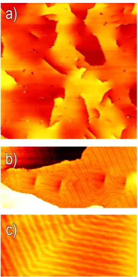
in Fig. 2b shows that the periodicity is drifted to 20 atomic positions. At larger scale (Fig. 3a) some defects of the reconstruction also appear, mainly due to surface morphology. The most visible signatures in STM im-ages comes from screw dislocations that appear as emerging steps. Edge dislocations leave weaker footprints on the morphology, but are responsi-ble for important deviations from the purely linear reconstruction. At the surface, edge dislocations split into two partial dislocations that elastically repel each other and form a stacking fault ribbon that appear like a short step segment high one third of the full step height. These defects have been seldomly seen on single crystal Au(111) surface [21]. More particularly, on the present sample these stacking fault ribbons are commonly arranged in sequences, like shown in Fig. 3b, probably arising from grain boundaries.
Figure 3: STM images of the Au(111) thin film surface (a) Large scale showing the presence of different defects (360 × 360 nm2). (b) Effect of a sequence of stacking fault ribbons
on Au(111) reconstruction (130 × 60 nm2). (c) The reconstruction deviates from its
unidimensional character and turns of 120◦ as in the herringbone reconstruction (60 × 30 nm2).
When the reconstruction deviates from its unidimensional character, it turns of 120◦ as in the herringbone reconstruction (Fig. 3c). Naturally, the re-construction elbows constitute the main trap for adatoms and represent the principal difficulty to overcome in order to obtain a homogeneous growth. 3.2. Growth of Co on Au(111)
Fig. 4 shows STM images of the Au(111) surface with unidirectional re-construction onto which Co was deposited at low coverage (θCo) in a wide
range of temperatures (50 K < T < 300 K). When the deposition of 0.25 ML of Co is carried out at room temperature (Fig. 4a) the diffusion channels are clearly activated along all directions and thus the diffusion of adatoms is es-sentially isotropic. Due to the long diffusion length at this temperature, the adatoms are eventually pinched at defects of the surface. These are mainly dislocations or random atomic impurities; their role must be underlined in the present case because of the thinness of the Au layer that requires reduced time of Ar sputtering as compared to a single crystal. As a consequence, the Co growth proceeds very differently compared to the growth on herringbone reconstruction [22]. While the profile analysis indicates that the dots retain the usual bilayer height, the absence of periodic elbows in the reconstruction makes the dots loose both their regular spacing and their narrow size distri-bution.
To obtain an anisotropic diffusion we have reduced the temperature of the substrate at 105 K for the deposition. the temperature of deposition is not very critical, as the processes described below were found to be identical at 135 K. As can be seen in Fig. 4b, Co (θCo= 0.15 ML) forms dots elongated
along the reconstruction direction, long up to 20 nm and with a mean aspect ratio equal to 5. The nucleation of this kind of dots takes place on the fcc zone of the reconstruction. From these observations we can deduce that at this temperature the diffusion is anisotropic, with the path of adatoms along a direction parallel to the discommensuration lines being favored. The dis-commensuration lines seem to act as energy barrier, repelling the diffusion of Co adatoms as observed for Al [23]. This contrasts with molecular dynam-ics simulations, which predicted a more favorable diffusion perpendicularly to the discommensuration lines, followed by slower diffusion on the top and along the discommensurations [24]. They act as attractors and constitute a favored path to kink sites, where Co nucleates and grows [25]. However, the sites along the sides of the discommensuration are found to show higher adsorption energies then fcc and hcp sites and thus could explain their
repul-Figure 4: STM images of Co deposited at different temperatures on Au(111) with uni-directional reconstruction (90 × 90 nm2). All the images are recorded at the deposition temperature. (a) T = 296 K, θCo = 0.25 ML, isotropic diffusion; this image has been
treated in order to enhance the contrast on the substrate. (b) T = 105 K, θCo= 0.15 ML,
anisotropic diffusion that leads to elongated dots. (c) T = 50 K, θCo = 0.15 ML,
post-nucleation regime.
sive character with respect to diffusing Co adatoms [26]. Due to the reduced mobility of adatoms, impurities and reconstruction defects constitute even more efficient traps than at room temperature. As a result, besides elongated dots also very small clusters of Co nucleate, leading to a wide distribution of dot size and shape. It is the same type of defects, including impurities, that are the source of nucleation at 300 K (Fig. 4). We believe that impurities are not only intrinsic, but are also due to the fact that the same template has been used several times to perform experiments at various temperatures. The etching procedure applied between each deposition to sputter away the Co atoms, was minimized so that the entire Au film (total initial thickness 8 nm) is not fully etched away. Probably this leaves Co impurities at the surface, which upon annealing are known to embed themselves in the upper-most plane of Au atoms, creating nucleations sites for the next deposition [27]. It is therefore likely that deposition at 105 K on a fresh template would yield a more homogeneous growth of elongated dots.
As expected, if we further decrease the temperature of deposition down to 50 K (θCo = 0.15 ML), the diffusion of adatoms is essentially completely
frozen and a post nucleation regime is observed, giving rise to frozen disorder. As shown in Fig. 4c, monolayer-thick dots with lateral size of few Angstr¨oms nucleate at spatially random locations with a high density, in a rather similar way than on Au(111) surface with herringbone reconstruction [28].
the self-assembly of nanostructures, and we successfully employed it to grow elongated dots also in the present case. However the high density of intrinsic and extrinsic defects on the surface disturbs the diffusion of Co adatoms, preventing a homogeneous growth. In order to have a better control on the number of nucleation centers on the surface, Pt seeding appears to be a suit-able candidate to elaborate a prepattening of the surface [29]. Pt is miscible with Au and previous studies showed that when deposited at room temper-ature on Au(111), Pt substitute Au atoms in the surface layer [30]. Since at room temperature the Au(111) surface could host a maximum of 0.03 ML of Pt, we increased the substrate temperature to 400 K to favor the exchange of Pt with Au atoms. Fig. 5a shows a room temperature STM image of 0.05 ML of Pt deposited on Au(111) at 400 K. The embedded Pt atoms are seen darker than the surrounding Au atoms, with an apparent height different of 0.2 ˚A with respect to the mean surface level. As a detrimen-tal consequence of the alloying, Au atoms are ejected from the surface and forms dots of 2.35 ˚A high that may perturb the regularity of the reconstruc-tion. More importantly, Pt atoms alloying does not occur randomly on the surface but takes place preferentially in the discommensuration regions (See inset of Fig. 5a). Thus the surface alloy could be regarded as a patterned surface in which Pt atoms constitute trapping sites that may permit to trig-ger the nucleation of Co. The ability of a seed layer to guide the growth of materials that otherwise do not show any order has been shown also for Pd on Ni3Al(111) [31] or Ir on graphene/Ir(111) [32] This is confirmed in Fig.
5b, where 0.15 ML of Co where deposited at room temperature on Au(111) seeded with Pt. Since the diffusion of adatoms is quenched by the presence of Pt atoms, Co dots nucleate with a high density in the form of monolayer dots with a mean lateral dimension of 1 nm. In the case of such small dots it has been shown that it is energetically more favorable to adopt a monolayer rather than a bilayer height in order to maximize the atomic coordination [33]. The Co follows the order imposed by the previous Pt deposition and forms hemispherical dots aligned along the reconstruction lines and not on the fcc stacking regions as we observed in the low-temperature growth. In particular Co nucleates alternatively in one over two discommensuration re-gions and thus the distance between neighbor nanodots is 7 nm. This is due to the fact that preferentially the alloying of Pt with Au takes place alter-natively in one single discommensuration lines of the reconstruction, as can be seen also in Fig. 5a. When θCo is increased, dots grow first vertically,
Figure 5: (a) STM image of Pt (θPt= 0.05 ML) deposited at 400 K on Au(111) with
unidi-rectional reconstruction; the Pt is embedded in the surface and follows the reconstruction lines. In inset, zoom (32 × 32 nm2) showing dark patches corresponding to Pt inserted atoms. (b) (θCo = 0.15 ML) and (c) (θCo = 1.1 ML), STM images of Co deposited on
Pt/Au(111): Pt acts like preferential nucleation sites for Co that forms small and sepa-rated dots at low Co coverage (b), that coalesce when the Co coverage is increased (c). All the images are 100 × 100 nm2and recorded at room temperature.
process the clusters percolate and, thanks to the small distance between dots on the same reconstruction line, form dots often very elongated along the reconstruction direction, with an aspect ratio up to 8. In some other cases even if two dots come very close together, the percolation does not take place probably due to an incommensurate growth of the dots [34]. This is observed even more systematically on Au(111) with herringbone reconstruction, and it is why the magnetic transition of Co nanodots to ferromagnetic films happen only at 1.5 ML. The problem of incommensurate growth in this case seems to be less stringent and several dots can coalesce together, as can be seen in Fig. 5c. Another relevant difference with Au(111) with herringbone is that the coalesce takes place in the same direction also at a macroscopic scale. This is due to the fact that the direction of the reconstruction is maintained fixed all over the sample, if surface defects are neglected. As an important consequence, the nanodots are preferentially elongated along the same axis. Significantly, this should permit to macroscopically measure the properties of such anisotropic objects along different directions. When applied to freshly re-prepared samples of uniaxial Au(111) reconstruction, the two procedures applied above (low temperature deposition or Pt seeding) are expected to provide a much cleaner template, efficient to fabricate uniaxial nanostruc-tures of various materials on a macroscopic scale. It is important to stress that no self-organized linear template with such a large period is currently
available starting from nominally-flat surfaces, i.e. compatible with a stan-dard wafer technology. This may open the way to the use of such templates for the investigation of physical properties of nanowires in a yet unexplored range of width.
4. Conclusions
In this work we employ a uniaxially strained Au(111) surface, whose re-construction is lifted to purely linear thanks to the epitaxial constraints of an underlying multilayer sample. We investigated the capability of this surface to act as a template to obtain unidimensional nanostructures. To achieve this, we focalized our attention on the growth of cobalt in submonolayer cov-erages. Elongated dots with an aspect ratio up to 8 were successfully obtained by following two different routes. In the first one we use the temperature to control the diffusion of adatoms across the surface. Below about 150 K the diffusion channel across the reconstruction is blocked and the anisotropic diffusion results in an elongated growth. Alternatively, we have successfully imposed order nucleation sites. This was done with the predeposition of a seed layer of Pt that forms a surface alloy with Au preferentially on the dis-commensuration lines. We believe that both these routes may be used to realize elongated nanodots of various materials (Mn, Fe, Ni, Pd, Pt...). The ability to control the diffusion and aggregation of adatoms combined with the skill to tailor a given surface structure is a powerful way to control the growth of metal nanostructures. This objective is of high importance in order to understand and exploit their physical properties.
Acknowledgement
The authors thank A. Sassella for fruitful discussions. V.R. acknowl-edges financial support from contract ANR-ETNAA (ANR-NANO-018-04) and Insitut Universitaire de France. O.F. acknowledges financial support from contract ANR-05-NANO-073.
References
[1] J. V. Barth, G. Costantini, K. Kern, Engineering atomic and molecular nanostructures at surfaces, Nature 437 (2005) 671.
[2] S. Rousset, B. Croset, Y. Girard, G. Pr´evot, V. Repain, S. Rohart, Self-organized epitaxial growth on spontaneously nano-patterned templates, C. R. Physique 6 (2005) 33.
[3] J. V. Barth, H. Brune, G. Ertl, R. J. Behm, Scanning tunneling mi-croscopy observations on the reconstructed Au(111) surface: Atomic structure, long-range superstructure, rotational domains, and surface defects, Phys. Rev. B 42 (1990) 9307.
[4] D. D. Chambliss, R. J. Wilson, S. Chiang, Nucleation of ordered Ni island arrays on Au(111) by surface-lattice dislocations, Phys. Rev. Lett. 66 (1991) 1721.
[5] H. A. D¨urr, S. S. Dhesi, E. Dudzik, D. Knabben, G. V. Laan, J. Goed-koop, F. U. Hillebrecht, Spin and orbital magnetization in self-assembled Co clusters on Au(111), Phys. Rev. B 59 (1999) R701.
[6] Y. Nahas, V. Repain, C. Chacon, Y. Girard, J. Lagoute, G. Rodary, J. Klein, S. Rousset, H. Bulou, C. Goyhenex, Dominant role of the epi-taxial strain in the magnetism of core-shell Co/Au self-organized nan-odots, Phys. Rev. Lett. 103 (6) (2009) 067202.
[7] S. Rohart, P. Campiglio, V. Repin, Y. Nahas, C. Chacon, Y. Girard, J. Lagoute, A. Thiaville, S. Rousset, Spin-wave-assisted thermal reversal of epitaxial perpendicular magnetic nanodots, Phys. Rev. Lett. 104 (13) (2010) 137202.
[8] P. Ohresser, N. Brookes, S. Padovani, F. Scheurer, H. Bulou, Mag-netism of small Fe clusters on Au(111) studied by x-ray magnetic cir-cular dichroism, Phys. Rev. B 64 (2001) 104429.
[9] H. R¨oder, E. Hahn, H. Brune, J.-P. Bucher, K. Kern, Building one- and two-dimensional nanostructures by diffusion-controlled aggregation at surfaces, Nature 366 (1993) 141.
[10] H. Sahaf, L. Masson, C. L´eandri, B. Aufray, G. L. Lay, F. Ronci, Forma-tion of a one-dimensional grating at the molecular scale by self-assembly of straight silicon nanowires, Appl. Phys. Lett. 90 (2007) 263110.
[11] P. Gambardella, M. Blanc, H. Brune, K. Kuhnke, K. Kern, One-dimensional metal chains on Pt vicinal surfaces, Phys. Rev. B 61 (2000) 2254.
[12] X.-D. Ma, D. I. Bazhanov, O. Fruchart, F. Yildiz, T. Yokoyama, M. Przybylski, V. S. Stepanyuk, W. Hergert, J. Kirschner, Strain re-lief guided growth of atomic nanowires in a Cu3N − Cu(110) molecular
network, Phys. Rev. Lett. 102 (20) (2009) 205503.
[13] T. Aste, U. Valbusa, Ripples and ripples: from sandy deserts to ion-sputtered surfaces, New J. Phys. 7 (2005) 122.
[14] B. Borca, O. Fruchart, P. David, A. Rousseau, C. Meyer, Kinetic self-organization of trenched templates for the fabrication of versatile ferro-magnetic nanowires, Appl. Phys. Lett. 90 (2007) 142507.
[15] O. Schaff, A. Schmid, N. Bartelt, J. de la Figuera, R. Hwang, In-situ STM studies of strain-stabilized thin-film dislocation networks under applied stress, Mater. Sci. Eng. A 319 (2001) 914.
[16] O. Fruchart, H. Tolentino, M. D. Santis, C. Goyhenex, H. Bulou, C. Clavero, M. Przybylski, J. Kirschner, to be published.
[17] O. Fruchart, P. Jubert, M. Eleoui, F. Cheynis, B. Borca, P. David, V. Santonacci, A. Li´enard, M. Hasegawa, C. Meyer, Growth modes of Fe(110) revisited: a contribution of self-assembly to magnetic materials, J. Phys.: Condens. Matter. 19 (2007) 053001.
[18] U. Harten, A. Lahee, J. Toennies, C. W¨oll, Observation of a soliton reconstruction of au(111) by high-resolution helium-atom diffraction, Phys. Rev. Lett. 54 (1985) 2619.
[19] A. Sandy, S. Mochrie, D. Zehner, K. Huang, D. Gibbs, Structure and phases of the au(111) surface: X-ray scattering measurements, Phys. Rev. Lett. 43 (1991) 4667.
[20] S. Narasimhan, D. Vanderbilt, Elastic stress domains and the herring-bone reconstruction on Au(111), Phys. Rev. Lett. 69 (1992) 1564. [21] J. Engbæk, J. Schiøtz, Atomic structure of screw dislocations
intersect-ing the Au(111) surface: A combined scannintersect-ing tunnelintersect-ing microscopy and molecular dynamics study, Phys. Rev. B 74 (2006) 195434.
[22] B. Voigtl¨ander, G. Meyer, N. Amer, Epitaxial growth of thin magnetic cobalt films on Au (111) studied by scanning tunneling microscopy, Phys. Rev. B 44 (18) (1991) 10354.
[23] B. Fischer, H. Brune, J. Barth, A. Fricke, K. Kern, Nucleation kinetics on inhomogeneous substrates: Al/Au (111), Phys. Rev. Lett. 82 (8) (1999) 1732.
[24] H. Bulou, Atomic diffusion on nanostructured surfaces, Superlatt. Mi-crostr 44 (2008) 533.
[25] H. Bulou, C. Massobrio, Dynamical behavior of co adatoms on the herringbone reconstructed surface of au (111), Superlattice Microst. 36 (2004) 305.
[26] C. Goyhenex, H. Bulou, Theoretical insight in the energetics of Co ad-sorption on a reconstructed Au(111) substrate, Phys. Rev. B 63 (2001) 235404.
[27] S. Padovani, I. Chado, F. Scheurer, J. P. Bucher, Transition from zero-dimensional superparamagnetism to two-zero-dimensional ferromagnetism of Co clusters on Au(111), Phys. Rev. B 59 (1999) 71815.
[28] S. Padovani, F. Scheurer, J. Bucher, Burrowing self-organized cobalt clusters into a gold substrate, Europhys. Lett. 45 (1999) 327.
[29] Y. Nahas, V. Repain, C. Chacon, Y. Girard, S. Rousset, Interplay be-tween ordered growth and intermixing of Pt on patterned Au surfaces, Surf. Sci. 604 (2010) 829.
[30] M. Pedersen, S. Helveg, A. Ruban, I. Stensgaard, E. Laegsgaard, J. K. Norskov, F. Besenbacher, How a gold substrate can increase the reac-tivity of a Pt overlayer, Surf. Sci. 426 (1999) 395.
[31] G. Hamm, C. Becker, C. Henry, Bimetallic Pd–Au nanocluster arrays grown on nanostructured alumina templates, Nanotechnology 17 (2006) 1943.
[32] A. N’Diaye, T. Gerber, C. Busse, J. Mysliveˇcek, J. Coraux, T. Michely, A versatile fabrication method for cluster superlattices, New J. Phys. 11 (2009) 103045.
[33] I. Chado, C. Goyhenex, H. Bulou, J. Bucher, Cluster critical size ef-fect during growth on a heterogeneous surface, Phys. Rev. B 69 (2004) 085413.
[34] O. Fruchart, G. Renaud, A. Barbier, M. Noblet, O. Ulrich, J. Deville, F. Scheurer, J. Mane-Mane, V. Repain, G. Baudot, S. Rousset, X-ray super-cell crystallography of self-organized Co/Au(111) deposits, Euro-phys. Lett. 63 (2003) 275.