ORIGINAL ARTICLE
Four-year results following treatment of intrabony
periodontal defects with an enamel matrix derivative
alone or combined with a biphasic calcium phosphate
Malgorzata Pietruska&Jan Pietruski&Katalin Nagy&
Michel Brecx&Nicole Birgit Arweiler&Anton Sculean
Received: 29 March 2011 / Accepted: 10 August 2011 / Published online: 1 September 2011 # Springer-Verlag 2011
Abstract The aim of this study was to evaluate the 4-year clinical outcomes following regenerative surgery in intrab-ony defects with either EMD+BCP or EMD. Twenty-four patients with advanced chronic periodontitis, displaying one-, two-, or three-walled intrabony defect with a probing depth of at least 6 mm, were randomly treated with either EMD+BCP (test) or EMD alone (control). The following clinical parameters were evaluated at baseline, at 1 year and at 4 years after regenerative surgery: plaque index, gingival index, bleeding on probing, probing depth, gingival recession, and clinical attachment level (CAL). The primary
outcome variable was CAL. No differences in any of the investigated parameters were observed at baseline between the two groups. The test group demonstrated a mean CAL change from from 10.8±1.6 mm to 7.4±1.6 mm (p<0.001) and to 7.6±1.7 mm (p<0.001) at 1 and 4 years, respectively. In the control group, mean CAL changed from 10.4±1.3 at baseline to 6.9±1.0 mm (p<0.001) at 1 year and 7.2± 1.2 mm (p<0.001) at 4 years. At 4 years, two defects in the test group and three defects in the control group have lost 1 mm of the CAL gained at 1 year. Compared to baseline, at 4 years, a CAL gain of≥3 mm was measured in 67% of the defects (i.e., in 8 out of 12) in the test group and in 75% of the defects (i.e., in 9 out of 12) in the control group. There were no statistically significant differences in any of the investigated parameters at 1 and at 4 years between the two groups. Within their limits, the present results indicate that: (a) the clinical improvements obtained with both treatments can be maintained over a period of 4 years, and (b) in two-and three-walled intrabony defects, the addition of BCP did not additionally improve the outcomes obtained with EMD alone. In two- and three-walled intrabony defects, the combination of EMD+BCP did not show any advantage over the use of EMD alone.
Keywords Regenerative periodontal therapy . Intrabony defects . Enamel matrix derivative . Biphasic calcium phosphate . Combination therapy . Controlled clinical study
Introduction
The goal of regenerative periodontal surgery is to com-pletely restore the tooth's supporting apparatus (i.e., root cementum, periodontal ligament and bone), which have been lost following periodontal disease or trauma [1].
M. Pietruska
Department of Periodontal and Oral Mucosa Diseases, Medical Academy Bialystok,
Bialystok, Poland J. Pietruski Dental Practice, Bialystok, Poland K. Nagy
Department of Oral Surgery, Szentgyörgyi Albert University, Szeged, Hungary
M. Brecx
Department of Periodontology, Catholic University of Louvain, Brussels, Belgium
N. B. Arweiler
Department of Periodontology, University of Marburg, Marburg, Germany
A. Sculean (*)
Department of Periodontology, Dental School University of Bern, Freiburgstrasse 7,
3010 Bern, Switzerland
Findings from histological studies in animals and humans have provided evidence for periodontal regeneration fol-lowing treatment with an enamel matrix derivative (EMD) [2–11]. Results from controlled clinical studies have demonstrated that in intrabony defects, open flap debride-ment (OFD) followed by application of EMD may result in significantly higher improvements in terms of clinical attachment level gain and bone fill when compared to OFD alone [12–16].
Concerns have been expressed regarding the viscous nature of EMD, which may not be sufficient to prevent flap collapse in periodontal defects with a complicated anatomy. A collapse of the mucoperiostal flap may limit the space available for the regeneration process, thus yielding less clinical improvements such as clinical attachment gain and defect fill [17–25]. In order to overcome this limitation and enhance the clinical outcomes obtained with EMD alone, different combinations of EMD and various types of grafting materials such as autogenous bone (AB) [17], demineralized freeze-dried bone allograft (DFDBA) [18, 19], anorganic bovine bone mineral (ABBM) [20–24], bioactive glass [25, 26], or beta tricalcium phosphate (β-TCP) [27] have been used with varying degree of success.
Recently, a biphasic calcium phosphate (BCP) with >99% crystallinity, consisting of 60% hydroxyapatite (HA) and 40% β-TCP in particulate form has been introduced as a grafting material in periodontal, peri-implant and bone defects [28–30]. Findings from animal studies have indicated that this ratio of HA and β-TCP may allow for a better control of the resorbability of the graft material, thus, resulting in accelerated new bone formation [28,29].
A recent controlled clinical study has evaluated the healing of intrabony defects treated with BCP alone, AB spongiosa, or OFD [30]. At 12 months following surgery, the results have shown similar clinical improvements in the groups treated with BCP and AB, which were superior to those obtained with OFD alone. On the other hand, results from another controlled, randomized multicenter clinical study comparing regenerative surgery with EMD alone to EMD + BCP have shown similar clinical outcomes following both treatments, without any advantage of the combination approach [31, 32]. Thus, at present, the potential advantage of combining EMD with BCP is still unclear. Moreover, to the best of our knowledge, no data are available evaluating over a longer period than 12 months the clinical outcomes obtained following this combination approach.
Therefore, the aim of this controlled clinical study was to evaluate the 4-year clinical outcomes following regenera-tive surgery in intrabony defects with either EMD+BCP or EMD.
Materials and methods
Study design and patient population
The study was designed as a prospective controlled clinical study with two treatment groups. Twenty-four non-smoking patients (14 females and 10 males) aged between 34 and 62 diagnosed from advanced chronic periodontitis [33] were included in this parallel design study (i.e., 12 patients in each group) after having signed an informed consent. The study was performed in accordance with the Helsinki Declaration of 1975, as revised in 2000 and was reviewed and approved by the local ethical committee (Ethics Committee Nr.: R-I-003/350/2005). All patients were treated at the Medical Academy Bialystok, Poland by the same experienced surgeon (MP). The patients were con-secutively enrolled in the study when following inclusion criteria were met: (1) no systemic diseases which could influence the outcome of the therapy, (2) a good level of oral hygiene (PI<1) [34], (3) compliance with the mainte-nance program, (4) presence of one intrabony defect with a probing depth of at least 6 mm and an intrabony component of at least 4 mm as detected on the radiographs. The following clinical parameters were assessed 1 week prior, at 1 year and at 4 years after the surgical procedure using the same periodontal probe (i.e., UNC 15, Hu-Friedy, Chicago IL, USA): plaque index (PI) [34], gingival index (GI) [34], bleeding on probing (BOP), pocket depth (PD), gingival recession (GR), and clinical attachment level (CAL). The measurements were made at six sites per tooth: mesioves-tibular (mv), midvesmesioves-tibular (v), distovesmesioves-tibular (dv), mesio-lingual (ml), midmesio-lingual (l), distomesio-lingual (dl) by a masked and calibrated investigator (JP). Examiner calibration was performed as follows: five patients, not enrolled in the study, and showing at least four teeth with probing depths ≥6 mm on at least one aspect of each tooth, were evaluated by the examiner on two separate occasions, 48 h apart. Calibration was accepted if measurements at baseline and at 48 h were similar to the millimeter at≥90%.
The cemento-enamel junction (CEJ) was used as the reference point. In cases where the CEJ was not visible, a restoration margin was used for these measurements. The study reports only the measurements at exactly the same site (at baseline the deepest) of the selected defect. Pre- and postoperative radiographs were taken with the long-cone parallel technique. Randomization was computer-generated by using a block approach.
Surgical procedure
Following local anesthesia and intracrevicular incisions mucoperiosteal flaps were raised vestibularly and orally. Vertical releasing incisions were performed if deemed
necessary for a better access to the surgical site or to achieve a better closure. All granulation tissue was removed from the defects and the roots were thoroughly scaled and planed by means of hand and ultrasonic instruments.
During surgery, the following measurements were made: distance from the CEJ to the bottom of the defect (CEJ– BD), distance from the CEJ to the most coronal extension of the alveolar bone crest (CEJ–BC). The intrabony component (INTRA) of the defects was defined as (CEJ– BD)-(CEJ–BC).
After defect debridement, in both groups, the root surfaces adjacent to the defects were conditioned for 2 min with ETDA gel (pH 6.7) (Straumann PrefGel™, Straumann, Basel, Switzerland) to remove the smear layer [35]. Subsequently, the defects and the adjacent mucoper-iosteal flaps were thoroughly rinsed with sterile saline to remove all EDTA remnants.
Following root conditioning, in both the test and the control group, EMD (Straumann® Emdogain, Straumann, Basel, Switzerland) was applied onto the root surfaces and into the defects with a sterile syringe. In the test group, the defects were additionally filled up with the mixture of EMD+BCP (Straumann® BoneCeramic, Straumann, Basel, Switzerland). If deemed necessary, periosteal releasing incisions were made to allow for a tension-free, coronally adaptation of the mucoperiosteal flaps. Finally, the mucoperiosteal flaps were closed with 5–0 monofilament vertical or horizontal mattress sutures and interrupted sutures.
Postoperative care
All patients received antibiotics for 1 week (3×500 mg amoxicillin per day) starting 1 h preoperatively. The
postoperative care consisted of 0.2% chlorhexidine rinses twice a day for 4 weeks. Sutures were removed 14 days after the surgery. Recall appointments were scheduled weekly during the first 4 weeks after surgery and every 3 months following the rest of the observation period of 4 years. The recall appointments consisted mainly of reinforcement of oral hygiene measures and professional supragingival tooth cleaning.
Statistical analysis
The statistical analysis was performed with the package for social sciences 12.0 for Windows (SPSS®, Chicago, IL, USA). The primary outcome variable was the change in CAL. The secondary variables were PD reduction and GR change. Only one measurement per tooth, at the deepest site of the selected defect at baseline, was included into the calculations. For the statistical evaluation of the changes from baseline to 1 and 4 years, the paired t test was used. For the comparisons between the groups, the unpaired t test was used. The alpha error was set at 0.05.
Power calculation has demonstrated that in order to detect a statistically significant difference between the two groups, a much higher number of patients would have been needed (i.e., 54 patients for each group). The power of the study, given 1 mm as a significant difference between the groups, was calculated to be 0.70.
Results
The early wound healing phase was uneventful. Only adverse events were minor and were limited to usual
Table 1 PI, GI, and BOP at baseline at 1 and at 4 years
No statistically significant differences between the EMD+BCP and EMD groups were found
PI Plaque index, GI gingival index, BOP bleeding on probing
Parameter Treatment Baseline 1 Year P value
(baseline—1 year)
4 Years P value (1–4 years)
PI EMD+BCP (n=12) Mean (±SD) 0.6±0.2 0.5±0.3 n.s. 0.9±0.8 n.s. EMD (n=12) Mean (±SD) 0.7±0.1 0.6±0.2 n.s. 1.0±0.7 n.s. GI EMD+BCP (n=12) Mean (±SD) 1.0±0.2 0.5±0.2 <0.05 1.0±0.8 n.s. EMD (n=12) Mean (±SD) 1.1±0.2 0.6±0.3 <0.05 1.0±0.9 n.s. BOP EMD+BCP(n=12) Mean (±SD) 36% 11% <0.05 30% n.s. EMD (n=12) Mean (±SD) 38% 12% <0.05 35% n.s.
postoperative discomfort, swelling, and pain during the first days following surgery. No adverse reactions against any of the used regenerative materials were observed. There were no differences in the gender distribution between the groups (i.e., eight females and four males in each of the two groups).
The PI, GI, and BOP for both treatment groups at baseline, after 1 and 4 years are summarized in Table 1. Mean PI did not reveal a statistically significant difference between the two groups at baseline and after 1 and 4 years. At 4 years, mean PI increased slightly in both groups, but this difference was not statistically significant compared to baseline or to the 1 year results.
A statistically significant difference was observed within both treatment goups, when comparing the 1 and 4 year GI and BOP to the baseline values (P<0.001). However, there were no statistically significant differences between the 1-and 4-year values (Table1).
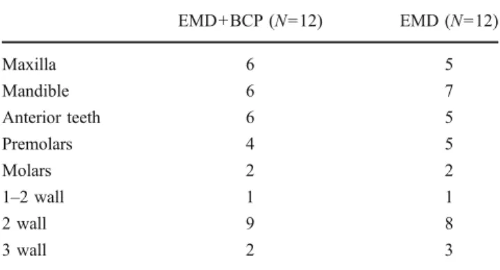
There were no differences in terms of defect distribution and configuration between the two groups (Table2). The baseline defect characteristics are presented in Table3. No statistically significant difference in the initial depth of the intrabony component was found between the two groups.
The PD, GR, and CAL at baseline, at 1 and at 4 years are shown in Table 4. At 1 year, mean PD decreased statistically highly significantly in both groups (P<0.001) (Table 4). Between the groups, no statistically significant difference was found. However, at 4 years, a slight, statistically not significant increase in PD was measured in both groups. At 4 years, the PD was still statistically highly improved compared to baseline (P<0.001) (Table4).
At 1 and 4 years in both groups, mean GR was statistically significantly increased compared to baseline
(P<0.01). No statistically significant differences between the two groups were found at 1 and 4 years (Table4).
Mean CAL demonstrated statistically significant improvements in both groups at 1 and at 4 years compared to the baseline (P < 0.001) (Table 4). No statistically significant differences were observed at 1 and at 4 years within and between the two treatment groups (Table4). The frequency distribution of CAL gain at 1 and 4 years are depicted in Tables5and6. At 1 year, a CAL gain of≥3 mm was measured in 75% of the defects (i.e., in 9 out of 12) in the EMD+BCP group and in 83% of the defects (i.e., in 10 out of 12) in the EMD group. At 4 years, a CAL gain of≥3 mm was measured in 67% of the defects (i.e., in 8 out of 12) in the test group and in 75% of the defects (i.e., in 9 out of 12) in the control group. At 4 years, two defects in the EMD+BCP group and three defects in the EMD group have lost 1 mm of the CAL gained at 1 year.
Discussion
The results of this study have shown that treatment of intrabony defects with both, a combination of EMD+BCP and EMD alone may lead to statistically significant PD reductions and CAL gains which can be maintained over a period of 4 years. However, the statistical analysis has failed to reveal significant differences between the two treatment modalities in any of the investigated clinical parameters at 1 and at 4 years. In both groups, a slight, statistically insignificant, loss of mean CAL was measured between the 1- and 4-year evaluation period. This loss of mean CAL was due to a CAL loss of 1 mm in two defects in the test group and in three defects in the control group. Compared to baseline, at 4 years, a CAL gain of≥3 mm was measured in 67% of the defects (i.e., in 8 out of 12) in the test group and in 75% of the defects (i.e., in 9 out of 12) in the control group.
It should, however, be emphasized that the study does not have the statistical power to rule out the possibility of a difference between the two groups. Further studies, with a higher number of patients and defects would be needed to detect an eventual difference between the treatments [36]. On the other hand, accord-ing to the best of our knowledge, at present, there are no other studies evaluating the clinical performance of a combination of EMD + BCP over a period of 4 years, and therefore, the findings may bear clinical relevance despite
Table 2 Distribution and configuration of treated defects
EMD+BCP (N=12) EMD (N=12) Maxilla 6 5 Mandible 6 7 Anterior teeth 6 5 Premolars 4 5 Molars 2 2 1–2 wall 1 1 2 wall 9 8 3 wall 2 3
Table 3 Baseline defect charac-teristics expressed in millimeters (mean±SD)
No statistically significant differ-ences between the EMD+BCP and EMD groups were found
Treatment PD (mm) CAL (mm) CEJ-BD (mm) CEJ-BC (mm) INTRA (mm)
EMD+BCP (N=12) 8.8±1.5 10.8±1.6 11.8±1.5 6.2±1.4 5.6±1.4
EMD (n=12) 8.8±1.0 10.4±1.3 11.7±1.2 6.0±1.2 5.7±1.3
the relatively small number of patients and defects. Further-more, the present results are in line with findings from previous controlled clinical studies which have failed to show significant differences in the clinical outcomes following regenerative surgery using EMD alone or combined with different alloplastic materials such as bioactive glass, beta tricalcium phosphate, or bone ceramic graft [26,27,31,32]. In a recent multicenter, randomized, controlled clinical study comparing treatment with EMD+BCP (test) to EMD alone (control), the obtained mean CAL gain measured at 1 year after therapy 1.7±2.1 mm in the test and 1.9±1.7 mm in the control group, respectively [32]. These values are somewhat lower than those obtained in the present study where treatment with EMD+BCP yielded a mean CAL gain of 3.4±1.0 mm, whereas the corresponding value in the EMD group was 3.5±0.9 mm. These slight differences may, on one hand, be related to differences in the initial depth of the defects and, on the other, to differences in defect configura-tion. It is well documented that in deeper defects, a greater CAL gain may be achieved [37,38]. In the present study, baseline PD was 8.8±1.5 mm in the test group and 8.8± 1.0 mm in the control group, while in the study referred to,
the corresponding values measured 6.9±1.8 mm and 7.1± 1.5 mm, respectively.
Moreover, when comparing the two studies, it needs to be mentioned that findings from preclinical and clinical studies have provided evidence for superior healing in so-called contained-type defects (i.e., three- and two-walled) compared to “non-contained” (i.e., one-walled or one- and two-walled) ones [39,40]. In the study referred to the great majority of the defects were classified as either one-walled, combined one- and two-walled or circumferential, while in the present study, most of the defects displayed a two-walled configuration [31,32].
Other key factors which have been shown to profoundly affect wound healing following conventional and regener-ative periodontal surgery are infection control and smoking [38, 41]. The pivotal role of careful patient selection and strict maintenance program is further supported by the present findings where the patient population did not include any smokers, while in both groups, the plaque and bleeding values remained unchanged throughout the entire observation period of 4 years.
Table 4 Clinical parameters at baseline at 1 and at 4 years
No statistically significant differences between the EMD+BCP and EMD groups were found
Parameter Treatment Baseline 1 Year P value
(baseline—1 year) 4 Years P value (1–4 years)
PD EMD+BCP (n=12) Mean (±SD) 8.8±1.5 4.3±0.9 <0.001 4.7±0.8 n.s. EMD (n=12) Mean (±SD) 8.8±1.0 4.1±0.5 <0.001 4.4±0.8 n.s. GR EMD+BCP (n=12) Mean (±SD) 2.1±1.0 3.2±1.1 <0.01 3.0±1.1 n.s. EMD (n=12) Mean (±SD) 1.6±1.0 2.9±0.9 <0.01 2.8±0.8 n.s. CAL EMD+BCP (n=12) Mean (±SD) 10.8±1.6 7.4±1.6 <0.001 7.6±1.7 n.s. EMD (n=12) Mean (±SD) 10.4±1.3 6.9±1.0 <0.001 7.2±1.2 n.s.
Table 6 Frequency distribution of CAL gains at 4 years in the EMD + BCP and EMD groups
CAL gain (mm) EMD+BCP (n=12) EMD (n=12)
No. Percent (%) No. Percent (%)
1 1 8
2 3 25 3 25
3 3 25 2 17
4 4 34 7 58
5 1 8
Table 5 Frequency distribution of CAL gains at 1 year in the EMD + BCP and EMD groups
CAL gain (mm) EMD+BCP (n=12) EMD (n=12)
No. Percent (%) No. Percent (%)
2 3 25 2 17
3 3 25 3 25
4 4 33 6 50
A recent human histologic study evaluating the healing of intrabony defects treated with EMD+BCP revealed formation of new cementum with an associated periodontal ligament in six out of the nine biopsies, while formation of new bone or of a mineralized bone-like tissue around the graft particles was only occasionally observed. Further-more, in most specimens, the graft particles were still present at 9 months following surgery without signs of resorption or bone formation [42]. These observations appear to suggest that adding BCP to EMD may not additionally improve the healing modulated by EMD alone, thus corroborating the present and previously reported findings [31,32].
When interpreting the present results, it should be pointed to the results from controlled clinical studies comparing various combination protocols of EMD with AB, ABBM, or DFDBA to EMD alone and have shown higher clinical improvements following the combination approach [17,18,22–24]. On the other hand, in most other studies where EMD was combined with different alloplastic grafts, no additional benefits were detected [25–27,31,32]. Thus, all these findings appear to suggest that the grafting material itself may also influence the healing process and, subsequently, the clinical outcomes. However, further research is necessary to elucidate the exact biological mechanisms behind these combinations.
Taken together, our results indicate that: (a) the clinical improvements obtained with both treatments can be main-tained over a period of 4 years, and (b) in two- and three-wall intrabony defects, the addition of BCP did not additionally improve the outcomes obtained with EMD alone.
Source of funding The study was funded by the author's own
institution.
Conflict of interest The authors declare that they have no conflict of
interests.
References
1. Wikesjö UME, Selvig KA (1999) Periodontal wound healing and
regeneration. Periodontol 2000 19:21–39
2. Hammarström L, Heijl L, Gestrelius S (1997) Periodontal regeneration in a buccal dehiscence model in monkeys after application of enamel matrix proteins. J Clin Periodontol 24:669– 677
3. Sculean A, Donos N, Brecx M, Reich E, Karring T (2000) Treatment of intrabony defects with enamel matrix proteins and guided tissue regeneration. An experimental study in monkeys. J
Clin Periodontol 27:466–472
4. Gkranias ND, Graziani F, Sculean A, Donos N (2010) Wound healing following regenerative procedures in furcation degree III defects: histomorphometric outcomes. Clin Oral Investig. Oct 22 (in press)
5. Heijl L (1997) Periodontal regeneration with enamel matrix derivative in one human experimental defect. A case report. J Clin Periodontol 24:693–696
6. Sculean A, Donos N, Windisch P, Gera I, Brecx M, Reich E, Karring T (1999) Healing of human intrabony defects following treatment with enamel matrix proteins or guided tissue
regenera-tion. J Periodontal Res 34:310–322
7. Sculean A, Chiantella GC, Windisch P, Donos N (2000) Clinical and histologic evaluation of treatment of intrabony defects with an enamel matrix protein derivative (Emdogain®). Int J Periodontics
Restorative Dent 20:375–381
8. Yukna RA, Mellonig J (2000) Histologic evaluation of periodontal healing in humans following regenerative therapy with enamel
matrix derivative. A 10-case series. J Periodontol 71:752–759
9. Sculean A, Stavropoulos A, Berakdar M, Windisch P, Karring T, Brecx M (2005) Formation of human cementum following different modalities of regenerative therapy. Clin Oral Investig 9:58–64
10. Bosshardt DD, Sculean A, Windisch P, Pietursson BE, Lang NP (2005) Effects of enamel matrix proteins on tissue formation along the roots of human teeth. J Periodontal Res 40:158–167 11. Mazjoub Z, Bobbo M, Atiyeh F, Cordioli G (2005) Two patterns
of histologic healing in an intrabony defect following treatment with an enamel matrix derivative: a human case report. Int J
Periodontics Restorative Dent 25:283–294
12. Heijl L, Heden G, Svardström G, Östgren A (1997) Enamel matrix derivative (Emdogain®) in the treatment of intrabony
periodontal defects. J Clin Periodontol 24:705–714
13. Pontoriero R, Wennström J, Lindhe J (1999) The use of barrier membranes and enamel matrix proteins in the treatment of angular bone defects. A prospective controlled clinical study. J Clin
Periodontol 26:833–840
14. Froum SJ, Weinberg MA, Rosenberg E, Tarnow D (2001) A comparative study utilizing open flap debridement with and without enamel matrix derivative in the treatment of periodontal intrabony defects: a 12-month re-entry study. J Periodontol 72:25– 34
15. Sculean A, Windisch P, Chiantella GC, Donos N, Brecx M, Reich E (2001) Treatment of intrabony defects with enamel matrix proteins and guided tissue regeneration. A prospective controlled
clinical study. J Clin Periodontol 28:397–403
16. Tonetti MS, Lang NP, Cortellini P, Suvan JE, Adriaens P, Dubravec D, Fonzar A, Fourmousis I, Mayfield L, Rossi R, Silvestri M, Tiedemann C, Topoll H, Vangsted T, Wallkamm B (2002) Enamel matrix proteins in the regenerative therapy of deep intrabony defects. A multicentre randomized controlled clinical trial. J Clin
Periodontol 29:317–325
17. Guida L, Annunziata M, Belardo S, Farina R, Scabbia A, Trombelli L (2007) Effect of autogenous cortical bone particulate in conjunction with enamel matrix derivative in the treatment of
periodontal intraosseous defects. J Periodontol 78:231–238
18. Gurinsky BS, Mills MP, Mellonig JT (2004) Clinical evaluation of demineralized freeze-dried bone allograft and enamel matrix derivative versus enamel matrix derivative alone for the treatment of periodontal osseous defects in humans. J Periodontol 75:1309– 1318
19. Aspriello SD, Ferrante L, Rubini C, Piemontese M (2011) Comparative study of DFDBA in combination with enamel matrix derivative versus DFDBA alone for treatment of periodon-tal intrabony defects at 12 months post-surgery. Clin Oral Invest
20. Yamamoto S, Masuda H, Shibukawa Y, Yamada S (2007) Combination of bovine-derived xenografts and enamel matrix derivative in the treatment of intrabony periodontal defects in
dogs. Int J Periodontics Restorative Dent 27:471–479
21. Sculean A, Windisch P, Keglevich T, Chiantella GC, Gera I, Donos N (2003) Clinical and histologic evaluation of human intrabony defects treated with an enamel matrix protein derivative combined with a bovine-derived xenograft. Int J Periodontics
Restorative Dent 23:47–55
22. Lekovic V, Camargo PM, Weinlaender M, Nedic M, Aleksic Z, Kenney EB (2000) A comparison between enamel matrix proteins used alone or in combination with bovine porous bone mineral in the treatment of intrabony periodontal defects in humans. J
Periodontol 71:1110–1116
23. Velasquez-Plata D, Scheyer ET, Mellonig JT (2002) Clinical comparison of an enamel matrix derivative used alone or in combination with a bovine-derived xenograft for the treatment of
periodontal osseous defects in humans. J Periodontol 73:433–440
24. Zucchelli G, Amore C, Montebugnoli L, De Sanctis M (2003) Enamel matrix proteins and bovine porous mineral in the treatment of intrabony defects: a comparative controlled clinical trial. J Periodontol 74:1725–1735
25. Kuru B, Yilmaz S, Argin K, Noyan U (2006) Enamel matrix derivative alone or in combination with a bioactive glass in wide intrabony defects. Clin Oral Invest 10:227–234
26. Sculean A, Pietruska M, Arweiler NB, Auschill TM, Nemcovsky C (2007) Four year results of a prospective controlled clinical study evaluating healing of intrabony defects following treatment with an enamel matrix protein derivative alone or combined with a
bioactive glass. J Clin Periodontol 34:507–513
27. Bokan I, Bill JS, Schlagenhauf U (2006) Primary flap closure combined with Emdogain® alone or Emdogain® and Cerasorb®
in the treatment of intra-bony defects. J Clin Periodontol 33:885–
893
28. Nery EB, LeGeros RZ, Lynch KL, Lee K (1992) Tissue response to biphasic calcium phosphate ceramic with different ratios of HA/ βTCP in periodontal osseous defects. J Periodontol 63:729–735 29. Jensen SS, Yeo A, Dard M, Hunziker E, Schenk R, Buser D
(2007) Evaluation of a novel biphasic calcium phosphate in standardized bone defects. A histologic and histomorphometric study in the mandibles of minipigs. Clin Oral Implants Res 18:752–760
30. Stein JM, Fickl S, Yekta SS, Hoischen U, Ocklenburg C, Smeets R (2009) Clinical evaluation of a biphasic calcium composite grafting material in the treatment of human periodontal intrabony defects: a 12-month randomized controlled clinical trial. J
Periodontol 80:1774–1782
31. Jepsen S, Topoll H, Rengers H, Heinz B, Teich M, Hoffmann T, Al-Machot E, Meyle J, Jervøe-Storm PM (2008) Clinical out-comes after treatment of intra-bony defects with an EMD/ synthetic bone graft or EMD alone: a multicentre randomized controlled clinical trial. J Clin Periodontol 35:420–428
32. Meyle J, Hoffmann T, Topoll H, Heinz B, Al-Machot E, Jervøe-Storm P-M, Meiß C, Eickholz P, Jepsen S (2011) A multi-centre randomized controlled clinical trial on the treatment of intra-bony defects with enamel matrix derivatives/synthetic bone graft or enamel matrix
derivatives alone: results after 12 months. J Clin Periodontol 38:652–
660
33. Armitage GC (1999) Development of a classification system for
periodontal diseases and conditions. Ann Periodontol 4:1–6
34. Löe H (1967) The gingival index, the plaque index and the
retention index systems. J Periodontol 38:610–616
35. Blomlöf JPS, Blomlöf LB, Lindskog SF (1996) Smear removal and collagen exposure after non-surgical root planing followed by
etching with EDTA gel preparation. J Periodontol 67:841–845
36. Gunsolley JC, Elswick RK, Davenport JM (1998) Equivalence and superiority testing in regeneration clinical trials. J Periodontol 69:521–527
37. Kahldahl WB, Kalkwarf KL, Patil KD, Molvar MP, Dyer JK (1996) Long-term evalution of periodontal therapy: I. Respone to 4 therapeutic modalities. J Periodontol 67:93–102
38. Tonetti MS, Pini Prato G, Cortellini P (1996) Factors affecting the healing response of intrabony defects following guided tissue
regeneration and access flap surgery. J Clin Periodontol 23:548–
556
39. Sculean A, Nikolidakis D, Schwarz F (2008) Regeneration of periodontal tissues: combinations of barrier membranes and
grafting materials—biological foundation and preclinical
evi-dence. A systematic review. J Clin Periodontol 35(suppl 8):106–
116
40. Iorio-Siciliano V, Andreuccetti G, Siciliano AI, Blasi A, Sculean A, Salvi GE (2011) Clinical outcome following treatment of non-contained intrabony defects with enamel matrix derivative (EMD) or guided tissue regeneration (GTR). A 12-month randomized
controlled clinical trial. J Periodontol 82:62–71
41. Tonetti MS, Pini-Prato G, Cortellini P (1995) Effect of cigarette smoking on periodontal healing following GTR in infrabony defects. A preliminary retrospective study. J Clin Periodontol 22:229–234
42. Sculean A, Windisch P, Szendröi-Kiss D, Horváth A, Rosta P, Becker J, Gera I, Schwarz F (2008) Clinical and histologic evaluation of an enamel matrix derivative combined with a biphasic calcium phosphate for the treatment of human intrabony