Comparative morphology of female flowers and systematics
in Geonomeae (Arecaceae)
F. W. Stauffer and P. K. Endress
Institute of Systematic Botany, University of Zurich, Zurich Switzerland
Received April 29, 2003; accepted May 2, 2003 Published online: November 6, 2003
Ó Springer-Verlag 2003
Abstract. Female floral structure is compared in Geonomeae (Arecaceae). A perianth is formed by two alternate whorls of three basally congenitally united and imbricate sepals and three basally congenitally united and apically valvate petals. A sterile androecium is formed by a variable number of staminodes, which are united into a tube. The gynoecium shows three more or less equally devel-oped carpels or is pseudomonomerous (Geonoma). The single anatropous ovule per carpel is median, either basal or at mid-height of the ovary. A septal nectary is present at the base and mid-height of the ovaries and exits at different levels of the ovary. Carpels in pseudomonomerous gynoecia seem to be ‘‘basistylous’’, but the styles are more lateral or apical in gynoecia with all three carpels equally developed. Stigmas expose unicellular or multicel-lular (Welfia) papillae at anthesis. Pollen tube transmitting tracts and a compitum are present in the ventral slits of the postgenitally united styles. Floral structure in Geonomeae is compared with other Arecaceae, especially Arecoideae, in a mor-phological and systematic context.
Key words: Arecaceae, Geonomeae, female floral structure, staminodia, morphology, systematics.
Introduction
Geonomeae (Arecoideae, Arecaceae) contain sixgenera:Pholidostachys,Welfia,Calyptronoma, Calyptrogyne, Asterogyne and Geonoma (Uhl and Dransfield 1987, Dransfield and Uhl 1998) and up to 70 species (Henderson et al. 1995). They are almost exclusively neotropical, with a distribution from south eastern Mexico through Central America, the Antilles, and the tropics of South America, south to Bolivia and Paraguay (Wessels Boer 1968). Geonomeae are mostly small to medium-sized understory palms, dom-inant and rich in species from the lowland rain forest up to the high elevation cloud forest. Only the monotypic Welfia grows in the sub-canopy. Flowers deeply sunken in pits, floral parts of all whorls united, at least at the base, and elongate and slender styles are in general considered to be the main shared characters within Geonomeae (Uhl and Dransfield 1987).
Geonomeae were found to be monophy-letic by Asmussen (1999a, 1999b) based on plastid rps16 DNA sequences, by Hahn (2002) based on atpB, rbcL, and 18S nrDNA
Plant Syst. Evol. 242: 171–203 (2003) DOI 10.1007/s00606-003-0030-1
with Euterpeinae warrants further investiga-tion, and may eventually suggest the reduction of Geonomeae to a subtribe within Areceae.
With the circumscription of Geonomeae, Drude (1881, 1889) described several aspects of the reproductive structures and emphasized the morphological diversity displayed by the sta-mens and staminodes. Floral morphology in the tribe has also been preliminarily described by Burret (1930), Wessels Boer (1968) and Uhl and Dransfield (1987). Detailed studies on the reproductive structures were carried out in less than eight species of Geonomeae, restricted to Asterogyne (Uhl 1966, Schmid 1970a, Stauffer et al. in press) and Geonoma (Gassner 1941, Uhl and Moore 1971). Floral development was only
(Wessels Boer 1968) their characteristics have largely been neglected in infratribal classifica-tions and generic treatments (e.g. Drude 1889, Burret 1930, Wessels Boer 1968, Moore 1973, Uhl and Dransfield 1987, Zona 1995). The aim of this study is to answer the following ques-tions: 1) What are the structural features in the female flowers in Geonomeae? 2) Do these features support recent molecular phylogenetic analyses? and 3) How does the floral structure of Geonomeae compare to other Arecoideae?
Material and methods
Female flowers of 15 species in six genera of Geonomeae (Table 1) were obtained from the
Table 1. List of material studied (species, collection, origin, stage of development)
Pholidostachys pulchraH. Wendl. ex Burret, J. Knudsen & C. Asmussen (655-AAU), Costa Rica, young buds; H. E. Moore & M. V. Parthasarathy (9410-BH), Costa Rica, anthetic flowers.
P. synanthera(Mart.) H. E. Moore, H. Balslev et al. (60678-AAU), Ecuador, anthetic flowers; R. Bernal (2588-AAU), Colombia, young buds.
Welfia regiaH. Wendl. ex Andre´, H. E. Moore & M. V. Parthasarathy (9412-BH), Costa Rica, old buds. Calyptronoma rivalis(O.F. Cook) L.H. Bailey, S. Zona (864-FTG), Puerto Rico, anthetic and pre-anthetic
flowers; S. Zona (81-292-FTG), Puerto Rico, young buds.
Calyptrogyne costatifrons(L.H. Bailey) Nevers, J. Knudsen & C. Asmussen (610-AAU), Panama, anthetic flowers.
C. ghiesbreghtiana.(Linden & H. Wendl.) H. Wendl., J. Knudsen & C. Asmussen (628-AAU), Panama, old buds; Murray (01-BH), Central America, old buds.
Asterogyne martiana(H. Wendl.) H. Wendl. ex Hemsl., J. Knudsen (00:65-private collection), Costa Rica, young buds; J. Knudsen & C. Asmussen (604-AAU), Panama, anthetic flowers.
A. ramosa(H.E. Moore) Wess. Boer, F. W. Stauffer (824-Z), Venezuela, old buds. A. spicata(H.E. Moore) Wess. Boer, F. W. Stauffer (822-Z), Venezuela, old buds. A. yaracuyenseHenderson and Steyerm., F. W. Stauffer (823-Z), Venezuela, old buds. Geonoma cuneataH. Wendl. ex Spruce, C. Listabarth (40-Z), Peru, old buds.
G. interrupta(Ruiz & Pav.) Mart., F. W. Stauffer (15-Z), Venezuela, young and old buds, anthetic flowers. G. macrostachysMart., C. Listabarth (s.n.-W), Peru, young buds.
G. maxima(Poit.) Kunth, J. Knudsen (98:118-AAU), Ecuador, young buds.
pickled collections deposited at AAU, BH, K and collections made by Christian Listabarth (Austrian Academy of Science), Jette Knudsen (University of Go¨teborg) and Scott Zona (Fairchild Tropical Garden). Flowers were also removed from herbar-ium specimens deposited at AAU and US for anatomical study. Reproductive structures of Geo-noma interrupta, G. simplicifrons and three species of Asterogyne were collected by the first author in Venezuela between 1999 and 2002. In order to evaluate structural diversity within Geonoma, which contains up to 55 species (Henderson et al. 1995), a preliminary screen based on 26 taxa (Appendix 1) was performed.
For anatomical investigations, flowers in late bud or at anthesis, fixed in FAA or ethanol (70%), were evacuated, dehydrated, and embedded in Kul-zer’s Technovit 7100 (2-hydroxyethyl methacrylate [HEMA]). Perianth and sometimes the sterile androe-cium were removed to facilitate infiltration. Further details on this technique are explained in Igersheim and Cichocki (1996). The material was transversally and longitudinally sectioned at six-seven lm using a rotary microtome (Microm HM-355, Microm La-borgera¨te GmbH, Walldorf, Germany), and then stained with ruthenium red and toluidine blue, and enclosed in Histomount (ä). For scanning electron microscopy, the dissected specimens were dehy-drated, critical-point dried, and sputter-coated with gold. All the permanent slides of the microtome sections are deposited at the Institute of Systematic Botany of the University of Zurich (Z). Arrangement of the genera follows Uhl and Dransfield (1987).
Results
Shared characters of the female flowers of Geonomeae. As in all Arecoideae (Uhl and Dransfield 1987), a female flower and two male flowers form a floral triad arranged in a cincinnus. Such floral triads in Geonomeae are sunken in pits of the inflorescence rachillae. Triads are protandrous. Anthesis of the sta-minate flowers occurs sequentially, or less commonly both flower at the same time (e.g. Pholidostachys dactyloides, Henderson 1986), but before the pistillate flower of the same triad is receptive. Subsequent development of the pistillate flower pushes the post-anthetic staminate flowers out of the pit. Perianth,
sterile androecium and gynoecium show some features that are shared by all species studied. The perianth is formed by two alternate whorls; the outer whorl has three basally congenitally united, apically imbricate sepals, and the inner whorl three basally congenitally united, apically valvate petals. Tanniferous tissue and cells with oxalate raphides are present in the sepals and petals, and are especially concentrated in the petal flanks. The perianth seems to be quite uniform in all taxa studied.
Staminodes are at least basally congenitally united into a tube and also have a common base with petals and carpels (Fig. 1). Tanni-ferous tissue and cells with oxalate raphides are present in the mesophyll of the staminodes. The three carpels alternate with the petals. The stigmatic branches are erect and more or less connivent in bud, but spreading at anthe-sis. Secretory papillae cover the entire ventral surface of the stigmatic branches and are exposed during anthesis. The ventral slits extend from the top of the ovary, at the base of the plicate zone, up to the base of the styles; they are papillate, secretory and open. The carpels are congenitally united at the base, free at mid-height of the ovary, or slightly above (Calyptrogyne), but postgenitally united up to mid-height of the styles. Two carpels are adaxial and one carpel is abaxial. The terms adaxial and abaxial are here applied in a purely topographical and not in a comparative morphological sense, and they are related with the proximity to the main axis of the inflores-cence rachilla. The gynoecium is synascidiate up to mid-height of the ovaries. Each ovary, if well developed, bears a crassinucellate, ana-tropous ovule, which has a median position (Fig. 1). In flowers at anthesis or shortly before, the presence of two integuments could not be ascertained (but see younger ovules in Geonoma maxima). The micropyle faces the ventral side of the locule (Fig. 1). A septal nectary is formed by incomplete union of the carpel flanks. The nectary is a triradiate cavity between the three carpels at mid-height of the ovaries and extends upwards and gradually
outwards to the apical region of the ovaries. It is formed by an epithelium of columnar, uninucleate cells. The openings of the nectary are non-secretory ducts with isodiametric epi-dermal cells. A pollen tube transmitting tract (PTTT) encompasses the epidermis of the ventral slits of the stigmas and styles. The individual tracts coming from the stigmatic branches join in a compitum at the level of postgenital union of the styles down to the
level of the placenta. The PTTT reaches the micropyle by surrounding both flanks of the funiculus.
The dorsal bundle of each carpel extends up to the top of the stigmatic branches. In the synascidiate zone a complex ventral bundle is present in each carpel. A branch derived from this ventral bundle, in the upper synascidiate region, serves the ovule. Tanniferous tissue is present throughout the gynoecium, but is
Fig. 1. Schematic LS of female flowers in Geonomeae. Arrows point to the different levels of separation of the corolla from the sterile androecium. Floral buds (Geonoma, Pholidostachys, Asterogyne and Welfia), flowers shortly before anthesis (Calyptrogyne costatifrons, C. ghiesbreghtiana and Calyptronoma)
especially concentrated towards the apex of the stigmatic branches.
Pholidostachys
The staminodial tube separates from the corolla above the base of the flower (Fig. 1). The upper part of the androecium has six (Pholidostachys synanthera) (Fig. 21) or seven (P. pulchra) (Figs. 2, 14) free parts (stamino-dial lobes, according to the terminology of Uhl and Moore 1971, Uhl and Dransfield 1987), which are connivent in bud (Fig. 22), and spreading at anthesis (Figs. 14, 21). The stam-inodes become free first from the inner surface of the tube (Figs. 3, 4) and then also from the outer surface. Each staminode has a vascular bundle near the inner surface (Fig. 6), which forms a vascular trace at the base of the flower. Tanniferous tissue is abundant in the free part of the staminodes and upper tube. Cells with oxalate raphides are present at the base
(P. pulchra) and upper part of the staminodial tube, and at the base of the free parts of the staminodes.
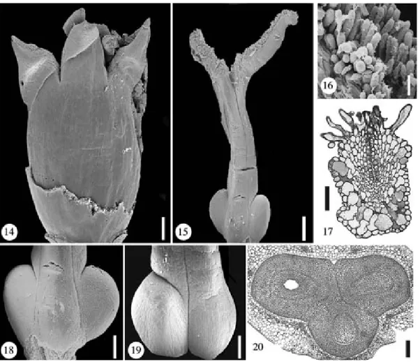
At the base the carpels are congenitally united (Figs. 8, 9) and become free from the staminodial tube first in their median areas (Fig. 12). The two adaxial ovaries are more or less the same size (Fig. 19), or one of them is larger than the other (P. pulchra). The abaxial ovary is always smaller (Figs. 7, 18, 23) All carpels have a well-developed ovule. The stigmatic branches may be equal (P. synan-thera) (Fig. 25) or unequal (P. pulchra) (Fig. 15) in length. If unequal, the branch of the abaxial carpel is always shorter. At anthe-sis the stigmatic branches are at the same level as the free parts of the staminodes. The stigmatic branches resemble the free part of the staminodes in size and shape (Fig. 21). Unicellular papillae (Fig. 17) cover the ventral surface of the stigmatic branches (Fig. 16). The styles appear to be lateral in all carpels, or
Figs. 2–13. Pholidostachys pulchra. Floral bud, TS series. Fig. 2. Level of free carpels (stigmas) and free staminodes. Figs. 3–5. Apocarpous styles; carpel flanks postgenitally united (dotted lines); staminodes congenitally united. Figs. 6–7. Apocarpous zone of ovaries above level of placentae; carpel flanks partially postgenitally united (dotted lines); zone with septal nectary. Figs. 8–11. Synascidiate zone at level of placentae and below; lower zone of septal nectary in 8. Figs. 12–13. Floral base, androecium and gynoecium are partially congenitally fused. Scale bar¼ 200 lm
more or less apical in the abaxial carpel (P. pulchra). At the base of the styles the dorsal surface is smooth (P. pulchra) (Fig. 15) or it has conspicuous protrusions (P. synanthera) (Fig. 24), which contain tissue with large and highly vacuolated cells (Fig. 26). The carpels
begin to separate from each other either from the periphery or the center of the syncarpous zone (Fig. 8). At the base of the plicate zone the contiguous surfaces of adjacent carpels are papillate (P. pulchra). A central protrusion is present between the three carpels (P.
synan-Figs. 14–20. Pholidostachys pulchra. Fig. 14. Flower at anthesis (perianth removed). Scale bar¼ 500 lm. Fig. 15. Gynoecium at anthesis. Scale bar¼ 500 lm. Fig. 16. Part of stigma. Scale bar ¼ 100 lm. Fig. 17. TS of stigma showing unicellular papillae and tanniferous mesophyll. Scale bar¼ 50 lm. Fig. 18. Abaxial view of lower gynoecium, note the reduced size of the abaxial ovary. Scale bar¼ 400 lm. Fig. 19. Adaxial view of lower gynoecium, note that both adaxial ovaries have the same size and shape. Scale bar¼ 400 lm. Fig. 20. TS of gynoecium at the level of the ovaries. Scale bar¼ 200 lm
Figs. 21–28. Pholidostachys synanthera. Fig. 21. Flower at anthesis. Scale bar¼ 500 lm. Fig. 22. Flower in late bud (perianth removed); the free staminodes are connivent. Scale bar¼ 500 lm. Fig. 23. Gynoecium in late bud. Scale bar¼ 500 lm. Fig. 24. Gynoecium at anthesis; the styles show irregular protrusions towards the base. Scale bar¼ 500 lm. Fig. 25. Stigmas at anthesis showing receptive papillae. Scale bar ¼ 500 lm. Fig. 26. TS of postgenitally united styles (towards the base); the irregular protrusions are formed by tanniferous parenchyma. Scale bar¼ 200 lm. Fig. 27. TS of ovaries (transition from synascidiate to apocarpous). Scale bar¼ 200 lm. Fig. 28. TS of ovaries showing central protrusion between the three carpels. Scale bar ¼ 100 lm
thera) (Fig. 28), or absent (P. pulchra) (Fig. 7); it extends slightly above mid-height of the ovaries, and is secretory. The ovule is attached at mid-height of the ovary (Fig. 1). The septal nectary forms simple slits between the carpels (Fig. 7) at the base of the apocarpous part of the gynoecium and becomes a triradiate cavity at mid-height of the ovaries (Fig. 7). The openings of the nectary are towards the top of the ovaries.
Four to six lateral procambial strands on each side of the carpels extend from the top to mid-height of the ovary and merge into three to four lateral bundles towards the base of the ovary; they are small and undifferentiated at anthesis (Figs. 2–13). Some of the lateral bundles connect with the dorsal bundle at about the top of the ovary (P. pulchra), or with the ventral bundles either near the base (P. synanthera) or near the top (P. pulchra) of the ovary. Between ten (P. pulchra) and 13 (P. syn-anthera) large vascular traces from the gynoe-cium, more or less equal in shape, rearrange into a ring-shaped stele at the base of the flower. Tanniferous tissue is common towards the apex of the stigmas, particularly concen-trated in the dorsal hypodermis, and also in the mesophyll of the styles, normally towards the dorsal surface, and throughout the mesophyll of the ovaries. Cells with oxalate raphides are concentrated in the mesophyll of the bulged upper part of the ovaries.
Welfia
The staminodial tube separates from the corolla in the upper half of the corolla, near the base of the free parts of the staminodes (Fig. 1). The 22 staminodes (15–16 according to Uhl and Dransfield 1987) are more or less connivent in bud (Fig. 38) and are spreading at anthesis. Tanniferous tissue is present through-out the mesophyll of the staminodes, including the tube and the free parts, particularly surrounding the vascular bundles. A few cells with oxalate raphides are present in the mesophyll of the staminodes (Fig. 45).
One of the two adaxial ovaries is the largest (Fig. 44) one and the abaxial ovary is the smallest (Fig. 43). All carpels have a well-developed ovule (Fig. 35). The ovaries are partially fused with the staminodial tube in the median areas of the carpels. The stigmatic branches are more or less equal in length (Figs. 39, 40). They have unicellular or multi-cellular papillae on their ventral surface (Figs. 41, 42, 46). The styles appear to be lateral in the two adaxial carpels and apical in the abaxial carpel. The carpels begin to sepa-rate from each other either from the periphery or the center of the syncarpous zone (Fig. 34). No central protrusion is present between the carpels. The ovule is attached at mid-height of the ovary (Fig. 1). The nectary forms a simple slit between the abaxial carpel and one of the
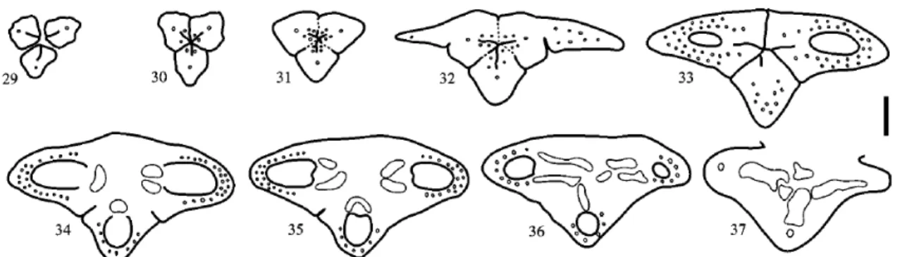
Figs. 29–37. Welfia regia. Floral bud, TS series. Fig. 29. Level of free carpels (stigmas). Figs. 30–32. Apocarpous styles; carpel flanks postgenitally united (dotted lines). Fig. 33. Apocarpous zone of ovaries above level of placentae; zone with septal nectary. Figs. 34–36. Synascidiate zone at level of placentae and below. Fig. 37. Floral base. Scale bar¼ 200 lm
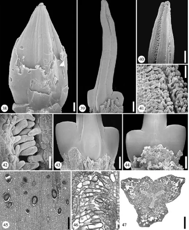
Figs. 38–47. Welfia regia. Fig. 38. Flower in late bud (calyx and upper part of corolla removed); the free staminodes are tightly connivent. Scale bar¼ 500 lm. Fig. 39. Gynoecium in late bud. Scale bar ¼ 500 lm. Fig. 40. Stigmas in late bud. Scale bar¼ 400 lm. Fig. 41. Detail of stigmas in late bud showing large papillae. Scale bar¼ 100 lm. Fig. 42. Stigmatic papillae. Scale bar ¼ 100 lm. Fig. 43. Abaxial view of lower gynoecium, note the reduced size of the abaxial ovary. Scale bar¼ 1 mm. Fig. 44. Adaxial view of lower gynoecium. Scale bar¼ 1 mm. Fig. 45. LS of the sterile androecium (detail) showing raphide idioblasts. Scale bar ¼ 150 lm. Fig. 46. LS of stigmas in late bud showing multicellular papillae. Scale bar¼ 50 lm. Fig. 47. TS of postgenitally united styles with tanniferous parenchyma. Scale bar¼ 200 lm
adaxial carpels at mid-height of the ovaries, and becomes a triradiate cavity towards the top (Fig. 33). The three openings of the nectary are towards the top of the ovaries.
Between two and three lateral bundles on each side of the ventral slit extend from the base of the stigmatic branches to the top of the ovary (Figs. 30, 32). In addition, at anthesis a double series of 10–12 procambial strands on each side of the carpel extend from the upper part of the ovary to mid-height of the ovary and merge into one series of three to four small and undifferentiated lateral bundles towards the base of the ovary (Figs. 32, 35). Some of the lateral bundles connect with the ventral bundle towards the base of the ovary. Two large branches derived from the ventral bundle complex in the upper synascidiate region serve the ovule. Up to 13 large vascular traces from
the gynoecium, more or less equal in shape, rearrange into a ring-shaped stele at the base of the flower. Tanniferous tissue is dominant in the mesophyll of the stigmas, at the base and mid-height of the styles (Fig. 47), and is surrounding the openings of the septal nectary. Cells with oxalate raphides are present at the base and mid-height of the styles and towards the top of the ovaries.
Calyptronoma
The staminodial tube separates from the corolla above the base of the flower (Figs. 1, 54). Towards the apex the tube becomes a thick, swollen, calyptrate structure that falls shortly before anthesis (Figs. 48, 61). At the apex of the calyptra six short projections, representing the non-united tips of the six
Figs. 48–60. Calyptronoma rivalis. Floral bud, TS series. Fig. 48. Level of free carpels (stigmas) and congenitally united bulged out staminodes. Figs. 49–52. Apocarpous styles; carpel flanks postgenitally united (dotted lines); staminodes congenitally united and separating from the corolla. Figs. 53–54. Apocarpous zone of ovaries above level of placentae; zone with septal nectary and central protrusion between carpels. Figs. 55–58. Synascidiate zone at level of placentae and below. Figs. 59–60. Lower synascidiate zone and floral base; calyx, corolla, androecium and gynoecium are congenitally fused; interrupted lines: part not complete. Scale bar¼ 200 lm
staminodes, are present. The staminodial tube contains a parenchymatous mesophyll, but at the level of the abscission zone of the calyptra, the tube is compressed, with col-lapsed tissue. The calyptra is formed by thickening of the abaxial region of the staminodial tube (Figs. 48, 49). Each stamin-ode is served by one vascular strand (Figs. 48–57). Tanniferous tissue surrounds
these vascular bundles and is especially con-centrated around the apical opening of the calyptrate region of the staminodial tube. A few cells with oxalate raphides are present throughout the calyptrate region.
At the base the carpels are congenitally united and, particularly the abaxial carpel, partially fused with the staminodial tube (Fig. 58). The three ovaries are more or less
Figs. 61–66. Calyptronoma rivalis. Fig. 61. Pre-anthetic flower showing the calyptrate upper part of the sterile androecium. Scale bar¼ 500 lm. Fig. 62. Gynoecium in late bud. Scale bar ¼ 500 lm. Fig. 63. Stigmas, showing the large abaxial branch. Scale bar¼ 200 lm. Fig. 64. Abaxial view of ovaries, note the reduced size of the abaxial ovary. Scale bar¼ 400 lm. Fig. 65. Adaxial view of ovaries. Scale bar ¼ 200 lm. Fig. 66. TS of ovaries (transition between apocarpous and synascidiate zone); corolla and sterile androecium are congenitally fused. Scale bar¼ 200 lm
equal in size (Figs. 55–57), and all have a well-developed ovule. The stigmatic branches are either the same size or the branch of the abaxial carpel is larger than the stigmatic branches of the adaxial carpels (Fig. 63). Unicellular papillae cover the ventral surface of the stigmatic branches. The stigmatic branches begin to spread while still in the calyptra and they spread fully once the calyptra has fallen. The styles appear to be lateral (Figs. 62, 64, 65). The carpels begin to separate from each other mainly from the center to the periphery of the syncarpous zone. No central protrusion is present between them. The ovule is attached at mid-height of the ovary (Fig. 1). The funi-culus shows a papillate surface on the side
that is turned away from the micropyle. The nectary forms a simple slit between the two adaxial carpels at mid-height of the ovaries (Fig. 66), but three slits farther up. The openings of the nectary are towards the top of the ovaries.
Up to four lateral procambial strands on each side of the carpel extend from the top to mid-height of the ovary and merge into two to three lateral bundles towards the base of the ovary. Some of these lateral strands connect with the ventral bundles at the base of the ovary (Figs. 48–59). Up to six large vascular traces from the gynoecium, more or less equal in shape, rearrange into a ring-shaped stele at the base of the flower. A few tanniferous cells surround the vascular
Figs. 67–77. Calyptrogyne costatifrons. Floral bud, TS series. Fig. 67. Level of free carpels (stigmas) and congenitally united bulged-out staminodes. Figs. 68–69. Apocarpous styles; carpel flanks postgenitally united (dotted lines). Figs. 70–72. Apocarpous zone of ovaries above level of placentae (postgenital union of carpels shown with dotted lines); zone with septal nectary. Figs. 73–75. Synascidiate zone at level of placentae and below. Figs. 76–77. Floral base; corolla, androecium and gynoecium are congenitally fused. Scale bar¼ 200 lm
bundles at the apex of the stigmatic branches, the styles, and throughout the ovaries (Fig. 66). Cells with oxalate raphides are lacking in the gynoecium.
Calyptrogyne
The staminodial tube separates from the corolla at the base of the flower (Calyptrogyne costatifrons) (Figs. 1, 75) or slightly above it (C. ghiesbreghtiana) (Figs. 1, 87). Six slits from the inner side are present at mid-height of the tube (C. ghiesbreghtiana) (Fig. 99). Towards the apex the tube forms a thick, swollen and calyptrate part (Figs. 67, 78, 82, 95), which falls off shortly before anthesis (Fig. 79). On top of the calyptra six short projections, representing the non-united tips of the six staminodes, are present (Figs. 78, 95). The
abscission zone at the base of the calyptra, particularly towards the abaxial region, is formed by compressed tissue. Irregular pro-trusions in the inner surface of the staminodial tube are present in C. ghiesbreghtiana (Fig. 100). Tanniferous tissue is nearly absent (C. costatifrons) or is present in the calyptrate region of the staminodial tube, especially surrounding the six staminodial vascular bun-dles, which reach the six apical tips (C. ghies-breghtiana). Cells with oxalate raphides con-centrate towards the outer epidermis of the staminodial tube, at the base and mid-height, and are almost lacking in the calyptrate region. The gynoecium is tricarpellate (Figs. 80, 96), but in one of two collections of Calyptro-gyne ghiesbreghtiana (Murray 01-BH) studied, all female flowers had only two carpels (Fig. 104). In tricarpellate gynoecia the two
Figs. 78–81. Calyptrogyne costatifrons. Fig. 78. Pre-anthetic flower showing the calyptrate upper part of the sterile androecium. Scale bar¼ 500 lm. Fig. 79. Anthetic flower in which the calyptrate upper part of the sterile androecium has fallen. Scale bar¼ 300 lm. Fig. 80. Abaxial view of lower gynoecium, note the extreme reduction of the abaxial ovary. Scale bar¼ 400 lm. Fig. 81. Adaxial view of lower gynoecium. Scale bar¼ 400 lm
adaxial ovaries are more or less equal (C. ghies-breghtiana) (Figs. 97, 102) or unequal in size (C. costatifrons) (Fig. 81). In C. costatifrons the abaxial ovary is conspicuously smaller than the adaxial ones (Figs. 72, 80). All carpels have a well-developed ovule (Figs. 87, 91). The stigmatic branches are unequal in length, the abaxial one being shorter than adaxial ones (Figs. 79, 96). The stigmatic branches are connivent while still enclosed by the calyptrate region of the staminodial tube and become
completely reflexed at anthesis once the calyp-tra has fallen. Unicellular papillae cover the ventral surface of the stigmatic branches (Fig. 96). The styles appear to be lateral (Figs. 81, 97), or the abaxial one almost apical (C. costatifrons) (Fig. 80). The dorsal surface of the styles is smooth (C. costatifrons) or has irregular protrusions from the base to mid-height (C. ghiesbreghtiana) (Figs. 96, 97), which consist of large-celled, highly vacuolated tissue (Figs. 100, 101). The carpels begin to
Figs. 82–94. Calyptrogyne ghiesbreghtiana. Floral bud, TS series. Fig. 82. Level of free carpels (stigmas) and congenitally united bulged-out staminodes. Figs. 83–87. Apocarpous styles; carpel flanks postgenitally united (dotted lines); congenitally united staminodes separating from the corolla. Figs. 88–90. Apocarpous zone of ovaries above level of placentae; carpel flanks are partially postgenitally united (dotted lines), septal nectary and central protrusion between carpels. Figs. 91–92. Synascidiate zone at level of placentae. Figs. 93–94. Floral base; calyx, corolla, androecium and gynoecium are congenitally fused. Scale bar¼ 200 lm
Figs. 95–104. Calyptrogyne ghiesbreghtiana. Fig. 95. Pre-anthetic flower showing the calyptrate upper part of the sterile androecium. Scale bar¼ 500 lm. Fig. 96. Gynoecium at anthesis; the styles show irregular protrusions towards the base. Scale bar¼ 500 lm. Fig. 97. Adaxial view of lower gynoecium showing the irregular protrusions at the base of the styles. Scale bar¼ 400 lm. Fig. 98. Overgrowth between the abaxial ovary and one of the adaxial ovaries. Scale bar¼ 200 lm. Fig. 99. TS of postgenitally united styles and congenitally united staminodes. Scale bar¼ 200 lm. Fig. 100. LS of postgenitally united styles (base) and staminodial tube showing the irregular protrusions. Scale bar¼ 300 lm. Fig. 101. TS of styles and abaxial ovary. Scale bar¼ 300 lm. Fig. 102. TS of ovaries (synascidiate zone). Scale bar ¼ 300 lm. Fig. 103. TS of adaxial ovaries showing one of the openings of the septal nectary. Scale bar¼ 300 lm. Fig. 104. TS of flower with bicarpellate gynoecium. Scale bar¼ 300 lm
A ventral outgrowth of the locule wall, which points toward the micropyle, may represent a short obturator. In C. ghiesbreghtiana the funiculus and the micropyle are not placed exactly in the same plane; the funiculus seems to be slightly bent toward one of the flanks of the ovary wall. The nectary forms a simple slit between the two adaxial carpels at mid-height of the ovaries (Figs. 90, 103) and becomes triradiate higher up. Openings of the nectary are at different levels of the ovaries. The opening between the adaxial carpels is towards the base (C. ghiesbreghtiana) (Fig. 91) or top of the ovaries (C. costatifrons) (Fig. 71); those between the abaxial carpel and the two adaxial carpels are at mid-height of the ovaries. In C. ghiesbreghtianathe openings are sometimes between two conspicuous outgrowths formed by the adaxial ovaries (Figs. 91, 98) or between the abaxial ovary and one of the adaxial ovaries.
Six to seven procambial strands, on each side of the carpel, extend from the top to mid-height of the ovary and merge into three to four lateral bundles towards the base of the ovary (Figs. 70–75). Some of the lateral bun-dles connect with the dorsal bundle towards the top of the ovary in an upward direction (C. costatifrons) and also with the ventral bundles at mid-height and at the base of the ovary. Up to six (C. ghiesbreghtiana) or 11 (C. costatifrons) large vascular traces, more or less equal in size, are arranged into a ring-shaped stele at the base of the flower. Tanniferous tissue is restricted to the top of the stigmatic branches, the dorsal surface of the carpels, particularly at the upper level of the locules, and in the integument and nucellus (Figs. 103, 104). Cells with oxalate raphides are absent in
(Fig. 1). The upper part of the tube is slightly swollen and ends with six (Asterogyne marti-ana) (Fig. 105), seven to eight (A. yaracuyense) (Fig. 146), eight to nine (A. ramosa), or 21 (A. spicata) (Fig. 126) free parts, which are connivent in bud (Figs. 118, 138, 157) and spreading at anthesis (Fig. 117). Separation of these free parts begins as slits from the inner surface of the staminodial tube (Figs. 128, 148). The epidermis of the free parts of the staminodes is papillate in A. spicata (Figs. 141, 142), but not in the other species. Some of the free parts of the staminodes show possibly secretory slits, which differentiate from the ventral sides (A. spicata, A. yaracuyense) (Figs. 126, 138, 141, 142). Tanniferous tissue is concentrated at the apex of the staminodes or is almost absent (A. ramosa). Cells with oxalate raphides are present in the mesophyll of the staminodial tube.
The two adaxial carpels are of about the same size; the adaxial one has a smaller ovary and a shorter stigma (Figs. 122, 123, 139, 140). However, all carpels have a well-developed ovule (Figs. 124, 144, 160). The stigmas are exserted from the free parts of the staminodes at anthesis (Fig. 117). The dorsal side of the stigmatic branches is flat (Asterogyne martiana) or rounded (A. ramosa, A. spicata, A. yaracuy-ense). Unicellular papillae cover the ventral side of the stigmatic branches (Fig. 120). The styles appear to be lateral (Figs. 139, 158). In A. martiana the dorsal side of the styles has irregular, large-celled protrusions up to mid-height (Figs. 119, 121). The carpels separate from each other beginning at the periphery of the syncarpous zone (A. spicata) or in the center (A. martiana, A. ramosa, A. yaracuyense) (Figs. 114, 154). A central protrusion is present
between the three carpels, it extends shortly above mid-height of the ovaries and is secretory (Figs. 113, 124). The ovule is attached at mid-height of the ovary (Fig. 1). The nectary forms a simple slit between the bases of the adaxial carpels (A. martiana, A. spicata, A. yaracuy-ense) or at mid-height of the ovaries (A. ramo-sa). All three openings of the nectary are towards the top of the ovaries (A. ramosa, A. spicata) (Fig. 130), or that between the adaxial carpels is at the base of the ovaries (A. martiana, A. yaracuyense) (Figs. 113, 153). In A. spicata, one lateral bundle on each side of the ventral slit extends from the base of
the stigmatic branch to the base of the ovary (Figs. 126–135, 143). Either two to three (A. martiana), four to five (A. ramosa, A. spi-cata), or six to seven (A. yaracuyense) procam-bial strands on each side of the carpel extend from the top to the base of the ovary. Some of the lateral procambial strands connect with the ventral bundle at the base of the ovary (A. martiana, A. yaracuyense) or merge into three to four lateral bundles downwards (A. ramosa, A. spicata). Between seven and 13 large vascular traces from the gynoecium, more or less equal in shape, rearrange into a ring-shaped stele at the base of the flower.
Figs. 105–116. Asterogyne martiana. Floral bud, TS series. Figs. 105–110. Apocarpous styles; carpel flanks postgenitally united (dotted lines); congenitally united staminodes separating from the corolla. Figs. 111–112. Apocarpous zone of ovaries above level of placentae; zone with septal nectary. Figs. 113–114. Synascidiate zone at level of placentae and below, zone with lower septal nectary and central protrusion between carpels. Figs. 115–116. Floral base; calyx, corolla, androecium and gynoecium are congenitally fused. Scale bar¼ 200 lm
Tanniferous tissue occurs throughout the gynoecium, especially concentrated at the apex of the stigmatic branches, towards the top of the ovaries, and particularly surrounding the openings of the septal nectary. Cells with oxalate raphides are absent in the gynoecium.
Geonoma
The staminodes are united into a tube, but their upper part is differentiated in various
ways (Figs. 1, 182–187). The tube separates from the corolla at the base of the flower (Fig. 1). Six vascular traces are present in the staminodial tube at the level of the ovary top (e.g. Geonoma interrupta, G. simplicifrons) (Figs. 162–166). Tanniferous tissue is present along all the tube or concentrated only towards the top (G. linearis). Cells with oxalate raphides are present at the base and mid-height of the tube (e.g. G. atrovirens, G. baculifera, G. congesta, G. irena, G. macrostachys).
Figs. 117–124. Asterogyne martiana. Fig. 117. Flower at anthesis. Scale bar¼ 500 lm. Fig. 118. Flower in late bud (calyx and upper part of corolla removed); the free parts of the staminodes are tightly connivent. Scale bar¼ 450 lm. Fig. 119. Gynoecium at anthesis, note the irregular protrusions up to mid-length of the styles. Scale bar¼ 500 lm. Fig. 120. Stigmas at anthesis showing the exposed papillae. Scale bar ¼ 1 mm. Fig. 121. Irregular protrusions of the styles (mid-length). Scale bar¼ 400 lm. Fig. 122. Abaxial view of lower gynoecium, note the reduced size of the abaxial ovary. Scale bar¼ 200 lm. Fig. 123. Adaxial view of lower gynoecium. Scale bar¼ 200 lm. Fig. 124. TS of flower showing septal nectary and central protrusion between the three carpels; the sterile androecium is partially congenitally fused with the gynoecium. Scale bar¼ 300 lm
Morphological diversity of the free stami-nodial tips has been, at least externally, described by Spruce (1871), Burret (1930) and Wessels Boer (1968) in Geonoma. In the first two studies, two types of tips were observed (truncate to dentate, and lobed), in the latter five types (crenulate, slightly crenu-late, truncate, shortly dentate, and digitately lobed). In our study we found that in some species the tips show intermediate forms, which are difficult to fit into these proposed categories; therefore, we distinguish only three forms: truncate, crenulate-dentate and lobed.
In the truncate form, the individual stam-inode tips and thus the number of stamstam-inodes, is not evident from a superficial view
(Fig. 170). In some species (e.g. G. baculifera and G. cuneata) the truncate androecium is apically swollen and slightly calyptrate (Figs. 174, 184, 187). The crenulate-dentate form is the most common in the material studied and the genus as a whole. Six longitu-dinal furrows, corresponding to the interspaces between staminodes, differentiate at the top of the tube (Figs. 183, 186). Deviations from the completely crenulate to the completely dentate apex were seen in some flowers of G. congesta. In the lobed form the staminodes appear as six or, less commonly, ten short free lobes (Figs. 182, 185). In bud, the lobes are conni-vent and enclose the stigmatic branches, at anthesis they are widely spreading. Of the ten
Figs. 125–136. Asterogyne spicata. Floral bud, TS series. Fig. 125. Level of apocarpous stigmas. Figs. 126–129. Symplicate zone of styles; carpel flanks partially postgenitally united (dotted lines). Figs. 130–133. Apocarpous zone of ovaries above level of placentae; zone with septal nectary and central protuberance between the carpels. Figs. 134–135. Synascidiate zone at level of placentae and below in 135; androecium and gynoecium are partially congenitally fused. Fig. 136. Floral base; calyx, corolla, androecium and gynoecium are congenitally fused. Scale bar¼ 200 lm
Figs. 137–144. Asterogyne spicata. Fig. 137. Pre-anthethic flower (calyx and upper part of corolla removed) showing connivent staminodes. Scale bar¼ 500 lm. Fig. 138. Ventral view of staminodes with longitudinal slits. Scale bar¼ 450 lm. Fig. 139. Gynoecium in late bud (the abaxial ovary is reduced in size). Scale bar¼ 500 lm. Fig. 140. Adaxial view of lower gynoecium. Scale bar ¼ 400 lm. Fig. 141. TS of postgenitally united styles and free staminodes. Scale bar¼ 300 lm. Fig. 142. TS of free staminodes showing ventral slits and papillate epidermis. Scale bar¼ 150 lm. Fig. 143. TS of congenitally united staminodes and postgenitally united styles. Scale bar¼ 200 lm. Fig. 144. TS of androecium and gynoecium showing openings of the septal nectaries. Scale bar¼ 300 lm
Geonomaspecies with a lobed form, eight have six staminodes and two (G. chococola, G. poly-andra) have up to ten. In G. polyandra the additional staminodes seem to come about by double position of organs.
The gynoecium is tricarpellate but appears pseudomonomerous later in development, only one of the adaxial ovaries fully develops, whereas the other two remain as small knobs at the base of the gynoecium (Figs. 165, 171, 172). In Geonoma cuneata and G. interrupta one (or rarely two) sterile ovaries develop up to 3=4 the size of the fertile ovary (Figs. 177– 178). Separation among the carpels begins in the center and proceeds to the periphery of the syncarpous zone (Figs. 167, 172). The stig-matic branches are equal in length (Figs. 169, 175). Unicellular papillae, especially large in G. macrostachysand G. supracostata, cover the ventral surface of the stigmatic branches (Figs. 171, 176). The style of the fertile carpel appears to be basal, and the styles of the sterile carpels appear to be apical (Fig. 171); the
styles are especially long in G. congesta. A central protrusion is present between the three carpels (G. interrupta) or absent (e.g. G. cune-ata, G. simplicifrons). It extends shortly above mid-height of the fertile ovary and is secretory. The sterile carpels do not contain ovules, only in G. cuneata, both sterile locules sometimes show aborted ovules. The ovule is basal and ascendent (Figs. 1, 173). In the ovule of a younger floral bud of G. maxima two integu-ments could be distinguished (Fig. 181). The nectary begins as a simple slit, at the base of the gynoecium, between the fertile ovary and one or two sterile ovaries. The openings of the nectary are at mid-height of the fertile ovary (Figs. 165, 179–180).
Towards the top of the ovary the meso-phyll is highly vascularized. Up to ten (two to three in the sterile carpels) small and undiffer-entiated procambial strands are present on each side of the fertile carpel at mid-height of the ovary. The procambial strands merge into four to six vascular bundles towards the base
Figs. 145–156. Asterogyne yaracuyense. Floral bud, TS series. Fig. 145. Level of stigmas and free staminodes. Figs. 146–149. Apocarpous styles; carpel flanks partially postgenitally united (dotted lines); separation of corolla from androecium. Figs. 150–152. Apocarpous zone of ovaries above and at the level of placentae; zone with septal nectary. Figs. 153–154. Synascidiate zone below level of placentae; opening of the septal nectary between two adaxial carpels. Figs. 155–156. Floral base; calyx, corolla, androecium and gynoecium are congenitally fused. Scale bar¼ 200 lm
of the ovary. Up to eleven large vascular traces from the gynoecium, more or less equal in shape, rearrange into a ring-shaped stele at the base of the flower (Figs. 161–168). Tannifer-ous tissue is present throughout the gynoe-cium, especially concentrated in styles and stigmas, ovary wall and surrounding the nec-tary ducts (Figs. 172, 178–181). Cells with oxalate raphides are present at mid-height and towards the top of the fertile ovary.
Discussion
Structure of the female flowers in Geonomeae. Flowers with organs variously congenitally or
postgenitally united in all whorls are present in all subfamilies of Arecaceae. In Geonomeae, perianth parts are congenitally united at least at the base, the staminodes are congenitally united into a tube, and the carpels are basally congenitally united, free at mid-length and postgenitally united towards the upper part. The degree of fusion between androecium and corolla is diverse. In Asterogyne (A. martiana, Schmid, 1983), and Welfia the staminodes separate from the corolla in its upper half, at the base of the free parts of the corolla. In Pholidostachys, Calyptronomaand Calyptrogy-ne ghiesbreghtiana the staminodes separate from the corolla in the lower half, and in
Figs. 157–160. Asterogyne yaracuyense. Fig. 157. Pre-anthethic flower (calyx and upper part of corolla removed) showing connivent staminodes. Scale bar¼ 500 lm. Fig. 158. Gynoecium in late bud. Scale bar¼ 500 lm. Fig. 159. Adaxial view of lower gynoecium. Scale bar ¼ 400 lm. Fig. 160. TS of united staminodes and ovaries showing the adaxial openings of the septal nectary. Scale bar¼ 300 lm
Calyptrogyne costatifrons and Geonoma at the base of the corolla.
A staminodial tube with a thick, bulging calyptra, which abscises at anthesis, is only present in Calyptrogyne and Calyptronoma among Arecaceae; in Calyptronoma the abscis-sion zone at the base of the calyptra is more conspicuous than in Calyptrogyne. In Geonoma baculiferaand G. cuneata the upper part of the staminodial tube is also thick and bulging, but without an abscission zone at its base. In Calyptronomathe free parts of the staminodes are longer than in Calyptrogyne but shorter than in some Geonoma species (e.g. G. polyan-dra, G. supracostata). In three of the six genera there are constantly six staminodes; but in Welfia, Asterogyne (except for A. martiana) and a few Geonoma species the number is increased; staminodial number becomes highly labile in androecia with 20 staminodes or more. In all species studied the number of staminodes was equal or lower than the number of fertile stamens, except for Pholido-stachys, in which there were six to seven, rarely eight, staminodes (Uhl and Dransfield 1987), but only six fertile stamens. Production of secretion at the surface of the free staminode tips was observed in Asterogyne martiana by Schmid (1970a). However, in our material the
staminodes did not show an apparent secretory tissue. Only in A. spicata a potentially secre-tory epidermis differentiates in the free parts of the staminodes, suggesting that the presence of secretory tissues in the staminodes may be variable within the genus.
The tricarpellate gynoecium of Geonomeae is at least basally syncarpous. The two adaxial ovaries are almost equal in size and shape, and the abaxial ovary is slightly smaller. Only in Geonoma is the gynoecium pseudomonomer-ous (Eckardt 1937, Uhl and Moore 1971, Stauffer et al. 2002). In general, the term pseudomonomery is used in gynoecia that have a monomerous appearance but are composed of more than one carpel of which only one is well developed. Eckardt (1937) listed pseudo-monomery for species of 42 angiosperm families, among them ten of monocotyledons (eleven, according to APG 1998, APG II 2003), in which it is derived from a dimerous (Araceae, Sparganiaceae, Stemonaceae, Typh-aceae), or a trimerous or pleiomerous gynoe-cium (Arecaceae, Colchicaceae, Marantaceae, Poaceae, Pontederiaceae, Restionaceae, Smi-lacaceae) (see also Ronse Decraene and Smets 1998). The degree of reduction of the sterile carpels varies from still clearly developed, as in most palms or Restionaceae (Ronse Decraene
Figs. 161–168. Geonoma simplicifrons. Floral bud, TS series. Figs. 161–163. Apocarpous styles; carpel flanks postgenitally united (dotted lines); staminodes congenitally united. Figs. 164–165. Apocarpous zone of ovaries above level of placenta; zone with septal nectary and central protuberance between carpels. Figs. 166–167. Synascidiate zone at level of placenta and below; separation of carpels. Fig. 168. Floral base; perianth, androecium and gynoecium are congenitally fused. Scale bar¼ 200 lm
et al. 2001, 2002), to almost nothing, as, e.g. in Typhaceae (Mu¨ller-Doblies 1970) or Araceae (Barabe´ et al. 1987). The mode of separation of the carpels from each other above the syncar-pous zone seems to be variable in some of the genera studied. It is either from the center to the periphery (Calyptrogyne ghiesbreghtiana, Asterogynep.p., Geonoma), from the periphery to the center (Asterogyne spicata), or either
way (Pholidostachys, Welfia, Calyptrogyne co-statifrons).
The stigmatic branch of the abaxial (mostly reduced) carpel is almost always the smallest one. However, in Calyptronoma rivalis, the reverse is the case, the abaxial branch is unusually elongate and almost protrudes out of the calyptrate region of the androecium, whereas the adaxial stigmatic branches are
Figs. 169–173. Geonoma simplicifrons. Fig. 169. Flower at anthesis. Scale bar¼ 500 lm. Fig. 170. Pre-anthetic flower (perianth removed), note the truncate upper part of the sterile androecium. Scale bar¼ 450 lm. Fig. 171. Gynoecium at anthesis showing the apparently basifixed styles and the strongly bulged fertile ovary. Scale bar¼ 500 lm. Fig. 172. TS of flower at the base of the gynoecium showing lower part of the septal nectary. Scale bar¼ 100 lm. Fig. 173. Erect, anatropous ovule. Scale bar ¼ 100 lm
Figs. 174–180. Geonoma cuneata; 181. G. maxima. Fig. 174. Calyptrate sterile androecium and stigmas. Scale bar¼ 400 lm. Fig. 175. Pre-anthetic stigmas. Scale bar ¼ 125 lm. Fig. 176. Stigmatic papillae. Scale bar¼ 30 lm. Fig. 177. Lower gynoecium showing the fertile ovary and further development of one of the two sterile ovaries, and the postgenitally united styles. Scale bar¼ 200 lm. Fig. 178. TS of gynoecium showing the bulged-out fertile ovary and two further developed sterile ovaries. Scale bar¼ 200 lm. Fig. 179. LS of septal nectary showing the secretory epithelium. Scale bar¼ 100 lm. Fig. 180. TS of base of ovaries (synascidiate zone) showing the base of the septal nectary. Scale bar¼ 100 lm. Fig. 181. LS of ovule in which two integuments and the nucellus can be distinguished; arrows showing the outer and the inner integument. Scale bar¼ 50 lm
shorter and remain hidden at anthesis. This early protrusion of the abaxial and receptive stigmatic branch possibly enables pollination of the flower even before the calyptra falls. Unicellular, secretory papillae cover the entire ventral surface of the stigmatic branches, but in Welfia, the large papillae, especially those surrounding the ventral slits, are multicellular. The large amounts of secretion observed on the stigmas of Geonomeae indicate that they
are ‘‘wet‘‘, following the terminology of He-slop-Harrison and Shivanna (1977).
The styles appear to be lateral, apical, or (in Geonoma, that of the fertile carpel) basal, depending on the degree to which the ovary is bulged up. In Geonoma the apparent basal insertion of the style is due to the lack of expansion of the abortive ovaries (Uhl and Moore 1971, Stauffer et al. 2002); the styles look superficially free, but are postgenitally
Figs. 182–187. Female flowers of Geonoma. Fig. 182. Geonoma supracostata. Scale bar¼ 500 lm. Fig. 183. G. interrupta. Scale bar¼ 450 lm. Fig. 184. G. congesta. Scale bar ¼ 500 lm. Figs. 185–187. Schematic LS showing diversity of upper parts of the sterile androecia. Androecium gray, top of androecium marked with arrows, upper end of united part of androecium marked with line. Fig. 185. G. supracostata. Fig. 186. G. interrupta. Fig. 187. G. congesta
united along their flanks. In all Geonomeae studied, in the region of postgenital union of the styles, the PTTT of all three carpels is united into a compitum. The presence of a compitum, a centralized PTTT for all carpels of a gynoecium, by postgenital union of free carpels is not uncommon in angiosperms. It was reported from some other monocots, such as taxa of Tecophilaeaceae, Heliconiaceae, and Alliaceae (Hartl and Severin 1981), and To-fieldiaceae (Igersheim et al. 2001), and from several families of core eudicots (e.g. Endress et al. 1983).
Conspicuous irregular dorsal protrusions in the styles are present in Pholidostachys synan-thera, Calyptrogyne ghiesbreghtianaand Astero-gyne martiana.Schmid (1970a) mentioned that when slightly damaged the translucent stylar protrusions of Asterogyne martiana readily exude fluid, which gives a reading in a refrac-tometer identical to that obtained for nectar sugar concentration. However, according to our study this tissue contains highly vacuolated cells and does not resemble secretory tissue.
In all species studied, the gynoecium is syncarpous, synascidiate up to mid-height of the ovaries; above the syncarpous zone the carpels separate for a short length and then postgenitally unite up to mid-height of the styles (see also Uhl 1966 for Asterogyne spicata, and Uhl and Moore 1971, and Stauffer et al. 2002 for Geonoma interrupta). Thus, a symplicate zone is lacking. A central secretory protrusion is present between the three carpels, above the syncarpous zone, in Asterogyne and some species of Geonoma and Pholidostachys. Within Arecoideae the presence of such a central protrusion had previously only been reported in Butia (Uhl and Moore 1971).
Ovules are anatropous and crassinucellate, also in Welfia and Asterogyne, which were described as hemianatropous by Uhl and Dransfield (1987). Ovules are borne on a median placenta at mid-height of the ovary, but basal in Calyptrogyne ghiesbreghtiana and Geonoma. The papillate surface of the funicu-lus probably serves the transmission of pollen tubes towards the micropyle. Several studies
on palm embryology (e.g. Venkata Rao 1959a, 1959b; Coccuci 1964; Shirke and Mahabale 1972) and general morphology of palm flowers (Uhl and Moore 1971, Uhl and Dransfield 1987) have indicated that the ovules are bitegmic. However, the study by Robertson (1976) on Jubaeopsis caffra is probably the only one including anatomical micrographs that unequivocally show the presence of two integuments.
In mature or near mature ovules of Geono-meae we could not clearly distinguish two integuments, not even the borderline between nucellus and (inner) integument was clear. Only in Geonoma maxima of which younger appropiate stages were available two integu-ments could be distinguished. In G. maxima, in contrast to Jubaeopsis, the basis of the inner integument is unusually thick (11–13 cell layers). The micropyle in ovules of Geono-meae, as in most Arecaceae, is in the median plane of the ovary, but in Calyptrogyne ghies-breghtiana, it is directed to the side.
All species studied have a septal nectary formed by incomplete union of the carpel flanks and with a secretory epithelium of elongate, uninucleate cells. Asterogyne marti-ana has the most conspicuous epithelium, which is not only present on the carpel flanks but also on the central protrusion between the three carpels. The openings of the septal nectary are inconspicuous non-secretory ducts without an epithelium. From the literature one has the impression that the openings are at the same level in all three carpels in palms (see discussion in Uhl and Moore 1971). However, according to our observations this is not the case in Geonomeae. The openings of the nectaries are either towards the top of the ovary (Asterogyne, Calyptronoma Pholidosta-chys, Welfia), or at mid-height (Geonoma), or the two abaxial openings at the base and the adaxial one at mid-height of the ovary (Ca-lyptrogyne). This is different from earlier reports of a position near the base of the ovary in Asterogyne martiana (Uhl and Moore 1977), and Geonoma (Uhl and Moore 1971), but supports results by Schmid (1983) in
epidermis, but internal slits are only present in the filaments of abnormal stamens, frequently inserted at the periphery of the androecium (Stauffer et al. in press). Wessels Boer (1968) did not find nectaries in Geonomeae and pointed out that the exserted stamens and rather long, recurved, papillate stigmatic branches perhaps may be interpreted as an indication for wind pollination; however, this is contradicted by our finding of septal nectar-ies in all specnectar-ies studied. Moreover, several studies on the reproductive biology indicate insect or bat pollination in the tribe (e.g. Schmid 1970b; Henderson 1986; Olesen and Balslev 1990; Listabarth 1993, 1999; Borchse-nius 1997; Knudsen 1999; Knudsen et al. 1999).
Carpels in Geonomeae normally have a dorsal vascular bundle, several lateral bundles and a ventral bundle complex, as described for Geonoma interrupta (Uhl and Moore 1971, Stauffer et al. 2002). Styles and stigmas are only served by one dorsal bundle, but in Asterogyne spicata and Welfia regia, there are additionally one or two bundles on each side of the ventral slit of the styles and stigmas, probably because of the large size of the gynoecium and fruit in these taxa. According to Uhl and Moore (1971) lack of histological protective devices in the long styles of Geono-ma (the only genus they studied of Geono-meae, which all have long styles) is compensated by submergence of flowers in pits covered by thick fibrous bracts. However, our study shows that in Geonoma protective histological devices are also present in the styles, as in the other floral parts. In Geono-meae, tanniferous tissue is spread in the mesophyll of the gynoecium, especially
to-shared characters within Geonomeae (Uhl and Dransfield 1987). Of these three charac-ters, only the elongate styles may be considered as unique for Geonomeae among Arecoideae. Flowers deeply sunken in pits have also been described in Linospadicinae (Areceae) (Uhl and Dransfield 1987, Dransfield and Uhl 1998), and union of organs within all floral whorls is also present in Caryoteae p.p. and Cocoeae p.p. within Arecoideae.
Partial or total congenital union of sepals is present in all tribes of Arecoideae, except for Podococceae (with only one genus), but only characteristic for Bactridinae (Cocoeae) and Geonomeae. Congenital union of petals is restricted to Caryoteae, Areceae (only in Reinhardtia), Cocoeae (only in Gastrococos) and Geonomeae. The staminodes in the female flowers of Arecoideae show various degrees of differentiation and union (see Uhl and Drans-field 1987, for general review). Staminodes congenitally united at least at the base are present in all Cocoeae and Geonomeae, but only in a few Areceae and Caryoteae. Small and scale-like staminodes are present in several taxa of Areceae, e.g. species of Areca, Cyrto-stachys and Ptychosperma (Al-Rawi 1945, Moncur 1988), and Dypsis (Rudall et al. in press). More differentiated staminodes, resem-bling normal stamens, except for size, were observed in Areca sp. (Venkata Rao 1959b) and Acrocomia (Uhl and Dransfield 1987). In Geonomeae, the staminodes are always united at least at the base and are differentiated in various ways. In Pholidostachys, Welfia, As-terogyneand Geonoma p.p., pollinators may be attracted by the stamen-like appearance of the staminodes, as suggested by Olesen and Bals-lev (1990) for Geonoma macrostachys. In taxa
with a calyptrate androecium top (Calyptro-noma, Calyptrogyne), attraction of pollinators may be by the light color of the bulging calyptra, contrasting with the otherwise dark rachillae (in a scarcely illuminated understory where the palms grow). Attraction of pollina-tors in Geonoma species with a less conspicu-ous upper part of the staminodes may be mainly by scent (Knudsen 1999, Knudsen et al. 1999) and heavy stigmatic secretion. The significance of the androecium diversity in the reproductive biology of Geonomeae is unknown and requires further investigation.
Among Arecoideae, gynoecia with all three carpels more or less equally developed are present in all tribes. They were studied in some detail in some Cocoeae (Al-Rawi 1945, Coccuci 1964, Uhl and Moore 1971) and Iriarteeae (Uhl and Moore 1971). Pseudomonomery based on three carpels occurs in Areceae, Iriarteeae and Geonomeae (all in Arecoideae) (Uhl and Drans-field 1987). In Areceae, among 17 subtribes, all are pseudomonomerous except for four triovu-late genera in four subtribes (Dransfield and Uhl 1998). Geonoma is unique among Geono-meae in being pseudomonomerous (Eckardt 1937, Uhl and Moore 1971, Stauffer et al. 2002); the early retardation of two ovaries in Geonoma has traditionally been considered to be a derived character within the tribe (Uhl and Dransfield 1987). However, this was never tested in a phylogenetic context within Geonomeae or in Arecaceae in general. Uhl and Moore (1971) suggested that pseudomonomery had a separate origin in several groups of palms, e.g. in the ‘‘arecoid‘‘ and ‘‘iriarteoid‘‘ alliances (sensu Moore 1973). The pseudomonomerous gynoe-cium of Geonoma is externally more similar to that in Wettinia (Iriarteeae) than those in Areceae. As in Geonoma, in Wettinia quinaria (Uhl and Moore 1971) the styles appear to be basally attached and the gynoecium has a large fertile ovary and two small ovaries with reduced locules. In contrast, in the pseudomonomerous gynoecium of Ptychosperma (Areceae) (Uhl 1976b) and Dypsis (Areceae) (Rudall et al. in press), the stigmatic branches are on top of the ovary, and the sterile ovaries have the same size
and shape as the fertile ovary. In pseudomono-merous gynoecia of Areceae the fertile and the sterile ovaries are almost equal in size and shape. Whereas in Geonomeae always one of the adaxial carpels is fertile, in Dypsis (Areceae) the abaxial one is fertile (Rudall et al. in press). The stigmatic branches seem to be structur-ally uniform throughout Arecoideae. Even in pseudomonomerous taxa all three carpels have a well developed stigmatic branch. A stigmatic surface with unicellular papillae may be the most common condition in the subfamily, but apart from Geonomeae (this study) they were only studied in Butia (Cocoeae) (Coccuci 1964, Uhl and Moore 1971). Multicellular papillae and an especially large receptive area, as in Welfia, had been previosly mentioned only in Calyptrocalyx hollrungii (as Paralinospadix hollrungii), Areca langloisiana and Elaeis guine-ensis(Areceae) (Uhl and Moore 1971).
The elongate styles in Geonomeae are an exception within Arecoideae, in which styles are commonly almost lacking. In Wettinia and Geonomathe styles appear to be basal because of the lack of further development of the two sterile ovaries. In the rest of the subfamily, in taxa with pseudomonomerous, tri- or poly-merous gynoecia the styles are either lateral or apical. Confluence of the secretory ventral slits of the stigmatic branches and formation of a compitum along the styles, which ends at the level of attachment of the ovules, have been occasionally reported in palms other than Geonomeae (e.g. Venkata Rao 1959b, Coccuci 1964, Uhl and Moore 1971, Rudall et al. in press).
Floral structure and systematics in Geono-meae. Some of the phylogenetic results by Roncal et al. (2002), based on molecular data, are supported by our study. Monophyly of Geonomeae is supported by the presence of an elongate style in all taxa studied. Monophyly of Geonoma is supported by the presence of a pseudomonomerous gynoecium, basal attach-ment of the ovule (in addition only observed in Calyptrogyne ghiesbreghtiana), and septal nec-taries with openings at mid-height of the ovary. A close relationship between
respect to Calyptrogyne, based on the nuclear gene phosphoribulokinase (PRK) and on intron 23 of RNA polymerase II subunit 2 (RPB2). It seems to be clear that there is a close relationship between these two taxa, which is further supported by the presence of a calyptrate synandrium (Uhl and Moore 1987, this study). Neither monophyly of Pholi-dostachys nor the sister relationship between Asterogyneand Geonoma are supported by our structural analysis. The isolated position of Welfia is supported by the presence of multi-cellular stigmatic papillae, which seems to be a rare feature. Nevertheless, the extensive fusion between the staminodial tube and the corolla, presence of numerous (more than six) stamin-odes, ovules attached at mid-height of the
and Urs Jauch for guidance with the SEM. Conny Asmussen, William Baker, Henrik Balslev, Finn Borchsenius, John Dransfield, Jette Knudsen, Christian Listabarth, Scott Zona and Natalie Uhl kindly provided important material for the com-parative study. Julissa Roncal is greatly thanked for sharing unpublished data on the molecular phylogeny of Geonomeae. Special thanks go to the curators of the herbaria AAU, BH, K and US. Visits of Fred Stauffer to AAU and K were kindly granted by the Royal Botanic Gardens Kew Latin America Research Fellowships Program, and to US by the Program for Short Term Visits of the Smithsonian Institute (NMNH). The Ministerio del Ambiente y de los Recursos Naturales Renovables (MARNR) and the Instituto Nacional de Parques (Inparques) kindly provided the botanical permits for the collections in Venezuela.
Appendix 1. Additional material of Geonoma studied (species, collection, origin, stage of development) Geonoma atrovirensBorchs. & Balslev, H. Balslev (6430-AAU), Ecuador, old buds.
G. baculifera(Poit.) Kunth, F. W. Stauffer (18-Z), Venezuela, young buds. G. brevispathaBarb. Rodr., D. Philcox (4606-K), Brazil, anthetic flowers.
G. congestaH. Wendl. ex Spruce, J. Knudsen (00:51-AAU), Costa Rica, anthetic flowers. G. deversa(Poit.) Kunth, R. Bernal (2605-AAU), Colombia, anthetic flowers.
G. irenaBorchs., H. Balslev (62018-AAU), Ecuador, young buds. G. jussieuanaMart., T. Plowman (4977-K), Peru, old buds.
G. leptospadixTrail, B. Bergmann & S. Laegaard (67236-AAU), Ecuador, old buds. G. linearisBurret, A. Barfod & F. Skov (60115-AAU), Ecuador, old buds.
G. longepedunculataBurret, H. Balslev et al. (62046-AAU), Ecuador, young buds. G. orbignyanaMart., H. Balslev et al. (6457-AAU), Ecuador, old buds.
G. paradoxaBurret, F. Skov et al. (64716-AAU), Ecuador, old buds. G. poeppigianaMart., R. Bernal (2558-AAU), Colombia, anthetic flowers. G. pohlianaMart., R. Harley (17853-K), Brazil, young buds.
G. polyandraSkov, J. Knudsen (96:18-AAU), Ecuador, young buds. G. spinescensH. Wendl., F. W. Stauffer (19-Z), Venezuela, old buds.
G. stricta(Poit.) Kunth, J. Knudsen & B. Stahl (96:55-AAU), Ecuador, anthetic flowers. G. supracostata Svenning, B. Stahl (s.n.-AAU), Ecuador, anthetic flowers.
G. triglochinBurret, J. Knudsen (98:82-AAU), Ecuador, anthetic flowers. G. undataKlotzsch, H. Balslev et al. (62489-AAU), Ecuador, old buds.
References
Al-Rawi A. (1945) Blu¨tenmorphologische und zytologische Untersuchungen an Palmen der Unterfamilie Ceroxyloideae. Ph.D. dissertation, University of Zurich. Orell Fu¨ssli, Zurich. APG (1998) An ordinal classification for the
families of flowering plants. Ann. Missouri Bot. Gard. 85: 531–553.
APG II (2003) An update of the Angiosperm Phylogeny Group classification for the orders and families of flowering plants: APG II. Bot. J. Linn. Soc. 141: 399–436.
Asmussen C. (1999a) Relationships of the tribe Geonomeae (Arecaceae) based on plastid rps16 DNA sequences. Acta Bot. Venez. 22: 65–76. Asmussen C. (1999b) Toward a chloroplast DNA
phylogeny of the Geonomeae (Palmae). In: Henderson A., Borchsenius F. (eds.) Evolution, variation and classification of palms. Mem. New York Bot. Gard. 83: 121–129.
Asmussen C., Chase M. (2001) Coding and non-coding DNA in palm systematics. Amer. J. Bot. 88: 1103–1117.
Barabe´ D., Chre´tien L., Forget S. (1987) On the pseudomonomerous gynoecia of the Araceae. Phytomorphology 37: 139–143.
Borchsenius F. (1997) Flowering biology of Geo-noma irena and G. cuneata var. sodiroi (Areca-ceae). Plant Syst. Evol. 208: 187–196.
Burret M. (1930) Geonomeae Americanae. Bot. Jahrb. Syst. 63: 123–270.
Cocucci A. E. (1964) Sobre la embriologı´a de Butia paraguayensis (Barb. Rodr.) Bailey (Pal-mae) con especial referencia a la taxonomı´a de la subfamilia Cocoideae. Trab. Mus. Bot. Co´rdoba 25: 15–19.
Dransfield J., Uhl N. (1998) Palmae. In: Kubitzki K. (ed.) Families and genera of vascular plants, flowering plants: monocotyledons, vol. 4, Springer, Berlin, pp. 306–389.
Drude O. (1881) Palmae. In: Martius K. F. P. v. Flora Brasiliensis 3: 253–283.
Drude O. (1889) Palmae. In: Engler A., Prantl K. (eds.) Die natu¨rlichen Pflanzenfamilien 11, 3. Engelmann, Leipzig, pp. 1–93.
Endress P. K., Jenny M., Fallen M. (1983). Convergent elaborations of apocarpous gynoe-cia in higher advanced dicotyledons (Sapindales, Malvales, Gentianales). Nord. J. Bot. 3: 293– 300.
Eckardt T. (1937) Untersuchungen u¨ber Morpho-logie, Entwicklungsgeschichte und systematische Bedeutung des pseudomonomeren Gynoeceums. Nova Acta Leop. 5: 1–112.
Gassner G. (1941) U¨ber den Bau der ma¨nnlichen Blu¨ten und die Pollenentwicklung einiger Palmen der Unterfamilie der Ceroxylinae. Ph.D. dissertation, University of Zurich. Hahn W. (2002) A molecular phylogenetic study
of the Palmae (Arecaceae) based on atpB, rbcL and 18S nrDNA sequences. Syst. Biol. 51: 92–112. Hartl D., Severin I. (1981) Verwachsungen im Umfeld des Griffels bei Allium, Cyanastrum und Heliconia und den Monokotylen allgemein. Beitr. Biol. Pfl. 55: 235–260.
Henderson A. (1986) A review of pollination studies in the Palmae. Bot. Rev. 52: 221–259. Henderson A., Galeano G., Bernal R. (1995) Field
guide to the palms of the Americas. Princeton University Press, Princeton, New Jersey. Heslop-Harrison Y., Shivanna K. R. (1977) The
receptive surface of the angiosperm stigma. Ann. Bot. 41: 1233–1258.
Igersheim A., Cichocki O. (1996) A simple method for microtome sectioning of prehistoric charcoal specimens, embedded in 2-hydroxyethyl methac-rylate (HEMA). Review Palaeobot. Palynol. 92: 389–393.
Igersheim A., Buzgo M., Endress P. K. (2001). Gynoecium diversity and systematics in basal monocots. Bot. J. Linn. Soc. 136: 1–65.
Knudsen J. (1999) Floral scent differentiation among coflowering, sympatric species of Geono-ma(Arecaceae). Pl. Spec. Biol. 14: 137–142. Knudsen J., Andersson S., Bergman P. (1999)
Floral scent attraction in Geonoma macrosta-chys, an understorey palm of the Amazonian rain forest. Oikos 85: 409–418.
Listabarth C. (1993) Pollination in Geonoma mac-rostachys and three congeners, G. acaulis, G. gracilis and G. interrupta. Bot. Acta 106: 496–506.
Listabarth C. (1999) Pollination studies of palm populations: a step toward the application of a biological species concept. In: Henderson A., Borchsenius F. (eds.) Evolution, variation, and classification of palms. Mem. New York Bot. Gard. 83: 81–93.
Moncur M. (1988) Floral development of tropical and subtropical fruit and nut species. Natural F. W. Stauffer and P. K. Endress: Comparative morphology and systematics in Geonomeae 201
pollinators of the Amazonian monoecious palm, Geonoma macrostachys: a case of Bakerian mimicry. Principes 34: 181–190.
Robertson B. (1976) Embryology of Jubaeopsis caffra Becc.: 2. Megasporangium, megasporo-genesis and megagametomegasporo-genesis. J. S. Afr. Bot. 42: 173–184.
Roncal J., Lewis C., Asmussen C., Francisco-Ortega J. (2002). Molecular phylogenetics of the palm tribe Geonomeae (Arecaceae). Botany 2002 – Meeting of the Botanical Society of America, Madison. Abstract 581.
Ronse Decraene L. P., Smets E. F. (1998) Meristic changes in gynoecium morphology, exemplified by floral ontogeny and anatomy. In: Owens S. J., Rudall P. J. (eds.) Reproductive biology. Royal Botanic Gardens, Kew, pp. 85–112.
Ronse Decraene L. P., Linder H. P., Smets E. F. (2001) Floral ontogenetic evidence in support of the Willdenowia clade of South African Restion-aceae. J. Plant Res. 114: 329–342.
Ronse Decraene L. P., Linder H. P., Smets E. F. (2002) Ontogeny and evolution of the flowers of South African Restionaceae with special empha-sis on the gynoecium. Plant Syst. Evol. 231: 225– 258.
Rudall P., Abranson K., Dransfield J., Baker W. (2003) Floral anatomy in Dypsis (Arecaceae-Areceae): a case of complex synorganization and stamen reduction. Bot. J. Linn. Soc. (in press). Schmid R. (1970a) Notes on the reproductive
biology of Asterogyne martiana (Palmae). I. Inflorescence and floral morphology, phenology. Principes 14: 3–9.
Schmid R. (1970b) Notes on the reproductive biology of Asterogyne martiana (Palmae). II. Pollination by syrphid flies. Principes 14: 39–49.
Schmid R. (1983) Septal nectaries of Asterogyne martiana and other Palmae. Principes 27: 168– 174.
ceae). Amer. J. Bot. 89: 220–229.
Stauffer F. W., Asmussen C. B., Henderson A., Endress P. K. (2003). A revision of Asterogyne (Arecaceae: Arecoideae: Geonomeae). Brittonia (in press).
Uhl N. (1966) Morphology and anatomy of the inflorescence axis and flowers of a new palm, Aristeyera spicata. J. Arnold Arbor. 47: 9– 22.
Uhl N. (1988) Floral organogenesis in palms. In: Leins P., Tucker S. C., Endress P. K. (eds.) Aspects of floral development. Cramer, Berlin, pp. 25–44.
Uhl N., Dransfield J. (1984) Development of the inflorescence, androecium and gynoecium with reference to palms. In: White R. A., Dickison W. C. (eds.) Contemporary problems in plant anatomy. Academic Press, New York, pp. 397– 449.
Uhl N., Dransfield J. (1987) Genera Palmarum: a classification of palms based on the work of H. E. Moore Jr., L. H. Bailey Hortorium, and the International Palm Society. Allen Press, Lawrence, Kansas.
Uhl N., Moore H. E. (1971) The palm gynoecium. Amer. J. Bot. 58: 945–992.
Uhl N., Moore H. E. (1977) Correlations of inflorescence, flower structure and floral anat-omy with pollination in some palms. Biotropica 9: 170–190.
Uhl N., Moore H. E. (1980) Androecial develop-ment in six polyandrous genera representing five major groups of palms. Ann. Bot. 45: 57–75.
Uhl N., Dransfield J., Davis J. I., Luckow M. A., Hansen K. S., Doyle J. J. (1995) Phylogenetic relationships among palms: Cladistic analyses of morphological and chloroplast DNA restriction site variation. In: Rudall P., Cribb P. J., Cutler D. F., Humphries C. J. (eds.) Monocotyledons,
systematics and evolution. Royal Botanic Gar-dens, Kew, pp. 623–661.
Venkata Rao C. (1959a) Contributions to the embryology of Palmae. I. Sabaleae. Proc. Nat. Inst. Sci. India 25:143–169.
Venkata Rao C. (1959b) Contributions to the embryology of Palmae. II. Ceroxylinae. J. Indian Bot. Soc. 38: 46–75.
Wessels Boer J. G. (1968) The geonomoid palms. Verh. Kon. Ned. Akad. Wetensch., Afd. Natu-urk., Tweede Sect. 58: 1–202.
Zona S. (1995) A revision of Calyptronoma (Arec-aceae). Principes 39: 140–151.
Address of the authors: Fred W. Stauffer (e-mail: f.stauff[email protected]), Peter K. Endress, Institute of Systematic Botany, University of Zurich, Zollikerstrasse 107, CH-8008 Zurich, Swit-zerland.