HAL Id: inserm-02439365
https://www.hal.inserm.fr/inserm-02439365
Submitted on 14 Jan 2020
HAL is a multi-disciplinary open access
archive for the deposit and dissemination of
sci-entific research documents, whether they are
pub-lished or not. The documents may come from
teaching and research institutions in France or
abroad, or from public or private research centers.
L’archive ouverte pluridisciplinaire HAL, est
destinée au dépôt et à la diffusion de documents
scientifiques de niveau recherche, publiés ou non,
émanant des établissements d’enseignement et de
recherche français ou étrangers, des laboratoires
publics ou privés.
Alpha-synuclein spreading in Parkinson’s disease
Ariadna Recasens, Benjamin Dehay
To cite this version:
Ariadna Recasens, Benjamin Dehay. Alpha-synuclein spreading in Parkinson’s disease. Frontiers in
Neuroanatomy, Frontiers, 2014, 8, pp.159. �10.3389/fnana.2014.00159�. �inserm-02439365�
Alpha-synuclein spreading in Parkinson’s disease
Ariadna Recasens
1 †and Benjamin Dehay
2,3*
1Neurodegenerative Diseases Research Group, Vall d’Hebron Research Institute – Center for Networked Biomedical Research on Neurodegenerative Diseases, Barcelona, Spain
2
Institut des Maladies Neurodégénératives, Université de Bordeaux, UMR 5293, Bordeaux, France 3
Institut des Maladies Neurodégénératives, Centre National de la Recherche Scientifique, UMR 5293, Bordeaux, France
Edited by:
Javier Blesa, Columbia University, USA
Reviewed by:
Hardy Rideout, Biomedical Research Foundation – Academy of Athens, Greece
Maria E. Herva, University of Cambridge, UK
*Correspondence:
Benjamin Dehay, Institut des Maladies Neurodégénératives, Université de Bordeaux, 146 rue Léo Saignat, 33076 Bordeaux cedex, France
e-mail: [email protected]
†Present address:
Ariadna Recasens, Department of Neurogenetics, Kolling Institute of Medical Research, Royal North Shore Hospital and the University of Sydney, Sydney, NSW, Australia
Formation and accumulation of misfolded protein aggregates are a central hallmark of
several neurodegenerative diseases. In Parkinson’s disease (PD), the aggregation-prone
protein alpha-synuclein (
α-syn) is the culprit. In the past few years, another piece of
the puzzle has been added with data suggesting that
α-syn may self-propagate, thereby
contributing to the progression and extension of PD. Of particular importance, it was the
seminal observation of Lewy bodies (LB), a histopathological signature of PD, in grafted
fetal dopaminergic neurons in the striatum of PD patients. Consequently, these findings
were a conceptual breakthrough, generating the “host to graft transmission” hypothesis,
also called the “prion-like hypothesis.” Several in vitro and in vivo studies suggest that
α-syn can undergo a toxic templated conformational change, spread from cell to cell and
from region to region, and initiate the formation of “LB–like aggregates,” contributing to
the PD pathogenesis. Here, we will review and discuss the current knowledge for such a
putative mechanism on the prion-like nature of
α-syn, and discuss about the proper use of
the term prion-like.
Keywords:α-synuclein, spreading, aggregation, Parkinson disease, neurodegenerative diseases
INTRODUCTION
Parkinson’s disease (PD) is a common neurodegenerative
dis-order of unknown origin mainly characterized by the loss of
neuromelanin-containing dopaminergic neurons in the
substan-tia nigra pars compacta (SN) and the presence of intraneuronal
proteinaceous cytoplasmic inclusions called Lewy bodies (LB).
One of the main protein components of the LB is the protein
α-synuclein (α-syn). Accompanying LB (which are located in
neu-ronal perikarya), gross distrophic neurites containing
α-syn and
ubiquitin inclusions and called Lewy neurites (LN) are common
in PD pathology. Besides SN dopaminergic neurons, a
signifi-cant number of other central and peripheral neuronal populations
exhibit Lewy pathology (combination of LB and LN), phenotypic
dysregulation, or degeneration in PD patients (
Dickson, 2012
).
UPDATE ON
α-SYNUCLEIN AND PD
α-synuclein is a 14 kDa protein consisting of 140 amino acids
which is localized to presynaptic terminals and the nucleus
(
Maroteaux et al., 1988
), cytosol and in some cellular
mem-branes, such as the mitochondria-associated membrane in the
endoplasmic reticulum (ER;
Guardia-Laguarta et al., 2014
). To
date, six different missense mutations – p.A53T, p.A30P, p.E64K,
p.H50Q, p.G51D, p.A53E – in the gene encoding for
α-syn (SNCA)
have been identified to cause autosomal-dominant forms of PD
(
Polymeropoulos et al., 1997
;
Kruger et al., 1998
;
Athanassiadou
et al., 1999
;
Spira et al., 2001
;
Zarranz et al., 2004
;
Ki et al.,
2007
;
Choi et al., 2008
;
Puschmann et al., 2009
;
Appel-Cresswell
et al., 2013
;
Lesage et al., 2013
). Although the exact function of
α-syn remains unknown, substantial evidence suggest that α-syn
function is related to its capacity to interact directly with
mem-brane phospholipids, particularly highly curved memmem-branes such
as vesicles. In particular,
α-syn seems to play a role in the vesicle
trafficking during the neurotransmission release.
In aqueous solution
α-syn does not have a defined
struc-ture and is normally referred as a natively unfolded protein.
However, the
α-syn protein adopts oligomeric and/or fibrillar
conformations in certain pathological conditions (such as
muta-tions in the SNCA gene, oxidative stress and post-translational
modifications). Mounting evidence suggests that the pathological
α-syn species include the post-translationally modified, mutant,
oligomeric or aggregated forms. These pathological species may
induce toxicity by several mechanism such as (i) disrupting the
normal function of
α-syn in neurotransmission release, where
it may act as a negative regulator of DA release (
Jenco et al.,
1998
;
Abeliovich et al., 2000
;
Murphy et al., 2000
;
Cabin et al.,
2002
;
Chandra et al., 2005
;
Larsen et al., 2006
;
Chen et al., 2013
;
DeWitt and Rhoades, 2013
), (ii) impairing mitochondrial
struc-ture and complex I activity, as well as mitochondrial dynamics
and mitophagy (
Martin et al., 2006
;
Devi et al., 2008
;
Liu et al.,
2009
;
Chinta et al., 2010
;
Kamp et al., 2010
;
Loeb et al., 2010
;
Nakamura et al., 2011
), (iii) disrupting ER-Golgi vesicular
trans-port, which results in toxic ER stress (
Cooper et al., 2006
;
Gitler
et al., 2008
;
Thayanidhi et al., 2010
) and (iv) impairing the
efficiency of some protein-degradation mechanisms (
Martinez-Vicente and Vila, 2013
), thereby interfering with the normal
physiology of the cell, and eventually leading to cell injury
and death. However, it is worth noting that two recent studies
contend that some
α-syn oligomers may also serve an important
Recasens and Dehay Alpha-synuclein spreading in Parkinson disease
physiological function as synaptic vesicle wranglers (
Burre et al.,
2014
;
Wang et al., 2014
).
The notion that
α-syn in PD may self-propagate and spread
progressively between interconnected brain regions via a
cell-to-cell transmission mechanism has been strongly promoted
recently (
Table 1).
Braak et al. (2003)
described the presence of
pathological
α-syn aggregates in different brain regions, such as
caudal raphe nuclei, coeruleus–subcoeruleus complex and SN.
Based on this finding,
Braak et al. (2003)
suggested the
possi-bility that sporadic PD might progress in six stages that follow
a caudo-rostral pattern. Although other groups have confirmed
some of these PD stages (
Bloch et al., 2006
;
Dickson et al., 2010
;
Halliday et al., 2012
) not all sporadic PD cases follow this
the-oretical caudo-rostral pattern of progression (
Burke et al., 2008
;
Alafuzoff et al., 2009
). Moreover, this staging does not explain
the absence of clinical symptoms in subjects who on autopsy
have widespread
α-syn pathology. Regardless of the validity of
Braak staging, this model has the merit of showing that
α-syn
lesions in PD are not only present in the SN, but in several
other brain areas including both the peripheral nervous system
(PNS) and central nervous system (CNS). According to the Braak
staging hypothesis, PD might originate outside of the CNS by
a causative pathogen capable of entering the CNS by way of
retrograde axonal and transneuronal transport, with misfolded
α-syn being a possible candidate for such a pathogen.
Support-ing this idea,
α-syn pathology is abundant in the peripheral
autonomic nervous system (pANS) of patients with LB diseases
(
Gelpi et al., 2014
). Interestingly, epicardial fat tissue obtained
during cardiac surgery from patients without parkinsonism but
with some premotor symptoms such as constipation and
act-ing dreams, exhibited
α-syn pathology (
Navarro-Otano et al.,
2013
).
Soon after Braak’s hypothesis, two groups independently
reported that embryonic mesencephalic neurons grafted into the
striatum of PD patients develop LB many years after grafting
(
Kordower et al., 2008
;
Li et al., 2008
) suggesting a host-to-graft
transmission of the LB pathology in the human brain.
Follow-ing these findFollow-ings, the terms “prion” and “prion-like” started
being widely used to describe the potential pathogenic
mech-anism of the
α-syn protein. In this scenario, α-syn could be
released by living cells (via an active process such as
exocyto-sis), or by dying cells into the surrounding extracellular milieu.
Thereafter, grafted neurons could take up this released
α-syn
through different pathways, including endocytosis. Once inside
the grafted neurons, the exogenous
α-syn could act as a
tem-plate that promotes misfolding of endogenously produced
α-syn,
ultimately leading to the formation of LB (
Brundin et al., 2008
).
However, we believed that there are still few unsolved
ques-tions that should be answered before using confidently the term
“prion” to describe the
α-synuclein protein (
Table 2). In this way,
it is worth noting that a third group reported no LB
pathol-ogy in a patient 14-year after graft transplantation (
Mendez
et al., 2008
). The differences between the LB presence or not in
the grafts could be associated with differences in the histology
protocols used, the graft environment, the years post-grafting
and/or individual differences between PD patients (
Brundin et al.,
2008
).
CELL-TO-CELL TRANSMISSION OF
α-SYNUCLEIN
All these previous histopathological findings in human samples
suggested the transmission of
α-syn between cells. The
ques-tion remains “can
α-syn really be secreted and internalized by
cells?” Since
α-syn lacks an ER signal sequence that would direct
it to secretory pathways, it was initially though that
α-syn was
exclusively an intracellular protein. However, the finding that
α-syn species (monomeric and oligomeric) can be detected in
human plasma and cerebrospinal fluid (CSF;
Borghi et al., 2000
;
El-Agnaf et al., 2003
) suggested the idea that
α-syn can be secreted.
Currently, it is well known that
α-syn can be secreted into the
cul-ture medium by several types of neuronal cells (
El-Agnaf et al.,
2003
;
Lee et al., 2005
;
Sung et al., 2005
;
Emmanouilidou et al.,
2010
;
Danzer et al., 2011
). Although the exact mechanism of
α-syn release has not been fully elucidated, recent results point
toward a non-classic secretory pathway. In particular, it seems
that
α-syn may be released by exosomes in a calcium-dependent
manner (
Lee et al., 2005
;
Emmanouilidou et al., 2010
) and
fur-ther exacerbated after lysosomal inhibition (
Alvarez-Erviti et al.,
2011b
).
On the other hand, several studies demonstrated that
α-syn can be internalized by cells (
Sung et al., 2001
;
Zhang
et al., 2005
;
Danzer et al., 2007
,
2009
;
Luk et al., 2009
;
Non-aka et al., 2010
;
Waxman and Giasson, 2010
), probably by
a classical endocytic mechanism (
Sung et al., 2001
;
Lee et al.,
2008a
;
Hansen et al., 2011
;
Volpicelli-Daley et al., 2011
) that
could include dynamin-dependent receptor-mediated endocytosis
(
Desplats et al., 2009
;
Hansen et al., 2011
). However, considering
the size of
α-syn fibrillar aggregates, receptor-mediated
endocy-tosis, which requires specific interactions between ligands and
cell-surface receptors, seems unlikely to be the principal mode
of fibril internalization. Other mechanisms could potentially
mediate the transcellular movement of cytosolic
α-syn
aggre-gates [e.g., tunnel-like structures connecting two cells, called
nanotubes (
Gousset et al., 2009
)], although these have not been
fully demonstrated. Finally,
α-syn monomers could potentially
enter cells via passive diffusion by interacting with membranes
and lipids (
Ahn et al., 2006
;
Lee et al., 2008a
;
Auluck et al.,
2010
).
Recently, in vitro studies demonstrated that synthetic
recom-binant preformed
α-syn fibrils (PFFs) could act as a seed to
induce the recruitment of endogenous soluble
α-syn into
insol-uble pathologic aggregates in cells overexpressing
α-syn (
Luk
et al., 2009
;
Hansen et al., 2011
;
Volpicelli-Daley et al., 2011
).
The formation of these
α-syn aggregates within recipient cells
leads to alterations in synaptic functions, compromising
neu-ronal excitability and connectivity, and culminates in neuneu-ronal
death.
One of the first in vivo studies demonstrating that
α-syn
can be spread via a cell-to-cell transmission mechanism was
by
Desplats et al. (2009)
. GFP-labeled mouse cortical neuronal
stem cells were injected into the hippocampus of transgenic
mice expressing human
α-syn under the control of the
Thy-1 promoter. Four weeks after transplantation, Thy-15% of the
grafted cells exhibited human
α-syn immunoreactivity.
Inter-estingly, few of these cells exhibited inclusion bodies within
the cytoplasm. In a separate study, 5% of fetal post-mitotic
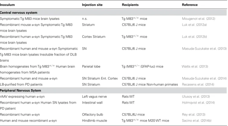
Table 1 | Summary of in vivo studies representing the major milestones in theα-synuclein-injected toxicity.
Inoculum Injection site Recipients Reference
Central nervous system
Symptomatic Tg M83 mice brain lysates n.s. Tg M83+/+mice Mougenot et al. (2012)
Recombinant mouseα-syn Symptomatic Tg M83 mice brain lysates
Striatum C57BL/6 J mice Luk et al. (2012a)
Recombinant humanα-syn Symptomatic Tg M83 mice brain lysates
Cortex Striatum Tg M83+/+mice Luk et al. (2012b)
Recombinant human and mouseα-syn Symptomatic Tg M83 mice brain lysates Insoluble fraction of DLB
brains
SN C57BL/6 J mice Masuda-Suzukake et al. (2013)
Brain homogenates from Tg M83+/+Human brain homogenates from MSA patients
Parietal lobe Tg (M83+/−:GFAP-luc) mice Watts et al. (2013)
Recombinant human and mouseα-syn SN Striatum Ent. Cortex C57BL/6 J mice Masuda-Suzukake et al. (2014)
LB-purified from PD patients SN Striatum C57BL/6 J mice Non-human primates Recasens et al. (2014)
Peripheral Nervous Sytem
rAAV expressing humanα-syn Left vagus nerve Rats WT Ulusoy et al. (2013)
Recombinant humanα-syn Human SN lysates from PD patient
Intestinal wall Rats WT Holmqvist et al. (2014)
Recombinant humanα-syn Olfactory bulb C57BL/6J mice Rey et al. (2013)
Human and mouse recombinantα-syn Hindlimb muscle Tg M83+/+mice M20 WT mice Sacino et al. (2014b)
α-syn, α-synuclein; DLB, dementia with Lewy body; Ent. Cortex, entorhinal cortex; GFAP, Glial fibrillary acidic protein; LB, Lewy body; Luc, luciferase; MSA, multiple
system atrophy; n.s., not specified; PD, Parkinson’s disease; SN, substantia nigra; rAAV, recombinant adeno-associated virus; Str, striatum; Tg, transgenic; WT, wild-type.
Table 2 | Missing evidences or open questions aboutα-synuclein spreading in PD.
Open questions
What is the composition and structure of recombinantα-syn seeds, brain homogenates samples or LB-purified samples?
What are theα-syn species responsible for toxicity and spreading in recombinant α-syn seeds, brain homogenates samples or LB-purified samples? Are there differences in biophysical or structural properties betweenα-syn species responsible for toxicity and spreading?
Does spreading implies infectivity?
Areα-syn species specific from a synucleinopathy to another? Is there a strain notion? Are cofactors (intracellular or extracellular) necessary for self-propagation?
What is the contribution of the axonal transport in the spreading process? Is glia involved in propagation to interconnected brain structures? Is there a common pathway/pattern for tissue migration?
What is the mechanism of cell death in thoseα-syn spreading based models? Does the immune response play a role?
How to improve the reproducibility of recombinantα-syn seeds? α-syn assembly by PMCA or qRT-QuIC might overcome this obstacle. Can we extrapolate the results obtained inα-syn spreading based models into human diseases?
Does the other neurodegenerative-associated proteins (Aβ, tau, huntingtin . . .) share the same spreading-toxic properties of α-syn?
α-syn, α-synuclein; Aß, amyloid-beta; LB, Lewy body; PMCA, protein misfolding cyclic amplification; qRT-QuIC, quantitative real-time quaking-induced conversion.
dopaminergic neurons grafted into the striatum of mice
over-expressing human
α-syn, exhibited human α-syn
immunoreac-tivity 6 months after transplantation (
Hansen et al., 2011
), thus
confirming the transfer of human
α-syn from host-to-graft in
vivo. In addition, this study also demonstrated that different
forms of human
α-syn, including monomers, oligomers and
fibrils, could be taken up by neurons in vivo by endocytosis
(
Hansen et al., 2011
). In addition, host-to-graft transmission of
human
α-syn has also been reported in rats (
Kordower et al.,
2011
).
Recasens and Dehay Alpha-synuclein spreading in Parkinson disease
Once demonstrated that
α-syn could be transmitted between
cells, the next step was to explore the potential pathogenic
effect of
α-syn transmission in vivo. In this context, both
syn-thetic and murine disease-associated forms of
α-syn were able
to induce a PD-like
α-syn pathology in vivo (
Luk et al., 2012b
).
Luk and colleagues reported that the intracerebral injection of
brain homogenates derived from old
α-syn transgenic mice (which
exhibited
α-syn pathology) into the neocortex and striatum of
young asymptomatic transgenic mice induced a widespread
accu-mulation of pathological
α-syn throughout the anterior/posterior
extent of the neural axis spanning the CNS, from olfactory bulb
(OB) to the spinal cord. These effects were mostly observed by
90 days post-injection, although at 30 days post-injection some
α-syn pathology was already evident. Similar results were obtained
after the injection of synthetic recombinant
α-syn PFFs, providing
the first evidence that PFFs alone were sufficient to initiate and
propagate the
α-syn pathology in vivo. Furthermore, the
inocula-tion of either symptomatic brain lysates or
α-syn PFFs accelerated
and increased the accumulation of
α-syn in these transgenic mice
and reduced their lifespan.
Mougenot et al. (2012)
reproduced part
of these results. In this case, the injection of brain homogenates
from symptomatic
α-syn transgenic mice into the brains of healthy
transgenic mice accelerated the characteristic clinical signs of
paralysis observed in this mouse model and reduced the lifespan
of injected animals. In addition, insoluble phosphorylated
α-syn
at Ser129 was also found in the brains of inoculated mice.
The pathological spreading of
α-syn was also reported in
wild-type (WT) mice (
Luk et al., 2012a
). The injection of synthetic
recombinant
α-syn PFFs into the striatum of WT mice induced
a pathological time-dependent accumulation of endogenous
α-syn that was associated with cell loss in the SN and impaired
motor coordination. The formation of an LB/LN-like pathology
in PFFs-inoculated mice occurred upstream of SN DA neuron
loss, indicating that the
α-syn pathology was sufficient to induce
the cardinal behavioral and pathological features of sporadic PD.
The injection of human and mouse PFFs directly into the SN
(
Masuda-Suzukake et al., 2013
) or hippocampus (
Sacino et al.,
2014a
) of WT mice also induced a time-dependent widespread
accumulation of
α-syn pathology, although no neuronal loss in
the SN or motor impairment was found in this case. It is
note-worthy that the
α-syn spreading efficiency observed in different
laboratories depends heavily on several factors which include the
preparation of synthetic recombinant
α-syn, the choice of the
strain of mice (
Sacino et al., 2014c
) as well as the brain areas of
inoculation (
Masuda-Suzukake et al., 2014
) and overall the
pos-sibility of a species barrier. Furthermore,
Sacino et al. (2014c)
raised an important point about the non-specific
immunohisto-chemical staining of the Ser129-phosphorylated
α-syn antibody
(mAB81A). This antibody reacts with phosphor-Ser129 but also
with phosphorylated neurofilament subunit L (NFL). To overcome
this obstacle, this antibody has to be used cautiously associated
with an optimizing protocol including (i) the use of very low
antibody concentrations for minimal background; (ii) the
con-firmation with other phosphor-Ser129
α-syn specific antibodies
and amyloid dyes such as Thioflavine S; and (iii) the combination
with biochemical procedures to separate the proteins by size to
detect phosphorylated
α-syn.
More recently,
Recasens et al. (2014)
demonstrated that human
α-syn species contained in PD-derived LB are pathogenic and
have the capacity to initiate a PD-like pathological process, not
only in rodents but also in non-human primates. Nigral LB
containing pathological
α-syn were purified from postmortem
PD brains by sucrose gradient fractionation and subsequently
inoculated into the SN or striatum of WT mice and macaque
monkeys. In both mice and monkeys, intranigral or intrastriatal
inoculations of PD-derived LB extracts resulted in progressive
nigrostriatal neurodegeneration starting at striatal
dopaminer-gic terminals. In LB-injected animals, exogenous human
α-syn
was quickly internalized within host neurons and triggered the
pathological conversion of endogenous
α-syn. At the onset of
LB-induced neurodegeneration, host pathological
α-syn diffusely
accumulated within nigral neurons and anatomically
intercon-nected brain regions. LB-induced pathogenic effects required both
human
α-syn present in LB extracts and host expression of α-syn.
Similarly, the injection of brain homogenates from patients with
other synucleinopathies, such as dementia with Lewy bodies (DLB;
Masuda-Suzukake et al., 2013
) and multiple system atrophy (MSA;
Watts et al., 2013
), triggered
α-synuclein pathology in mice. While
the DLB homogenate did not induce a glial response or neuronal
loss, mice injected with MSA exhibited prominent astrocytic and
microglial activation and developed progressive signs of
neuro-logic dysfunction. These contradictory results concerning human
α-syn-induced neurodegeneration might be explained by
differ-ences in: (i) mouse strain (WT vs. transgenic), (ii) injection site
(SN vs. parietal lobe), and (iii) sample sonication (non-sonicated
vs. sonicated). A further possibility is that different
α-syn strains
might exist in each disease (PD, MSA, and DLB), thus explaining
the differences observed after the injection of each synucleinopathy
sample. Supporting this concept, distinct
α-syn strains generated
through repetitively seeded fibrillization in vitro exhibited
differ-ent seeding properties both in vitro (
Bousset et al., 2013
) and in
vivo (
Guo et al., 2013
).
PERIPHERAL TRANSMISSION OF
α-SYNUCLEIN PATHOLOGY
TO THE BRAIN
While the studies mentioned above involved a direct intracerebral
inoculation of pathological
α-syn, other studies have addressed the
possible transmission of
α-syn pathology from the periphery to the
brain. For example, recombinant adeno-associated virus (rAAV)
serotype 2/6-expressing human WT
α-syn has been injected into
the left vagus nerve in the neck of rats (
Ulusoy et al., 2013
). This
injection induced a strong expression of human
α-syn in the
medulla oblongata (MO), leading to a caudo-rostral spreading of
the
α-syn pathology into other interconnected brain regions, such
as the pontine coeruleus-subcoeruleus complex, the dorsal raphe,
the hypothalamus and the amygdala. In addition,
α-syn
accumu-lation present in the aforementioned areas was accompanied by
morphological evidence of neuronal abnormalities (i.e.,
thread-like axons with irregularly spaced, densely labeled varicosities).
Surprisingly, the transmission of
α-syn did not reach the SN, and
neuronal damage was not induced in this brain region for at least
18 weeks after the injection.
In another study,
Brundin et al. (2008)
examined if
α-syn
could transfer from the OB to other brain structures through
neuronal connections (
Rey et al., 2013
). To answer this question,
different molecular species (monomers, oligomers composed of
soluble high molecular weight species, and fibrils) of
recombi-nant human
α-syn were injected into the OB of normal mice. The
authors reported that cells in different layers of the OB (i.e., the
glomerular layer, mitral cell layer and granule cell layer) readily
take up recombinant monomeric and oligomeric
α-syn. Fibrillar
α-syn was also taken up, but to a much lesser extent within the
time frame of the experiments. Soon after the injection (1.5 h
and 3 h), soluble and oligomer, but not fibrillar,
α-syn species
were detected in several interconnected brain regions,
includ-ing the anterior olfactory nucleus, the frontal cortex, the tenia
tecta, the olfactory tubercle, the periform cortex, the striatum
and the amygdala. At these time points, few microglial cells in
the OB, anterior olfactory nucleus and frontal cortex were
posi-tive for human
α-syn. α-Syn in microglial cells was present only
locally, and not in other brain regions 12 h after injection into
the OB. In contrast, at later time points,
α-syn was extensively
detected in microglial cells, suggesting that microglia might clear
the human
α-syn released into the extracellular space by the
neurons. Recently, a study from the group of Giasson reported
that in vitro-generated PFFs induced
α-syn pathology by a single
peripheral intramuscular injection of
α-syn in transgenic mice,
associated with robust gliosis and motor impairments (
Sacino
et al., 2014b
).
The gastrointestinal pathway has also been extensively studied.
Pan-Montojo and colleagues reported that intragastric
admin-istration of the environmental toxin rotenone induced
α-syn
accumulation in both the enteric nervous system (ENS) and
CNS following the same pattern of progression as hypothesized
by Braak (
Pan-Montojo et al., 2010
). Firstly, they reported
α-syn accumulations in ENS neurons as soon as 1–5 months after
rotenone treatment. Next, they determined whether the local
effect of rotenone on the ENS could lead to alterations in the
synaptically connected ANS centers in the spinal cord and
brain-stem [i.e., in the intermediolateral nucleus in the spinal cord
(IML) and dorsal motor nucleus of the vagus (DMV)]. Both
the IML and DMV exhibited accumulation and aggregation of
syn 1.5 and 3 months after rotenone treatment, although
α-syn pathology in these areas was not associated with neuronal
death. Interestingly, the SN also exhibited
α-syn accumulation,
phosphorylation and inflammatory signs 3 months after rotenone
treatment. Unlike the DMV and IML,
α-syn increments in the
SN were associated with neuronal loss. After intragastric rotenone
administration, pesticide was not detected in the blood or brain,
and no inhibition of complex I activity in muscle or brain was
found, suggesting that the reported alterations in the mentioned
brain regions were not due to a systemic effect of rotenone.
Remarkably, the rotenone-induced
α-syn pathology was specific,
as only neuronal subpopulations with direct connections to the
ENS showed alterations, while nearby areas (e.g., striatum,
cere-bellum, and cortex) remained unaffected. This specificity together
with the fact that the appearance of
α-syn accumulations in the
SN were only detected at the last treatment time-point, raised
the possibility of a direct mechanism between cells being
respon-sible for this pattern of progression of the
α-syn pathology. To
confirm this hypothesis,
Pan-Montojo et al. (2012)
severed some
of the connecting nerves between the CNS and the gut, which
delayed the appearance of motor symptoms after oral rotenone
treatment. This treatment also stopped the progression of
α-syn
pathology into the IML and DMV, and prevented cell death in the
SN (
Pan-Montojo et al., 2012
). Recently,
Holmqvist et al. (2014)
have demonstrated that both human
α-syn present in the SN
of PD patients and distinct recombinant
α-syn forms (including
monomers, oligomers and fibrils) can be transported via the vagal
nerve to the CNS after the injection into the intestinal wall of WT
adult rats.
α-SYNUCLEIN TRANSMISSION AND NEUROINFLAMMATION
The secretion of
α-syn by neurons may not only induce
toxic-ity once inside the cytoplasm of neighboring cells, but also in
the extracellular space; this may activate glial cells and induce
chronic inflammation (i.e., a common pathological feature of
PD), thereby contributing to the progression of the pathology
throughout the brain. Supporting this idea, glial cells (i.e.,
astro-cytes and microglia) are able to take up and degrade synthetic
recombinant
α-syn aggregates even more efficiently than neurons
(
Lee et al., 2008b
). Indeed,
α-syn can be transmitted between
neurons and glial cells in vitro (
Lee et al., 2010
;
Alvarez-Erviti
et al., 2011a
). Interestingly, the exposure of neuron-derived
α-syn
induced an inflammatory reaction in rat primary astrocytes (
Lee
et al., 2010
) and microglia (
Zhang et al., 2005
;
Reynolds et al., 2008
;
Alvarez-Erviti et al., 2011a
). The direct transfer of
α-syn from
neurons to astrocytes was demonstrated in vivo using transgenic
mice overexpressing human
α-syn under a neuronal promoter. In
these transgenic mice, abundant human
α-syn accumulation was
observed not only in neurons but also in glial cells (
Lee et al., 2010
).
Consistent with these results, recombinant
α-syn oligomers and
monomers injected into the neocortex of WT mice were taken up
by oligodendrocytes (
Reyes et al., 2014
). Similarly, in rAAV-treated
rats overexpressing human
α-syn, embryonic oligodendrocytes
grafted into the striatum were found to contain this human
α-syn,
thus further demonstrating the neuron-to-astrocyte transmission
of
α-syn (
Reyes et al., 2014
).
α-SYNUCLEIN AND SIDEKICKS
Recently, several studies have provided convincing evidence that
this same self-propagating mechanism of the
α-syn protein may
be applicable to a wide range of neurodegenerative associated
proteins, including A
β, tau, huntingtin, superoxide dismutase
1 (SOD1) and TDP-43 (see
Guo and Lee, 2014
, for review).
Each of these proteins (i.e., recombinant proteins or contained
in brain lysates) have been shown to act as a template or seed
that could efficiently recruit their soluble counterparts into
elon-gating fibrils in cultured cells and/or living animals. Recently,
Cicchetti et al. (2014)
described the presence of mutant
hunt-ingtin (mHtt) in tissue grafted into the brains of three patients
with Huntington ‘s disease (HD) who received their transplants
9–12 years before they died. Similarly to the embryonic
mesen-cephalic neurons grafted into the striatum of PD patients which
develop LB many years after grafting, the presence of mHtt in
this graft tissue could be explained by the host-to-graft
transmis-sion of the neurodegenerative-associated protein Htt. However,
it is worth noting that the mHtt in this study was localized
Recasens and Dehay Alpha-synuclein spreading in Parkinson disease
to the extracellular matrix of the transplant tissue, unlike the
mHtt protein aggregates found within the non-grafted regions,
which localized to the neurons and neuropil (
Cicchetti et al.,
2014
).
CONCLUDING REMARKS AND FUTURE DIRECTIONS
Mounting evidence suggests the concept that
α-syn may be
respon-sible for initiating and spreading the pathological process in PD.
Notably, cellular and animal models developed so far based on
the transmission (or spreading) properties might allow to screen
therapeutic approaches against
α-syn pathology (
Sato et al., 2014
).
Of interest, a recent study using the PFFs-based model of PD
demonstrated that immunotherapy with antibodies specifically
targeting misfolded
α-syn is able to block the entrance and
prop-agation of
α-syn in neurons, and hence prevents the development
of neuropathological abnormalities in the brain (
Tran et al., 2014
).
However, several important questions remain to be solved
(
Table 2): (i) it is currently unknown whether the pathological
conversion of endogenous
α-syn triggered by PD-derived
mate-rial or recombinant
α-syn fibrils actually occurs directly through a
seeding process or indirectly as a general response to cellular stress;
(ii) the association between pathological
α-syn accumulation and
neuron cell death remains so far correlative. In addition, there is no
definitive evidence to support the idea that PD can be contagious
from one person to another, as is characterized for prion diseases
(
Beekes et al., 2014
). In this line, a retrospective, postmortem study
of recipients of cadaver-derived human growth hormone (hGH)
found no reported incidence of PD, although the donors of
pitu-itary glands used for hGH preparation probably included people
with PD, and pathological
α-syn is frequently found in the
post-mortem pituitary glands of people with PD (
Irwin et al., 2013
).
One of the possible experiments would be to isolate
α-syn
aggre-gates developed in PD-derived material or recombinant
α-syn
fibrils injected animals, and injecting again in a healthy animals.
These experiments would allow us to differentiate between
infec-tious and self-propagating properties. Some approaches should
be tested to evaluate the transmission of these disorders between
animals (mice and monkeys) in order to study species-barrier
properties or the use of different administration routes
(intrac-erebrally, intranasal or fluids). All these studies should answer
the unavoidable question of infectivity and/or contagiousness,
the last missing criterion that defines a prion disease. However,
until the issues mentioned above around nature and mechanisms
of
α-syn prion-like properties are better understood, we believe
that the term prion for
α-syn has to be used and considered
cautiously. A new term referring as self-propagating pathogenic
protein for
α-syn needs to emerge and this is a mechanism well
worth considering.
ACKNOWLEDGMENTS
We thank Mathieu Bourdenx and Daniel Ko for valuable
com-ments on the manuscript. The University of Bordeaux and the
Centre National de la Recherche Scientifique provided
infras-tructural support. This work was supported by the Fondo de
Investigación Sanitaria-Instituto de Salud Carlos III, Spain, Marie
Curie Reintegration Grant from the European Commission
FP7-PEOPLE-2009-ERG256303 (to Benjamin Dehay) and by a grant
from the Fondation pour la Recherche Médicale (to Benjamin
Dehay).
REFERENCES
Abeliovich, A., Schmitz, Y., Farinas, I., Choi-Lundberg, D., Ho, W. H., Castillo, P. E., et al. (2000). Mice lacking alpha-synuclein display functional deficits in the nigrostriatal dopamine system. Neuron 25, 239–252. doi: 10.1016/S0896-6273(00)80886-7
Ahn, K. J., Paik, S. R., Chung, K. C., and Kim, J. (2006). Amino acid sequence motifs and mechanistic features of the membrane translocation of alpha-synuclein.
J. Neurochem. 97, 265–279. doi: 10.1111/j.1471-4159.2006.03731.x
Alafuzoff, I., Ince, P. G., Arzberger, T., Al-Sarraj, S., Bell, J., Bodi, I., et al. (2009). Staging/typing of lewy body related alpha-synuclein pathology: a study of the brainnet Europe consortium. Acta Neuropathol. 117, 635–652. doi: 10.1007/s00401-009-0523-2
Alvarez-Erviti, L., Couch, Y., Richardson, J., Cooper, J. M., and Wood, M. J. (2011a). Alpha-synuclein release by neurons activates the inflammatory response in a microglial cell line. Neurosci. Res. 69, 337–342. doi: 10.1016/j.neures.2010.12.020 Alvarez-Erviti, L., Seow, Y., Schapira, A. H., Gardiner, C., Sargent, I. L., Wood, M. J., et al. (2011b). Lysosomal dysfunction increases exosome-mediated alpha-synuclein release and transmission. Neurobiol. Dis. 42, 360–367. doi: 10.1016/j.nbd.2011.01.029
Appel-Cresswell, S., Vilarino-Guell, C., Encarnacion, M., Sherman, H., Yu, I., Shah, B., et al. (2013). Alpha-synuclein p.H50Q, a novel pathogenic mutation for Parkinson’s disease. Mov. Disord. 28, 811–813. doi: 10.1002/mds.25421 Athanassiadou, A., Voutsinas, G., Psiouri, L., Leroy, E., Polymeropoulos, M. H., Ilias,
A., et al. (1999). Genetic analysis of families with Parkinson disease that carry the Ala53Thr mutation in the gene encoding alpha-synuclein. Am. J. Hum. Genet. 65, 555–558. doi: 10.1086/302486
Auluck, P. K., Caraveo, G., and Lindquist, S. (2010). Alpha-synuclein: membrane interactions and toxicity in Parkinson’s disease. Annu. Rev. Cell Dev. Biol. 26, 211–233. doi: 10.1146/annurev.cellbio.042308.113313
Beekes, M., Thomzig, A., Schulz-Schaeffer, W. J., and Burger, R. (2014). Is there a risk of prion-like disease transmission by Alzheimer- or Parkinson-associated protein particles? Acta Neuropathol. 128, 463–476. doi: 10.1007/s00401-014-1324-9 Bloch, A., Probst, A., Bissig, H., Adams, H., and Tolnay, M. (2006). Alpha-synuclein
pathology of the spinal and peripheral autonomic nervous system in neurologi-cally unimpaired elderly subjects. Neuropathol. Appl. Neurobiol. 32, 284–295. doi: 10.1111/j.1365-2990.2006.00727.x
Borghi, R., Marchese, R., Negro, A., Marinelli, L., Forloni, G., Zaccheo, D., et al. (2000). Full length alpha-synuclein is present in cerebrospinal fluid from Parkinson’s disease and normal subjects. Neurosci. Lett. 287, 65–67. doi: 10.1016/S0304-3940(00)01153-8
Bousset, L., Pieri, L., Ruiz-Arlandis, G., Gath, J., Jensen, P. H., Habenstein, B., et al. (2013). Structural and functional characterization of two alpha-synuclein strains.
Nat. Commun. 4:2575. doi: 10.1038/ncomms3575
Braak, H., Del Tredici, K., Rub, U., de Vos, R. A., Jansen Steur, E. N., and Braak, E. (2003). Staging of brain pathology related to sporadic Parkinson’s disease.
Neurobiol. Aging 24, 197–211. doi: 10.1016/S0197-4580(02)00065-9
Brundin, P., Li, J. Y., Holton, J. L., Lindvall, O., and Revesz, T. (2008). Research in motion: the enigma of Parkinson’s disease pathology spread. Nat. Rev. Neurosci. 9, 741–745. doi: 10.1038/nrn2477
Burke, R. E., Dauer, W. T., and Vonsattel, J. P. (2008). A critical evaluation of the braak staging scheme for Parkinson’s disease. Ann. Neurol. 64, 485–491. doi: 10.1002/ana.21541
Burre, J., Sharma, M., and Sudhof, T. C. (2014). Alpha-synuclein assembles into higher-order multimers upon membrane binding to promote SNARE complex formation. Proc. Natl. Acad. Sci. U.S.A. 111, E4274–E4283. doi: 10.1073/pnas.1416598111
Cabin, D. E., Shimazu, K., Murphy, D., Cole, N. B., Gottschalk, W., McIlwain, K. L., et al. (2002). Synaptic vesicle depletion correlates with attenuated synaptic responses to prolonged repetitive stimulation in mice lacking alpha-synuclein.
J. Neurosci. 22, 8797–8807.
Chandra, S., Gallardo, G., Fernandez-Chacon, R., Schluter, O. M., and Sud-hof, T. C. (2005). Alpha-synuclein cooperates with CSPalpha in preventing neurodegeneration. Cell 123, 383–396. doi: 10.1016/j.cell.2005.09.028 Chen, R. H., Wislet-Gendebien, S., Samuel, F., Visanji, N. P., Zhang, G., Marsilio, D.,
recycling machinery and presynaptic activity. J. Biol. Chem. 288, 7438–7449. doi: 10.1074/jbc.M112.439497
Chinta, S. J., Mallajosyula, J. K., Rane, A., and Andersen, J. K. (2010). Mitochon-drial alpha-synuclein accumulation impairs complex I function in dopaminergic neurons and results in increased mitophagy in vivo. Neurosci. Lett. 486, 235–239. doi: 10.1016/j.neulet.2010.09.061
Choi, J. M., Woo, M. S., Ma, H. I., Kang, S. Y., Sung, Y. H., Yong, S. W., et al. (2008). Analysis of PARK genes in a Korean cohort of early-onset Parkinson disease.
Neurogenetics 9, 263–269. doi: 10.1007/s10048-008-0138-0
Cicchetti, F., Lacroix, S., Cisbani, G., Vallieres, N., Saint-Pierre, M., St-Amour, I., et al. (2014). Mutant huntingtin is present in neuronal grafts in Huntington disease patients. Ann. Neurol. 76, 31–42. doi: 10.1002/ana.24174
Cooper, A. A., Gitler, A. D., Cashikar, A., Haynes, C. M., Hill, K. J., Bhullar, B., et al. (2006). Alpha-synuclein blocks ER-Golgi traffic and Rab1 rescues neuron loss in Parkinson’s models. Science 313, 324–328. doi: 10.1126/science.1129462 Danzer, K. M., Haasen, D., Karow, A. R., Moussaud, S., Habeck, M., Giese, A., et al.
(2007). Different species of alpha-synuclein oligomers induce calcium influx and seeding. J. Neurosci. 27, 9220–9232. doi: 10.1523/JNEUROSCI.2617-07.2007 Danzer, K. M., Krebs, S. K., Wolff, M., Birk, G., and Hengerer, B. (2009).
Seed-ing induced by alpha-synuclein oligomers provides evidence for spreadSeed-ing of alpha-synuclein pathology. J. Neurochem. 111, 192–203. doi: 10.1111/j.1471-4159.2009.06324.x
Danzer, K. M., Ruf, W. P., Putcha, P., Joyner, D., Hashimoto, T., Glabe, C., et al. (2011). Heat-shock protein 70 modulates toxic extracellular alpha-synuclein oligomers and rescues trans-synaptic toxicity. FASEB J. 25, 326–336. doi: 10.1096/fj.10-164624
Desplats, P., Lee, H. J., Bae, E. J., Patrick, C., Rockenstein, E., Crews, L., et al. (2009). Inclusion formation and neuronal cell death through neuron-to-neuron transmission of alpha-synuclein. Proc. Natl. Acad. Sci. U.S.A. 106, 13010–13015. doi: 10.1073/pnas.0903691106
Devi, L., Raghavendran, V., Prabhu, B. M., Avadhani, N. G., and Anan-datheerthavarada, H. K. (2008). Mitochondrial import and accumulation of alpha-synuclein impair complex I in human dopaminergic neuronal cul-tures and Parkinson disease brain. J. Biol. Chem. 283, 9089–9100. doi: 10.1074/jbc.M710012200
DeWitt, D. C., and Rhoades, E. (2013). Alpha-synuclein can inhibit SNARE-mediated vesicle fusion through direct interactions with lipid bilayers.
Biochem-istry 52, 2385–2387. doi: 10.1021/bi4002369
Dickson, D. W. (2012). Parkinson’s disease and parkinsonism: neuropathology. Cold
Spring Harb. Perspect. Med. 2:a009258. doi: 10.1101/cshperspect.a009258
Dickson, D. W., Uchikado, H., Fujishiro, H., and Tsuboi, Y. (2010). Evidence in favor of Braak staging of Parkinson’s disease. Mov. Disord. 25(Suppl. 1), S78–S82. doi: 10.1002/mds.22637
El-Agnaf, O. M., Salem, S. A., Paleologou, K. E., Cooper, L. J., Fullwood, N. J., Gibson, M. J., et al. (2003). Alpha-synuclein implicated in Parkinson’s disease is present in extracellular biological fluids, including human plasma. FASEB J. 17, 1945–1947. doi: 10.1096/fj.03-0098fje
Emmanouilidou, E., Melachroinou, K., Roumeliotis, T., Garbis, S. D., Ntzouni, M., Margaritis, L. H., et al. (2010). Cell-produced alpha-synuclein is secreted in a calcium-dependent manner by exosomes and impacts neu-ronal survival. J. Neurosci. 30, 6838–6851. doi: 10.1523/JNEUROSCI.5699-09.2010
Gelpi, E., Navarro-Otano, J., Tolosa, E., Gaig, C., Compta, Y., Rey, M. J., et al. (2014). Multiple organ involvement by alpha-synuclein pathology in Lewy body disorders. Mov. Disord. 29, 1010–1018 doi: 10.1002/mds.25776
Gitler, A. D., Bevis, B. J., Shorter, J., Strathearn, K. E., Hamamichi, S., Su, L., et al. (2008). The Parkinson’s disease protein alpha-synuclein disrupts cellular rab homeostasis. Proc. Natl. Acad. Sci. U.S.A. 105, 145–150. doi: 10.1073/pnas.0710685105
Gousset, K., Schiff, E., Langevin, C., Marijanovic, Z., Caputo, A., Browman, D. T., et al. (2009). Prions hijack tunnelling nanotubes for intercellular spread. Nat. Cell
Biol. 11, 328–336. doi: 10.1038/ncb1841
Guardia-Laguarta, C., Area-Gomez, E., Rub, C., Liu, Y., Magrane, J., Becker, D., et al. (2014). Alpha-synuclein is localized to mitochondria-associated ER membranes.
J. Neurosci. 34, 249–259. doi: 10.1523/JNEUROSCI.2507-13.2014
Guo, J. L., Covell, D. J., Daniels, J. P., Iba, M., Stieber, A., Zhang, B., et al. (2013). Distinct alpha-synuclein strains differentially promote tau inclusions in neurons.
Cell 154, 103–117. doi: 10.1016/j.cell.2013.05.057
Guo, J. L., and Lee, V. M. (2014). Cell-to-cell transmission of pathogenic proteins in neurodegenerative diseases. Nat. Med. 20, 130–138. doi: 10.1038/nm.3457 Halliday, G., McCann, H., and Shepherd, C. (2012). Evaluation of the Braak
hypoth-esis: how far can it explain the pathogenesis of Parkinson’s disease? Expert Rev.
Neurother. 12, 673–686. doi: 10.1586/ern.12.47
Hansen, C., Angot, E., Bergstrom, A. L., Steiner, J. A., Pieri, L., Paul, G., et al. (2011). Alpha-synuclein propagates from mouse brain to grafted dopaminergic neurons and seeds aggregation in cultured human cells. J. Clin. Invest. 121, 715–725. doi: 10.1172/JCI43366
Holmqvist, S., Chutna, O., Bousset, L., Aldrin-Kirk, P., Li, W., Bjorklund, T., et al. (2014). Direct evidence of Parkinson pathology spread from the gastrointestinal tract to the brain in rats. Acta Neuropathol. 128, 805–820. doi: 10.1007/s00401-014-1343-6
Irwin, D. J., Abrams, J. Y., Schonberger, L. B., Leschek, E. W., Mills, J. L., Lee, V., et al. (2013). Evaluation of potential infectivity of Alzheimer and Parkinson disease proteins in recipients of cadaver-derived human growth hormone. JAMA Neurol. 70, 462–468. doi: 10.1001/jamaneurol.2013.1933
Jenco, J. M., Rawlingson, A., Daniels, B., and Morris, A. J. (1998). Regulation of phos-pholipase D2: selective inhibition of mammalian phosphos-pholipase D isoenzymes by alpha- and beta-synucleins. Biochemistry 37, 4901–4909. doi: 10.1021/bi972776r Kamp, F., Exner, N., Lutz, A. K., Wender, N., Hegermann, J., Brunner, B., et al. (2010). Inhibition of mitochondrial fusion by alpha-synuclein is rescued by PINK1, Parkin and DJ-1. EMBO J. 29, 3571–3589. doi: 10.1038/emboj.2010.223 Ki, C. S., Stavrou, E. F., Davanos, N., Lee, W. Y., Chung, E. J., Kim, J. Y.,
et al. (2007). The Ala53Thr mutation in the alpha-synuclein gene in a Korean family with Parkinson disease. Clin. Genet. 71, 471–473. doi: 10.1111/j.1399-0004.2007.00781.x
Kordower, J. H., Chu, Y., Hauser, R. A., Freeman, T. B., and Olanow, C. W. (2008). Lewy body-like pathology in long-term embryonic nigral transplants in Parkinson’s disease. Nat. Med. 14, 504–506. doi: 10.1038/nm1747
Kordower, J. H., Dodiya, H. B., Kordower, A. M., Terpstra, B., Paumier, K., Madhavan, L., et al. (2011). Transfer of host-derived alpha synuclein to grafted dopaminergic neurons in rat. Neurobiol. Dis. 43, 552–557. doi: 10.1016/j.nbd.2011.05.001
Kruger, R., Kuhn, W., Muller, T., Woitalla, D., Graeber, M., Kosel, S., et al. (1998). Ala30Pro mutation in the gene encoding alpha-synuclein in Parkinson’s disease.
Nat. Genet. 18, 106–108. doi: 10.1038/ng0298-106
Larsen, K. E., Schmitz, Y., Troyer, M. D., Mosharov, E., Dietrich, P., Quazi, A. Z., et al. (2006). Alpha-synuclein overexpression in PC12 and chromaffin cells impairs catecholamine release by interfering with a late step in exocytosis. J. Neurosci. 26, 11915–11922. doi: 10.1523/JNEUROSCI.3821-06.2006
Lee, H. J., Patel, S., and Lee, S. J. (2005). Intravesicular localization and exo-cytosis of alpha-synuclein and its aggregates. J. Neurosci. 25, 6016–6024. doi: 10.1523/JNEUROSCI.0692-05.2005
Lee, H. J., Suk, J. E., Bae, E. J., Lee, J. H., Paik, S. R., and Lee, S. J. (2008a). Assembly-dependent endocytosis and clearance of extracellular alpha-synuclein.
Int. J. Biochem. Cell Biol. 40, 1835–1849. doi: 10.1016/j.biocel.2008.01.017
Lee, H. J., Suk, J. E., Bae, E. J., and Lee, S. J. (2008b). Clearance and deposition of extracellular alpha-synuclein aggregates in microglia. Biochem. Biophys. Res.
Commun. 372, 423–428. doi: 10.1016/j.bbrc.2008.05.045
Lee, H. J., Suk, J. E., Patrick, C., Bae, E. J., Cho, J. H., Rho, S., et al. (2010). Direct transfer of alpha-synuclein from neuron to astroglia causes inflam-matory responses in synucleinopathies. J. Biol. Chem. 285, 9262–9272. doi: 10.1074/jbc.M109.081125
Lesage, S., Anheim, M., Letournel, F., Bousset, L., Honore, A., Rozas, N., et al. (2013). G51D alpha-synuclein mutation causes a novel parkinsonian-pyramidal syndrome. Ann. Neurol. 73, 459–471. doi: 10.1002/ana.23894
Li, J. Y., Englund, E., Holton, J. L., Soulet, D., Hagell, P., Lees, A. J., et al. (2008). Lewy bodies in grafted neurons in subjects with Parkinson’s disease suggest host-to-graft disease propagation. Nat. Med. 14, 501–503. doi: 10.1038/nm1746 Liu, G., Zhang, C., Yin, J., Li, X., Cheng, F., Li, Y., et al. (2009). Alpha-synuclein is
differentially expressed in mitochondria from different rat brain regions and dose-dependently down-regulates complex I activity. Neurosci. Lett. 454, 187–192. doi: 10.1016/j.neulet.2009.02.056
Loeb, V., Yakunin, E., Saada, A., and Sharon, R. (2010). The transgenic over-expression of alpha-synuclein and not its related pathology associates with complex I inhibition. J. Biol. Chem. 285, 7334–7343. doi: 10.1074/jbc.M109. 061051
Recasens and Dehay Alpha-synuclein spreading in Parkinson disease
Luk, K. C., Kehm, V., Carroll, J., Zhang, B., O’Brien, P., Trojanowski, J. Q., et al. (2012a). Pathological alpha-synuclein transmission initiates Parkinson-like neurodegeneration in nontransgenic mice. Science 338, 949–953. doi: 10.1126/science.1227157
Luk, K. C., Kehm, V. M., Zhang, B., O’Brien, P., Trojanowski, J. Q., and Lee, V. M. (2012b). Intracerebral inoculation of pathological alpha-synuclein initiates a rapidly progressive neurodegenerative alpha-synucleinopathy in mice. J. Exp.
Med. 209, 975–986. doi: 10.1084/jem.20112457
Luk, K. C., Song, C., O’Brien, P., Stieber, A., Branch, J. R., Brunden, K. R., et al. (2009). Exogenous alpha-synuclein fibrils seed the formation of lewy body-like intracellular inclusions in cultured cells. Proc. Natl. Acad. Sci. U.S.A. 106, 20051– 20056. doi: 10.1073/pnas.0908005106
Maroteaux, L., Campanelli, J. T., and Scheller, R. H. (1988). Synuclein: a neuron-specific protein localized to the nucleus and presynaptic nerve terminal.
J. Neurosci. 8, 2804–2815.
Martin, L. J., Pan, Y., Price, A. C., Sterling, W., Copeland, N. G., Jenkins, N. A., et al. (2006). Parkinson’s disease alpha-synuclein transgenic mice develop neu-ronal mitochondrial degeneration and cell death. J. Neurosci. 26, 41–50. doi: 10.1523/JNEUROSCI.4308-05.2006
Martinez-Vicente, M., and Vila, M. (2013). Alpha-synuclein and protein degrada-tion pathways in Parkinson’s disease: a pathological feed-back loop. Exp. Neurol. 247, 308–313. doi: 10.1016/j.expneurol.2013.03.005
Masuda-Suzukake, M., Nonaka, T., Hosokawa, M., Kubo, M., Shimozawa, A., Akiyama, H., et al. (2014). Pathological alpha-synuclein propagates through neural networks. Acta Neuropathol. Commun. 2:88. doi: 10.1186/s40478-014-0088-8
Masuda-Suzukake, M., Nonaka, T., Hosokawa, M., Oikawa, T., Arai, T., Akiyama, H., et al. (2013). Prion-like spreading of pathological alpha-synuclein in brain.
Brain 136(Pt 4), 1128–1138. doi: 10.1093/brain/awt037
Mendez, I., Vinuela, A., Astradsson, A., Mukhida, K., Hallett, P., Robertson, H., et al. (2008). Dopamine neurons implanted into people with Parkinson’s disease survive without pathology for 14 years. Nat. Med. 14, 507–509. doi: 10.1038/nm1752
Mougenot, A. L., Nicot, S., Bencsik, A., Morignat, E., Verchere, J., Lakhdar, L., et al. (2012). Prion-like acceleration of a synucleinopathy in a transgenic mouse model.
Neurobiol. Aging 33, 2225–2228. doi: 10.1016/j.neurobiolaging.2011.06.022
Murphy, D. D., Rueter, S. M., Trojanowski, J. Q., and Lee, V. M. (2000). Synu-cleins are developmentally expressed, and alpha-synuclein regulates the size of the presynaptic vesicular pool in primary hippocampal neurons. J. Neurosci. 20, 3214–3220.
Nakamura, K., Nemani, V. M., Azarbal, F., Skibinski, G., Levy, J. M., Egami, K., et al. (2011). Direct membrane association drives mitochondrial fission by the Parkinson disease-associated protein alpha-synuclein. J. Biol. Chem. 286, 20710– 20726. doi: 10.1074/jbc.M110.213538
Navarro-Otano, J., Gelpi, E., Mestres, C. A., Quintana, E., Rauek, S., Ribalta, T., et al. (2013). Alpha-synuclein aggregates in epicardial fat tissue in living subjects without parkinsonism. Parkinsonism Relat. Disord. 19, 27–31. doi: 10.1016/j.parkreldis.2012.07.005
Nonaka, T., Watanabe, S. T., Iwatsubo, T., and Hasegawa, M. (2010). Seeded aggregation and toxicity of {alpha}-synuclein and tau: cellular models of neu-rodegenerative diseases. J. Biol. Chem. 285, 34885–34898. doi: 10.1074/jbc.M110. 148460
Pan-Montojo, F., Anichtchik, O., Dening, Y., Knels, L., Pursche, S., Jung, R., et al. (2010). Progression of Parkinson’s disease pathology is reproduced by intragastric administration of rotenone in mice. PLoS ONE 5:e8762. doi: 10.1371/journal.pone.0008762
Pan-Montojo, F., Schwarz, M., Winkler, C., Arnhold, M., O’Sullivan, G. A., Pal, A., et al. (2012). Environmental toxins trigger PD-like progression via increased alpha-synuclein release from enteric neurons in mice. Sci. Rep. 2:898. doi: 10.1038/srep00898
Polymeropoulos, M. H., Lavedan, C., Leroy, E., Ide, S. E., Dehejia, A., Dutra, A., et al. (1997). Mutation in the alpha-synuclein gene identified in families with Parkinson’s disease. Science 276, 2045–2047. doi: 10.1126/science.276. 5321.2045
Puschmann, A., Ross, O. A., Vilarino-Guell, C., Lincoln, S. J., Kachergus, J. M., Cobb, S. A., et al. (2009). A Swedish family with de novo alpha-synuclein A53T mutation: evidence for early cortical dysfunction. Parkinsonism Relat. Disord. 15, 627–632. doi: 10.1016/j.parkreldis.2009.06.007
Recasens, A., Dehay, B., Bove, J., Carballo-Carbajal, I., Dovero, S., Perez-Villalba, A., et al. (2014). Lewy body extracts from Parkinson disease brains trigger alpha-synuclein pathology and neurodegeneration in mice and monkeys. Ann. Neurol. 75, 351–362. doi: 10.1002/ana.24066
Rey, N. L., Petit, G. H., Bousset, L., Melki, R., and Brundin, P. (2013). Trans-fer of human alpha-synuclein from the olfactory bulb to interconnected brain regions in mice. Acta Neuropathol. 126, 555–573. doi: 10.1007/s00401-013-1160-3
Reyes, J. F., Rey, N. L., Bousset, L., Melki, R., Brundin, P., and Angot, E. (2014). Alpha-synuclein transfers from neurons to oligodendrocytes. Glia 62, 387–398. doi: 10.1002/glia.22611
Reynolds, A. D., Glanzer, J. G., Kadiu, I., Ricardo-Dukelow, M., Chaud-huri, A., Ciborowski, P., et al. (2008). Nitrated alpha-synuclein-activated microglial profiling for Parkinson’s disease. J. Neurochem. 104, 1504–1525. doi: 10.1111/j.1471-4159.2007.05087.x
Sacino, A. N., Brooks, M., McKinney, A. B., Thomas, M. A., Shaw, G., Golde, T. E., et al. (2014a). Brain Injection of alpha-synuclein induces multiple pro-teinopathies, gliosis, and a neuronal injury marker. J. Neurosci. 34, 12368–12378. doi: 10.1523/JNEUROSCI.2102-14.2014
Sacino, A. N., Brooks, M., Thomas, M. A., McKinney, A. B., Lee, S., Regen-hardt, R. W., et al. (2014b). Intramuscular injection of alpha-synuclein induces CNS alpha-synuclein pathology and a rapid-onset motor phenotype in transgenic mice. Proc. Natl. Acad. Sci. U.S.A. 111, 10732–10737. doi: 10.1073/pnas.1321785111
Sacino, A. N., Brooks, M., Thomas, M. A., McKinney, A. B., McGarvey, N. H., Rutherford, N. J., et al. (2014c). Amyloidogenic alpha-synuclein seeds do not invariably induce rapid, widespread pathology in mice. Acta Neuropathol. 127, 645–665. doi: 10.1007/s00401-014-1268-0
Sato, H., Kato, T., and Arawaka, S. (2014). Potential of cellular and animal models based on a prion-like propagation of alpha-synuclein for assessing antiparkin-son agents. Mol. Neurobiol. doi: 10.1007/s12035-014-8858-7 [Epub ahead of print].
Spira, P. J., Sharpe, D. M., Halliday, G., Cavanagh, J., and Nicholson, G. A. (2001). Clinical and pathological features of a Parkinsonian syndrome in a fam-ily with an Ala53Thr alpha-synuclein mutation. Ann. Neurol. 49, 313–319. doi: 10.1002/ana.67
Sung, J. Y., Kim, J., Paik, S. R., Park, J. H., Ahn, Y. S., and Chung, K. C. (2001). Induc-tion of neuronal cell death by Rab5A-dependent endocytosis of alpha-synuclein.
J. Biol. Chem. 276, 27441–27448. doi: 10.1074/jbc.M101318200
Sung, J. Y., Park, S. M., Lee, C. H., Um, J. W., Lee, H. J., Kim, J., et al. (2005). Proteolytic cleavage of extracellular secreted {alpha}-synuclein via matrix metalloproteinases. J. Biol. Chem. 280, 25216–25224. doi: 10.1074/jbc.M503 341200
Thayanidhi, N., Helm, J. R., Nycz, D. C., Bentley, M., Liang, Y., and Hay, J. C. (2010). Alpha-synuclein delays endoplasmic reticulum (ER)-to-Golgi transport in mammalian cells by antagonizing ER/Golgi SNAREs. Mol. Biol. Cell 21, 1850– 1863. doi: 10.1091/mbc.E09-09-0801
Tran, H. T., Chung, C. H., Iba, M., Zhang, B., Trojanowski, J. Q., Luk, K. C., et al. (2014). Alpha-synuclein immunotherapy blocks uptake and templated propaga-tion of misfolded alpha-synuclein and neurodegenerapropaga-tion. Cell Rep. 7, 2054–2065. doi: 10.1016/j.celrep.2014.05.033
Ulusoy, A., Rusconi, R., Perez-Revuelta, B. I., Musgrove, R. E., Helwig, M., Winzen-Reichert, B., et al. (2013). Caudo-rostral brain spreading of alpha-synuclein through vagal connections. EMBO Mol. Med. 5, 1051–1059. doi: 10.1002/emmm.201302475
Volpicelli-Daley, L. A., Luk, K. C., Patel, T. P., Tanik, S. A., Riddle, D. M., Stieber, A., et al. (2011). Exogenous alpha-synuclein fibrils induce Lewy body pathol-ogy leading to synaptic dysfunction and neuron death. Neuron 72, 57–71. doi: 10.1016/j.neuron.2011.08.033
Wang, L., Das, U., Scott, D. A., Tang, Y., McLean, P. J., and Roy, S. (2014). Alpha-synuclein multimers cluster synaptic vesicles and attenuate recycling. Curr. biol. 24, 2319–2326. doi: 10.1016/j.cub.2014.08.027
Watts, J. C., Giles, K., Oehler, A., Middleton, L., Dexter, D. T., Gentleman, S. M., et al. (2013). Transmission of multiple system atrophy prions to transgenic mice. Proc. Natl. Acad. Sci. U.S.A. 110, 19555–19560. doi: 10.1073/pnas.1318 268110
Waxman, E. A., and Giasson, B. I. (2010). A novel, high-efficiency cellular model of fibrillar alpha-synuclein inclusions and the examination of mutations that
inhibit amyloid formation. J. Neurochem. 113, 374–388. doi: 10.1111/j.1471-4159.2010.06592.x
Zarranz, J. J., Alegre, J., Gomez-Esteban, J. C., Lezcano, E., Ros, R., Ampuero, I., et al. (2004). The new mutation, E46K, of alpha-synuclein causes Parkinson and Lewy body dementia. Ann. Neurol. 55, 164–173. doi: 10.1002/ana.10795
Zhang, W., Wang, T., Pei, Z., Miller, D. S., Wu, X., Block, M. L., et al. (2005). Aggre-gated alpha-synuclein activates microglia: a process leading to disease progression in Parkinson’s disease. FASEB J. 19, 533–542. doi: 10.1096/fj.04-2751com Conflict of Interest Statement: The authors declare that the research was conducted in the absence of any commercial or financial relationships that could be construed as a potential conflict of interest.
Received: 30 October 2014; accepted: 04 December 2014; published online: 18 December 2014.
Citation: Recasens A and Dehay B (2014) Alpha-synuclein spreading in Parkinson’s disease. Front. Neuroanat. 8:159. doi: 10.3389/fnana.2014.00159
This article was submitted to the journal Frontiers in Neuroanatomy.
Copyright © 2014 Recasens and Dehay. This is an open-access article dis-tributed under the terms of the Creative Commons Attribution License (CC BY). The use, distribution or reproduction in other forums is permitted, provided the original author(s) or licensor are credited and that the original publica-tion in this journal is cited, in accordance with accepted academic practice. No use, distribution or reproduction is permitted which does not comply with these terms.