HAL Id: tel-01259476
https://tel.archives-ouvertes.fr/tel-01259476
Submitted on 20 Jan 2016HAL is a multi-disciplinary open access archive for the deposit and dissemination of sci-entific research documents, whether they are pub-lished or not. The documents may come from teaching and research institutions in France or abroad, or from public or private research centers.
L’archive ouverte pluridisciplinaire HAL, est destinée au dépôt et à la diffusion de documents scientifiques de niveau recherche, publiés ou non, émanant des établissements d’enseignement et de recherche français ou étrangers, des laboratoires publics ou privés.
gamma-hydroxybutyrate and neurosteroids on cellular
models of Alzheimer’s disease
Guillaume Wendt
To cite this version:
Guillaume Wendt. Assessment of neuroprotective effects of gamma-hydroxybutyrate and neurosteroids on cellular models of Alzheimer’s disease. Neurobiology. Université de Strasbourg, 2014. English. �NNT : 2014STRAJ120�. �tel-01259476�
UNIVERSITÉ DE STRASBOURG
Ecole Doctorale des Sciences de la Vie et de la Santé (ED 414)
INSERM UMR_S U1119 – Biopathologies de la Myéline, Neuroprotection etStratégies Thérapeutiques
THÈSE
En cotutelle avec l’Universität des Saarlandes, Allemagne
présentée par :
Guillaume WENDT
soutenue le 30 octobre 2014pour obtenir le grade de :
Docteur de l’Université de Strasbourg
Discipline / Spécialité : NeurosciencesAssessment of neuroprotective effects of
gamma-hydroxybutyrate and neurosteroids
on cellular models of Alzheimer’s disease
THÈSE dirigée par :
M. MENSAH-NYAGAN Ayikoé, Guy Professeur, Université de Strasbourg, France M. SCHMITT Manfred J. Professeur, Universität des Saarlandes, Allemagne
RAPPORTEURS :
M. MÜLLER Uli Professeur, Universität des Saarlandes, Allemagne M. KIRSCH Matthias HDR, Universität Freiburg, Allemagne
!"#$%&'$'(#$%&)
&&&&&&&&&&&&&&&&&&&&&&&&&&&&&&&&&&&&&&&&&&&&&&&&&&'*&+!,-!#& ./01&&&&&&&&&&&&&&&&&&&&&&&&&&&&&&&&&&&&&&&!"#$%&&%'"( )*+,%"&+-. /% 0-"1&2#'"3( 4"1*5% &&&&&&&&&&&&&&&&&&&&&&&&&&&&&&&&&&&&&&&&&&&&&&&&&&&&&&&&&&&&&&&&&&&&&&&&'*&+2$34&(560.&&&&&&&&&&&&&&&&&&&&&&&&&&&&&&&&&&&&&&&&&&&&&67( )*+,%"&+-8- /%& 011"91*/%&( :99%;13*% &&&&&&&&&&&&&&&&&&&&&&&&&&&&&&&&&&&&&&&&&&&&&&&&&&&&&&&&&&&&&&&&&&&&&&&&&&&&&&&&&&&&&&&&&&&&&&&&&&&&&&&&&&&&'78&9$''$4&:10;.<=>8&&&&&&&&&&&&&&&&&&&&&&&&&&&&<64( )*+,%"&+-. /% 0-"1&2#'"3( 4"1*5%
A mon père A ma famille A mon épouse et mes deux filles
REMERCIEMENTS
Tout d’abord, je souhaite adresser mes remerciements à Monsieur le Professeur André Dufour, Monsieur le Professeur Uli Müller, Monsieur le Professeur Matthias Kirsch et le Docteur Björn Diehl d’avoir accepté de siéger dans le jury de ma thèse. Je les remercie de m’accorder l’opportunité d’échanger scientifiquement avec eux. Ensuite, je tiens à remercier chaleureusement Monsieur le Professeur Ayikoé Guy Mensah-Nyagan de m’avoir offert la possibilité de faire ce projet de thèse franco-allemand en cotutelle et de m’avoir fait confiance depuis ma première année de Master. Je souhaite lui exprimer ma gratitude pour son encadrement, ses enseignements, ses conseils, sa disponibilité et son soutien en toutes circonstances. Son investissement et sa patience dans mon suivi m’ont énormément appris et touché.
Je souhaite également remercier sincèrement Monsieur le Professeur Manfred J. Schmitt, mon co-directeur de thèse, de m’avoir permis de mener à bien ce projet franco-allemand de cotutelle de thèse. Merci de m’avoir accueilli en stage en troisième année de Licence (ERASMUS) et de m’avoir soutenu dans mon projet d’études franco-allemand. Je le remercie également pour sa disponibilité, son soutien et les opportunités d’enseignements qu’il m’a offertes.
Un grand merci au Docteur Véronique Kemmel, ma co-encadrante, pour son aide, sa disponibilité, son implication dans le projet et aussi les possibilités d’enseignements qu’elle m’a proposées.
Un grand merci aussi au Docteur Christine Patte-Mensah qui m’a régulièrement aidé et soutenu tout au long de ma thèse et avec qui nous avons souvent bien ri.
Merci également au Docteur Laurence Meyer pour son soutien, son aide dans de nombreuses circonstances et les moments de rigolade partagés.
Un merci particulier au Docteur Jörn Pütz qui m’a convaincu de me lancer dans une année ERAMUS et un parcours franco-allemand et qui m’a aidé dans de nombreuses démarches durant cette cotutelle de thèse.
Je remercie chaleureusement Amandine Grimm pour sa disponibilité, son soutien et les nombreuses discussions (scientifiques ou non) que nous avons eu depuis la première année de Master.
Je tiens à remercier l’ensemble des membres et collègues de l’équipe strasbourgeoise qui ont pu m’aider d’une manière ou d’une autre lors de ce long périple qu’a été cette thèse ; entre autres, merci à Angeline Gaucherot, David Hollinger, Julien Graff, Chritiane Marchand, Christine Stutz et Désirée Gigan pour votre aide et votre soutien. Un merci particulier au Docteur Béatrice Uring-Lambert qui m’a permis d’apprivoiser la cytométrie de flux et, par la même, de collecter des
sans difficultés.
Un grand merci au Docteur Björn Diehl pour son aide, ses nombreux conseils et son soutien durant cette thèse. Je le remercie aussi pour ses enseignements concernant le « saarländisch » et la culture attenante.
Merci beaucoup à tous mes collègues et amis thésards du laboratoire allemand qui, pour certains d’entre eux ont déjà terminé leur thèse : Esther Giesselmann, Nina Müller, Thorsten Hoffmann et Björn Becker. Vous m’avez été d’une aide précieuse et les bons moments passés ensemble resteront dans les annales. Un merci particulier à Björn Becker avec qui nous avons sportivement évacué le stress des expériences sans résultats et qui a été d’un soutien sans faille en toutes circonstances (notamment à mon enterrement de vie de garçon).
Un grand merci à l’ensemble des membres du laboratoire allemand, notamment le Docteur Frank Breinig, pour leur aide durant les quatre dernières années. Merci à Nicole Jundel pour son aide dans toutes mes démarches administratives et pour sa curiosité en matière de vins français. Merci à Roswitha Schepp pour son soutien technique et merci aux stagiaires qui m’ont formidablement aidé dans mon travail ; !"#$%&"' (&")*' +",$%&"' ()$-' "!'+.,ène Lanter. Merci à Dommenik Rammo pour les savoureux débats scientifiques autour de la qualité des digestifs et autres cigares dégustés pour fêter les expériences fructueuses.
Merci également à Sebastian Krug et à Kathrin Doerr ainsi qu’à leurs directeurs de laboratoire (Prof. Hartmann et le Prof. Hoth) qui, même si les expériences étaient infructueuses, m’ont permis d’aborder les techniques d’HPLC et de calcium imaging et avec qui j’ai passé de très bons moments.
Puis, je souhaite remercier mes amis qui m’ont soutenu (et supporté) pendant ces quatre dernières années. Je remercie notamment Catherine et Jonathan Van Loo pour leur écoute, François Rivière pour ses délicieuses crêpes, Patrick et Alexia Herrmann pour les bonnes soirée jeux passées ensemble. Merci à Lionel Untereiner pour son soutien. Un merci particulier à Marc Bourgeois-Bour alias Marco, co-fondateur (avec moi) du petit cercle des « épi-curistes » pour son écoute, son soutien constant et les magnifiques « gueuletons » improvisés. Merci également à Patrick Ovono avec lequel nous avons passé de bon moments entre thésards. Merci à Guillaume (Choukette) Thomassin pour sa présence et son soutien. Merci également à Thierry et Slavka Marchadour ainsi que Thierry Champougny pour leur présence et les superbes soirées passées ensembles à Luxembourg. Un grand merci aussi à l’ensemble des membres du groupe de jujitsu que j’anime à Phalsbourg et qui m’ont permis de me défouler (souvent à leurs dépens) et de recharger les batteries. Evidemment, merci à Jean-Christophe Ayot, mon vieil ami et professeur de jujitsu, pour son écoute et son soutien, et grâce à qui la charpenterie n’a plus aucun secret pour moi.
Je remercie sincèrement ma famille, particulièrement ma mamie et ma mère pour leur présence, leur soutien sans faille et leurs encouragements depuis toujours et durant ces quatre dernières années qui n’ont pas été les plus simples. Un grand merci à mon petit frère David, qui malgré son jeune âge a fait preuve de la plus grande sagesse et qui a su me réconforter dans les moments difficiles alors que ce n’était pas forcément son rôle, merci vieux frère ! Je remercie également du fond du cœur Anne-Sophie, qui durant cette thèse est devenue mon épouse et qui a le mérite de me supporter depuis de longues années. Merci pour ta patience, ton soutien et ton amour, cette thèse est un peu la tienne aussi. Merci aussi de m’avoir fait le plus beau des présents : Agathe et Lucie (les plus beaux bébés du monde), qui ont un mois et demi à l’heure où j’écris cette page et qui ont changé notre vie. Merci également à ma belle famille avec laquelle nous passons régulièrement de bons moments (culinaires entre autres).
Enfin, je souhaite remercier mon père, parti trop tôt, qui m’a toujours soutenu, réconforté et qui m’a transmis sa curiosité, sans laquelle je ne serai pas allé jusqu’à une thèse. J’espère que de là où il se trouve, il verra que cette thèse se termine avec succès. Papa, cette thèse est pour toi…
Zuerst möchte ich mich bei Prof. Dr. Uli Müller, Prof. Dr. Matthias Kirsch, Prof. Dr. André Dufour und Dr. Björn Diehl für die Zusage, in meinem Prüfungsausschuss zu sitzen, bedanken. Ich danke Ihnen, dass Sie mir die Gelegenheit gaben, mit Ihnen wissenschaftliche Themen auszutauschen.
Einen herzlichen Dank an Prof. Dr. Manfred J. Schmitt, meinem deutschen Doktorvater, für die Möglichkeit, meine deutsch-französische Promotion in seiner Arbeitsgruppe durchzuführen. Vielen Dank für Ihre Unterstützung und Ihr Vertrauen seit meinem ERASMUS-Jahr an der Uni Saarland. Vielen Dank für Ihr Interesse an einem Thema, das nicht direkt zu Ihrem Laborthema gehört. Vielen Dank für Ihre Hilfe, Ihre Ratschläge und die konstruktive Kritik, die mir viel gebracht haben. Herzlichen Dank noch für die Gelegenheit, die Sie mir gegeben haben, Vorlesungen zu halten und für Ihre Unterstützung im Erwerben des Hochschuldidaktik-Zertifikats. Ein großer Dank an Prof. Dr. Ayikoé Guy Mensah-Nyagan, meinem französischen Doktorvater, für die Gelegenheit, meine deutsch-französische Promotion in seiner Arbeitsgruppe durchzuführen. Vielen Dank für Ihre Unterstützung seit meinem ersten Master-Jahr, für Ihre Hilfe, Ihre konstruktive Kritik und für die ansteckend gute Laune. Danke auch für Ihre Bereitschaft und Mithilfe am Erarbeiten meines Papers und der Dissertation.
Einen herzlichen Dank an Dr. Véronique Kemmel, meiner Betreuerin, für ihre Verfügbarkeit, Ihre Hilfe und Unterstützung am Fortschritt meiner Experimente. Vielen Dank auch an Dr. Christine Patte-Mensah und Dr. Laurence Meyer für ihre Unterstützung.
Ein besonderer Dank gilt Prof. Dr. Uli Müller für die freundliche Übernahme der wissenschaftlichen Koordination und die Bereitschaft, sich mit meinen Ergebnissen und meinen wissenschaftlichen Fragen auseinanderzusetzen.
Vielen Dank an Dr. Jörn Pütz, der mir die Gelegenheit gab, ein ERASMUS-Jahr in Saarbrücken zu verbringen, und der mich bei meiner deutsch-französischen Doktorarbeit unterstützt hat.
Herzlichen Dank an meine deutschen Lieblings- Laborkollegen/in Esther Giesselmann, Nina Müller, Björn Becker und Thorsten Hoffmann für ihre Hilfe und die lustigen und witzigen Momente, die wir zusammen verbracht haben. Vielen Dank an meinen Freund Björn Becker, der etwas zu schnell saarländisch redet, aber der mich immer zum Lachen gebracht und sportlich (Firmenlauf) motiviert hat und für sein Kommen zu meinem Junggesellenabschied (gell Björn ??). Danke auch an Dr. Frank Breinig für alle Antworten, die er auf meine (manchmal blöden) Fragen gegeben hat. Ein besonderer Dank an Dr. Björn Diehl für die Unterstützung, die Ratschläge und die Hilfe bei vielen wissenschaftlichen- (oder nicht-wissenschaftlichen) und
Computer-Fragen sowie für die schönen sportlichen Momente, die wir zusammen verbracht haben.
Bei Nicole Jundel (meiner Lieblingssekretärin) bedanke ich mich für die hervorragende Hilfe bei bürokratischen und privaten Anliegen. Danke auch für Deine Neugier im Wein-Bereich.
Ein großes Dankeschön geht auch an meine ehemaligen Praktikantinnen Stefanie Gier, Melanie Graß und Mylène Lanter, die sich während ihrer Praktika bzw. Bachelor-Arbeiten mit grossem Einsatz an meiner Forschung beteiligt haben und somit zum Gelingen dieser Doktorarbeit beigetragen haben. Danke auch an Dommenik Rammo für die interessanten wissenschaftlichen Debatten über Whiskys und Zigarren.
Bei Roswitha Schepp möchte ich mich ganz herzlich für ihre Hilfe in Zellkultur und Western Blots bedanken.
Für die wertvolle technische und freundliche Hilfe im Bereich Calcium Imaging und HPLC danke ich Kathrin Doerr und Sebastian J. Krug und auch ihren jeweiligen Laborleitern Prof. Dr. R.W. Hartmann und Prof. Dr. M. Hoth.
Vielen Dank an alle meine Freunde, die auch zum Gelingen dieser Arbeit beitrugen. Zum Schluss möchte ich den wichtigsten Menschen in meinem Leben danken, die diese Arbeit auch zum Teil erworben haben. Ich danke ganz herzlich meiner Familie, die mich immer unterstützt hat. Besonders danke ich meiner Mutter und meiner Grossmutter, für ihre Liebe, ihr Verständnis und ihre Geduld. Meinem kleinen Bruder David für seine Unterstützung und seine Hilfe in zahlreichen Bereichen. Ich danke auch von ganzem Herzen Anne-Sophie, die während meiner Promotion meine Frau geworden ist. Danke für Deine Geduld, Dein Verständnis und Deine unvergängliche Unterstützung im Laufe meiner (nicht so einfachen) Promotion. Danke von ganzem Herzen für die bestmöglichen Geschenke, die unser Leben verändert haben: Agathe und Lucie, die zwei schönsten Babies der Welt. Danke auch an meine Schwiegereltern, Isabelle und Klaus (Roger) für ihre Unterstützung und die tollen (kulinarischen) und lustigen Momente, die wir zusammen verbracht haben.
Mein letzter Dank geht an meinen verstorbenen Papa, der immer an mich geglaubt hat und der mir die Neugier für alles beigebracht hat. Vielen Dank für Deine Liebe, ich werde Dich nie vergessen. Diese Doktorarbeit ist für Dich…
1
ABSTRACT ... 4
ABREVIATIONS ... 5
FIGURES AND TABLES LISTS ... 11
FIGURES LIST ... 11
TABLES LIST ... 14
1. INTRODUCTION ... 15
1.1. ALZHEIMER’S DISEASE ... 15
1.1.1. Clinical definition and etiological factors ... 15
1.1.2. Biological hallmarks and mechanisms ... 16
1.1.2.1. Amyloid peptides ... 17
1.1.2.1.1. Biological clearance of amyloid peptides from the brain ... 22
1.1.2.2. Neurofibrillary tangles ... 23
1.1.3. Biological consequences of Amyloid peptides and neurofibrillary tangles ... 26
1.1.3.1. Synaptic failure and axonal transport impairment ... 26
1.1.3.2. Neuroinflammation ... 28
1.1.3.3. Loss of calcium regulation... 28
1.1.4. Oxidative stress in Alzheimer’s disease ... 29
1.1.4.1. Amyloid peptides and oxidative stress ... 31
1.1.4.2. Tau and oxidative stress ... 32
1.1.4.3. Oxidative stress-induced apoptosis ... 34
1.1.4.4. Oxidative stress, amyloidogenesis and Tau : a vicious circle ... 37
1.1.5. Endoplasmic reticulum stress in Alzheimer’s disease ... 39
1.1.5.1. ER functions under normal and pathological conditions ... 39
1.1.5.2. Endoplasmic reticulum stress in Alzheimer’s disease ... 43
1.1.5.3. Interplay between Endoplasmic Reticulum and Mitochondria in Alzheimer’s disease ... 45
1.2. NEUROSTEROIDS ... 48
1.2.1. Definition and mode of actions ... 48
1.2.1.1. Genomic actions of neurosteroids ... 49
1.2.1.2. Non genomic actions of neurosteroids ... 51
1.2.2. Evidence for neuroprotective effects of neurosteroids in neurodegenerative disorders ... 52
1.3. GAMMA-HYDROXYBUTYRATE (GHB) ... 54
1.3.1. GHB : an endogenous neuromodulator ... 54
1.3.2. Gamma-hydroxybutyrate as potential neuroprotective agent ... 58
1.4. GHB AND NEUROSTEROIDS : A POTENTIAL LINK ... 59
1.5. PHD PROJECT ... 60
1.5.1. Objectives ... 60
1.5.2. Experimental models ... 61
2. MATERIALS AND METHODS ... 63
2.1. NEUROBLASTOMA CELL LINE SH-SY5Y ... 63
2.2. YEAST :PICHIA PASTORIS ... 64
2.3. MATERIAL ... 65
2.3.1. Antibodies ... 65
2.3.2. RNA oligonucleotides ... 65
Table of contents
2
2.4. CELL CULTURE ... 68
2.4.1. SH-SY5Y cells ... 68
2.4.1.1. Routine culture ... 68
2.4.1.2. Freezing and unfreezing of SH-SY5Y cells ... 68
2.4.2. Pichia Pastoris ... 70
2.4.2.1. Culture media ... 70
2.4.2.2. Pichia pastoris culture ... 71
2.4.3. Cell counting ... 72
2.5. CELL VIABILITY ASSAYS ... 73
2.5.1. Trypan blue exclusion method ... 73
2.5.2. MTT viability assay ... 73
2.6. RT-QPCR ... 76
2.6.1. RNA extraction ... 76
2.6.2. RNA concentration and quality determination ... 78
2.6.3. Reverse transcription ... 78
2.6.4. Real Time quantitative PCR (RT-qPCR) ... 79
2.7. PROTEIN BASED ANALYSIS ... 81
2.7.1. Samples preparation – Protein Extraction ... 81
2.7.2. Assessment of protein level with BCA assay ... 83
2.7.3. SDS-PAGE ... 83
2.7.4. Western analysis ... 85
2.7.4.1. “Semi dry” blotting ... 86
2.7.4.2. Immunodetection ... 86
2.8. MMP-2/-9 ACTIVITY ASSAY WITH RECOMBINANT YEAST ... 88
2.9. FLOW CYTOMETRY- AND MICROSCOPY-BASED METHODS ... 90
2.9.1. Flow cytometry (FACS) assessment of activated Caspase-3 and TUNEL labeling ... 90
2.9.2. Confocal microscope analysis of apoptotic signals ... 91
2.9.3. Calcium [Ca2+]i imaging ... 92
2.10. STATISTICAL ANALYSIS ... 94
3. RESULTS ... 95
3.1. EFFECTS OF GHB AND/OR NEUROSTEROIDS AGAINST OXIDATIVE STRESS- AND APPWT -OVEREXPRESSION-INDUCED CELL DEATH ... 95
3.1.1. Effect of H2O2-induced oxidative stress on native and genetically modified SH-SY5Y cell viability ... 95
3.1.2. Trypan blue exclusion and MTT assessments of control and APPwt-overexpressing SH-SY5Y cell viability and survival ... 97
3.1.3. Assessment of basal level of apoptotic signal in native, control vector-pCEP4- and APPwt-transfected cells ... 98
3.1.4. Protective effect of GHB against APPwt-overexpression-induced decreased cell viability ... 100
3.1.5. Protective effect of GHB against H2O2-induced cell death ... 100
3.1.6. Protective effect of GHB against APPwt-overexpression and H2O2-evoked apoptosis ... 103
3.1.7. Protective effects of neurosteroids against H2O2-induced cell loss ... 109
3.1.7.1. Effects of Allopregnanolone ... 109
3.1.7.2. Effects of Estradiol ... 110
3
3.2. EFFECTS OF GHB AND/OR NEUROSTEROIDS AGAINST ER STRESS-INDUCED
CELL DEATH ... 115
3.2.1. Effects of GHB or neurosteroids against tunicamycin-induced cell death ... 115
3.2.1.1. Effects of tunicamycin on native SH-SY5Y cell viability ... 115
3.2.1.2. Effects of GHB or allopregnanolone against tunicamycin-induced cell death ... 116
3.2.2. Effects of GHB or neurosteroids against thapsigargin-induced cell death ... 117
3.2.2.1. Effects of thapsigargin on native and genetically modified SH-SY5Y cell viability 117 3.2.2.2. Effects of GHB against thapsigargin-induced cell death ... 119
3.2.2.3. Effects of allopregnanolone against thapsigargin-induced cell death ... 120
3.2.2.4. Assessment of concomitant actions of GHB and allopregnanolone against thapsigargin-induced cell death ... 121
3.2.2.5. Effects of estradiol against thapsigargin-induced cell death ... 122
3.2.3. Effects of GHB and neurosteroids on thapsigargin-induced cytosolic calcium changes ... 123
3.2.3.1. Effects of allopregnanolone... 123
3.2.3.2. Effects of GHB ... 125
3.2.4. Assessment of ER stress signaling proteins evoked by thapsigargin ... 126
3.3. NEUROPROTECTION BY GHB AND NEUROSTEROIDS : ADDITIVE OR SYNERGISTIC ACTION ? ... 127
3.3.1. Assessment of the neuroprotective action of GHB and neurosteroid co-treatments ... 128
3.3.2. Evaluation of GHB capacity to induce neuroprotection via the modulation of neurosteroid production ... 129
3.3.3. Effect of GHB on aromatase expression in SH-SY5Y cells ... 130
3.4. EFFECTS OF GHB AND/OR NEUROSTEROIDS ON THE ACTIVITY AND EXPRESSION OF BETA AMYLOID DEGRADING ENZYMES (MMP-2 AND MMP-9) ... 131
3.4.1. Effects of GHB and/or neurosteroids on human MMP-2 and MMP-9 activity in yeast ... 131
3.4.1.1. Validation of the yeast-based assay ... 131
3.4.1.2. Effects of GHB and/or neurosteroids on MMP-2 and MMP-9 activity ... 133
3.4.2. Effects of GHB on human MMP-2 and MMP-9 mRNA expression in SH-SY5Y cells ... 138
4. DISCUSSION ... 139
4.1. PROTECTIVE EFFECTS OF GHB AGAINST OXIDATIVE STRESS-INDUCED CELL DEATH .. 139
4.2. PROTECTIVE EFFECTS OF ALLOPREGNANOLONE AND ESTRADIOL AGAINST OXIDATIVE STRESS-INDUCED CELL DEATH ... 145
4.3. EFFECTS OF GHB AND NEUROSTEROIDS AGAINST ER STRESS-INDUCED CELL LOSS . 147 4.4. INTERACTIONS BETWEEN GHB AND NEUROSTEROIDS FOR NEUROPROTECTIVE STRATEGY ... 148
4.5. EFFECTS OF GHB AND NEUROSTEROIDS ON MMP-2 AND MMP-9 ACTIVITY/EXPRESSION ... 151
5. CONCLUSIONS ... 155
6. PERSPECTIVES ... 158
7. REFERENCES ... 160
DESCRIPTIF SYNTHETIQUE EN FRANÇAIS DES TRAVAUX DE LA THESE ... 177
Abstract
4
Abstract
This PhD work showed that GHB and neurosteroids efficiently protect neuroblastoma cells against nerve cell death caused by Alzheimer's disease etiological factors including amyloid precursor protein overexpression and oxidative stress. Interestingly, an additive action of GHB and allopregnanolone was identified that may result from the combination of partial stimulation of anti-apoptotic protein expression induced by both compounds. GHB protective effect was blocked by aromatase inhibitors, suggesting that GHB may also induce neuroprotection via the activation of neurosteroidogenesis. Finally, we have used a yeast-based MMP activity assay to check whether GHB and neurosteroids can regulate the activity of human MMP-2 and MMP-9, which both /0%!)0,' 1-' 2"2!&3"' 3"4)$3$!&0%5' Although we cannot yet conclude from our preliminary results, further improvement of the experimental setup in combination with RT-qPCR and western analyzes in human neuroblastoma cells will help to determine the modulatory action of GHB and neurosteroids on MMP activity and/or expression. Together, our data suggest that GHB and neurosteroids may be used to develop combined neuroprotective strategies against neuronal loss in Alzheimer disease.
Zusammenfassung
In der vorliegenden Doktorarbeit konnte gezeigt werden, dass Gamma-Hydroxybutyrat (GHB) und Neurosteroide effektiv in der Lage sind, Neuroblastoma Zellen vor den ätiologischen Faktoren der Alzheimer-Krankheit, darunter insbesondere durch oxidativen Stress und Überexpression von Amyloid-Precursor-Proteinen verursachten Zelltod, zu schützen. Interessanterweise wurde eine additive neuroprotektive Wirkung von GHB und Allopregnanolon gegen den durch oxidativen Stress induzierten Zelltod beobachtet. Diese additive Wirkung ist vermutlich auf eine spezifische Aktivierung von anti-apoptotischen Signalwegen durch GHB und Allopregnanolon zurückzuführen. Die Schutzwirkung von GHB wurde durch Aromatase-Inhibitoren blockiert, was darauf schließen lässt, dass GHB möglicherweise die Neurosteroidogenese aktiviert. Abschließend wurde mit Hilfe eines Hefe-basierten MMP-Aktivitätstests überprüft, ob GHB und/oder Neurosteroide die Aktivität von humanem MMP-2 bzw. MMP-6*'7",/8"'3"%'199$:';0%'1--Peptiden kontrollieren, direkt beeinflussen. Auch wenn mit dem verwendeten Testsystem noch kein signifikanter Effekt von GHB und Neurosteroiden beobachtet wurde, sollte eine weitere Optimierung des Testsystems kombiniert mit RT-qPCR und Western-Analysen an humanen Neuroblastoma Zellen dazu beitragen, mögliche regulatorische Effekte von GHB und Neurosteroiden auf die MMPAktivitat und -Expression zu bestimmen. Zusammenfassend deuten die vorliegenden Daten darauf hin, dass GHB und Neurosteroide möglicherweise als kombinierte Neuroprotektiva in der Alzheimer-Therapie Anwendung finden könnten.
5
Abreviations
AD Alzheimer’s disease
[Ca2+]i Intracellular calcium
<=--HSD <=--Hydroxysteroid dehydrogenase
>?*@?-THP >?*'@?-Tetrahydroxyprogesterone or allopregnanolone >?-DIOL >?-androstanediol
>?-HSOR >?-Hydroxysteroid oxidoreductase >--HSD >--Hydroxysteroid dehydrogenase
@?-R @?-Reductase
ABAD 1-'9&%3&%4'2)0!"&%'$,/00,'3"8.3)04"%$A" ACE Angiotensin converting enzyme
acetyl-CoA Acetyl-Coenzyme A
ADAM A-disintegrin and metalloprotease AF1 Activation function 1 domain AF2 Activation function 2 domain AICD APP intracellular domain
AMPAR ?-amino-3-hydroxy-5-methyl-4- isoxazoleproprionic acid receptor ANOVA Analysis of variance
AOX1 Alcohol oxidase 1
AOX2 Alcohol oxidase 2
AP Allopregnanolone
Apaf-1 Apoptotic protease activating factor 1 APH1 Anterior pharynx defective
APP Amyloid precursor protein APP-CTF- APP carboxy-terminal fragment
APPwt Wild-type APP
APS Ammonium persulfate
AR Androgens receptors
ARG Arginine
ATF4 Activating transcription factor 4 ATF6 Activating transcription factor 6
Abreviations
6
1- Beta-amyloid peptides
BACE-1 --site APP cleaving enzyme 1 Bax Bcl-2 associated X protein
BCA Bicinchoninic acid
Bcl-2 B-cell Lymphoma 2
BiP Binding immunoglobulin protein BIS N,N'-methylene-bis-acrylamide BMG Buffered Minimal Glycerol
BMM Buffered Minimal Methanol
BSA Bovine serum albumine
cAMP Cyclic adenosine monophosphate
caspases Cysteine-dependent, aspartate-specific proteases
CAT Catalase
CDK5 cyclin-dependent kinase 5
cDNA Complementary DNA
CHOP C/EBP-homologous protein
CNS Central nervous system
COX Cytochrome c oxidase
CREB cAMP response element binding protein
Cyt c Cytochrome c
DBD DNA-binding domain
DHEA Dehydroepiandrosterone
DHP Dihydroprogesterone
DHT Dihydrotestosterone
DMEM Dubelcco’s modified eagle medium
DMSO dimethylsulfoxide
DNA Deoxyribonucleic acid
dsDNA Double strand DNA
E2 Estradiol
EC50 Half maximal effective concentration EDTA Ethylenediaminetetraacetic acid EGTA Ethylene glycol tetraacetic acid
7
ER Endoplasmic reticulum
E2R Estrogens receptor
ERAD ER associated degradation
ERE Estrogen response elements
ERK2 Extracellular signal-related kinase 2 ETC Electron transport chain
FACS Fluorescence activated cell sorting FAD Familial Alzheimer’s disease FADH2 Flavin adenine nucleotide
FITC Fluorescein
FURA-2 AM FURA-2 acetoxymethyl ester GABA F-aminobutyric acid
GABAA-R F-aminobutyric acid type A receptors GABAB-R F-aminobutyric acid type B receptors
GABA-T GABA-transminase
GADD153 DNA damage inducible gene 153
GHB Gamma-hydroxybutyrate
GHBh1 GHB human receptor type 1
GHB-R GHB-receptor
GHB-T GHB-transporter
GPCRs G protein-coupled receptors
GPX Glutathione peroxidase
GR Glucocorticoids receptor
GRP78 Glucose-regulated protein 78kDa ( G>- G,./04"%'A.%!8$A"'E&%$A"'>-
HIS Histidine
HMG-CoA 3-hydroxy-3-methylglutaryl-CoA
HMGR HMG-CoA reductase
HRP Horse raddish peroxidase
IDE Insulin degrading enzyme
IMM Inner membrane
IMS Inter-membrane space
Abreviations
8 BHI<? Inositol-)"J:&)&%4'"%K.L"'<?
JNK JUN amino-terminal kinase
LBDs Ligand binding regions
LTD Long term depression
LTP Long term potentiation
MAMs Mitochondria-ER associated membranes MAPK Mitogen activated protein kinase
MAPs Microtubule-associated proteins MCI Mild cognitive impairment
MMP-2 Matrix Metalloproteinase 2 MMP-9 Matrix Metalloproteinase 9
MOMP Mitochondrial outer membrane permeabilization MR Mineralocorticoids receptor
mRNA Messenger ribonucleic acid
MTT 3-[4,5-dimethylthiazol-2-yl]-2,5 diphenyl tetrazolium bromide NADH Nicotine adenine dinucleotide
NEP Neprylysin
NFTs Neurofibrillary tangles
MCNO Nuclear factor kappa-light-chain-enhancer of activated B cells NLS Nuclear localization signal
NMDAR N-methyl-D-aspartate receptor
NR2A NMDAR subunit 2A
NR2B NMDAR subunit 2B
NR Nuclear receptors
OD Optical density
OxPhos Oxidative phosphorylation
P450c17 P.!0/8)0L"'QR@S/<='0)'<=?-hydroxylase P450scc Cytochrome P450side chain-cleavage
PBS Phosphate buffered saline
PCR Polymerase chain reaction
PDH Pyruvate dehydrogenase
PERK Protein kinase RNA-like ER kinase
9 PI3K/Akt Phosphatidylinositol 3-kinase and protein kinase B
PNS Peripheral nervous system
PPP Pentose phosphate pathway
PR Progestins receptors
PSD95 Post-synaptic density protein 95
PSEN1 Presenilin 1
PSEN2 Presenilin 2
PTPC Permeability transition pore complex
PVDF Polyvinylidene
PXR Pregnane xenobiotic receptor RFU Relative fluorescence intensity
RIPA buffer Radio Immunoprecipitation assay buffer RNS Reactive nitrogen species
ROS Reactive oxygen species
RPM Revolutions per minute
RT-qPCR Real time quantitative polymerase chain reaction
RyR Ryanodine receptor
SAD Sporadic Alzheimer’s disease
SDS Sodium dodecyl sulfate
SDS-PAGE Sodium dodecyl sulfate - Polyacrylamide Gel Electrophoresis SEM Standart error of the means
SERCA pumps Sarcoplasmic or endoplasmic reticulum Ca2+-ATPases
SOD Superoxide dismutase
SRCs Steroid receptor coactivators
SSA Succinic semialdehyd
SSADH Succinic semialdehyde dehydrogenase SSR Succinic semialdehyde reductase StAR Steroidogenic acute regulatory protein
STAT3 Signal transducer and activator of transcription 3
SUC Succinate
TBS Tris Buffered Saline
TC Tunicamycin
Abreviations
10 TEMED -N,N,N',N'-tetramethylethylene diamine
THDOC Tetrahydrodeoxy-corticosterone
THG Thapsigargin
TIMPs Tissue inhibitors of metalloproteinases TNF-? T:L0)'%"/)0A&A'#$/!0)'?
TRIzol Guanidinium thiocyanate-phenol-chloroform Trxr-1 Thioredoxin reductase 1
TSPO Translocator protein (18-kDa)
TUNEL Terminal deoxynucleotidyl transferase dUTP nick end labeling
UPR Unfolded protein response
UQ Ubiquinone
VDAC Voltage-dependent anion channel
VIAAT Vesicular inhibitory amino acid transporter XBP-1 X-box binding protein 1
YNB Yeast nitrogen base
?=-nAChR ?=-nicotinic-acetylcholine receptor ?G(T ?-ketoglutarate dehydrogenase
11
Figures and tables lists
Figures list
Figure Title Page
1 Clinical course of Alzheimer’s disease. 16
2 Amyloid plaques and neurofibrillary tangles in human cerebral
cortex of Alzheimer’s disease patient 17
3 Proteolysis of Amyloid Precursor Protein 19
4 Generation of A- peptides from APP 20
5 Aggregation of A- into oligomers, fibrils and plaques 21 6 Formation of paired helical filaments and neurofibrillary tangles 24 7 Schematic representation of the oxidative phosphorylation system 30 8 Impairment of the electron transport chain in Alzheimer’s disease 33 9 Intrinsic mitochondrial pathway of apoptosis induced by oxidative
stress or calcium overload 36
10 Central role of oxidative stress in Alzheimer’s disease 38
11 ER stress and its induction of apoptosis 42
12 Endoplasmic reticulum stress-mediated cell death in Alzheimer’s
disease 44
13 Interplay between endoplasmic reticulum and mitochondria in
Alzheimer’s disease 47
14 Biochemical pathways leading to neurosteroids biosynthesis 49 15 General outline of functional and structural domains of steroid
hormones nuclear receptors 50
16 Overview of a GABA/GHB synapse 47
17 Morphology of native and transfected SH-SY5Y cells 64
Figures and tables lists
12
Figure Title Page
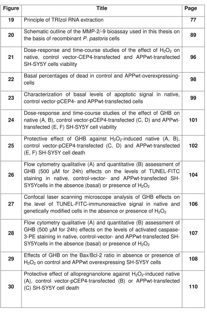
19 Principle of TRIzol RNA extraction 77
20 Schematic outline of the MMP-2/-9 bioassay used in this thesis on
the basis of recombinant P. pastoris cells 89
21
Dose-response and time-course studies of the effect of H2O2 on native, control vector-CEP4-transfected and APPwt-transfected SH-SY5Y cells viability
96
22 Basal percentages of dead in control and
APPwt-overexpressing-cells 98
23 Characterization of basal levels of apoptotic signal in native,
control vector-pCEP4- and APPwt-transfected cells 99 24
Dose-response and time-course studies of the effect of GHB on native (A, B), control vector-pCEP4-transfected (C, D) and APPwt-transfected (E, F) SH-SY5Y cell viability
101
25
Protective effect of GHB against H2O2-induced native (A, B), control vector-pCEP4-transfected (C, D) and APPwt-transfected (E, F) SH-SY5Y cell death
102
26
Flow cytometry qualitative (A) and quantitative (B) assessment of GHB (500 µM for 24h) effects on the levels of TUNEL-FITC staining in native, control-vector- and APPwt-transfected SH-SY5Ycells in the absence (basal) or presence of H2O2
104
27
Confocal laser scanning microscope analysis of GHB effects on the level of TUNEL-FITC-immunoreactive signal in native and genetically modified cells in the absence or presence of H2O2
106
28
Flow cytometry qualitative (A) and quantitative (B) assessment of GHB (500 µM for 24h) effects on the levels of activated caspase-3-PE staining in native, control-vector- and APPwt-transfected SH-SY5Ycells in the absence (basal) or presence of H2O2
107
29 Effects of GHB on the Bax/Bcl-2 ratio in absence or presence of
H2O2 on control and APPwt overexpressing SH-SY5Y cells 108
30
Protective effect of allopregnanolone against H2O2-induced native (A), control vector-pCEP4-transfected (B) or APPwt-transfected
13
Figure Title Page
31
Protective effect of Estradiol against H2O2-induced native (A), control vector-pCEP4-transfected (B) or APPwt-transfected (C) SH-SY5Y cell death
111
32
Dose-response study of the effect of allopregnanolone on native (A), control vector-pCEP4-transfected (B) or APPwt-transfected (C) SH-SY5Y cell viability
113
33
Dose-response study of the effect of estradiol on native (A), control vector-pCEP4-transfected (B) or APPwt-transfected (C) SH-SY5Y cell viability
114
34 Dose-response effect of tunicamycin on native SH-SY5Y cell
viability 115
35 Effects of GHB (A, C) and allopregnanolone (B, D) against
tunicamycin-induced native SH-SY5Y cell death 117 36 Dose-response effect of tunicamycin on native (A), control vector
(B) or APPwt transfected SH-SY5Y cell viability 118 37 Effects of GHB against thapsigargin-induced native (A), pCEP4-
(B) or APPwt-transfected (C) SH-SY5Y cell death 119 38 Effects of allopregnanolone against thapsigargin-induced native
(A), pCEP4- (B) or APPwt-transfected (C) SH-SY5Y cell death 120 39
Effects of separate and concomitant treatments of GHB and allopregnanolone against thapsigargin-induced native (A), control vector (B) and APPwt-transfected (C) SH-SY5Y cell death
122
40 Effects of estradiol against thapsigargin-induced SH-SY5Y cell
death 123
41 Effects of allopregnanolone on thapsigargin-induced cytosolic
calcium elevations 124
42 Effects of GHB on thapsigargin-induced cytosolic calcium
elevations 125
43 Western blot analysis of CHOP protein levels in control vector- (A)
and APPwt-transfected SH-SY5Y cells (B) 126
44 Western blot analysis of XBP-1 protein levels in control vector- (A)
Figures and tables lists
14
Figure Title Page
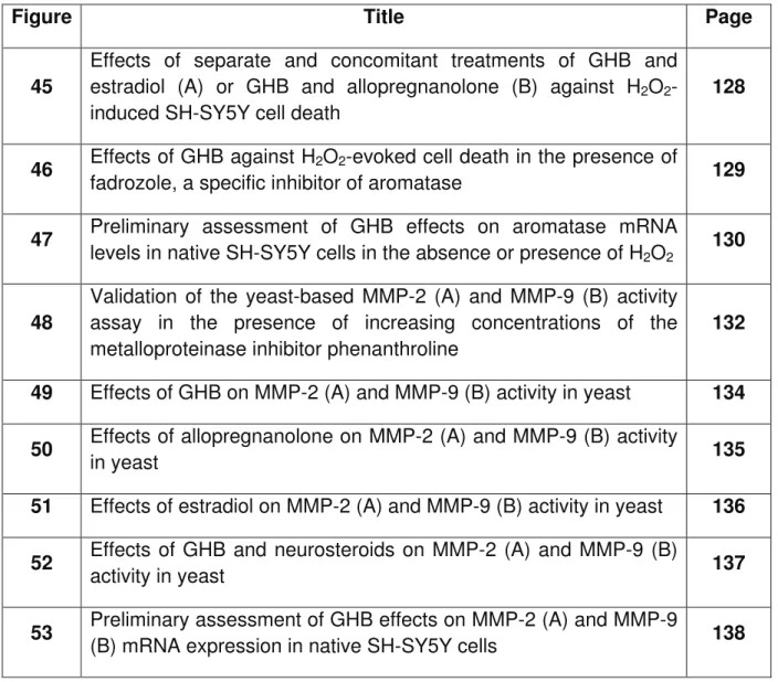
45
Effects of separate and concomitant treatments of GHB and estradiol (A) or GHB and allopregnanolone (B) against H2O2-induced SH-SY5Y cell death
128
46 Effects of GHB against H2O2-evoked cell death in the presence of
fadrozole, a specific inhibitor of aromatase 129
47 Preliminary assessment of GHB effects on aromatase mRNA
levels in native SH-SY5Y cells in the absence or presence of H2O2 130 48
Validation of the yeast-based MMP-2 (A) and MMP-9 (B) activity assay in the presence of increasing concentrations of the metalloproteinase inhibitor phenanthroline
132 49 Effects of GHB on MMP-2 (A) and MMP-9 (B) activity in yeast 134 50 Effects of allopregnanolone on MMP-2 (A) and MMP-9 (B) activity
in yeast 135
51 Effects of estradiol on MMP-2 (A) and MMP-9 (B) activity in yeast 136 52 Effects of GHB and neurosteroids on MMP-2 (A) and MMP-9 (B)
activity in yeast 137
53 Preliminary assessment of GHB effects on MMP-2 (A) and MMP-9
(B) mRNA expression in native SH-SY5Y cells 138
Tables list
Table Title Page
1 Most proteases involved in A- turnover 22
2 Antibodies 65
3 Oligonucleotide sequences 66
4 Reagents, kits and devices 67
15
1. Introduction
1.1.
Alzheimer’s disease
1.1.1. Clinical definition and etiological factors
Alzheimer’s disease (AD), which was first described by Alois Alzheimer in 1907 (Alzheimer, 1907), is the most frequent cause of dementia in Western societies affecting more than 35 million people worldwide, including 860,000 in France and 1.2 million in Germany (Sperling et al., 2011). Thus, AD represents a major health concern identified as a research priority in several countries. The clinical course of AD represents a continuum including three phases; the Preclinical phase, the mild cognitive impairment (MCI) phase and the dementia itself (see Fig. 1) (Sperling et al., 2011). MCI is an intermediate state in which persons have more memory problems than subtle mnesic disturbances considered as normal for their age, but their symptoms are not severe as in AD patients. As the disease progresses (usually a period of 7 to 10 years), AD patients become vegetative and totally dependent for all bodily functions (Petersen et al., 1999). Since there are no curative treatments available yet, the final issue of the pathology is always the death of patients due to secondary complications.
Introduction
16
Figure 1 : Clinical course of Alzheimer’s disease. The preclinical phase is
asymptomatic. Thereafter, mild cognitive impairment is an intermediate state in which persons have more memory problems than normal cases, but the symptoms are not as severe as the symptoms of Alzheimer’s dementia. In dementia, symptoms intensify and people become vegetative and totally dependent for all bodily functions.
1.1.2. Biological hallmarks and mechanisms
Many molecular lesions have been detected in AD, but two major hallmarks are the accumulation of missfolded proteins such as extra-cellular hydrophobic beta-amyloid peptides U1-V and intra-cellular neurofibrillary tangles (NFTs) (Fig 2). Amyloid plaques and NFTs are common features of rare familial AD (FAD) forms (less than 1% of all cases) as well as of sporadic AD (SAD) patients representing the majority of all cases (Querfurth and LaFerla, 2010, Ballard et al., 2011).
17 Figure 2 : Amyloid plaques and neurofibrillary tangles in human cerebral cortex of Alzheimer’s disease patient. (A) Beta-amyloid peptides deposits either in diffuse plaques and in plaques containing elements of degenerating cells, termed neuritic plaques. (B) Neurofibrillary tangles results of intracellular deposition of hyperphosphorylated tau protein. (Adapted from http:// http://library.med.utah.edu/).
1.1.2.1. Amyloid peptides
Beta-amyloid peptides result from the multistep proteolytic cleavage by secretases of amyloid precursor protein (APP), a membrane-bound precursor. APP can be processed in two different cleavage-pathways : (i) the amyloidogenic pathway leads to the formation of hydrophobic A-' 2"2!&3"A (De Strooper et al., 2010) and (ii) the non-amyloidogenic pathway, generating hydrophilic fragments. These two pathways are physiologically balanced in non pathological conditions, but in case of AD, the amyloidogenic pathway is over-activated. Although several hypotheses are suggested in the literature, the mechanisms leading to this over-activation remain
Introduction
18 unclear (De Strooper, 2010, De Strooper et al., 2010, Querfurth and LaFerla, 2010, Ballard et al., 2011).
In the amyloidogenic pathway, APP is cleaved by -secretase and !-secretase (Fig. 3). -secretase releases the ectodomain APPs-, and the remaining APP carboxy-terminal fragment (APP-CTF-) is subsequently cleaved by !-secretase which generates extracellular A-' 2"2!&3"A' $%3' 1QQ' &%!)$/",,:,$)' 30L$&%' U1BPTV5' W8"' biological functions of APPA-, A-' $%3 AICD are poorly understood, although A-' release is associated with decreased synaptic activity and abnormal neurotransmission (Kamenetz et al., 2003). AICD has been proposed to be a transcription factor (Cao and Sudhof, 2001) but this suggestion is controversial (Hebert et al., 2006, Waldron et al., 2008).
The non-amyloidogenic pathway involves "-secretase activity which releases APPs"
ectodomain and carboxy-terminal APP-CFT" is thereafter processed by !-secretase
generating a small p3 fragment and AICD (Fig. 3). It has been shown that APPs"
ectodomain may exert neuroprotective effect and synapse-promoting action, but the mechanisms involved are most non elucidated (Bandyopadhyay et al., 2007). "-Secretase is a family of membrane-bound metalloproteases. Several members of the “A disintegrin and metalloprotease” or ADAM family have been implicated as "-secretases. ADAM-9, -10, and -17 appear to be the major members of the "-secretase family (De Strooper, 2010). In contrast to the "-Secretase family, -Secretase activity is mainly due to one enzyme : the -site APP cleaving enzyme 1 (BACE-1). BACE-1 is a membrane-bound aspartyl protease, and its genetic inactivation results in a dose dependent decrease of A-'4"%")$!&0%'$%3'3"20A&!&0%'&%' various APP-overexpressing mouse models (Laird et al., 2005, McConlogue et al., 2007, Kobayashi et al., 2008). !-Secretase is a multi-enzymatic complex composed
19 of four proteins that are present at equal stoechiometry : Presenilin 1 (PSEN1) or Presenilin 2 (PSEN2), nicastrin, anterior pharynx defective 1 (APH1) and presenilin enhancer 2. In total, there are four different !-secretase complexes allowing therefore several cleavage possibilities of APP (De Strooper, 2010).
Figure 3 : Proteolysis of Amyloid Precursor Protein. Full length APP can be
cleaved by , - and !-secretases. Cleaving sites are indicated. Cleavage by "-secretase yields a soluble ectodomain (APPs?) and a membrane-bound
carboxyterminal fragment (APP-CTF?) which after cleavage by !-secretase yields p3
and the APP intracellular domain (AICD). -secretase (also known as -site APP cleaving enzyme 1, BACE-1) yields APPs- and APP-CTF-, which is then processed
by !-secretase into A- and AICD. Adapted from De Strooper, 2010.
It is important to realize that A-' 2"2!&3"A' /0%A&A!' 0#' $' 8"!")04"%0:A' L&X!:)"' 0#' peptides having different solubility, stability and biological/toxic properties. C-terminal heterogeneity is generated by the !-secretase complex itself. This protease cleaves
Introduction
20 APP at different positions (Fig. 4), generating a variety of peptides consisting of 38 to 43 amino acids. Additional heterogeneity is due to further extracellular enzymes (Kumar et al., 2011) resulting in a mix of more than 20 A-'2"2!&3"A'!8$!'$,,'2$)!&/&2$!"' to different putative functions in the normal brain and also to oligomerization and fibrillization in the AD brain. A-RS'&A'/0%!&%:0:A,.'2)03:/"3'&%'90!8'8"$,!8.'$%3'1T' brain, whereas other A-'2"2!&3"A such as A-RD are continuously produced at much higher levels in AD brains (Benilova et al., 2012).
Figure 4 : Generation of A peptides from APP. The sites of secretases-mediated cleavage are indicated with arrows, and the transmembrane domain of APP is 8&48,&48!"3'&%'4)".5'F-secretase-mediated cleavages produce a pool of A- peptides, varying in their lenght and hydrophobicity. Adapted from Benilova et al., 2012.
The well known peptide is A-42 which was firstly described in FAD. C-terminal fragments of A-42 provided the conditions initiating polymerization mechanisms leading to the formation of amyloid plaques (Jarrett et al., 1993a, b).
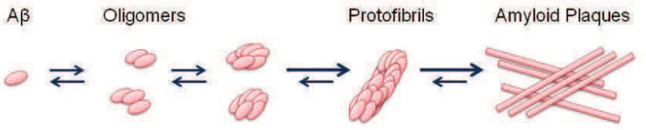
A- monomers (~4kDa) spontaneously self aggregates into multiple coexisting physical forms. One form consists of oligomers (2 to 6 peptides), which merge into intermediate assemblies (Kayed et al., 2003). A- can also grow into fibrils, which
21 assembly --sheets to form the insoluble fibers of advanced amyloid plaques (Fig. 5). Soluble oligomers and intermediate amyloid are the most neurotoxic form of A-.
Figure 5 : Aggregation of A into oligomers, fibrils and plaques. Due to its hydrophobicity, A- monomers self aggregate into various oligomers or larger conformation until Amyloid Plaques. Most of toxicity is assumed by the oligomeric forms. Adapted from De Strooper, 2010.
The fact that many research studies used solutions containing only a single type of A- in order to investigate aggregation and toxicity properties may not be appropriate to clarify AD pathogenesis. Indeed, A-' $//:L:,$!&0%' $%3' !0X&/&!. are likely to be strongly mediated by the concomitant presence of various A- species. In addition to A-42, A-43 is increased in some FAD, while shorter peptides are decreased (Portelius et al., 2010). Moreover, recent evidence show that A- polymerization represents a complex “melting pot“ of different A- species which occurs via metastable intermediaries in a process called “nucleated conformational conversion” (Benilova et al., 2012).
Introduction
22 1.1.2.1.1. Biological clearance of amyloid peptides from the brain The physiological A- fractional clearance rate is estimated to be about 8% per hour (Bateman et al., 2006). Physiological parameters, like blood and cerebrospinal fluid in the brain together with various “clearance receptors” are implicated in the removal of A-, which can also be taken up by endothelial cells of the blood-brain-barrier (De Strooper, 2010). Defects in these clearance processes appear to be relevant for the accumulation of A- on the blood vessels walls, causing vascular abnormalities and angiopathy observed in AD brains.
The proteolytic machinery in cerebral tissues also contribute to the clearance of A- peptides and fibrils. Knockout experiments of specific proteases demonstrated the increase of cerebral concentrations of A- peptides (Table 1).
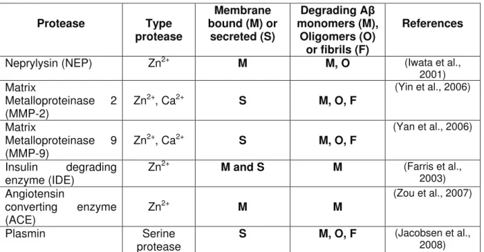
Table 1 !"#$%&"'($&)*%)%"+,-$.-)/"+,"0 "&1(,$-)(2
Protease Type protease Membrane bound (M) or secreted (S) Degrading A monomers (M), Oligomers (O) or fibrils (F) References
Neprylysin (NEP) Zn2+ M M, O (Iwata et al.,
2001) Matrix Metalloproteinase 2 (MMP-2) Zn 2+, Ca2+ S M, O, F (Yin et al., 2006) Matrix Metalloproteinase 9 (MMP-9) Zn 2+, Ca2+ S M, O, F (Yan et al., 2006) Insulin degrading enzyme (IDE) Zn
2+ M and S M (Farris et al.,
2003) Angiotensin converting enzyme (ACE) Zn 2+ M M (Zou et al., 2007) Plasmin Serine protease S M, O, F (Jacobsen et al., 2008)
23 Matrix-Metalloproteinases are secreted zinc- and calcium-dependent endopeptidases. MMP-2 and MMP-9, also known as gelatinases, are secreted and activated in the extra-cellular compartment. They modulate various physiological functions including neuritic outgrowth and matrix remodeling (Page-McCaw et al., 2007, Ould-yahoui et al., 2009) but they are also involved neuroinflammation or tumor invasiveness (Curran and Murray, 1999, Yong, 2005). MMP-2 and MMP-9 are expressed at low levels in the brain, but their astrocytal expression can be stimulated by A- peptides. Knockout of MMP-2 results in increased concentration of A- peptides in the soluble fraction of hippocampus and cortex, while infusion of broad-spectrum of MMPs inhibitor in the brain also induced an increase of A- levels (Yin et al., 2006).
In both FAD and SAD, there is a clear unbalance between the production and the clearance of A- peptides. Therefore, one therapeutic strategy may be the up-regulation of the brain expression/activity of amyloid degrading enzymes such as MMP-2 or MMP-9.
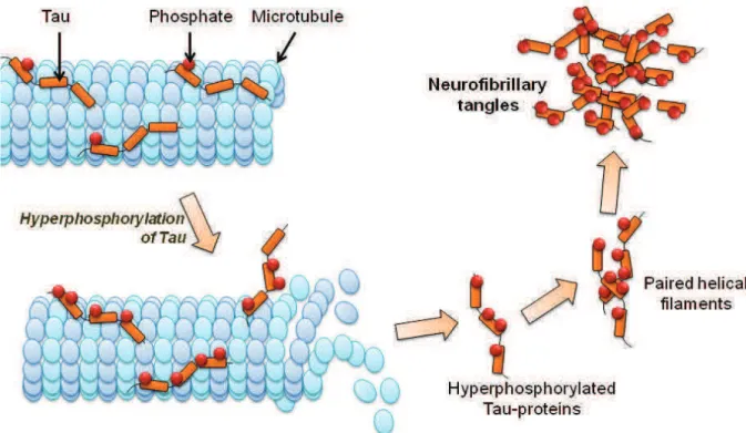
1.1.2.2. Neurofibrillary tangles
Neurofibrilary tangles (NFTs) are filamentous inclusions found in cell bodies and apical dendrites in AD and other neurodegenerative disorders generally termed tauopathies (Lee et al., 2001). Usually, the number of NFTs reflects the severity of the disease. NFTs are mainly composed of highly phosphorylated and aggregated forms of the tau protein which belongs to the family of microtubule-associated proteins (MAPs) abundant in axons (Grundke-Iqbal et al., 1986). Tau promotes the assembly and stability of microtubules and vesicle transport in axons. Hyperphosphorylation of Tau leads to an insoluble form which lacks affinity for
Introduction
24 microtubules and self-associate into paired helical filaments and finally form the NFTs (Fig. 6). Both A- and Tau undergo nucleation-dependent fibril formation.
Figure 6 : Formation of paired helical filaments and neurofibrillary tangles. Tau binding promotes microtubules assembly and stability. Excessive kinases and reduced phosphatases activities lead to an hyperphosphorylation of Tau and induces its detachment and self-aggregate. Adapted from Querfurth and LaFerla, 2010.
Like A- oligomers, intermediate aggregates of hyperphosphorylated Tau are cytotoxic and impair cognition (Santacruz et al., 2005, Khlistunova et al., 2006). Tau mutations do not occur in AD and increased levels of phosphorylated Tau and total Tau in the cerebrospinal fluid correlate with the reduction of cognitive performance (Wallin et al., 2006). Evidence show that A- peptides accumulation precedes and drives Tau-aggregation (Gotz et al., 2001). Moreover, A- toxicity requires the presence of endogenous Tau (Roberson et al., 2007, Ittner et al., 2010, Ittner and Gotz, 2011). Tau contains 80 serine and threonine residues and 5 tyrosines that can
25 be phosphorylated. In physiological conditions, there are 2-3 Mol of phosphate per Mol of Tau. But in AD brain, there are 7-8 Mol of phosphate per Mol of Tau. Many phosphokinases, including glycogen synthase kinase 3-'U( G3-V, cyclin-dependent kinase 5 (CDK5), and extracellular signal-related kinase 2 (ERK2) and mitogen activated protein kinase (MAPK) have been investigated as potential targets to reduce Tau phosphorylation (Takashima, 2009).
If A- peptides colonize synchronously predilection sites in the brain of AD patients (in hippocampus, amygdala and prefrontal cortex), NFTs develop an spread in a predictable manner. Braak and collaborators organized AD progression into six stages based on the distribution of NFTs (Braak and Braak, 1996). During the first stage, NFTs are observed in the transentorhinal and Cornu Ammonis (CA) regions of the hippocampus. The number of NFTs increases in Braak stage II. Stages I and II together are called the “transentorhinal stage”. Brains of normal non-demented aged subjects are often categorized as Braak stages I and II. In Braak stage III and IV, called the “limbic stage”, many NFTs appear in the entorhinal cortex, and tangles are found in the entire limbic system, including hippocampal regions CA1-4 and the amygdala. During the limbic stage, patients show various AD-specific symptoms, such as memory impairment, reduced spatial cognition and increased anhedonia, as a result of neuronal dysfunctions in the limbic system. In Braak stages V and VI, called the “isocortical stage”, NFTs are present in the cerebral cortex, where they impair neuronal functions, leading to dementia. The increasing spread of NFTs from the transentorhinal cortex to the limbic system, and finally to the cortex, correlates with the severity of cognitive impairment. Samuel et al. reported that the number of NFTs in the hippocampal formation correlates with the degree of dementia and that synapse loss is a key determinant of dementia in AD (Samuel et al., 1994).
Introduction
26 1.1.3. Biological consequences of Amyloid peptides and neurofibrillary
tangles
1.1.3.1. Synaptic failure and axonal transport impairment
Aging itself causes synaptic loss (Masliah et al., 2006), therefore, it is easy to understand that AD is primarily a disorder of synaptic transmission (Selkoe, 2002). As aforementioned, NFTs and synaptic loss are pivotally involed in AD physiopathology. In parallel to Braak spreading of the disease, hippocampal synapses begin to decline in MCI patients (Scheff et al., 2007). Some evidence reveal a 25% decrease of presynaptic vesicle synaptophysin in mild AD (Masliah, 2001, Masliah et al., 2001). In the last Braak stages, dramatic synaptic loss is positively correlated with dementia (Terry et al., 1991).
A- peptides are known to impair the “long term potentiation” (LTP), an experimental indicator of memory formation (Palop and Mucke, 2010). Subsequently, signaling molecules important to memory are also inhibited. Many studies investigated the effects of A- peptides on excitatory synaptic transmission, that is tightly regulated by the number of active N-methyl-D-aspartate receptor (NMDAR) and the ?-amino-3-hydroxy-5-methyl-4-isoxazoleproprionic acid receptor (AMPAR). NMDAR activation has a central role in memory, by inducing LTP or long term depression (LTD), depending on the extent of the resultant intracellular calcium ([Ca2+]i) rise in the dendritic spines and the downstream activation of intracellular cascades (Kullmann and Lamsa, 2007). Activation of post-synaptic NMDARs and large increases in ([Ca2+]i) are necessary for LTP, whereas internalization of NMDARs, activation of perisynaptic NMDARs and lower increase in [Ca2+]i are necessary for LTD. LTP induction promotes recruitment of AMPARs and growth of dendritic spines, whereas LTD induces spine shrinkage and synaptic loss (Kullmann and Lamsa, 2007).
27 Pathologically elevated A- may indirectly cause a partial block of NMDARs and induce LTD and synaptic loss. Although the mechanisms underlying A--induced LTD remain poorly understood, they may involve the internalization or desensitization of receptors. A- effects on synaptic function may be mediated by the activation of presynaptic ?7-nicotinic-acetylcholine receptor (?7-nAChR) and perisynaptic activation of NMDARs, inducing excessive Ca2+ inwards and downstream effects on many intracellular cascades including the activation of GSK3-' $%3' +1QG' A&4%aling pathways. A--induced synaptic depression may result from NMDAR activation followed by NMDAR desensitization, internalization and by the stimulation of perisynaptic NMDARs or metabotropic glutamate receptors. These processes lead to a chronic increase in excitotoxic Ca2+ inwards, which can also lead to cell death. Several data involve oligomeric A- in synapse failure, but until recently, only little was was known about the role of Tau in these dysfunctions. Ittner and colleagues described a dendritic function of Tau, mediating A- toxicity via NMDAR conformation (Ittner et al., 2010). Tau is able to target the src kynase Fyn to the dendrite, where it phosphorylates the NMDAR subunit NR2B, thereby facilitating complex formation with the post-synaptic density protein 95 (PSD95) and allowing excitotoxic over-activation of NMDAR by A-.
Moreover, neurons are elongated cells, and in order to maintain neuronal functions and excitability, they need efficient delivery of cellular organelles (such as mitochondria, endoplasmic reticulum, lysosome..) from the soma to the axon, dendrite and synapse. The delivery of organelles is purely based on microtubules, which serves as “rail tracks”, motor protein (such as kynesin or dynein) that represent engines, and organelles as cargoes which are directed to the cell-periphery or back again to the soma. Tau is known to facilitate the anterograde axonal transport of
Introduction
28 organelles such as mitochondria (Gotz et al., 2006). In AD neurons, the detachment of hyperphosphorylated tau from the microtubules leads to impaired axonal transport of organelles (such as mitochondria targeted to the synapses).
1.1.3.2. Neuroinflammation
Biochemical markers of activated microglia and reactive astrocytes are increased in the brain of AD patients (Wyss-Coray and Mucke, 2002). In physiological conditions, phagocytic microglia engulf and degrade A-. However, the large and constant production of A- in the pathological case chronically activate microglia, leading to the release of a myriad of damaging chemokines and cytokines such as interleukin-1, interleukin-6 and tumor necrosis factor ? (TNF-?V (Akiyama et al., 2000). The binding of A- peptides to advanced glycosylation end products receptors (expressed by microglia) amplifies the generation of cytokines, glutamate and nitric oxide (Yan et al., 1996).
1.1.3.3. Loss of calcium regulation
Loss of calcium regulation is common to several neurodegenerative disorders. In AD, elevated concentrations of cytosolic calcium stimulate the amyloidogenic pathway (Isaacs et al., 2006). The chronic state of glutamatergic receptor activation is thought to aggravate neuronal damage in AD. NMDAR activation increases cytosolic calcium, which in turn stimulates calcium release channels in the endoplasmic reticulum. Evidence also show that A- could form voltage-independent cation channels in lipid membranes, resulting in calcium uptake and degeneration of neuritis (Lin et al., 2001). Deregulation of cytosolic calcium concentration can lead to a “calcium
29 overload”, which is capable to induce mitochondrial dysfunctions, oxidative stress and also cell death.
1.1.4. Oxidative stress in Alzheimer’s disease
The brain requires 20% of total blood flow and 25% of the body’s glucose utilization. Due to their excitable properties and their important need in energy, neurons have a large number of mitochondria either in the soma or in synaptic ends. Since neurons have a limited glycolytic activity, mitochondria play an essential role in bioenergetics thanks to adenosine triphosphate (ATP) production. Also, mitochondria are key producers of reactive oxygen species (ROS). Unbalance between ROS formation and reduction may lead to the deleterious oxidation of lipids, proteins and DNA, also called “oxidative stress”.
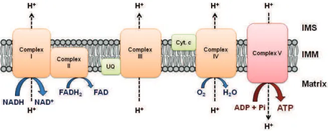
In neurons or glial cells, ATP is produced aerobically through the tricarboxilic acid (TCA) cycle and the oxidative phosphorylation (OxPhos), also called respiratory chain, in mitochondria. TCA cycle is composed of 8 enzymatic steps and converts carbohydrates and free fatty acids into ATP. It also yields electrons in form of reduced hydrogen carriers, nicotine adenine dinucleotide (NADH) and flavin adenine nucleotide (FADH2). These compounds enter subsequently as coenzymes into the OxPhos, which is composed of electron transport chain or ETC (enzymatic complexes I-IV) and an ATP-synthase (complex V). These five enzyme complex are functionally interconnected via mobile electron carriers ; cytochrome c and ubiquinone/coenzyme Q. Enzymatic complexes and electron carriers are localized at the inner mitochondrial membrane (Fig. 7). In addition to flavin and nicotinamides, ETC involves cytochromes and metal ions clusters (iron and copper) to transfer electrons in a sequence of oxido/reductions steps, leading to a proton gradient
Introduction
30 across the inner membrane which finally drives ATP synthesis via the complex V. In parallel, the complexes transfer electrons to oxygen and produce water. OxPhos is not totally efficient and up to 2% of electrons are incompletely reduced to yield O2¯ ,
instead of H2O (Eckert et al., 2013, Sutherland et al., 2013).
Figure 7 : Schematic representation of the oxidative phosphorylation system. Complex I (NADH-ubiquinone oxidoreductase) and II (succinate dehydrogenase) also belong to the tricarboxilic acid cycle (TCA) and receive electrons from NADH and FADH2, respectively. Thereafter, electrons are driven from complexes by the mobile
electron carrier ubiquinone (UQ) and cytochrome c (Cyt c) to the final acceptor, molecular oxygen. In parallel, a proton gradient is established across the inner membrane (IMM) in complexes I, III and IV. This gradient is utilized by complex V to generate ATP. IMS : inter-membrane space. Adapted from Eckert et al., 2013.
Low molecular weight anti-oxidant as glutathione and metal-containing enzymes including superoxide dismutase (SOD), catalase (CAT) and glutathione peroxidase (GPX), act to control the cellular redox environment. The first reducing barrier against ROS is SOD which converts O2¯ to H2O2. Then, CAT and GPX transfer H2O2 to
oxygen and water. Iron and copper, which are also abundant in the brain, may facilitate H2O2 conversion into other ROS or may promote A- peptide formation (von
31 1.1.4.1. Amyloid peptides and oxidative stress
In the ageing brain, OxPhos not only becomes less efficient, but there is a substantial decrease in anti-oxidant enzymes expression/activity (Kowald, 2001). Also, A- peptides are potent mitochondrial poisons, which especially affect the synaptic pool (Mungarro-Menchaca et al., 2002). A- and APP itself were reported to accumulate in mitochondria in the brain of AD patients (Pavlov et al., 2009). A- peptides alter key mitochondrial enzymes (Hauptmann et al., 2006) such as cytochrome c oxidase (COX/Complex IV), pyruvate dehydrogenase (PDH) and ?-ketoglutarate dehydrogenase (?KGD), leading to bioenergetic disturbances as evidenced in AD patients (Mosconi et al., 2008). Exposure of human neuroblastoma SH-SY5Y cells to A- peptides revealed a significant decrease of Complex IV activity (Lim et al., 2010). Moreover, decreased brain levels of antioxidant enzymes such as SOD or CAT are also observed in AD (Aksenov et al., 1998). The combination of ROS elevation to antioxidant enzyme decrease evidenced in brain tissues from AD patients suggests that oxidative stress may be a central element of AD physiopathology. Cu2+ ions have also been shown to render A- more toxic. Indeed, dimeric conformation of A-42 appear as potent inhibitor of COX, but only in the presence of Cu2+ (Crouch et al., 2005).
The mitochondrial A- binding protein alcool dehydrogenase (ABAD) can also bind to A-42 present in the cortical mitochondria of APP transgenic mice (Lustbader et al., 2004, Yan et al., 2007). In mice double transgenic for ABAD and APP, toxic effects of A- was increased compared to APP single transgenic mice. This interaction promotes the leakage of ROS, mitochondrial dysfunction, cell death, as well as spatial learning and memory deficits (Lim et al., 2010, Lim et al., 2011, Grimm et al.,