HAL Id: hal-00978024
https://hal.archives-ouvertes.fr/hal-00978024
Submitted on 14 Apr 2014
HAL is a multi-disciplinary open access
archive for the deposit and dissemination of
sci-entific research documents, whether they are
pub-lished or not. The documents may come from
teaching and research institutions in France or
abroad, or from public or private research centers.
L’archive ouverte pluridisciplinaire HAL, est
destinée au dépôt et à la diffusion de documents
scientifiques de niveau recherche, publiés ou non,
émanant des établissements d’enseignement et de
recherche français ou étrangers, des laboratoires
publics ou privés.
Structural and electronic relaxations around
substitutional Cr3+ and Fe3+ ions in corundum
Emilie Gaudry, Amonmat Kiratisin, Philippe Sainctavit, Christian Brouder,
Francesco Mauri, A. Ramos, Andrei Rogalev, José Goulon
To cite this version:
Emilie Gaudry, Amonmat Kiratisin, Philippe Sainctavit, Christian Brouder, Francesco Mauri, et al..
Structural and electronic relaxations around substitutional Cr3+ and Fe3+ ions in corundum. Physical
Review B: Condensed Matter and Materials Physics (1998-2015), American Physical Society, 2003,
67, pp.094108-094118. �10.1103/PhysRevB.67.094108�. �hal-00978024�
Structural and electronic relaxations around substitutional Cr
3¿and Fe
3¿ions in corundum
Emilie Gaudry,*Amonmat Kiratisin, Philippe Sainctavit, Christian Brouder, Francesco Mauri, and Aline Ramos Laboratoire de Mine´ralogie Cristallographie de Paris, Universite´ Paris 6, Case 115, 4 Place Jussieu, 75252 Paris Cedex 05, France
Andreı¨ Rogalev and Jose´ Goulon
European Synchrotron Radiation Facility, BP 220, F-38043 Grenoble Cedex, France
~Received 11 July 2002; revised manuscript received 7 October 2002; published 17 March 2003!
The local arrangement of atoms surrounding a substitutional Cr31and Fe31ion ina-Al
2O3: Cr31~ruby!
and ina-Al2O3:~Fe-Ti! ~blue sapphire! has been studied experimentally using natural x-ray linear dichroism
absorption at the Cr and Fe K edge. The isotropic and dichroic signals have been recorded using single crystals and a reliable method has been applied to remove diffraction peaks. Results given by the analysis of both signals are compared with ab initio density functional calculations. This study reveals that the introduction of an impurity ion in the structure leads to relaxations that are very local in nature. The oxygen atoms in the coordination shell relax to an arrangement similar to that for Cr ina-Cr2O3or for Fe ina-Fe2O3. Aluminum
or oxygen atoms at a farther distance are weakly affected by the presence of the impurity.
DOI: 10.1103/PhysRevB.67.094108 PACS number~s!: 78.40.Ha, 78.70.Dm, 61.72.Bb
I. INTRODUCTION
The color of allochromatic minerals results from minor impurities and is usually interpreted within the framework of ligand field theory. Indeed, the color diversity in minerals doped with the same impurity is explained by a modification of the crystal field, due to a change of the local environment around this impurity. For example, ruby (a-Al2O3: Cr31)
and emerald (Be3Al2Si16O18:Cr31) are both doped by
chro-mium in the same valence state and in similar octahedral environments, but ruby is red and emerald is green. The dif-ference of color is explained by a smaller ligand field in emerald than in ruby, which is related to a supposed weaker Cr-O bonding in emerald than in ruby.1Likewise, a weaken-ing of the ligand field due to a longer Cr-O bond length in eskolaite (a-Cr2O3) than in ruby is invoked to interpret the
transition of color from red in ruby to green in eskolaite when increasing the chromium proportion in a-Al2O3: Cr31.2To enforce these arguments, a precise description of the local environment around an impurity is needed.
On the other hand, the calculation of the optical properties of materials has made great progress recently.3– 6These cal-culations use a combination of the GW approximation and the Bethe-Salpeter equation to describe accurately the elec-tronic excitations and the excitonic processes in semiconduc-tors and insulasemiconduc-tors. However, these methods were not able to calculate the optical spectra of a transition metal impurity. There are two reasons for this failure. First, transition metal impurities exhibit strong electronic correlations, secondly, the crystallographic structure around an impurity in a crystal is not known. The theory of strongly correlated systems im-proves steadily and a realistic modeling of the optical spectra of transition metal impurities does not appear out of reach. Therefore, an accurate determination of the atomic position around a transition metal impurity becomes an urgent task.
In this paper, we are interested in the local environment around chromium in ruby (a-Al2O3: Cr31) and around iron
in a-Al2O3: Fe31. Corundum (a-Al2O3) is a transparent and highly insulating material with a wide band gap energy
@measured value of 8.8 eV ~Ref. 7!#. When some transition
metal impurities substitute for aluminum in the structure, it becomes colored. Ruby is red anda-Al2O3: Fe31is yellow.
To investigate the local structure around chromium or iron, we use a powerful experimental technique that not only gives precise metal-oxygen distances in the coordination shell, but gives metal-aluminum second neighbors distances. All these results, with additional density functional calculations, allow us to have a complete picture of the local environment of chromium and iron impurities ina-Al2O3.
The crystal structure of a-Al2O3 belongs to the space
group R3¯ 2/c or D3d6 , thus making it a uniaxial birefringent material.8 The rhombohedral unit cell contains two Al2O3
species. Aluminum ions are linked to six oxygen ions in a distorted octahedron, with respect to pure octahedral geom-etry. The ionic radius of the paramagnetic impurities is larger than the ionic radius of aluminum atoms (rCr3150.615 Å,
rFe3150.645 Å, and rAl3150.535 Å in an octahedral site9!. This imposes strong relaxation in the material. Since the Al-O bond in corundum is known to be ionocovalent,10 the relaxation is not simply given by the difference between the ionic radii of the impurity and the aluminum ion. Many stud-ies have been undertaken to determine the angular and radial relaxations. Single crystal x-ray diffraction,11,12 UV-VIS spectroscopy,13,14and EPR spectroscopy15,16have shown the existence of such relaxations. However the metal-oxygen ( M -O! bond lengths in the coordination shell were not de-termined, merely the averaged M-O distances. This can hide probable differences between oxygen atoms in the coordina-tion shell. For instance, the mean Cr-O distance ina-Cr2O3
is 1.99 Å ~Ref. 17! and the mean Fe-O distance ina-Fe2O3is
2.02 Å.18 ~the mean Al-O distance ina-Al2O3 is 1.92 Å.17!
Although the two mean bond lengths are quite similar, the trigonal distortion of the octahedral site in a-Cr2O3 is very
different from that in a-Fe2O3. The two M-O distances in
the coordination shell are 1.97 and 2.02 Å ina-Cr2O3while
ina-Fe2O3the values are 1.94 and 2.11 Å ~they are 1.86 and
1.97 Å ina-Al2O3). To determine precisely the positions of
PHYSICAL REVIEW B 67, 094108 ~2003!
the nearest and next nearest neighbors, x-ray absorption fine structure ~XAFS! has been performed at the Cr K edge in powders19 or single crystals20 of a-Al2O3: Cr31. The
ex-perimental determination of the precise Cr-O distances ina -Al2O3: Cr31 powders is difficult to obtain from isotropic
EXAFS signals, due to the large amount of parameters needed to account for the fit. In addition, the difference be-tween two fits performed using either one averaged Cr-O distance or two Cr-O1 and Cr-O2 distances leads to little
improvement of the fit. The extended XAFS ~EXAFS! re-cording of single crystal spectra is difficult due to the pres-ence of diffraction peaks that strongly distort the experimen-tal information.20 No XAFS experiments have been performed at the Fe K-edge in sapphire. At the same time, relaxation calculations around chromium in ruby have been performed using parametrized19 or first principles21 density functional calculations. The results support the existence of the trigonal distortion of the octahedral site of the impurity. No calculation on the structural relaxation around other 3d impurities in corundum has been reported to date.
Our goal is to measure very precisely the distances around paramagnetic impurities ina-Al2O3. To do that, it is
neces-sary to record XAFS signals on single crystals, despite the presence of unavoidable diffraction peaks. In this study, a powerful experimental technique22 presented in Sec. II is used to record XAFS spectra of single crystals at the chro-mium K edge ofa-Al2O3: Cr31and at the iron K edge ofa
-Al2O3: Fe31. The data treatment gives, simultaneously, the
isotropic spectra and the dichroic spectra, without any dif-fraction peak. The isotropic signal is then classically ana-lyzed to yield averaged bond lengths in the coordination shell. To obtain accurate structural information about the im-purity site, it is necessary to analyze the dichroic signal, which is more sensitive to the difference of distances be-tween the two types of oxygen atoms in the coordination shell. Such an analysis is not typical, and details are pre-sented in Sec. III. At the same time we have carried out first principles density functional calculations on a-Al2O3: Cr31anda-Al2O3: Fe31~Sec. IV!. The calculated M-O and
M-Al distances are compared to experimental values. Results are discussed in Sec. V.
II. X-RAY ABSORPTION MEASUREMENTS
In order to examine the local environment around the im-purity ions in ruby and sapphire, it is necessary to measure EXAFS signal on single crystals for two independent orien-tations of the linear x-ray polarization. We also use the di-chroic signal, which gives direct evidence of the inequiva-lence between atoms around the impurity site.
Two types of single crystals synthesized by the Verneuil process are studied. They are rubies (a-Al2O3: Cr31)
con-taining 60 (610), 800 (630), 10 000 (6500) wt. ppm of chromium and a sapphire (a-Al2O3:Fe,Ti! containing 1500
(650) wt. ppm of iron and 750 (630) wt. ppm of titanium. These compositions were analyzed using the Cameca Micro-beam electron microprobe at the CAMPARIS analytical fa-cility of the Universities of Paris 6/7. A 30 kV acceleration with a 15 nA beam current, defocused to 10mm, was used.
X-ray intensities were corrected for dead-time, background, and matrix effects using the Cameca ZAF routine. The stan-dards used were a-Al2O3, a-Cr2O3, and a-Fe2O3. The
single crystals of ruby were cylindrically shaped ~diameter 15 mm and thickness 3 mm!. They were cut so that the C3
axis ~@111# direction in the rhombohedral lattice! was or-thogonal to the normal of the disk surface. The single crystal of blue sapphire was cut in a similar way with the C3 axis
orthogonal to the normal of the surface. We made the XAFS experiments on synthetic blue sapphire (a-Al2O3:Fe,Ti!
in-stead of on yellow sapphire (a-Al2O3: Fe31) because
syn-thetic yellow sapphires (a-Al2O3: Fe31) with high iron
con-centrations are often strongly inhomogeneous. We assume that the low concentration of titanium compared to that of iron in blue sapphire does not affect the site relaxation around iron.
In doing XAFS measurements on single crystals, one is faced with the problem of very intense diffraction peaks. These arise from either diffracted rays striking a fluorescence detector or from the occurrence of diffraction inside the crystal.20 These diffraction rays may drag on and have dev-astating effects on the whole absorption spectrum. Without the use of the rotating sample technique described in the following, many diffraction peaks were present on the XAFS signal, comparable to what was found by Emura et al.20 These diffraction peaks forbade any unambiguous EXAFS analysis.
In this study, the chromium K edge ~energy range 5900– 6700 eV! in rubies and the iron K edge ~energy range 7050– 7500 eV! in sapphire have been measured on the ID12A beam line ~European Synchrotron Radiation Facility, France! dedicated to polarization dependent spectroscopies.23A spe-cial technique called the rotating sample technique, proposed by Goulon,22 is applied to register the absorption spectra. It is an unusual but powerful method to eliminate diffraction peaks and to measure natural linear dichroism on single crys-tals. The monochromatic x-ray beam is obtained through a double Si~111! crystal monochromator, having a resolution
DE/E;1024. The photon flux is linearly polarized with the
polarization vectoreW orthogonal to the normal of the sample surface. Single crystals are placed on a rotating holder that allows for the samples to be rotated around the x-ray beam direction, perpendicular to the face of the sample. The @111# direction stays perpendicular to the x-ray beam direction when the samples are turned. For each energy \vof incident photon, the fluorescence intensity is measured by eight de-tectors for 400 angles of the rotating holder from 0 to 2p
radians. This process is carried out for 700 energy points. Then, the isotropic spectra and the linear dichroism have to be extracted from this amount of data. A filtering algorithm24 is used to obtain, simultaneously, both the isotropic absorp-tion coefficientmiso and the dichroic signalmdichro. This
al-gorithm operates as follows.24The experimental dependence of the intensity recorded by each detector at each energy \v
as a function of angle is fitted to the function f~u,\v!5a~\v!1b~\v!cosu1c~ \v!sinu
where u is the rotation angle. This function takes various phenomena into account. The dependence on cosu and sinu
describes the fact that the x-ray beam is not circular, and that the rotation axis of the sample holder might not be perfectly aligned with the x-ray beam direction. In principle, the spec-trum averaged over angles is given by a(\v), and the an-gular dependence is given by d(\v) and e(\v). It must be checked that b(\v) and c(\v) are small and do not vary much with \v. For each detector and each energy, the func-tion f (u,\v) is first fitted to all of the data ~i.e., intensities as a function of angle!. A root mean square errors, between fit and data is obtained. Then, each data point i is in turn selected from the experimental data. A fit is calculated skip-ping point i and the corresponding root mean square errorsi is calculated. The experimental point i0, causing the largest
error, is determined by choosing i0such thatsi0is the small-est among thesi. Then,si0/s is calculated. If the ratio is greater than 0.95, it is considered that the fit is not signifi-cantly improved by suppressing any point in the data, and therefore the data is taken to be correct. If the ratio is smaller than 0.95, then point i0 is removed from the experimental
data and the process is started again. Once the filtering pro-cedure is finished, the functions a(\v), b(\v), c(\v), d(\v), and e(\v) of all of the detectors are then summed over. The experimental signal may be written as
S~u,\v!5Sa~ \v!1Sb~ \v!cosu1Sc~ \v!sinu
1Sd~ \v!cos 2u1Se~ \v!sin 2u. ~2!
Given that a-Al2O3 is uniaxial, with an appropriate choice
for the origin ofu, Se(\v) can be reduced almost to null so that the isotropic part of the signal is @3Sa(\v)
2Sd(\v)#/3 and 2Sd(\v) is the dichroic XAFS signal.25
III. EXAFS ANALYSIS
The measured isotropic and dichroic x-ray absorption spectra from the 10 000 wt. ppm Cr31and 1500 wt. ppm Fe31doped alumina samples are reproduced in Figs. 1 and 2. We have also measured the EXAFS signal of the other two
a-Al2O3: Cr31single crystals with chromium concentration
equal to 800 and 60 wt. ppm. The Cr K-edge EXAFS signals of the threea-Al2O3: Cr31samples are essentially identical,
except for signal to noise ratio. We also measured the XANES spectra for the three chromium concentrations. It appears that the chromium site must be largely independent of the chromium concentration in a range between 60 and 10 000 wt. ppm. In the following, the EXAFS analysis is performed on the 10 000 wt. ppm a-Al2O3: Cr31and the
1500 wt. ppma-Al2O3:~Fe-Ti! samples.
Given that the isotropic signal is not very sensitive to the small difference between the two M-O distances in the coor-dination shell, structural information determined by its analysis only gives an averaged picture. The interesting point is the analysis of the dichroic spectra, which provides accu-rate results concerning the inequivalence between atoms.
A. Isotropic signal
A standard procedure is used to analyze the isotropic part of the two absorption spectra.26,27The preedge background is removed by applying a polynomial fit to the pre-edge data and extrapolating it into the post-edge region. Data normal-ization and energy shift is performed using an error function
~step function! and a pseudo-Voigt function ~peak function!.
The energy scale is converted to reciprocal space units using k5(1/\)
A
2m(E2E0), where E0 is the threshold energy ofthe absorption edge. Then, a cubic spline function is fitted to the kn weighted data by a non linear least square fit of the spline knots. These normalized EXAFS signalsx(k) are fur-ther analyzed as a Fourier series of plane wavelets. Each has a scattering amplitude of Aj, a back-scattering function
Fj(k) and a total phase function Fi j(k)52d1
8
(k)1uj(k). The first termd18
(k), arises from the potential of the central atom and uj(k) is the phase function of the neighboring atoms28x~ k !521
k
(
jAjsin@2krj1Fi j~ k !#. ~3!
The summation is over j shells and the scattering amplitudes Aj, are related to the electron mean free path lj(k), the
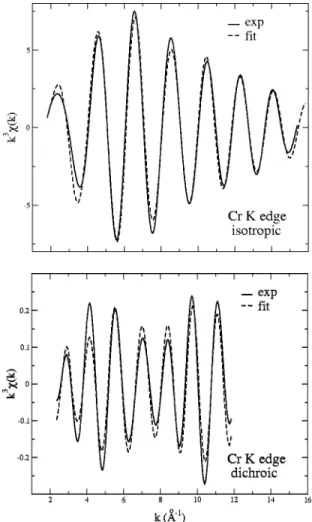
FIG. 1. Isotropic ~top! and dichroic ~bottom! x-ray absorption spectra (K edge! for Cr in ruby.
STRUCTURAL AND ELECTRONIC RELAXATIONS . . . PHYSICAL REVIEW B 67, 094108 ~2003!
coordination number Nj, the bond distances rj, and the Debye-Waller factor sj: Aj5 Nj r2j Fj~ k !e 22sj2k2e22rj/lj(k). ~4!
The radial structure function in real space F(r), is ob-tained by the Fourier transform of thex(k) spectra, using the equation F(r)5(1/2p)*k
min
kmax
W(k)knx(k)e2ikrdk, where W(k) is a Kaiser window function to minimize truncation effects in the Fourier transform.29
We are interested in the determination of structural pa-rameters of both the first shell ( M -O bonding! and the sec-ond shell ( M -Al bsec-onding!. We carry out the analysis in two steps. First we select an r range in which only M-O contri-butions are expected and we separate these EXAFS oscilla-tions from the total experimental signal using back Fourier transform. It is well known that in a-Al2O3, the six Al-O distances in the first coordination shell are gathered in two groups to comply with the C3 symmetry. In addition, EPR
measurements suggest that the chromium site in a-Al2O3:
Cr31is trigonal.30Then, the isotropic signal is analyzed with one mean metal-oxygen distance for the fit. The fit gives initial information on Nj, rj,sj and DE0
j that can be useful for the second step. For the second stage, the same separation
and fit procedure is applied to determine structural informa-tion on both the first shell ( M -O bonding! and the second shell ( M -Al bonding!. In fact, it is very difficult to separate the M-O contribution from the M-Al because of the displace-ment of peak positions due to phase shifts Fi j(k). The EX-AFS oscillations of particular interest are separated from the total experimental signal by a back Fourier transform of the signals. This gives a signal ~solid line in Figs. 3 and 4! that can be fitted by a model function ~dotted line in Figs. 3 and 4!.
B. Dichroic signal
The analysis of the dichroic signal is novel. In keeping with the formalism developed in the preceding paragraph, it is necessary to turn the linear dichroic absorption coefficient
mdichro(k) into a xdichro(k) signal
xdichro~ k !5 mdichro~ k ! m0~ k ! 5mi~ k !2m m0~ k ! 0~ k ! 2m'~ k !2m m0~ k ! 0~ k ! . ~5!
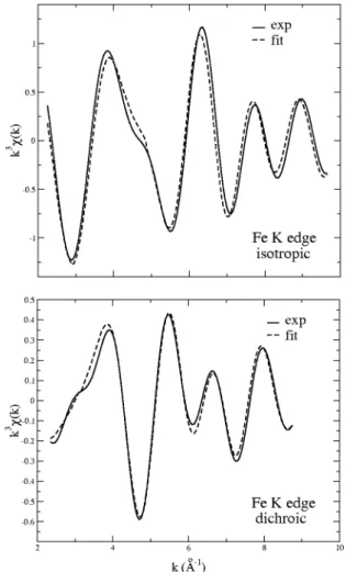
We consider a fully linearly polarized x-ray beam.31The normalized dichroic EXAFS signal is then given by28 FIG. 2. Isotropic ~top! and dichroic ~bottom! x-ray absorption
spectra (K edge! for Fe in sapphire.
xdichro~ k !52 1 k
(
j A8
jsin@2krj1Fi j~ k !#, ~6! where A8
j53Nj r2j Fj~ k !e 22sj 2k2 e22rj/lj(k)@^
~eˆ i• rˆj!2&
2^
~eˆ'• rˆj!2&
# ~7! and eˆi andeˆ' are unit vectors in the @111# direction and a
perpendicular direction (@1¯ 21¯#, for example!,
^
(eˆ• rˆj)2
&
51/Nj(i(eˆ• rˆj
i)2 with i running over atoms in the jth shell.
Let us introduce N
8
j53Nj@^
(eˆi• rˆj)2&
2^
(eˆ'• rˆj)2&
#. Equa-tion ~6! becomes xdichro~ k !52 1 k(
j N8
j r2j Fj~ k !e 22sj 2k2 e22rj/lj(k)sin@2kr j 1Fi j~ k !#. ~8!Equation ~8! is similar to Eq. ~3! in the case of an isotro-pic signal except that N
8
j is no longer a positive integer rep-resenting a number of atoms. The parameter N8
j is fixed by the geometry and it can be positive or negative. In C3sym-metry, the shell of neighbors are clustered in packs of three atoms, for which the angle uj between the bond rˆj and the
C3 axis is constant. For such a shell, N
8
j5 3 2(3 cos2u
j21).
The dichroic signalxdichro(k) is further analyzed as a Fourier
series of plane wavelets with the same technique described in Sec. III A ~see Figs. 3 and 4!. Because the number of param-eters used to fit the experimental spectrum is large, the num-bers of neighbors Njis fixed ~3 O1, 3 O2, 1 Al1, and 3 Al2).
The Debye Waller factorssj, are fitted, however, we impose
sO15sO2 andsAl15sAl2. The mean free paths are also
fit-ted but we constrain lO15lO2 and lAl15lAl2. Finally, the
inner potential shift DE0j is fitted, where we set DE0O1
5DE0 O2
and DE0Al1
5DE0 Al2
. Final results are shown in Tables I and II.
IV. COMPUTATIONS
The structure relaxation calculations were performed us-ing the density functional theory and the local spin density approximation ~LSDA! with the parametrization of Ref. 32. We described the atomic cores by norm conserving pseudopotentials33in the Kleinman-Bylander form.34For the aluminum pseudopotential we considered 1s, 2s, and 2 p as core states, and 3s, 3 p, 3d as valence states with the 3s23 p03d0 configuration and with core radii of 2.00 atomic units. For the oxygen pseudopotential we considered 1s as core state, and 2s and 2 p as valence states with core radii of 1.45 a.u. and with the 2s22 p4 configuration. For the
chro-mium pseudopotential we considered 1s, 2s, 2 p as core states and 3s, 3 p, and 3d as valence states with core radii of FIG. 4. Fourier back-transformed data k3x(k) for Fe in
sap-phire.
TABLE I. Distances Cr-O and Cr-Al ~Å! from the analysis of the isotropic and dichroic signals and from calculation. These distances are compared to Al-O and Al-Al distances ina-Al2O3and to Cr-O and Cr-Cr in a-Cr2O3.
Al2O3 Al2O3: Cr31 Al2O3: Cr31 Al2O3: Cr31 Cr2O3
Ref. 17 isotropic dichroic ab initio Ref. 17
data data data
M -O1 1.86 1.97 1.92 1.95 1.97
M -O2 1.97 1.97 2.01 2.00 2.02
M -Al ~face! 2.65 2.76 2.65 2.64 2.65
M-Al ~edge! 2.79 2.76 2.85 2.80 2.89
STRUCTURAL AND ELECTRONIC RELAXATIONS . . . PHYSICAL REVIEW B 67, 094108 ~2003!
1.00 (3s) a.u. and 1.70 a.u. (3 p and 3d), and with the 3s23 p63d3 configuration. For the iron pseudopotential we considered 1s, 2s, 3s as core states and 3s, 3 p, 3d as valence states with core radii of 0.90 a.u. (3s) and 1.50 a.u. (3 p and 3d), and with the 3s23 p63d5 configuration. Since we have treated 3s, 3 p, and 3d as valence states in Fe and Cr, we do not need to include nonlinear core corrections. We expanded the wave functions and the charge density in plane waves with a 80 Rydberg cutoff and a 320 Rydberg cutoff, respectively. The cell used in the density functional calcula-tions was a supercell built on the vectors 2aWR, 2bWR, 2cWR (aWR, bWR, cWR are the base vectors of the rhombohedral unit cell! and contained 80 atoms as follows, 1 chromium or iron atom, 31 aluminum atoms, and 48 oxygen atoms. The lattice constants are those resulting from the calculation of Duan et al.:35 aR59.66 a.u. ~5.11 Å! and u555.41°. We start the relaxation with aluminum atoms at 6(u,u,u;u112,u1
1 2,u
112) where u50.352, and with oxygen atoms at 6(u, 1 2 2u,14; 1 22u, 1 4,u; 1 4,u, 1
22u) where u50.555. The supercell is large enough to minimize interaction between two para-magnetic ions: the minimal distance between two of them is 10.43 Å. The spin multiplet degeneracy imposed on the trivalent paramagnetic ions was 4 for Cr31and 6 for Fe31. The Brillouin zone was sampled at the G point. We verified the convergence of this calculation by making the difference with a 8 k points computation. The difference between the two calculations is very small since the discrepancies in the atomic forces are less than 0.0015 Ry/a.u. Atomic relaxations were carried out with Car-Parrinello molecular dynamics and simulated annealing. All atoms were allowed to relax. The calculations were performed with the CPMDprogram.36The calculated metal-oxygen and metal-aluminum distances are presented in Tables I and II. Our results for the chromium impurity are similar to those obtained in Ref. 21 using the local density approximation.
V. DISCUSSION A. Experimental analysis
Our ultimate purpose is the determination of the precise environment of a paramagnetic impurity present in a crystal of corundum. To do so, we have analyzed two independent sets of experimental data ~isotropic and dichroic EXAFS spectra! and performed ab initio density functional
calcula-tions. The ionic radius for chromium @rCr3150.615 Å ~Ref. 9!# and iron (rFe3150.645 Å) are almost similar and much larger than the ionic radius for aluminum (rAl31
50.535 Å ). One then expects the averaged bond distance
between the paramagnetic ion and oxygen atoms to be greater than the aluminum-oxygen bond distance in a -Al2O3. This effect has been captured successfully by the
isotropic EXAFS. From the conventional analysis of the iso-tropic signal, it has been possible to extract an averaged Cr-O and Fe-O distance in the coordination shell. We found the Cr-O and the Fe-O mean distances to be 1.97 and 2.01 Å respectively. This is in good agreement with what is expected from the simple application of the ionic radii and also from other data reported in the literature.11,12,19,37,38From EXAFS analysis, Emura et al.20 and Kizler et al.19 found the aver-aged Cr-O distances in ruby to be 2.00 and 1.96 Å. From optical measurements interpreted in the framework of the ligand field multiplet approach, Langer14 found that the av-eraged Cr-O distance was about 1.96 Å. Density functional calculations results from this work also yield mean cation-oxygen distances to be in agreement with our isotropic EX-AFS data.
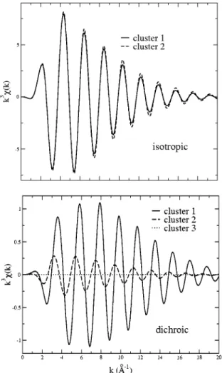
In order to experimentally determine the bond distances around the paramagnetic impurities, the analysis of the di-chroic EXAFS signal is essential. This is best seen in Fig. 5 where the calculated isotropic and dichroic EXAFS signals using FEFF8 ~Ref. 39! program have been represented for
three different chromium environments described in the fol-lowing and labeled cluster 1, 2, and 3. The three clusters contain seven atoms ~one chromium atom and six oxygen atoms!. The geometry in cluster 1 is given by the density functional calculations results ~Sec. IV!. It is built with three oxygen neighbors at 1.95 Å making an angle of 62.8° with the C3 axis and a further three at 2.00 Å with an angle of
48.4°. In cluster 2, the chromium-oxygen bond angles are the same as for the preceding cluster and the bond distances for the six oxygen are set to the averaged value 1.975 Å. In cluster 3, the chromium atom is at the center of a pure oxy-gen octahedron ~1.975 Å for the Cr-O bond lengths! and the dichroic EXAFS signal is zero ~at least in the electric dipole approximation!. One sees that dichroic EXAFS signals are much more sensitive to differences between chromium-oxygen bonds in the coordination shell than the isotropic signals. This proves that inequivalences between oxygen
at-TABLE II. Distances Fe-O and Fe-Al ~Å! from the analysis of the isotropic and dichroic signals and from calculation. These distances are compared to Al-O and Al-Al distances ina-Al2O3and to Fe-O and Fe-Fe in a-Fe2O3.
Al2O3 Al2O3: Fe31 Al2O3: Fe31 Al2O3: Fe31 a-Fe2O3
Ref. 17 isotropic dichroic ab initio Ref. 18
data data data
M -O1 1.86 2.01 1.90 1.92 1.94
M -O2 1.97 2.01 2.05 2.10 2.11
M -Al ~face! 2.65 2.85 2.70 2.72 2.90
oms can reliably been addressed only through the EXAFS analysis of the dichroic signal.
From the dichroic EXAFS signal, it has been found that despite similar mean M-O distances ( M 5Cr, Fe!, the chro-mium and the iron site in a-Al2O3 are very different. If
dM -O22dM -O1is the difference between the long ( M -O2) and
the short ( M -O1) metal-oxygen distances, then dCr-O2
2dCr-O150.09 Å and dFe-O22dFe-O150.15 Å. From the
pre-vious experimental findings, it is clear that the six oxygen
atoms group into two distance groups, thus it is realistic to believe that the C3 axis of corundum crystal is conserved at
the impurity site. This is in agreement with what was found from density functional calculations and also from the EPR measurements ina-Al2O3: Cr31.
30
It should be noted that if two oxygen shells at the two different distances from the central chromium atom, as determined by the analysis of the dichroic EXAFS signal, are introduced in the analysis of the isotropic EXAFS signal, then the experimental signal can be fitted with just a few modifications of the apparent Debye Waller factors. The contribution of the first aluminum neigh-bors around the paramagnetic impurity can also be deter-mined by the analysis of the dichroic EXAFS signal. There are two types of aluminum neighbors. The first aluminum neighbor is on the C3 axis in an octahedron joined by a face
with the impurity octahedron. The second shell of aluminum neighbors consist of three ions in an octahedron, joined by three edges with the impurity octahedron. This strongly an-isotropic distribution yields a large contribution to the di-chroic EXAFS signal, from which it is possible to determine with a good reliability, the rather large cation-aluminum tances. It is found that for the chromium site, the Cr-Al dis-tances are 2.65 Å for the aluminum on the C3 axis and 2.79
Å for the three farther aluminum neighbors. In the same way, the Fe-Al distances are determined to be 2.75 Å for the first aluminum atom and 2.88 Å for the three farther aluminum atoms.
From the point of view of EXAFS measurements, our finding gives the following picture for the relaxation around the paramagnetic impurity. The symmetry of the relaxed site for Cr or Fe is compatible with the C3point group that is the
site symmetry of aluminum atoms in corundum. The local environment around the chromium ion in ruby is nearly the same as that ina-Cr2O3. The Cr-O bond lengths in ruby are
closer to Cr-O bond lengths ina-Cr2O3 rather than to Al-O
bond length ina-Al2O3~see Table I!. However, Cr-Al ~edge!
distances are closer to Al-Al bond lengths ina-Al2O3than to
Cr-Cr bond lengths ina-Cr2O3. The relaxations seem to be very local and do not affect atoms far from the chromium ion, within an error bar of about 1%. The case of iron in corundum is not exactly the same as that of chromium. The Fe-O bond lengths in blue sapphire are closer to the Fe-O bond lengths in a-Fe2O3rather than to Al-O bond lengths in
a-Al2O3 and the Fe-Al bond lengths are in between Al-Al
bond lengths in a-Al2O3 and Fe-Fe bond lengths in a
-Fe2O3. Seen from perspective of the impurity, the relaxation
extends farther in a-Al2O3: Fe31than in a-Al2O3: Cr31.
To have a precise description of the site departure from pure octahedral geometry, the parameter t is constructed. It is defined as t( M )5200@(dM -O22dM -O1)/(dM -O21dM -O1)#.
FIG. 5. Calculated isotropic and dichroic EXAFS signals of CrO6clusters. These clusters are made of seven atoms ~one
chro-mium atom and six oxygen atoms!. Cluster 1 is given by density functional calculations ~three oxygen atoms at 1.95 Å making an angle of 62.85° with the C3axis and three other at 2.00 Å with the
angle 48.36°). In cluster 2, the Cr-O bond angles are the same as in cluster 1 and the bond distances for the six oxygen atoms are set to the averaged value 1.975 Å. Cluster 3 is a perfect octahedral cluster
~1.975 Å for the Cr-O bond lengths!.
TABLE III. Departure from pure octahedral geometry. The r1and the r2distances are the M -O2and
M -O1distances, respectively.
Al2O3 Al2O3: Cr31 Cr2O3 Al2O3: Fe31 a-Fe2O3
Ref. 17 dichro. calc. Ref. 17 dichro. calc. Ref. 18 2(r12r2)
r11r2
100 5.74 4.58 2.53 2.51 7.59 8.96 8.40
STRUCTURAL AND ELECTRONIC RELAXATIONS . . . PHYSICAL REVIEW B 67, 094108 ~2003!
It is clear from Table III that t(Cr31),t(Al31),t(Fe31) in a-Al2O3. In fact, the spectroscopic term of the ground state of Cr31 is 4A2g and it is 6A1g ~essentially from 6S
spherical spectroscopic term! for Fe31. The crystal field is then expected to have less impact on the 6A1g term than on
the 4A2g term. The Fe31ground state energy is almost
inde-pendent of the crystal field, so that large lowering of sym-metry has almost no influence on the Fe31 ion. On the con-trary, the 4A2g term is affected by the crystal field and
t(Cr31),t(Al31). The determination of the impurity site departure from pure octahedral symmetry is worthwhile con-cerning the color. Ruby ora-Al2O3: Fe31presents two
dis-tinct colors depending on the angle of the polarization vector of light with the C3 axis. This property of dichroism goes
directly with the two kinds of metal-oxygen distances in the coordination shell. Their precise determination is essential for next optical spectra calculations.
When considering distances extracted from EXAFS data, all the distances around the impurity are centered around the impurity site. This can hide the actual size of the perturbation imposed by the impurity. This is what has been examined in the following section.
B. Size of the relaxation around the paramagnetic impurity
We would like to use the density functional calculations results for further analysis regarding information on atoms far from the absorbing atom. Such analysis is unobtainable
with EXAFS measurements but can be undertaken success-fully with a DFT calculation. From the position of atoms in the supercell given by the density functional calculations, it is possible to generate all possible positions of atoms in a crystal by using lattice translation vectors. This collection of atoms is then sorted by increasing distances from the central atom. At the end, a cluster is obtained by keeping atoms inside a sphere S of radius 5.2 Å. In our computation, the super-cell is such that the shortest impurity-impurity distance is 10.43 Å. It is then reasonable to limit any analysis con-cerning the size of the relaxation around a specific impurity to a sphere S of radius 5.2 Å around this impurity. In doing so, relaxations imposed by other impurities are minimized.
One needs to compare the relaxed coordinates of atoms in the sphere S to coordinates that equivalent atoms have in the pure corundum structure. This pure corundum structure was obtained by a convergent density functional calculation, starting from the positions given by Duan et al.21The differ-ences between the converged positions and the initial posi-tions were always less than 0.01 Å. Given that the supercell may be translated and rotated by the DFT calculation, it is necessary to align the relaxed and the pure corundum struc-ture. This is done in a number of steps. First, the atoms of the two clusters are rotated so that the C3axis of the two clusters
are parallel to the same axis ~called z axis!. The azimuthal angle is determined so as to minimize the quantity Q, where Q is the sum over all the atoms Pi inside S of the norms of the displacement vectors VWi:
Q5
(
i uV Wiu5(
iA
@Xrelaxi 2Xunrelax i #2 1@Yrelax i 2Yunrelax i #2 1@Zrelax i 2Zunrelax i #2,where Xunrelaxi , Yiunrelax, and Ziunrelax are the coordinates of the vector joining the central aluminum atom to the neigh-boring oxygen or aluminum atoms i of the corundum struc-ture and Xirelax, Yirelax, and Zirelaxare the respective counter-parts joining the impurity to the neighboring atoms of the relaxed structure. In doing so, one finds that relaxation ina -Al2O3: Cr31 is small although not negligible ~aluminum neighbors at 4.2 Å are moved by 0.04 Å! while ina-Al2O3:
Fe31, the relaxation is larger as aluminum and oxygen neighbors at 4.2 Å are displaced by more than 0.1 Å. Such a middle range relaxation is simply due to the displacement of the impurity itself. To eliminate the influence of the impurity displacement all atoms in the relaxed and unrelaxed clusters are translated so as to superimpose the centers V (OVW
5 (iO PWiwhere i runs over all atoms Pi inside the sphere S except the central atom! of the two clusters. The norm of the displacement vector is evaluated again for each atom in sphere S and results are reported in Figs. 6 and 7. With this method, one considers the relaxation as a whole and not merely as seen by the impurity. This is especially important when the impurity is not precisely located at the aluminum site. One sees that the relaxations around Cr or Fe are very
local. The oxygen coordination shell absorbs almost all the relaxation. Atoms further than 2.5 Å are displaced by less than 0.035 Å. Relaxation around iron can be described as follows. Iron is repelled by the nearest aluminum neighbor along the C3 axis. The oxygen coordination shell is strongly relaxed by 0.08 and 0.11 Å, and further neighbors remain almost nondisplaced. That is in line with some previous EPR measurements where Fe is said to be moved by 0.04 Å
60.02 Å on the C3 axis farther from the first aluminum
neighbors.16Around chromium, the picture is slightly differ-ent. The chromium atom is almost at the aluminum site and oxygen atoms of the coordination shell move to accommo-date for the larger ionic radius. Again, the relaxation is very local. The literature is controversial concerning the direction of the displacement of the chromium atom along the C3
axis.11,12,19,37,38 In all cases the displacement is said to be small. In addition, we have analyzed the angular relaxations. To this end, we evaluatedu5uunrelax2urelax, whereu is the
angle between the V PWi direction and the C3 axis. In both
cases, angular relaxation is weak.
Thanks to all of these results, it is now possible to picture the relaxation ~see Fig. 6!. Chromium as an impurity in a
-Al2O3tends to impose the large site it has ina-Cr2O3. This
mismatch between Al-O1bond distances ina-Al2O3and Cr-O1 bond distances in Cr2O3 is 10.11 Å and between Al-O2
and Cr-O2it is 10.05 Å. In the actuala-Al2O3: Cr31crystal
the mismatch is compensated by a displacement of O1 type
atoms by 0.09 Å nearly in the Cr-O1bond direction. The O2
oxygen atoms are displaced by 0.05 Å in the Cr-O2 bond
direction. The same considerations apply to Fe ina-Al2O3.
The mismatch between Al-O1 distances ina-Al2O3 and
Fe-O1 distances ina-Fe2O3is 10.08 Å, while between Al-O2
and Fe-O2 it is 10.14 Å. The three O2 type oxygen atoms
determine the shared face between Fe and its nearest Al neighbor. A displacement as large as 0.14 Å would consider-ably shorten Al-O2 bond lengths and perturb the crystal. For iron to accommodate its relaxed site, it moves away by 0.09 Å along the C3, axis farther from its nearest Al neighbors
with a concomitant displacement of 0.09 Å for O2 type at-oms and 0.11 Å for O1atoms. As in a-Al2O3: Cr31, atoms
farther than the coordination shell do not move much.
VI. CONCLUSION
Iron and chromium are known to be at the origin of the color of many minerals. In this paper, we have shown that the analysis of the isotropic and dichroic x-ray absorption spectra yields significant information about relaxations around impurities in single crystals. Ab initio calculations confirm results given by the experimental analysis and pro-vide an overall view of the relaxation. These structural de-terminations are an essential first step toward ab initio cal-culations of optical spectra. Concerning ruby, this study contributes to the understanding of the change of color from red to green when increasing the chromium concentration.40 It is usually believed that the mean metal-ligand distance plays a considerable role in the color process. Contrary to what can be found in textbooks,1,2 we have shown that the difference of the Cr-O distances in ruby and in a-Cr2O3 is not at the origin of the modification of the color. Other pa-rameters such as electron and magnetic pairing between chromium neighbors certainly play a role in the color pro-cess. These parameters could for instance be investigated by spectroscopic measurements at chromium L2,3edges.
ACKNOWLEDGMENTS
We acknowledge the careful reading of the manuscript by Delphine Cabaret during its preparation. We are very grateful to Siaˆn Joyce for a critical reading of the manuscript. We are glad to thank Franc¸oise Vilain and Christophe Gauthier for their skillful help during preliminary experiments at LURE
~Orsay! and ESRF ~Grenoble!. Ruby and blue sapphire
single crystals were kindly provided by Bruno Boizot and Georges Calas, who are at the origin of this piece of work. Chemical analysis has been performed under supervision by E´ tienne Balan. One of us ~A.K.! thanks Thailand government for financial support during her Ph.D. thesis. Computing time was partially supplied by the ‘‘Institut du De´veloppement et des Ressources en Informatique Scientifique.’’ This is IPGP No. 1891.
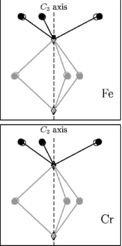
FIG. 6. Environment around the paramagnetic impurity. All at-oms are projected on a plane containing the C3 axis. The circles
represent the oxygen atoms, the diamonds the aluminum atoms, the square the impurity atom. Empty signs represent the positions of atoms ina-Al2O3, full signs represent the positions of atoms in
a-Al2O3: Cr31or ina-Al2O3: Fe31. As seen from the upper picture,
the iron atom in a-Al2O3: Fe31 does not take exactly the same
place as the aluminum ion ina-Al2O3.
FIG. 7. Relaxations in a-Al2O3: Cr31and a-Al2O3: Fe31.
Norms of displacement vectors as a function of distances from the chromium or iron atom.
STRUCTURAL AND ELECTRONIC RELAXATIONS . . . PHYSICAL REVIEW B 67, 094108 ~2003!
*Electronic address: [email protected]
1K. Nassau, The Physics and Chemistry of Color ~Wiley, New
York, 1983!.
2
R. G. Burns, Mineralogical Applications of Cristal Field Theory, Vol. 5 of Cambridge Topics in Mineral Physics and Chemistry, 2nd ed. ~Cambridge University Press, Cambridge, 1993!.
3L. X. Benedict, E. L. Shirley, and R. B. Bohn, Phys. Rev. Lett. 80,
4514 ~1998!.
4S. Albrecht, L. Reining, R. DelSole, and G. Onida, Phys. Rev.
Lett. 80, 4510 ~1998!.
5M. Rohlfing and S. G. Louie, Phys. Rev. B 62, 4927 ~2000!. 6V. Olevano and L. Reining, Phys. Rev. Lett. 86, 5962 ~2001!. 7R. H. French, J. Am. Ceram. Soc. 73, 477 ~1990!.
8J. F. Nye, Physical Properties of Crystals ~Clarendon, Oxford,
1957!.
9R. D. Shannon, Acta Crystallogr., Sect. A: Cryst. Phys., Diffr.,
Theor. Gen. Crystallogr. 32, 751 ~1976!.
10
W. Y. Ching and Y.-N. Xu, J. Am. Ceram. Soc. 77, 404 ~1994!.
11J. W. McCauley and G. V. Gibbs, Z. Kristallogr. 135, 453 ~1972!. 12S. C. Moss and R. E. Newnham, Z. Kristallogr. 120, 359 ~1964!. 13D. S. McClure, J. Chem. Phys. 36, 2757 ~1962!.
14K. Langer, Z. Kristallogr. 216, 87 ~2001!.
15R. Bu¨scher, K. P. Such, and G. Lehmann, Phys. Chem. Miner. 14,
553 ~1987!.
16Z. Wen-Chen, Physica B 245, 119 ~1998!.
17W. B. Pearson, Structure Reports ~International Union of
Cristal-lography, Utrecht, 1962!, Vol. 27.
18L. W. Finger and R. M. Hazen, J. Appl. Phys. 51, 5362 ~1980!. 19P. Kizler, J. He, D. R. Clarke, and P. R. Kenway, J. Am. Ceram.
Soc. 79, 3 ~1996!.
20S. Emura, H. Maeda, Y. Kuroda, and T. Murata, Jpn. J. Appl.
Phys. 32, 734 ~1993!.
21W. Duan, G. Paiva, R. M. Wentzcovitch, and A. Fazzio, Phys.
Rev. Lett. 81, 3267 ~1998!.
22J. Goulon, C. Goulon-Ginet, A. Rogalev, G. Benayoun, C.
Mal-grange, and C. Brouder, Proc. SPIE 3773, 316 ~1999!.
23J. Goulon, A. Rogalev, C. Gauthier, C. Goulon-Ginet, S. Paste, R.
Signorato, C. Neumann, L. Varga, and C. Malgrange, J. Syn-chrotron Radiat. 5, 232 ~1998!.
24Ch. Brouder, D. Cabaret, Ph. Sainctavit, A. Kiratisin, J. Goulon,
and A. Rogalev, Radiat. Eff. Defects Solids 155, 89 ~2001!.
25C. Brouder, J. Phys.: Condens. Matter 2, 701 ~1990!. 26M. Winterer, J. Phys. IV 7, 243 ~1997!.
27D. Bonnin, G. Calas, H. Suquet, and H. Pezerat, Phys. Chem.
Miner. 12, 55 ~1985!.
28B. K. Teo, EXAFS: Basic Principles and Data Analysis ~Springer
Verlag, Berlin, 1985!.
29D. C. Koningsberger and R. Prins, X-ray Absorption. Principles,
Applications, Techniques of EXAFS, SEXAFS, and XANES
~Wiley, New York, 1988!, Vol. 92.
30B. Boizot, Ph.D. thesis, Universite´ Paris VI, 1996. 31
L. Varga, C. Giles, C. Neumann, A. Rogalev, C. Malgrange, J. Goulon, and F. D. Bergevin, J. Phys. IV 7, 309 ~1997!.
32S. Goedecker, M. Teter, and J. Hutter, Phys. Rev. B 54, 1703 ~1996!.
33N. Troullier and J. L. Martins, Phys. Rev. B 43, 1993 ~1991!. 34L. Kleinman and D. M. Bylander, Phys. Rev. Lett. 48, 1425
~1982!.
35W. Duan, R. M. Wentzcovitch, and K. T. Thomson, Phys. Rev. B
57, 10 363 ~1998!.
36J. Hutter, P. Ballone, M. Bernasconi, P. Focher, E. Fois, S.
Goedecker, M. Parrinello, and M. Tuckerman ~unpublished!.
37N. Laurance, E. C. McIrvine, and J. Lambe, J. Phys. Chem.
Sol-ids 23, 515 ~1962!.
38L. L. Lohr and W. H. Lipscomb, J. Chem. Phys. 38, 1607 ~1963!. 39A. L. Ankudinov, B. Ravel, J. J. Rehr, and S. D. Conradson, Phys.
Rev. B 58, 7565 ~1998!.