Permeability changes in heartwood of Picea abies and Abies
alba induced by incubation with Physisporinus vitreus*
Francis W.M.R. Schwarze
1,2,**, Helge
Landmesser
1,2, Bruno Zgraggen
1and Markus
Heeb
11
Wood Laboratory, Swiss Federal Laboratories for
Materials Testing and Research (EMPA), St. Gallen,
Switzerland
2
Institut fu¨r Forstbotanik und Baumphysiologie,
Albert-Ludwigs-Universita¨t Freiburg, Freiburg i.Br., Germany
**Corresponding author.Wood Laboratory, Swiss Federal Laboratories for Materials Testing and Research (EMPA), Lerchenfeldstr. 5, 9014 St. Gallen, Switzerland
E-mail: francis.schwarze@empa.ch
Abstract
The present study shows that isolates of P. vitreus have
an extraordinary capacity to induce substantial
permea-bility changes in heartwood of P. abies without causing
significant losses in impact bending strength. The
deg-radation of pit membranes by P. vitreus is an important
aspect that could also have significant benefits in wood
protection processes. Further studies are currently in
progress with the objective of optimising the uniformity
of wood colonisation and duration of incubation, so as
to improve the permeability of water-borne wood
preser-vatives or hydrophobic substances applied by brushing,
dipping and impregnation.
Keywords: Abies alba; heartwood; Physisporinus vitreus;
Picea abies; wood permeability.
Introduction
The permeability of conifer wood is enhanced by
bio-degradation of pit membranes, which usually occurs in
the course of bacterial activity in water-stored logs
(Schmidt and Liese 1994). Permeability of sapwood has
been increased after such a treatment and as a
conse-quence the uptake of preservatives was improved (Bauch
et al. 1970; Johnson 1979; Schmidt and Liese 1994).
Treatment of wood with enzymes that degrade the
prin-cipal constituents of the pit membranes (pectin,
hemi-celluloses and hemi-celluloses) is also promising (Nicholas and
Thomas 1968; Bauch et al. 1970; Sharma and Kumar
1979). More recent findings indicate that the most
effec-tive enzymes are hydrolases with a broad spectrum of
cellulolytic and hemicellulolytic activity when finely
ground Picea abies wood was the substrate (Militz
1993a,b). It was, however, also noted that the practical
*Dedicated to Prof. Dr. (Dr. hc mult.) Walter Liese, to mark his 80th birthday.importance of enzymes in this context is limited: their
diffusion into wood is slow and the extractives adhering
to aspirated pits have inhibitory effects (Militz 1993a,b).
Rosner et al. (1998) demonstrated that basidiomycetes
are capable of colonising sapwood and heartwood and
can improve the permeability. A major disadvantage is
that fungi also weaken the wood by degrading the
sec-ondary walls as well as the pit membranes (Tucker et al.
1998). The white rot fungus Physisporinus vitreus is an
exception in this regard; it preferentially degrades the pit
membranes of bordered pits in the heartwood of conifers
under controlled conditions, and causes only negligible
mass losses (Schwarze and Landmesser 2000). This
fun-gus is, however, capable of strongly degrading
water-sat-urated timber in cooling towers (Schmidt et al. 1996,
1997).
The objective of the present study was to investigate
whether P. vitreus can significantly enhance the
perme-ability of conifer heartwood without inducing significant
strength losses. If this approach is successful, several
processes for wood modification and finishing could
be significantly improved. The improved permeability of
waterborne solvents into conifer wood is one useful
application.
Materials and methods
Specimens from the heartwood of living 40–50-year-old trees of Abies alba Mill. and Picea abies Karst were incubated with two isolates of P. vitreus and then assessed for quantitative and spatial alterations in their water uptake and impact bending strength. Wood specimens of two different dimensions, but cut exactly the same in the radial and tangential planes, were inves-tigated: (1) 100 mm (longitudinal)=10 mm (tangential)=15 mm (radial) (15 cm3); and (2) 100 mm (longitudinal)=25 mm
(tangential)=15 mm (radial) (37.5 cm3). A total of 448 test
spe-cimens were prepared according to European standard EN 113 (1997), comprising 16 specimens for each treatment and incu-bation period. Mass loss and water uptake were measured for each specimen before and after incubation. Before water uptake measurements, wood specimens were dried at 1038C for 18–24 h and then cooled in a desiccator and weighed. Strength tests were performed only with the smaller specimens, and microscopy was carried out exclusively on the larger specimens.
Selection of heartwood specimens
The water uptake of the blocks was measured (in kg my3)
according to EN 113 before incubation. Based on the results, only specimens that showed the typical water uptake for heart-wood of each of the two species were selected. Wood speci-mens with atypical water uptake were rejected. This ensured that all specimens contained a high proportion of heartwood. To ensure that water was taken up via the tangential and radial surfaces only, the end grain of each dried and weighed speci-men was first sealed with soft silicone grease and firmly fixed
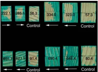
Figure 1 Heartwood specimens of Picea abies (top) and Abies alba (bottom) impregnated with a 0.1% aqueous solution of Neolan Glaucin E-A using vacuum treatment after 6-week incu-bation with Physisporinus vitreus (left, 15 cm3; right, 37.5 cm3).
Numbers refer to radial and tangential uptake of water in kg my3.
In comparison to weakly stained controls, cross-sections of incubated P. abies specimens are moderately stained and those of A. alba are strongly stained. Arrows indicate the direction from which wood samples were colonised by hyphae.
between metal mounts to prevent the formation of cracks and shakes. Retention of water taken up via the tangential and radial surfaces was then measured by immersing the blocks in distilled water at reduced pressure (7 mbar) for 20 min in a vacuum des-iccator according to EN 113. Then the vacuum vessel was brought back to atmospheric pressure and the specimens were left for a further 120 min in water before re-weighing (Figure 1).
Incubation of wood specimens
Before incubation, the blocks were sterilised with ethylene oxide for approximately 1 h. Incubation was carried out with two iso-lates of Physisporinus vitreus (EMPA 642 and EMPA 643) in Kolle flasks according to EN 113. The flasks were incubated in a ran-dom array at 22"18C and 75"5% RH for 6, 12 and 18 weeks. After incubation, the wood blocks were dried for 48 h at 308C and permeability changes were measured. To evaluate qualita-tive changes in uptake and distribution of the synthetic organic textile dye Neolan Glaucin E-A (Ciba Geigy), the end grain of each block was sealed with silicone grease as described above and the wood specimens were then impregnated with a 0.1% aqueous solution of the dye according to EN 113. After impreg-nation, incubated wood blocks were dried at 1038C for 18–24 h and then cooled in a desiccator and weighed for measurement of mass loss. The smaller wood specimens (15 cm3) were
pre-conditioned in an incubator at 208C and 65% RH for 4 weeks. Their impact bending strength, Aw (kJ m
y2), was measured in
the tangential direction using a Zwick pendulum impact testing machine in accordance with DIN standard 52 189 (1981). For statistical evaluation of differences between control and incu-bated blocks of each tree species for water retention and impact bending strength, t-tests (P-0.05–P-0.001) were performed using Origin 7.5 (Microcal Software Inc.).
Electron microscopy
Radial longitudinal specimens were extracted from stained and unstained regions of the larger (37.5 cm3) incubated wood
blocks. These were then dried in a vacuum oven at 408C and 10 mbar for 12 h, glued on a specimen holder using carbon-adhesive and sputtered with a platinum layer of approximately
10 nm for investigation using a field-emission SEM (Jeol 6300F) at an acceleration voltage of 5 kV and working distance of 24 mm.
Results and discussion
The mass losses in heartwood of P. abies and A. alba
were negligible after 6-week incubation (-1%, Table 1).
After 12 and 18 weeks, there were significant
differenc-es between the mass lossdifferenc-es in P. abidifferenc-es and A. alba
(P-0.05). Only after 6-week incubation, as the mass
losses induced by both P. vitreus isolates were slight,
was the wood permeability increased, as manifest by the
increase in water uptake in the test blocks to
approxi-mately 300–400 kg m
y3in P. abies and 400–680 kg/m
y3in A. alba (Table 1). The differences before and after
incu-bation of the specimens were highly significant
(P-0.001). The two P. vitreus isolates showed no significant
differences in their effect on permeability. Conspicuous
qualitative changes in permeability were also apparent
from the uptake of the bluish dye Neolan Glaucin E-A.
Uptake of the dye by A. alba test blocks incubated with
P. vitreus was homogeneous on visual inspection, but the
homogeneity was less pronounced in corresponding P.
abies blocks.
FE-SEM studies revealed that uptake of Neolan
Glau-cin E-A was attributable to preferential degradation of pit
membranes (Figures 2 and 3). Complete or partial
hydrol-ysis of bordered pits and crossfield pits was apparent in
regions stained with Neolan Glaucin E-A. Unstained
regions of pit membranes remained intact (Figure 3).
Hyphae entered the pit chamber via apertures, and
mem-branes were subsequently degraded (Figures 2 and 3).
Degradation commenced from the thickened central part
of the membrane (torus). Calcium oxalate crystals were
regularly observed on hyphae. In wood of A. alba,
crys-tals accumulations were often apparent within bordered
pits in close proximity to hyphae (Figure 3). Production
of polygalacturonase (PG) and hydrolysis of bordered pit
membranes during incipient decay has been described
in detail (Green et al. 1991, 1995a,b; Green and Clause
1999). A key factor for pectin hydrolysis by plant
patho-gens has been shown to be fungal production of oxalic
acid, which lowers the pH of the substrate and chelates
calcium ions. Production of oxalic acid may serve a
similar role for P. vitreus isolates during incipient wood
decay.
Crossfield pits of the xylem ray parenchyma also
showed signs of extensive degradation. In decayed
wood specimens of A. alba, degradation of taxodioid pit
membranes was found throughout the stained wood
regions. In contrast, degradation of piceoid pit
mem-branes in P. abies was more localised and was restricted
to cells near the outer surfaces of the specimens.
Pre-vious studies have demonstrated that most brown- and
white-rot fungi have the capacity to hydrolyse the pectin
in pit membranes during incipient decay, which facilitates
colonisation (Cowling 1961; Wilcox 1968; Green and
Clausen
1999;
Schwarze
and
Landmesser
2000;
Schwarze et al. 2004).
The impact bending strength of incubated P. abies
wood specimens was not significantly reduced in
T able 1 Mass loss, water uptake and str ength loss in heartwood specimens of Picea abies and Abies alba after 6-, 12-and 18-week incubation with two isolates of Physisporinus vitreus . Sample Mass loss (g) W ater uptake (kg m –3 ) (befor e and after incubation) Str ength (kJ m –2) 6 weeks 12 weeks 18 weeks 6 weeks 12 weeks 18 weeks 6 weeks 12 weeks 18 weeks P . abies Contr ol, 15 cm 3 – 25.7 " 4.05 EMP A 642, 15 cm 3 0.9 "0.35 NS 6.50 "2.30*** 10.7 "4.26*** 174.8 " 66.15 170.60 "69.20 55.6 " 64.90 22.7 " 2.00 NS 13.0 "3.26*** 9.9 "4.77*** 418.7 " 176.64*** 598.30 "167.20*** 738.80 "156.18*** EMP A 643, 15 cm 3 0.8 "0.29 NS 4.2 "0.97*** 8.1 " 3.78* 185.30 "70.66 328.90 "188.50 150.4 " 62.60 23.2 " 3.57 NS 16.3 "3.26*** 11.0 "2.90*** 378.90 "160.32*** 449.50 "154.85*** 760.10 "86.18*** EMP A 642, 37.5 cm 3 0.8 "0.24 NS 5.30 "1.37*** 7.0 " 2.41*** 34.7 " 44.77 50.40 " 70.71 69.8 " 39.4 – – – 327.0 " 75.22*** 650.3 " 113.78*** 782.20 "59.87*** EMP A 643, 37.5 cm 3 0.8 "0.24 NS 3.9 "1.24*** 6.3 " 3.19* 98.90 " 40.96 54.2 " 64.30 53.20 " 58.80 – – – 375.80 "83.78*** 571.90 "155.09*** 772.30 "56.15*** A. alba Contr ol – – 25.7 " 4.13 EMP A 642, 15 cm 3 0.9 "0.36 3.41 "1.25 5.8 " 2.70 162.80 "70.71 174.10 "70.71 135.90 "49.30 17.6 " 2.00** 18.0 "3.18*** 14.6 "2.81*** 660.7 " 64.04*** 672.9 " 174.10*** 819.8 " 70.71*** EMP A 643, 15 cm 3 0.7 "0.24 1.60.8 "1.51 5.0 " 3.48 332.60 "75.48 219.3 " 158.30 157.9 " 76.60 20.5 " 3.57* 17.3 "2.16*** 11.0 "2.90*** 536.40 "120.30*** 532.7 "112.26*** 754.20 "51.84*** EMP A 642, 37.5 cm 3 0.7 "0.24 1.40 "1.39 3.2 " 2.70 168.10 "37.77 168.10 "37.80 285.40 "46.30 – – – 647.70 "88.41*** 654.7 " 75.18*** 758.90 "47.82*** EMP A 643, 37.5 cm 3 0.7 "0.35 1.1 "0.96 3.50 "3.37 165.60 "40.89 173.7 " 36.80 303.60 "43.90 – – – 631.0 " 87.25*** 664.6 " 89.86*** 769.40 "39.85*** Significant dif fer ences between contr ol and tr eatment (water uptake and str ength) or species (mass loss) within the same incubation periods ar e p re sented, accor ding to a t-test (n s 16): NS, not significant; *P -0.05; **P -0.01; ***P -0.001.
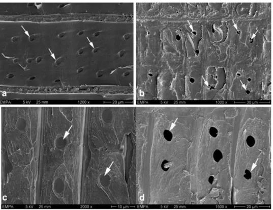
Figure 2 Scanning electron micrographs (5 kV) showing bordered pits (a–d) in Picea abies and Abies alba after 6-week incubation with Physisporinus vitreus. Hyphae entered the bordered pits via the aperture and degraded the torus; lysis of the warty layer (pointer) and calcium oxalate crystals (arrows) occurred in close proximity to hyphae within bordered pit chambers (b). After 12 weeks, most bordered pits showed partial to complete dissolution (c,d) of pit membranes, so that both pit apertures in pit pairs were occasionally exposed (arrow).
Figure 3 Scanning electron micrographs showing crossfield pits in sound and degraded heartwood of Picea abies (a,b) and Abies alba (c,d), respectively. After 6-week incubation with Physisporinus vitreus, some crossfield pits showed partial dissolution (b; arrows) in P. abies, whereas most pit membranes of A. alba were completely degraded (d; arrows).
comparison to controls after 6 weeks, whereas there was
a significant reduction in A. alba specimens (EMPA 642,
P-0.01; EMPA 643, P-0.05; Table 1). After 12 and
18 weeks, wood strength was significantly lower in all
cases in comparison to the controls (P-0.001).
Colonisation of wood specimens of A. alba and P.
abies by P. vitreus was more homogeneous when they
were incubated with their tangential surfaces, rather than
their radial surfaces, in contact with the mycelium. The
latter orientation was used for the smaller specimens, to
allow them to rest on their broader (radial) surfaces. The
radial surfaces of wood specimens are relatively resistant
to colonisation by fungal hyphae (Kleist and Seehann
1997; Schwarze et al. 2004). Thus, a more rapid hyphal
growth in the radial than tangential direction resulted in
a higher increase of water uptake in larger wood
mens irrespective of the wood species. In wood
speci-mens of A. alba, water uptake was also facilitated by
homogenous degradation of the large taxodioid pit
mem-branes in the xylem ray parenchyma. The results appear
to suggest that P. vitreus colonises wood of A. alba more
rapidly, but on the basis of losses in mass, it seems to
show a preference for degrading heartwood of P. abies.
Although permeability increased with incubation time,
significant strength losses were recorded after 12 and
18 weeks. The potentially disadvantageous strength loss
in A. alba appears to be correlated with a more intense
colonisation by P. vitreus, which could be mitigated
sim-ply by using shorter incubation periods and higher
growth temperatures.
Acknowledgements
We would like to thank the Fond zur Fo¨rderung der Wald- und Holzforschung (FFWH), Bern, for partly funding these studies (Projektnumbers 2005.01 and 2004.16). A German patent has been issued (No. 10 2005 002 366.5) and a European application (No. 05027812.6) has been submitted regarding the use of P. vitreus to increase the permeability of conifer heartwood. The authors wish to thank E. Strub for technical assistance with FE-REM and Dr. D. Lonsdale for comments on the paper.
References
Bauch, J., Liese, W., Berndt, H. (1970) Biological investigations for the improvement of the permeability of softwoods. Holz-forschung 24:199–205.
Cowling, E.B. (1961) Comparative biochemistry of the decay of sweetgum sapwood by white-rot and brown-rot fungi. WO-TB-1258. USDA Forest Service, Washington, DC.
DIN standard 52189 (1981) Testing of wood; Impact bending test; Determination of impact bending strength.
European standard (1997) EN 113. Wood preservatives – test method for determining the protective effectiveness against wood destroying basidiomycetes. Determination of toxic values.
Green, F., Clausen, C.A. (1999) Production of polygalacturonase and increase of longitudinal gas permeability in southern pine by brown-rot and white-rot fungi. Holzforschung 53: 563–568.
Green, F., Larsen, M.J., Winandy, J.E., Highley, T.L. (1991) Role of oxalic acid in incipient brown-rot decay. Mater. Organis-men 26:191–213.
Green, F., Tschernitz, J., Kuster, T.A., Highley, T.L. (1995a) Hydrolysis of bordered pits during colonization of conifers by brown-rot decay. Document No. IRG/WP/95-10103. Inter-national Research Group on Wood Protection, Stockholm, Sweden.
Green, F., Clausen, C.A., Kuster, T.A., Highley, T.L. (1995b) Induc-tion of polygalacturonase and the formaInduc-tion of oxalic acid by pectin in brown-rot fungi. World J. Microbiol. Biotechnol. 11:519–524.
Johnson, B.R. (1979) Permeability changes induced in three western conifers by selective bacterial inoculation. Wood Fibre 11:10–21.
Kleist, G., Seehann, G. (1997) Colonization patterns and topo-chemical aspects of sap streak caused by Stereum sangui-nolentum in Norway spruce. Eur. J. For. Pathol. 27:351–361. Militz, H. (1993a) Der Einfluss enzymatischer Behandlungen auf die Tra¨nkbarkeit kleiner Fichtenproben. Holz Roh Werkst. 51:135–142.
Militz, H. (1993b) Der Einfluss enzymatischer Behandlungen von Fichtenrund- und Schnittholz zur Verbesserung der Tra¨nk-barkeit. Holz Roh Werkst. 51:339–346.
Nicholas, D.D., Thomas, R.J. (1968) The influence of enzymes on the structure and permeability of loblolly pine. Am. Wood-Preserv. Assoc. Proc. 64:70–76.
Rosner, B., Messner, K., Tucker, E., Bruce, A. (1998) Improve preservative penetration of spruce after pre-treatment with selected fungi. I. Fungal pre-treatment of pole sections. Doc-ument No. 98-40117. International Research Group on Wood Protection, Stockholm, Sweden.
Schmidt, O., Liese, W. (1994). Occurrence and significance of bacteria in wood. Holzforschung 48:271–277.
Schmidt, O., Liese, W., Moreth-Kebernik, U. (1996) Decay of tim-ber in a water cooling tower by the basidiomycete Physis-porinus vitreus. Mater. Organismen 30:161–177.
Schmidt, O., Schmitt, U., Moreth, U., Potsch, T. (1997) Wood decay by the white-rotting basidiomycete Physisporinus vitreus. Holzforschung 51:193–200.
Schwarze, F.W.M.R., Landmesser, H. (2000) Preferential degra-dation of pit membranes within tracheids by the basidiomy-cete Physisporinus vitreus. Holzforschung 54:461–462. Schwarze, F.W.M.R., Engels, J., Mattheck, C. (2004) Fungal
strategies of wood decay in trees. 2nd ed. Springer Verlag, Heidelberg. 218 pp.
Sharma, M., Kumar, S. (1979) Degradation of wood pectin by micro-organisms. Int. J. Wood Preserv. 1:87–90.
Tucker, E., Bruce, A., Staines, H.J., Rosner, B., Messner, K. (1998). Improve preservative penetration of spruce after pre-treatment with selected fungi. II. Creosote pre-treatment, analysis and strength testing. Document No. 98-40106. International Research Group on Wood Protection, Stockholm, Sweden. Wilcox, W.W. (1968) Changes in the microstructure through
pro-gressive stages of decay. FLP-RP-70. US Department of Agriculture, Forest Service, Forest Products Laboratory, Madison, WI.