Nephrol Dial Transplant (2010) 25: 2970–2976 doi: 10.1093/ndt/gfq088
Advance Access publication 18 February 2010
Original Articles
Nineteen novel
NPHS1 mutations in a worldwide cohort of patients
with congenital nephrotic syndrome (CNS)
Dominik S. Schoeb
1,2, Gil Chernin
1, Saskia F. Heeringa
1, Verena Matejas
2, Susanne Held
1,
Virginia Vega-Warner
1, Detlef Bockenhauer
3, Christopher N. Vlangos
1, Khemchand N. Moorani
4,
Thomas J. Neuhaus
5, Jameela A. Kari
6, James MacDonald
1, Pawaree Saisawat
1,
Shazia Ashraf
1, Bugsu Ovunc
1, Martin Zenker
2, Friedhelm Hildebrandt
1,7and Members of the Gesellschaft für Paediatrische Nephrologie (GPN) Study Group*
1
Department of Pediatrics and Department of Human Genetics, University of Michigan, 1150 W. Medical Center Drive Drive, Ann Arbor, MI, USA,2Institute of Human Genetics, University of Erlangen–Nuremberg, Schwabachanlage 10, Erlangen, Germany,
3
Paediatric Nephrology, Great Ormond Street Hospital, Great Ormond Street, London, WC1 3JH, UK,4Department of Pediatric Medicine, National Institute of Child Health, Rafiqi H J Road, Karachi, Pakistan,5Nephrology Unit, University Children's Hospital
Zurich, Steinwiesstrasse 75, 8032 Zurich, Switzerland,6Department of Pediatrics, King Abdul-Aziz University Hospital, Ali Al-Murtada Road, Jeddah, Saudi Arabia and7Howard Hughes Medical Institute, Chevy Chase, Maryland
Correspondence and offprint requests to: Friedhelm Hildebrandt; E-mail: fhilde@umich.edu
*Dr. Albalwi (Riyadh, Saudi Arabia), Dr. Ariceta Iraola (Baracaldo-Bizkaia, Spain), Dr. Attrach and Dr. Shibli (Al Ain, Abu Dhabi, United Arab Emirates), Dr. Basak (Istanbul, Turkey), Dr. Böhm (Dortmund, Germany), Dr. Bogdanovic (Belgrad, Serbia), Dr. Chadha (Richmond, USA), Dr. Clothier and Dr. Macdonald (Birmingham, UK), Dr. Conley (Chapel Hill, USA), Dr. Cucer and Dr. Rusu (Iasi, Romania), Dr. Dixon (Cincinnati, USA), Dr. Grillenberger (Linz, Austria), Dr. Hanan (Alexandria, Egypt), Dr. Hanevold (Augusta, USA), Dr. Hempel (Munich, Germany), Dr. Herman (Sacramento, USA), Dr. Hodson (Sydney, Australia), Dr. Hoppe (Cologne, Germany), Dr. Keng (Penang, Malaysia), Dr. Khoury (Sydney, Australia), Dr. Lehmann and Dr. Laube (Zürich, Switzerland), Dr. Loza (Lima, Peru), Dr. Milford (Birmingham, UK), Dr. Montoya (Munich, Germany), Dr. Mueller (Berlin, Germany), Dr. Nayir (Istanbul, Turkey), Dr. Nissel (Rostock, Germany), Dr. Ozaltin (Ankara, Turkey), Dr. Peco-Antic (Belgrad, Serbia), Dr. Pohl (Freiburg, Germany), Dr. Querfeld (Berlin, Germany), Dr. Rademacher (Minneapolis, USA), Dr. Serdaroglu (Izmir, Turkey), Dr. Soliman (Cairo, Egypt), Dr. Soran (Sanliurfa, Turkey), Dr. Soylu (Izmir, Turkey)
Abstract
Background. Recessive mutations in the NPHS1 gene encoding nephrin account for∼40% of infants with con-genital nephrotic syndrome (CNS). CNS is defined as ste-roid-resistant nephrotic syndrome (SRNS) within the first 90days of life. Currently, more than 119 different mutations of NPHS1 have been published affecting most exons. Methods. We here performed mutational analysis of NPHS1 in a worldwide cohort of 67 children from 62 dif-ferent families with CNS.
Results. We found bi-allelic mutations in 36 of the 62 fam-ilies (58%) confirming in a worldwide cohort that about one-half of CNS is caused by NPHS1 mutations. In 26 families, mutations were homozygous, and in 10, they were compound heterozygous. In an additional nine pa-tients from eight families, only one heterozygous mutation was detected. We detected 37 different mutations. Nine-teen of the 37 were novel mutations (∼51.4%), including 11 missense mutations, 4 splice-site mutations, 3 nonsense mutations and 1 small deletion. In an additional patient with later manifestation, we discovered two further novel mutations, including the first one affecting a glycosylation site of nephrin.
Conclusions. Our data hereby expand the spectrum of known mutations by 17.6%. Surprisingly, out of the two siblings with the homozygous novel mutation L587R in NPHS1, only one developed nephrotic syndrome before the age of 90 days, while the other one did not manifest until the age of 2 years. Both siblings also unexpectedly experienced an episode of partial remission upon steroid treatment.
Keywords: mutation analysis; nephrotic syndrome; NPHS1
Introduction
The protein nephrin [1] is an essential component of the renal glomerular slit diaphragm [2], which is formed by adjacent glomerular epithelial cells (podocytes). The zip-per-like structure of the glomerular slit membrane consists of complexes that contain the molecules neph1 and ne-phrin, which interact between neighbouring podocyte foot processes [3]. Nephrin contains eight immunoglobulin-like domains, a fibronectin type III-immunoglobulin-like domain, a trans-membranous domain and a short intracellular domain [1]
© The Author 2010. Published by Oxford University Press on behalf of ERA-EDTA. All rights reserved. For Permissions, please e-mail: journals.permissions@oxfordjournals.org
(Figure 1). It plays a significant role in signalling between podocytes by interacting with molecules like CD2AP and podocin [3]. Phosphorylated nephrin binds to Nck, an adapter protein, hereby reorganizing the cell's actin fila-ment network [4]. Recently, an interaction of the intracel-lular domain of nephrin with β-arrestin was shown to attenuate nephrin signalling [5].
Congenital nephrotic syndrome (CNS) is defined as ne-phrotic syndrome with onset before the 90thday of postna-tal life [6]. Recessive mutations of the nephrin encoding gene NPHS1 were initially described in the renal histopath-ological entity of nephrotic syndrome of the‘Finnish type’ (CNF) [1]. However, they have more recently also been found outside Finland [7]. Recently, mutations in nephrin were shown to cause∼40% of all cases of CNS [6].
The disease is characterized by massive proteinuria caused by a disruption of the filtration barrier [8]. Due to the mas-sive protein loss, patients often require central venous albu-min replacement as well as parental nutrition, leading to a high mortality from septicaemia. End-stage kidney disease (ESKD) before the age of 2–3years and resistance to stan-dard steroid treatment are the rules. To avoid infectious, thromboembolic and other complications from massive loss of protein, including immunoglobulins and coagulation fac-tors, bilateral nephrectomy, dialysis and renal transplanta-tion at a body weight of 10kg are recommended [9].
‘Congenital nephrotic syndrome of the Finnish type’ (CNF) [10,11] is exclusively caused by mutations in NPHS1 [1]. Renal histology shows microcystic dilatation of the proximal tubules and a progressive mesangial sclerosis [12]. Very recently, rare cases with a manifestation beyond the age of 90days have also been published, indicating that different mutations in NPHS1 might cause a spectrum of clinical severity [13].
To date, 119 different mutations in NPHS1 are known. To expand the spectrum of known mutations, we per-formed mutational analysis of NPHS1 by direct
sequenc-ing of all exons in 67 patients from 62 different families with CNS.
Materials and methods
Patients and data recruitment
DNA samples and clinical data of a worldwide cohort of 2 056 children with nephrotic syndrome (NS) were ascertained between 1996 and 2008. The diagnosis was made by paediatric nephrologists on the basis of pub-lished criteria [14]. Nephrotic-range proteinuria was defined as protein-uria >40 mg/m2/h. After informed consent was obtained, detailed clinical data and pedigree information were referred to us by the specia-lists through a standardized clinical questionnaire (www.renalgenes.org) [15]. For all the patients, we performed mutational analysis of NPHS2 encoding podocin and WT1, the most frequent monogenic causes of child-hood NS. Human subject research was approved by the University of Mi-chigan Institutional Review Board and the Ethics Commission of the University of Freiburg, Germany. Out of this worldwide cohort, we selected 65 children from 62 different families who had CNS and in whom NPHS2 (podocin) and WT1 were excluded. Two patients manifested later, but their siblings had CNS. Also, mutation analysis in phospholipase C epsilon 1 (PLCE1) was negative for the six patients with CNS who had a renal his-tology of diffuse mesangial sclerosis (DMS) [16]. In all 67 patients, muta-tion analysis for NPHS1 was performed by PCR with exon-flanking primers followed by direct sequencing. When evaluating frequency of mu-tations, we relate them to families rather than patients because siblings have identical mutations. When evaluating clinical data, we relate them to pa-tients because siblings might differ in clinical phenotype.
Mutation analysis
Genomic DNA was isolated from blood samples using the Puregene®DNA purification kit (Gentra, Minneapolis, MN) following the manufacturer's guidelines. Mutation analysis was performed by direct sequencing of all 29 exons of NPHS1, all eight exons of NPHS2 and exons 8 and 9 of WT1. WT1 analysis was limited to exons 8 and 9 because mutations of this gene accounting for isolated NS have only been reported in these two exons [17,18]. Additionally, for seven patients with a renal histology of DMS, all exons of PLCE1 were examined by direct sequencing. Exon-flanking pri-mers for NPHS1, PLCE1, NPHS2 and WT1 have been published previously [15,16,18,19]. For sequence analysis, the software Sequencher 3.8 (Gene Codes, Ann Arbor, MI) was used. As reference for NPHS1, the published wild-type sequence (NM_004646) was used for nucleotide and amino acid Fig. 1. Localization of mutations in nephrin. The nephrin protein consists of eight extracellular Ig-like domains (Ig 1–8), a fibronectin type III-like module (Ig, FN3), a transmembrane domain (Ig) and a C-terminal (C) cytoplasmic domain (curled line). The grey/white background delimits the exons coding for the corresponding protein domains. All mutations found in this study are listed (novel mutations—white on black; known mutations—black on white). Note that mutations were spread throughout the protein with predominance of Ig-like domain 5. The patient harbouring these mutations was not included in the study cohort (asterisk).
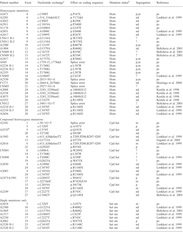
Table 1. All NPHS1 mutations detected
Patient number Exon Nucleotide exchangea Effect on coding sequence Mutation statusb Segregation Reference
Homozygous mutations
A2471 4 c.C500T p.P167L Hom p,m ps
A2201 4 c.514_516delACC p.T172del Hom nd Lenkkeri et al. 1999
A3023 8 c.C896T p.R299C Hom nd ps
A2911 9 c.C1019A p.P340H Hom nd ps
A1176 9 c.G1040A p.G347E Hom nd ps
A2031 9 c.A1096C p.S366R Hom nd Lenkkeri et al. 1999
A2617 9 c.C1099T p.R367C Hom nd Lenkkeri et al. 1999
CNS11 II-1 9 c.G1134A p.W378X Hom ? ps
CNS11 II-2 9 c.G1134A p.W378X Hom ? ps
A2300 10 c.C1219T p.R407W Hom p,m ps
A1804 11 c.G1379A p.R460Q Hom nd Beltcheva et al. 2001
CNS09 II-1 13 c.C1672T p.R558C Hom ? Beltcheva et al. 2001
CNS09 II-2 13 c.C1672T p.R558C Hom ? Beltcheva et al. 2001
A2417 13 c.A1757G p.R586G Hom p,m ps
A945 14 c.1759-15_1778del Splice error Hom p,m ps
A2236 II-1 14 c.T1760G p.L587R Hom p,m ps
A2236 II-2 14 c.T1760G p.L587R Hom p,m ps
A2355 14 c.T1760G p.L587R Hom p,m ps
CNS05 14 c.G1868T p.C623F Hom ? Lenkkeri et al. 1999
A2330 20 c.2815+5G>A Splice error Hom p,m ps
F1273 20 c.2664-4_2670del Splice error Hom nd Heeringa et al. 2008
CNS03 21 c.2927+1G>A Splice error Hom ? ps
A2088 24 c.3243_3250insG p.1084fsX12 Hom nd Kestila et al. 1998
A2538 24 c.3243_3250insG p.1084fsX12 Hom nd Kestila et al. 1998
A3075 24 c.3243_3250insG p.1084fsX12 Hom nd Kestila et al. 1998
A2553 26 c.C3325T p.R1109X Hom nd Kestila et al. 1998
CNS12 27 c.3481+1G>T Splice error Hom ? Beltcheva et al. 2001
A2210 II-1 28 c.C3478T p.R1160X Hom nd Lenkkeri et al. 1999
A2210 II-3 28 c.C3478T p.R1160X Hom nd Lenkkeri et al. 1999
A2036 28 c.C3478T p.R1160X Hom nd Lenkkeri et al. 1999
Compound heterozygous mutations
A2341 1 c.58+1G>T Splice error Cpd het m ps
3 c.C320T p.A107V p ps
A2535b 5 c.C574T p.Q193X Cpd het nd ps
20 c.T2728C p.S910P nd ps
A1943 6 c.613_620delinsTT p.T205,P206,R207>I205 Cpd het nd Lenkkeri et al. 1999
13 c.G1715A p.S572N nd Gigante et al. 2005
A2616 6 c.613_620delinsTT p.T205,P206,R207>I205 Cpd het m Lenkkeri et al. 1999
22 c.G2928T p.R976S p ps
CNS01 8 c.G886A p.W289X Cpd het ? ps
14 c.T1760G p.L587R ? ps
CNS08 9 c.T1048C p.S350P Cpd het ? Lenkkeri et al. 1999
19 c.G2625A p.W875X ? ps
A2030 9 c.A1096C p.S366R Cpd het nd Lenkkeri et al. 1999
24 c.C3478T p.R1160X nd Lenkkeri et al. 1999
A3083 9 c.C1019A p.P340H Cpd het nd ps
24 c.C3478T p.R1160X nd Lenkkeri et al. 1999
A2475A1981 15 c.G2043T c.W681C Cpd het p ps
17 c.2227delC p.R743fsX10 m ps
15 c.C2019A p.N673K Cpd het p ps
24 c.C3478T p.R1160X m Lenkkeri et al. 1999
A2249 17 c.C2227T p.R743C Cpd het nd Lenkkeri et al. 1999
27 c.C3442T p.Q1148X nd Beltcheva et al. 2001
Single mutations only
A2618 3 c.C320T c.A107V het sm m ps
A2380 10 c.G1223A c.R408Q het sm nd Lenkkeri et al. 1999
A1869 11 c.G1379A c.R460Q het sm nd Beltcheva et al. 2001
A1517 14 c.G1868T c.C623F het sm nd Lenkkeri et al. 1999
A2250 17 c.C2227T c.R743C het sm nd Lenkkeri et al. 1999
A2062 19 c.G2625A c.W875X het sm p ps
A2320 II-1 23 c.C3418T c.R1140C het sm nd Lenkkeri et al. 1999
A2320 II-2 23 c.C3418T c.R1140C het sm nd Lenkkeri et al. 1999
As relevant wild-type gene sequence, the published reference sequence of NPHS1 was used (NM_004646). het, heterozygous; Hom, homozygous mutation; cpd het, compound heterozygous mutation; sm, single mutation only detected; p, paternal; m, maternal; nd, not done; ?, information not available for this patient; ps, novel mutation detected in the present study.
a
All mutations were absent from 93 healthy controls.
b
numbering. For all detected mutations and variants, both strands were quenced. Whenever possible, segregation was confirmed by direct se-quencing of the parental samples. For novel mutations, their absence from 93 healthy control individuals was confirmed by direct sequencing.
Homozygosity mapping
Genome-wide homozygosity mapping for 12 families with CNS was per-formed and evaluated as described previously [20]. Single-nucleotide polymorphism (SNP) arrays (GeneChip®) from Affymetrix, Inc. with a resolution of 250K (Human Mapping 250K Styl Array) were used. Samples were processed, hybridized and scanned using the manufac-turer's standard methods at the University of Michigan Core Facility (www.michiganmicroarray.com). Using the software Allegro [21] and ALOHOMORA [22], non-parametric likelihood ratio Z-scores (ZLRs) were calculated using one marker every 100 000 markers. Allele frequen-cies for Caucasians as specified by Affymetrix®, a disease allele frequency of 0.001, and a standard pedigree structure assuming first-cousin marriage for parents of affected individuals were used. ZLRs were calculated under three different conditions, i.e. for minor allele frequencies of >0.2, >0.3 and >0.4, and a non-existent sibling was included to enable non-parametric Al-legro runs. If a peak was constantly exceeding the value of 2.0 in two out of the three conditions, we referred to it as a‘consistent ZLR peak’ (cZLR) and expected it to harbour the homozygous mutation of the recessive disease gene [20]. The ZLRs were plotted against genetic distance across the entire human genome using the Gnuplot software (http:// www.gnuplot.info) (Supplementary Figure 1; see online supplementary material for a colour version of this figure). In this way, the maxima of ZLR scores represent segments of homozygosity by descent.
Results and discussion
Clinical characteristics and ethnicity of patients
In this study, 67 patients (32 females, 34 males and 1 with unknown gender) from 62 different families with CNS were included. All patients were examined for NPHS1 mutations. Renal biopsy was performed in 24 patients, showing 10 with a pattern congruent to NS‘Finnish type’, 8 patients with DMS, 3 with minimal change nephrotic syndrome (MCNS), 2 with focal segmental glomerulosclerosis (FSGS), 1 with membranoproliferative glomerulonephritis (MPGN) and 1 with end-stage nephrosclerosis (Supplemen-tary Table 2). Altogether, 21 different ethnicities were repre-sented within the cohort; among these, the largest groups were of Turkish (15%), Arabic (15%), European (13%) and Caucasian (9%) descent (Supplementary Table 2). Consanguinity was reported in 20 families (Supplementary Table 1). In three cases, a nephrectomy had been per-formed, and 10 patients had received a renal transplant (Supplementary Table 2).
Modality of treatment and response
Because traditionally CNS is considered treatment refrac-tory, steroid treatment was only reported as attempted in 14 (20.8%) cases. Eleven of these patients showed no re-sponse (steroid-resistant nephrotic syndrome, SRNS), as expected for CNS, while three (A2236 II-1/II-2, A2380) did partially respond. In four cases, cyclosporin A was ap-plied in addition to steroids, while one patient was treated with cyclosporin A only (Supplementary Table 2). In none of these subjects was any response recorded (Supplemen-tary Table 2, Supplemen(Supplemen-tary Table 4).
‘Antiproteinuric therapy’ with angiotensin-converting enzyme (ACE) inhibitors or indomethacin was attempted
in 34 patients. In nine patients, exclusively ACE inhibitors were applied, of which two (22.2%) showed a partial remis-sion (A1804, A2475). Partial remisremis-sion is hereby under-stood as the permanent disappearance of oedema, an increase in the serum albumin concentration to >35 g/L and the persistence of proteinuria of >4 mg/m2/h [23]. In both patients, mutation analysis had revealed disease-caus-ing mutation in NPHS1. Surprisdisease-caus-ingly, A2475 showed com-pound heterozygosity for a missense mutation and a deletion. A combined therapy with indomethacin and ACE inhibitors was administered to 26 patients of which eight (30%) showed partial remission, while in 17 cases, no effect was observed. Of the eight patients showing a par-tial remission, a disease-causing mutation in NPHS1 was found in six patients. Of these six patients, missense muta-tions were disease causing in five cases. Unexpectedly, however, patient A2201 also showed partial remission, al-though a homozygous deletion was detected in this patient. He was treated with captopril for 40months followed by 10 months of treatment with losartan. Indomethacin was applied for 3months but did not show any benefit. These data show that ‘antiproteinuric’ therapy has a beneficial effect on 20–30% of patients with CNS and should not only be considered for patients with missense mutations but might also be positive for patients with a more severe type of mutation (Supplementary Table 2, Supplementary Table 4).
For 19 out of the 67 patients, no pharmacological treat-ment was reported.
NPHS1 mutations
In 67 children from 62 different families, mutation analysis by direct sequencing of all NPHS1 exons was performed. In 36 families, causative mutations in NPHS1 were de-tected on both alleles. We hereby confirm the results of former studies [6,19] showing that approximately one-half of CNS cases are caused by recessive mutations in NPHS1 (Table 1). In CNS, it has been shown that ∼85% of the cases are explained by mutations in four genes. The distri-bution among these four genes is: NPHS1 39.8%, NPHS2 39.8%, WT1 2.2% and LAMB2 4.4% [6]. Twenty-six of the 36 families showed homozygous mutations, and another 10 families had compound heterozygous mutations. In seven families, only one heterozygous NPHS1 mutation was detected (Table 1). As a reason for this relatively high number of patients with disease-causing mutations, only on one allele, one might speculate that deletions/duplications of whole exons as well as intronic mutations and mutations in the promoter region can explain these cases. A direct sequencing approach might not have been able to detect these mutations.
Out of the 37 different disease-causing NPHS1 mutations detected, 19 mutations were novel, consisting of 11 mis-sense mutations, 4 splice-site mutations, 3 nonmis-sense muta-tions and 1 deletion. They were found in exons 1, 3, 4, 8, 9, 10, 13, 14, 15, 17, 19 and 21 (Table 1 and Figure 1, Supple-mentary Figure 1; see online suppleSupple-mentary material for a colour version of this figure). The 18 previously published mutations consisted of 10 missense mutations, 3 nonsense mutations, 1 insertion, 1 deletion, 2 splice-site mutations
and 1 insertion/deletion. They were found in exons 4, 6, 9, 10, 11, 13, 14, 17, 20, 22, 24, 26 and 27 (Table 1, Figure 1). None of the patients had the Finmajor or the
Finminormutation. The mutations were broadly distributed
over the nephrin protein, affecting all domains. The most frequently affected domains were immunoglobulin (Ig)-like domain 5 and Ig-like domain 3 (Figure 1).
Genotype/phenotype correlations
While the CNS classical histology of‘Finnish type’ rapidly progresses into ESKD and shows no response to treatment, several cases of patients with NPHS1 mutations have been reported, whose histological phenotype was not as severe and who sometimes even showed a response to treatment [19,24].
From the 12 patients in our study, in whom renal biopsy was performed and whose disease was explained by two recessive mutations in NPHS1, eight patients showed NS ‘Finnish type’, three showed MCNS, one showed DMS and one showed FSGS (Supplementary Table 3). These da-ta confirm the previous finding that renal pathology does not exclusively appear as NS‘Finnish type’ in CNS caused by recessive NPHS1 mutations. A higher frequency of mu-tations in a certain ethnicity was not observed.
Out of the seven patients in our cohort with a biopsy of DMS, only one had disease-causing NPHS1 mutations (A2911 II-1). This patient showed a novel homozygous mis-sense mutation (c.1019C>A, p.P340H) (Table 1). The pa-tient was steroid resistant. Manifestation was at birth, and ESKD developed at the age of 4months (Supplementary Table 1). Another patient (A3083 II-1) showed the same mutation heterozygously together with a heterozygous known nonsense mutation [c.3478 C>T (h), p.R1160X] (Table 1). He manifested at the age of 2months and also showed steroid resistance. Biopsy was not performed, and ESKD was not reported by the age of 6months (Supple-mentary Table 1). DMS is seen in patients with mutations in PLCE1, WT1 and LAMB2. As WT1 and PLCE1 yielded no mutations in the remaining DMS patients, we speculate that they may have mutations in LAMB2 or PLCE1.
One patient (A2616 II-1) with a biopsy of FSGS showed compound heterozygosity for a known deletion (c.613_620delinsTT; p.T205, P206, R207>I205) and a novel missense mutation [c.2928G>T (h), p.R977S] (Table 1). The age of disease onset was 2 months, and steroid treatment was not attempted. The patient devel-oped ESKD by the age of 10years and was transplanted (Supplementary Table 1).
In two patients with a homozygous NPHS1 mutation (CNS03, CNS11 II-1), a renal biopsy of MCNS was re-ported. Patient CNS03 II-1 had a truncating mutation [c.1134G>A (H), p.W378X], while patient CNS11 II-1 had a splice-site error (c.2927 +1G>A, splice error) (Table 1). In both cases, no treatment was attempted. The age of onset for patient CNS03 was shortly after birth, and patient CNS11 II-1 manifested later at the age of 4 months. For patient CNS11 II-2, no biopsy was performed; both siblings were treated with steroids for 2months but did not respond (Sup-plementary Table 1). Patient CNS03 showed severe progres-sion with nephrectomy at the age of 6months and histology
of glomerulosclerosis as well as tubular atrophy, microcysts and interstitial fibrosis. As patients with an early biopsy of-ten show MCNS but progress rapidly, the aetiopathology of this patient is not surprising. However, patient CNS11 II-1 is reported to be stabile at the age of 2.5years now and, togeth-er with the late onset, is showing a rathtogeth-er unusual course of disease. Both siblings also showed partial remission to treat-ment with ACE inhibitors and indomethacin.
Although mutations in NPHS1 were thought to exclusive-ly cause CNF, these results confirm former findings indicat-ing that NPHS1 mutations can cause a somewhat broader variety of histological phenotypes in nephrotic syndrome. In a recent genetic study of patients with CNS [6], in a total of 21 patients with two NPHS1 mutations, the histological phenotypes were distributed as follows: ‘Finnish type’ (14%), MCNS (14%), FSGS (4.6%), DMS (3.6%), mesan-gial proliferation (9.2%), mesanmesan-gial sclerosis (3.6%) and no finding (3.6%) [6] (Supplementary Table 3). In 10 patients, no biopsy was performed. These results were also confirmed by another study of children with CNS, showing again ‘non-Finnish type’ manifestations [19] (Supplementary Table 3). While the CNS classical histology of‘Finnish type’ rap-idly progresses into ESKD and shows no response to treat-ment, several cases of patients with NPHS1 mutations have been reported, whose histological phenotype was not as severe and who sometimes even showed a response to treat-ment [19,24]. Recently, Phillipe et al. even reported several cases with childhood rather than congenital onset of ne-phrotic syndrome and confirmed mutations in NPHS1 [13]. In this study, two siblings were included who had dif-ferent age of onset. While the male sibling (A2236 II-2) manifested as CNS by the age of 2.5months, his elder sister (A2236 II-1) stayed healthy until the age of 24months. Both showed partial remission due to steroid treatment, and in both sibs, a novel homozygous missense mutation (c.1760T>G, p. L587R) was detected. Partial remission is hereby understood as the permanent disappearance of oe-dema, an increase in the serum albumin concentration to >35 g/L and the persistence of proteinuria of >4 mg/m2/h [23]. A third patient (A2355 II-1), who was classified as CNS and of the same ethnicity, showed the same mutation. Interestingly, he also was not diagnosed before the age of 5 months. Steroid treatment for this patient was not attempted. Additionally, one patient (CNS01) showed L587R hetero-zygously in combination with a heterozygous nonsense mu-tation [c.886G>A (h), W289X]. This patient showed the age of onset of 2months, and no form of steroid treatment was reported. We therefore conclude that homozygous L587R may be a milder mutation, causing a less severe form of ne-phrotic syndrome than other NPHS1 mutations with possi-ble childhood onset later than 90 days of life. It is, to our knowledge, the first homozygous mutation in NPHS1 to cause childhood onset (in A2236 II-1 and A2355 II-1), and the findings suggest that mutation analysis should also be sought in children who manifest after 90days of life. Detection of NPHS1 mutations by total genome homozygosity mapping
Mutations that are homozygous by descent have been described as being frequent (30–80%) in paediatric
dis-eases [20]. This can be mapped by homozygosity map-ping. In order to investigate if homozygosity mapping is a useful tool for screening, we performed homozygosity mapping in a subset of this cohort of 12 patients from 12 families with different background (Supplementary Table 1). All of them exhibited homozygous segments by inspection of their homozygosity plots, while only five (A2031 II-1, F1273 II-1, A1804 II-1, A2088 II-1, A2036 II-1) revealed homozygosity at the NPHS1 locus (Supplementary Figure 2; see online supplementary ma-terial for a colour version of this figure). We detected homozygous disease-causing mutations of NPHS1 in all five patients. Of the remaining seven patients, two (A1981 II-1, A2062 II-1) showed compound heterozy-gous disease-causing mutations, two (A1517 II-1, A1869 II-1) showed single heterozygous mutations and three (A1970 II-1, A1980 II-1, A2112 II-1) showed no mutations of NPHS1 (Table 1, Supplementary Table 1).
Mutation in N-glycosylation site
In addition to the results of our systematic mutation screening in a CNS cohort, we report on a single patient with onset of nephrotic syndrome at the age of 9months who was also found to be mutated in NPHS1. The patient was not included in the study, and his clinical data is not shown here. He was treated with steroids but did not re-spond (SRNS). His renal biopsy showed the histological features of IgM nephropathy.
Mutation analysis of NPHS1 in patient A2535 revealed two novel heterozygous mutations: c.574C>T; p.Q193X and c.2728T>C; p.S910P (Table 1, Figure 1, not included in the examined cohort). This finding is of interest as, to our knowledge, S910P is the first mutation described, which directly affects one of nephrin's ten known N-glycosylation sites [25]. Substitution of the serine residue by proline is predicted to prevent glycosylation at this site. Defects in post-translational modification may lead to decreased stability of impaired interaction with other molecules. Considering the late manifestation of nephrotic syn-drome in this patient, we speculate that the mutation S910P has some residual protein function and might be a ‘mild’ mutation. As it has been described recently, a ‘mild’ mutation in combination with a ‘severe’ muta-tion in NPHS1 may cause childhood onset of nephrotic syndrome [13].
Adding these mutations, we here report 21 novel muta-tions, expanding the number of published mutations in NPHS1 by 17.6%.
Supplementary data
Supplementary data is available online at http://ndt.oxford-journals.org.
Acknowledgements. We thank the patients and their physicians for con-tribution of blood samples and clinical data. This work was supported by a grant to F.H. from the National Institutes of Health (DK076683, RC1-DK086542), and the Thrasher Research Fund, and by a grant to M.Z. by the German Research Foundation (DFG; SFB 423). F.H. is an investigator of the Howard Hughes Medical Institute, a Doris Duke Distinguished Clinical Scientist and a Frederick G.L. Huetwell Professor.
Conflict of interest statement. None declared.
References
1. Kestila M, Lenkkeri U, Mannikko M et al. Positionally cloned gene for a novel glomerular protein—nephrin—is mutated in congenital nephrotic syndrome. Mol Cell 1998; 1: 575–582
2. Ruotsalainen V, Ljungberg P, Wartiovaara J et al. Nephrin is specif-ically located at the slit diaphragm of glomerular podocytes. Proc Natl Acad Sci U S A 1999; 96: 7962–7967
3. Khoshnoodi J, Sigmundsson K, Ofverstedt LG et al. Nephrin pro-motes cell–cell adhesion through homophilic interactions. Am J Pathol 2003; 163: 2337–2346
4. Jones N, Blasutig IM, Eremina V et al. Nck adaptor proteins link nephrin to the actin cytoskeleton of kidney podocytes. Nature 2006; 440: 818–823
5. Quack I, Rump LC, Gerke P et al. beta-Arrestin2 mediates nephrin endocytosis and impairs slit diaphragm integrity. Proc Natl Acad Sci U S A 2006; 103: 14110–14115
6. Hinkes BG, Mucha B, Vlangos CN et al. Nephrotic syndrome in the first year of life: two thirds of cases are caused by mutations in 4 genes (NPHS1, NPHS2, WT1, and LAMB2). Pediatrics 2007; 119: e907–e919
7. Beltcheva O, Martin P, Lenkkeri U, Tryggvason K. Mutation spec-trum in the nephrin gene (NPHS1) in congenital nephrotic syndrome. Hum Mutat 2001; 17: 368–373
8. Holmberg C, Tryggvason K, Kestila M, Jalanko H. Congenital Nephrotic Syndrome. In: Avner E, Harmon WE, Niaudet P (eds). Pediatric Nephrology. 5th edn. Philadelphia; Lippincott Williams & Wilkins, 2004
9. Qvist E, Laine J, Ronnholm K et al. Graft function 5–7 years after renal transplantation in early childhood. Transplantation 1999; 67: 1043–1049
10. Norio R. Heredity in the congenital nephrotic syndrome. A genetic study of 57 Finnish families with a review of reported cases. Ann Paediatr Fenn 1966; 12: 21–94
11. Huttunen NP. Congenital nephrotic syndrome of Finnish type. Study of 75 patients. Arch Dis Child 1976; 51: 344–348
12. Kuusniemi AM, Merenmies J, Lahdenkari AT et al. Glomerular sclerosis in kidneys with congenital nephrotic syndrome (NPHS1). Kidney Int 2006; 70: 1423–1431
13. Philippe A, Nevo F, Esquivel EL et al. Nephrin mutations can cause childhood-onset steroid-resistant nephrotic syndrome. J Am Soc Nephrol 2008; 19: 1871–1878
14. APN. Short versus standard prednisone therapy for initial treatment of idiopathic nephrotic syndrome in children. Lancet 1988; 380–383 15. Ruf RG, Lichtenberger A, Karle SM et al. Patients with mutations in NPHS2 (podocin) do not respond to standard steroid treatment of nephrotic syndrome. J Am Soc Nephrol 2004; 15: 722–732 16. Hinkes B, Wiggins RC, Gbadegesin R et al. Positional cloning
uncovers mutations in PLCE1 responsible for a nephrotic syndrome variant that may be reversible. Nat Genet 2006; 38: 1397–1405 17. Ruf RG, Schultheiss M, Lichtenberger A et al. Prevalence of
WT1 mutations in a large cohort of patients with steroid-resistant and steroid-sensitive nephrotic syndrome. Kidney Int 2004; 66: 564–570
18. Mucha B, Ozaltin F, Hinkes BG et al. Mutations in the Wilms' tumor 1 gene cause isolated steroid resistant nephrotic syndrome and occur in exons 8 and 9. Pediatr Res 2006; 59: 325–331
19. Heeringa SF, Vlangos CN, Chernin G et al. Thirteen novel NPHS1 mutations in a large cohort of children with congenital nephrotic syn-drome. Nephrol Dial Transplant 2008; 23: 3527–3533
20. Hildebrandt F, Heeringa SF, Ruschendorf F et al. A systematic approach to mapping recessive disease genes in individuals from out-bred populations. PLoS Genet 2009; 5: e1000353
21. Gudbjartsson DF, Jonasson K, Frigge ML, Kong A. Allegro, a new computer program for multipoint linkage analysis. Nat Genet 2000; 25: 12–13
22. Ruschendorf F, Nurnberg P. ALOHOMORA: a tool for linkage anal-ysis using 10K SNP array data. Bioinformatics 2005; 21: 2123–2125
23. ISKDC. Primary nephrotic syndrome in children: clinical signifi-cance of histopathologic variants of minimal change and of diffuse mesangial hypercellularity. A report of the International Study of Kidney Disease in children. Kidney Int 1981; 20: 765–771 24. Kitamura A, Tsukaguchi H, Hiramoto R et al. A familial
childhood-onset relapsing nephrotic syndrome. Kidney Int 2007; 71: 946–951
25. Khoshnoodi J, Hill S, Tryggvason K, Hudson B, Friedman DB. Iden-tification of N-linked glycosylation sites in human nephrin using mass spectrometry. J Mass Spectrom 2007; 42: 370–379
Received for publication: 3.8.09; Accepted in revised form: 2.2.10
Nephrol Dial Transplant (2010) 25: 2976–2981 doi: 10.1093/ndt/gfq119
Advance Access publication 10 March 2010
Long-term follow-up of patients with Bartter syndrome type I and II
Elena Puricelli
1,4,**, Alberto Bettinelli
1,**, Nicolò Borsa
2, Francesca Sironi
2, Camilla Mattiello
2,
Fabiana Tammaro
1, Silvana Tedeschi
2, Mario G. Bianchetti
3and Italian Collaborative Group for Bartter Syndrome
*1
Department of Pediatrics, San Leopoldo Mandic Hospital, Largo Mandic 1, Merate, Lecco, Italy,2Laboratory of Medical Genetics, Fondazione IRCCS Ca’Granda-Ospedale Maggiore Policlinico, Mangiagalli e Regina Elena, Milan, Italy,3Department of Pediatrics, Mendrisio and Bellinzona Hospitals, University of Bern, Bern, Switzerland and4Department of Pediatrics, F.Del Ponte Hospital, Via F. Del Ponte 19, Insubria University of Varese, Varese, Italy
Correspondence and offprint requests to: Alberto Bettinelli; E-mail: a.bet@libero.it; a.bettinelli@ospedale.lecco.it
*
Silvio Maringhini, Unit of Pediatric Nephrology, Civile Hospital, Palermo, Italy; Paolo Porcelli, Unit of Pediatric Endocrinology, Villa Sofia Hospital, Palermo, Italy; Marco Materassi, Department of Pediatrics, Meyer Hospital, University of Florence, Italy; Maria Renata Proverbio, Department of Pediatric Nephrology, Cardarelli Hospital, Naples, Italy; Nunzia Miglietti, Department of Pediatrics, University of Brescia, Italy; Maria Gabriella Porcellini, Department of Pediatric Nephrology, Regina Margherita Hospital, Turin, Italy; Carla Navone, Department of Pediatrics, Santa Corona Hospital, Pietra Ligure, Savona, Italy; Giuseppe Ruffa, Department of Pediatrics, Gaslini Hospital, Genoa, Italy; Aldo Rosini, Department of Pediatrics, B. Eustachio Hospital, San Severino Marche, Italy; Aurora Rossodivita, Department of Pediatrics, Gemelli Hospital, Rome, Italy.
**
These authors contributed equally to the work.
Abstract
Background. Little information is available on a long-term follow-up in Bartter syndrome type I and II. Methods. Clinical presentation, treatment and long-term follow-up (5.0–21, median 11 years) were evaluated in 15 Italian patients with homozygous (n = 7) or compound heterozygous (n = 8) mutations in the SLC12A1 (n = 10) or KCNJ1 (n = 5) genes.
Results. Thirteen new mutations were identified. The 15 children were born pre-term with a normal for gestational age body weight. Medical treatment at the last follow-up control included supplementation with potassium in 13, non-steroidal anti-inflammatory agents in 12 and gastropro-tective drugs in five patients. At last follow-up, body weight and height were within normal ranges in the patients. Glo-merular filtration rate was <90 mL/min/1.73 m2 in four patients (one of them with a pathologically increased uri-nary protein excretion). In three patients, abdominal ul-trasound detected gallstones. The group of patients with antenatal Bartter syndrome had a lower renin ratio (P < 0.05) and a higher standard deviation score (SDS) for height (P < 0.05) than a previously studied group of pa-tients with classical Bartter syndrome.
Conclusions. Patients with Bartter syndrome type I and II tend to present a satisfactory prognosis after a median
fol-low-up of more than 10 years. Gallstones might represent a new complication of antenatal Bartter syndrome.
Keywords: Bartter syndrome; cholelithiasis; growth retardation; KCNJ1 gene; SLC12A1 gene
Introduction
Bartter syndrome type I (BS I) and type II (BS II) are salt-wasting renal tubular disorders that are clinically character-ized by polyhydramnios leading to premature delivery, marked polyuria and a tendency towards nephrocalcinosis [1,2]. Loss-of-function mutations either in the furosemide-sensitive sodium–potassium–chloride cotransporter gene (SLC12A1; BS I, OMIM 601678) or in the inwardly rectify-ing potassium channel ROMK gene (KCNJ1; BS II, OMIM 241200) have been identified in the vast majority of patients with this autosomal recessive disorder [3,4]. Mutations in the CLCNKB chloride channel gene (Bartter syndrome type III also defined as classical Bartter syndrome—OMIM 607364) as well as in the BSND gene (Bartter syndrome form associated with sensorineural deafness—Bartter type IV— OMIM 602522) are also sometimes responsible for an iden-tical clinical phenotype but will not be treated in this report.
© The Author 2010. Published by Oxford University Press on behalf of ERA-EDTA. All rights reserved. For Permissions, please e-mail: journals.permissions@oxfordjournals.org