HAL Id: inserm-00799941
https://www.hal.inserm.fr/inserm-00799941
Submitted on 12 Mar 2013
HAL is a multi-disciplinary open access
archive for the deposit and dissemination of
sci-entific research documents, whether they are
pub-lished or not. The documents may come from
teaching and research institutions in France or
abroad, or from public or private research centers.
L’archive ouverte pluridisciplinaire HAL, est
destinée au dépôt et à la diffusion de documents
scientifiques de niveau recherche, publiés ou non,
émanant des établissements d’enseignement et de
recherche français ou étrangers, des laboratoires
publics ou privés.
Distributed under a Creative Commons Attribution| 4.0 International License
mammals by peroxisome proliferator-activated receptors.
Stéphane Mandard, Patsouris David
To cite this version:
Stéphane Mandard, Patsouris David. Nuclear control of the inflammatory response in mammals by
peroxisome proliferator-activated receptors.. PPAR Research, Hindawi Publishing Corporation, 2013,
2013, pp.613864. �10.1155/2013/613864�. �inserm-00799941�
Volume 2013, Article ID 613864,23pages
http://dx.doi.org/10.1155/2013/613864
Review Article
Nuclear Control of the Inflammatory Response in Mammals by
Peroxisome Proliferator-Activated Receptors
Stéphane Mandard
1and David Patsouris
2,31Centre de Recherche INSERM-UMR866 “Lipides, Nutrition, Cancer” Facult´e de M´edecine, Universit´e de Bourgogne 7,
Boulevard Jeanne d’Arc, 21079 Dijon Cedex, France
2Laboratoire CarMeN, UMR INSERM U1060/INRA 1235, Universit´e Lyon 1, Facult´e de M´edecine Lyon Sud,
165 Chemin du Grand Revoyet, 69921 Oullins, France
3Department of Chemical Physiology, he Scripps Research Institute, MB-24, 10550 North Torrey Pines Road, La Jolla, CA 92037, USA
Correspondence should be addressed to St´ephane Mandard; stephane.mandard@u-bourgogne.fr Received 15 October 2012; Revised 14 January 2013; Accepted 29 January 2013
Academic Editor: Massimo Bionaz
Copyright © 2013 S. Mandard and D. Patsouris. his is an open access article distributed under the Creative Commons Attribution License, which permits unrestricted use, distribution, and reproduction in any medium, provided the original work is properly cited.
Peroxisome proliferator-activated receptors (PPARs) are ligand-activated transcription factors that play pivotal roles in the regula-tion of a very large number of biological processes including inlammaregula-tion. Using speciic examples, this paper focuses on the inter-play between PPARs and innate immunity/inlammation and, when possible, compares it among species. We focus on recent discov-eries establishing how inlammation and PPARs interact in the context of obesity-induced inlammation and type 2 diabetes, mostly in mouse and humans. We illustrate that PPAR� ability to alleviate obesity-associated inlammation raises an interesting pharmaco-logic potential. In the light of recent indings, the protective role of PPAR� and PPAR�/� against the hepatic inlammatory response is also addressed. While PPARs agonists are well-established agents that can treat numerous inlammatory issues in rodents and humans, surprisingly very little has been described in other species. We therefore also review the implication of PPARs in inlamma-tory bowel disease; acute-phase response; and central, cardiac, and endothelial inlammation and compare it along diferent species (mainly mouse, rat, human, and pig). In the light of the data available in the literature, there is no doubt that more studies concerning the impact of PPAR ligands in livestock should be undertaken because it may inally raise unconsidered health and sanitary beneits.
1. Introduction
he peroxisome proliferator-activated receptors (PPARs) are ligand-activated transcription factors that play critical roles in very diferent biological pathways such as lipid, protein, glycerol, urea, glucose, glycogen and lipoprotein metabolism, adipogenesis, trophoblast diferentiation, and cell migration [1–6]. Notably, PPARs are also required to balance cell pro-liferation and cell death and therefore impact skin wound healing and proliferative diseases such as cancer [7–9]. PPARs are also prominent players in inlammation control [10,11]. PPAR�, the irst PPAR isotype identiied in mouse, was originally cloned in the early 1990s as a novel member of the steroid hormone receptor superfamily [12]. Shortly ater, a rat version of PPAR� as well as three novel members related to each other (xPPAR�, xPPAR�, and xPPAR�) and to
mouse PPAR� have been subsequently cloned from Xenopus (frog) [13]. Since then, substantial eforts have been made to identify other related receptors; several additional PPAR iso-forms and variants have been therefore isolated in a wide range of species including mammals (human, rabbit, mouse, rat, pig, rhesus and cynomolgus monkey, dog, guinea pig, hibernating ground squirrel, and hamster), ishes (grass carp, cobia not only but also marine ish such as the teleost red sea bream (Pagrus major) and the mullet Chelon labrosus), mar-ine gastropod mollusks (Cyclostoma), reptiles (leopard gecko, crocodile, and turtle), and birds (domestic chicken, goose) [14–51].
Since PPARs are ligand-activated transcription factors, a large part of our knowledge about their biological importance is coupled to the function of their target genes. At the molecu-lar level, it was shown that PPARs readily heterodimerize with
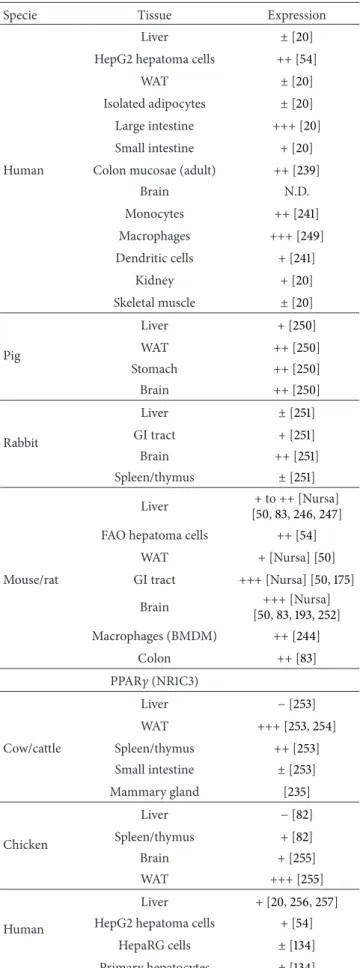
the Retinoid X Receptor (RXR) prior to ligand binding [52]. In all species tested so far, Ppar�, Ppar�/�, and Ppar� show speciic time- and tissue-dependent patterns of expression (Table 1).
Ater ligand treatment, the PPAR/RXR heterodimer sta-bly binds on genomic DNA at speciic sites called Peroxi-some Proliferator Response Element (PPRE) and upregulates gene transcription. Consensus PPREs are formed by two hexameric core binding motifs (AGGTCA) in a direct repeat orientation with an optimal spacing of one nucleotide (DR1). Molecular investigations have demonstrated that PPAR occu-pies the 5�motif of the DR1 [53]. Recent analyses have further revealed that even if DR1 PPREs can be located within the promoter sequences of target genes, about 50% of all target sites are located within genes (introns, exons) as well as in 3� downstream sequences of the target genes [4, 7, 54–
58]. he PPAR� (NR1C1), PPAR�/� (NR1C2), and PPAR� (NR1C3) genes encode proteins that share a highly conserved structure and molecular mode of action, yet the array of genes regulated by each PPAR isotype is divergent and may also difer from one species to another [59]. An extended analysis of the cross-species (mouse to human) conservation of PPREs brought support to this hypothesis because it revealed only limited conservation of PPRE patterns [60]. Strengthening this observation, only a minor overlap between the Wy14,643 (Wy: a speciic PPAR� agonist) regulated genes from mouse and human primary hepatocytes was found by Rakhshan-dehroo et al. demonstrating that some, but not all, genes are equally regulated by PPAR� in mouse and human hepato-cytes [61]. In this review, we explore and focus on the role of PPARs in the control of chronic (mediated by obesity) or acute (as a result of bacterial infection) inlammation in dif-ferent species, mainly from human, mouse, rat, pig, and cow.
2. PPARs and Obesity-Induced Inflammation:
Interplay with Adipose Tissue Macrophages
2.1. PPAR�. In spite of the relative weak expression level of Ppar� in white adipose tissue (WAT, mainly in adipocytes and not in stromal-vascular cells), several lines of evidence sup-port the notion that PPAR� and PPAR� agonists could play a functional role in the control of obesity-induced chronic inlammatory response in vivo. For instance, treatment of obese diabetic KKAy mice with Wy decreased the mRNA levels of Tnf-� (tumor necrosis factor-�), Mcp-1 (monocyte chemotactic protein-1, also referred to as chemokine (C-C motif) ligand 2, CCL2), and Mac-1 (macrophage antigen-1, also known as cluster of diferentiation molecule-11b, Cd11b) in epididymal fat, suggesting a reduction in macrophage inil-tration [62]. In addition, expression of inlammatory genes in adipose tissue such as Tnf-�, Mcp-1, and IL-1� (Interleukin-1 beta) as well as that of speciic macrophage markers such as Cd68 (macrophage antigen Cd68, also known as scavenger receptor class D member 1, Scard1), F4/80 (also referred to as lymphocyte antigen-71, Ly71), and Adam8 (ADAM metal-lopeptidase domain 8, also known as cluster of diferentiation molecule-156, Cd156) in the stromal vascular fraction was more pronounced in Ppar�-deicient mice compared to WT (wild-type) mice rendered obese with a high-fat feeding,
Table 1: Tissue distribution of the various PPARs in diferent species.
Specie Tissue Expression PPAR� (NR1C1) Liver ++ [237] WAT N.D. Cow/cattle GI tract N.D. Brain N.D. Spleen/thymus N.D. Liver ++ [23] Chicken WAT + [82] Brain ++ [82] Spleen + [82] Liver [20,61,118,+++162,175] Primary hepatocytes ± to +++ [61,134] HepG2 hepatoma cells + [54] HepaRG hepatoma cells ++ [134]
Human WAT + [20,118,238] Isolated adipocytes ± [20] GI tract ++ [20,118,175,239] Brain + [118,175,240] Monocytes + [241,242] Dendritic cells ++ [241,242] Kidney ++ [20,118] Heart +++ [118] Pig Liver ± [243] WAT + [243] Liver +++ [61,83,244–247] Hepatocytes ++ [61] GI tract ++[Nursa] [175] Mouse/rat Brain +[Nursa]
Spleen/thymus − [83] Macrophages (BMDM) − [244]
FAO hepatoma cells ++ [54] WAT + [62,248] PPAR�/� (NR1C2) Liver N.D. WAT N.D. Cow/cattle GI tract N.D. Brain N.D. Spleen/thymus N.D. Liver N.D. WAT N.D. Chicken GI tract N.D. Brain N.D. Spleen/thymus N.D.
Table 1: Continued.
Specie Tissue Expression Liver ± [20] HepG2 hepatoma cells ++ [54]
WAT ± [20] Isolated adipocytes ± [20] Large intestine +++ [20] Small intestine + [20] Human Colon mucosae (adult) ++ [239]
Brain N.D. Monocytes ++ [241] Macrophages +++ [249] Dendritic cells + [241] Kidney + [20] Skeletal muscle ± [20] Liver + [250] Pig WAT ++ [250] Stomach ++ [250] Brain ++ [250] Liver ± [251] Rabbit GI tract + [251] Brain ++ [251] Spleen/thymus ± [251] Liver + to ++[Nursa] [50,83,246,247] FAO hepatoma cells ++ [54]
WAT +[Nursa] [50] Mouse/rat GI tract +++[Nursa] [50,175]
Brain +++[Nursa] [50,83,193,252] Macrophages (BMDM) ++ [244] Colon ++ [83] PPAR� (NR1C3) Liver − [253] WAT +++ [253,254] Cow/cattle Spleen/thymus ++ [253] Small intestine ± [253] Mammary gland [235] Liver − [82] Chicken Spleen/thymus + [82] Brain + [255] WAT +++ [255] Human Liver + [20,256,257] HepG2 hepatoma cells + [54]
HepaRG cells ± [134] Primary hepatocytes ± [134]
Table 1: Continued.
Specie Tissue Expression WAT +++ [20,54,256,257] Isolated adipocytes +++ [20] Simpson-Golabi-Behmel Syndrome (SGBS) adipocytes +++ [84] Large intestine +++ [20] Small intestine ± [20] Brain N.D. Monocytes +++ [241] Dendritic cells +++ [241] Kidney + [20] Skeletal muscle ± [20] Pig Liver − [243] WAT ++ [243] Liver − to + [251,258] WAT +++ [258] Rabbit GI tract +++ [251] Brain − [251] Spleen/thymus ++ [251] Liver + to – [Nursa] [83,246,247] Hepatocytes + [259] FAO hepatoma cells − [54]
WAT +++ [Nursa] [83,256,260] Mouse/rat 3T3-L1 adipocytes +++ [84] GI tract + [Nursa] [83] Brain + [Nursa] [83,261,262] Spleen/thymus ++ [83] Macrophages (BMDM) +++ [244]
Abbreviations: GI: gastrointestinal; WAT: white adipose tissue; N.D.: not determined. BMDM: bone marrow-derived macrophages.
Symbols:−: absent; ±: barely detectable; +: weak; ++: moderate; +++: high. the citation link for Nursa ishttp://www.nursa.org/10.1621/datasets.02001.
reinforcing the notion that PPAR� is required for the control of the adipose inlammation process [63]. Another study has also examined the efects of ibrates on the inlammatory changes induced by the interaction between adipocytes and macrophages in obese adipose tissue. Systemic administra-tion of Wy or fenoibrate to genetically obese ob/ob mice signiicantly reduced Tnf-� and Mcp-1 mRNA expression in WAT [64]. Similar observation was also reported using adipose tissue explants from ob/ob mice suggesting a direct efect of PPAR� agonists. To check for the deinitive involve-ment of PPAR� in the efects of Wy-mediated reduction in the production of proinlammatory cytokines by white fat pads, adipose tissue explants obtained from PPAR�-deicient
mice were also used [64]. Compared to WT mice, induction of Mcp-1 mRNA expression by TNF-� (a major paracrine mediator of inlammation in adipocyte) was much robust in adipose tissue explants from Ppar�-deicient mice, sug-gesting that PPAR� is constitutively required to control the steady-state level of adipose Mcp-1 mRNA levels. Intriguingly, induction of adipose Mcp-1 mRNA expression by TNF-� was also suppressed by Wy in explants from Ppar�-deicient mice, suggesting that Wy can act independently of the presence of the receptor in fat, at least for the control of the inlammation process [64]. Because Ppar� is expressed in both mature adipocytes and macrophages, we cannot rule out that part of the efects of ibrates on adipose inlammation are mediated through this other PPAR isotype. Moreover, treating 3T3-L1 mouse adipocytes with Wy or fenoibrate suppressed bacterial lipopolysaccharides-(LPS-) mediated increased in Mcp-1 mRNA levels, indicating a cell autonomous efect [62]. Interestingly, pharmacological activation of PPAR� also reduced LPS-mediated induction of Mcp-1 mRNA level in peritoneal macrophages. herefore, it is possible that PPAR� agonists mediate reduction of the inlammatory response in both adipocytes and iniltrated macrophages in WAT. Whether adipose PPAR� is a critical factor for the control of adipose inlammation remains a matter for further study. To close this gap, it could be interesting in the future to check for the consequence of the selective deletion of Ppar� in WAT, using the Cre/loxP strategy and the adipocyte/macrophage-speciic aP2 (a-FABP) promoter [65].
2.2. PPAR�/�. While ubiquitously expressed, probably in all cells found in WAT, PPAR�/� is also the isotype whose exact roles in the control of WAT function and type-2 diabetes in general are the least clear. Firstly, PPAR�/� undoubtedly displays anti-inlammatory properties in numerous cell types present in WAT, such as macrophages, adipocytes, and endothelial cells [66]. In agreement, it was found that acti-vation of PPAR�/� prevents LPS-induced NF-�B (a key reg-ulatory proinlammatory transcription factor) activation by regulating ERK1/2 (Extracellular signal-Regulated Kinases) phosphorylation in adipocytes and WAT in mice [67]. PPAR�/� may therefore represent an interesting target for the treatment of inlammatory diseases such as atherosclerosis [68]. Secondly, several investigations aiming to determine the role of PPAR�/� in WAT mass have demonstrated that it probably only plays a moderate role in adipogenesis and an indirect role in the control of WAT mass [69–72]. For instance, feeding murine models of obesity and diabetes with a PPAR�/� agonist decreases their adiposity [73]. Yet, these efects are most likely mediated by Ppar�/� expression in other nonadipose tissues such as liver and skeletal muscle because WAT Ppar�/� conditional knockout mice do not exhibit any apparent adipose tissue phenotype [70]. Further-more, this indirect role of PPAR�/� is also provided in mice overexpressing Ppar�/� in skeletal muscle because these mice display decreased adiposity and adipocyte size [74]. Regard-ing WAT inlammation, several publications have led to discrepant indings as well. For instance, reconstitution with Ppar�/� null bone marrow of irradiated WT mice to generate Ppar�/� null animals lacking Ppar�/� in hematopoietic cells
had no clear efects on WAT inlammation and insulin sensitivity. If any beneits on insulin sensitivity were seen, these were diferent according to the genetic background of the mice and likely mediated by the liver where PPAR�/� switches the phenotype of Kupfer cells (liver macrophages-like cells) into an anti-inlammatory phenotype (also called M2 phenotype; this phenotype is acquired ater cell activa-tion by cytokines such as Interleukin-4 and Interleukin-13) [66,75]. Classically, activated macrophages (also known as M1 type) express high levels of proinlammatory mediators that elevate inlammation to a low, but chronic, grade and contribute to insulin resistance [76, 77]. In contrast, M2 “alternatively” activated macrophages are characterized by low production of proinlammatory cytokines (including IL-1�, TNF-�, and IL-6) and high production of anti-inlammatory cytokines (including IL-10), by a gene expres-sion proile distinct from other macrophage populations and by their capacity to scavenge debris, to promote angiogenesis, tissue repair, and remodeling [78]. However, the observations evoked above contrast with that of Kang et al. who describe that PPAR�/� is required for the polarization of adipose tissue macrophages (ATMs) into an M2 phenotype [79]. In summary, the exact role of PPAR�/� in the control of WAT inlammation requires further investigations.
2.3. PPAR�. In response to an inappropriate diet, insulin resistance settles in WAT further limiting its capacity to store fat. Consequently, excess fatty acids overlow into other organs such as skeletal muscle and liver (ectopic fat), which in turn alters proper functioning of these tissues [80]. PPAR� is strongly associated with obesity because it is highly expressed in white fat depots and it serves as a target for certain anti-diabetic drugs. A substantial amount of Ppar�1 mRNA level is detected in many tissues including white and brown adipose tissue, skeletal muscle, liver, colon, bone, and placenta and cell types such as pancreatic�-cells and macrophages in diferent species ranging from humans to rodents, sheep and cattle [81]. he other Ppar� isoform, Ppar�2, is highly expressed in WAT in rodents (mainly rats and mice) as well as in humans, chicken, and sheep [20,82–86].
A wealth of studies has established the critical role of PPAR� in adipose tissue biology and it is now widely accepted that PPAR� is a predominant nuclear receptor regulating the process of adipose diferentiation both in vivo and in vitro [87–89]. However, it now appears that it is more speciically the low-grade systemic inlammation associated with obesity that is central to the etiology of the disease. During devel-opment of obesity, the expansion of WAT is accompanied with increased iniltration of macrophages that accumulate around stressed mature adipocytes [90]. Several genetic and pharmacological manipulations have further revealed situa-tions in which obesity and inlammation were disconnected, demonstrating that obesity as such does not necessarily leads to type-2 diabetes as long as inlammation does not occur [77, 91–93]. In the context of obesity, adipocytes are exposed to excessive concentrations of free fatty acids. We and others have recently demonstrated that various fatty acids, especially arachidonic acid, induce the murine adipose transcription and secretion of chemokines such as MCP-1,
Regulated upon Activation, Normal T-cell Expressed and Secreted/chemokine (C-C motif) ligand 5 (RANTES/CCL5), and the chemokine Keratinocyte Chemoattractant (KC, also known as CXCL1) [94–96]. As chemokines govern the rec-ruitment of leukocytes such as macrophages, high-fat diets providing elevated levels of fatty acids are likely to cause the adipose secretion of chemokines. In turn, these chemokines will induce the recruitment of macrophages in WAT and elevate local inlammation (Figure 1).
Detailed analysis of the molecular mechanisms involved revealed that the activation of the Toll-like receptor 4 pathway (TLR4) by the fatty acids was required. Surprisingly, activa-tion of this pathway causes the decreased expression level of Ppar�, which was prevented by the cotreatment with ER stress inhibitors [94]. his observation adds up to other pub-lications demonstrating the key, yet unstable, role played by this specialized organelle in maintaining an adequate cellu-lar response to metabolic stresses [97,98]. Together, this led us to establish a model in which fatty acids, through a TLR4/ ER stress-dependent pathway, induce the recruitment of leu-kocytes by increasing the secretion of chemokines [99].
In spite of decreased Ppar� mRNA levels, pharmacolog-ical activation of PPAR� with rosiglitazone (RSG), a thiazo-lidinedione (TZD)/PPAR� agonist, prevents fatty acid-med-iated adipose induction of chemokines expression and secre-tion [94, 100]. hese observations were strengthened by in vivo experiments where treatments of mice fed a high-fat diet by RSG increased adiposity but decreased the expression of chemokines by adipocytes, the classically activated adipose tissue macrophages (M1 type) content and WAT inlamma-tion [77,94,101]. herefore, PPAR� maintains the expression of chemokines to a minimal level in adipocytes. As a mem-ber of the nuclear hormone receptor superfamily, PPAR� dis-plays both transactivational and transrepressional activities [59,102]. Interestingly, it is likely through transrepressional activity that PPAR� afects chemokines secretion by adipo-cytes [94]. In line with this, it is worth mentioning the recent discovery of MBX-102/JNJ39659100, a member of a novel non-TZD class of selective partial PPAR� agonist with weak transactivational activity, yet high transrepressional activ-ity for PPAR�, that conserves insulin-sensitizing properties without inducing well-known major side efect [103]. As PPAR� transrepressional activity is involved in the repression of proinlammatory cytokines and chemokines, it is tempting to think that part of TZDs therapeutic properties on type-2 diabetes could be explained by their anti-inlammatory properties. herefore, developing agents able to disconnect the transactivational activity of PPAR� from its transrepres-sional activity may represent an efective strategy to treat diferent inlammatory diseases such as type-2 diabetes. his hypothesis raises the fundamental question about how does PPAR� transrepressional activity work? Elucidation of the basic mechanism on how PPAR� controls inlammation has derived primarily from work performed in macrophages [104–106]. As PPAR� transrepressional activity is also involved in the repression of proinlammatory cytokines in the stromal vascular cells of WAT (i.e., the macrophage con-taining cellular fraction), similar molecular mechanisms of regulation may also occur in adipocytes and macrophages,
but it is a nonproven hypothesis at the moment. he scenario is probably as follows: in resting situation, constant binding of corepressors complexes such as nuclear receptor corepressor (NCOR) and silencing mediator for retinoid and thyroid hormone receptors (SMRT) on the gene promoter sequence of these cytokines and chemokines prevent their expression [106]. When an inlammatory stimulus is applied, NCOR becomes ubiquitinated further excluding these complexes from the nucleus. In addition, coactivators are recruited to the promoter of cytokines and transcription of the gene occurs. However, when activated by an agonist of the TZD family, PPAR� becomes SUMOylated and docked to the corepressor complexes [107,108]. Association between PPAR� and NCOR prevents its ubiquitination further maintaining the expres-sion of chemokines and cytokines in a repressed state. he contribution of PPARs in disconnecting obesity and inlam-mation is illustrated in genetic models where PPAR isotypes were selectively invalidated in macrophages and bone mar-row-derived cells. First, when Ppar� is invalidated in macro-phages, mice become more susceptible to develop insulin resistance, a state that is accompanied with elevated local inlammation in liver, adipose, and skeletal muscle tissues [109,110]. All the above observations were explained by the shit of macrophages into a proinlammatory (M1 type) phen-otype [110]. In consequence, one major role of PPAR� in macrophages is to maintain this population in an alternative anti-inlammatory state (M2 type) expressing genes such as the anti-inlammatory cytokine Interleukin-10, the IL-1 receptor antagonist (IL1-Ra), and arginase I [111,112].
Another mechanism by which PPAR� controls adipose tissue macrophage polarization in coordinating the metabo-lism of macrophages. Indeed, classical (M1) activation of macrophages is a highly energy demanding state, which is sustained by glycolytic activity. Alternative (M2) activation of macrophages is less energy demanding and represents a state in which energy supplies are provided by oxidation of fatty acids and glucose. Interestingly, Odegaard and Chawla demonstrated that PPAR� is required to coordinate the oxi-dative genetic program in macrophages [113]. In support of this notion, it was also demonstrated that the expression of Ppar� in macrophages is under the control of the pro-M2 cytokine Interleukin-4, which further involves the activation of STAT6 (signal transducer and activator of transcription 6). Finally, PPAR� requires the transcriptional coactivator PGC-1� (peroxisome proliferator-activated receptor-gamma coac-tivator-1) in order to induce the oxidative program support-ing macrophages alternative activation. Altogether, this series of observations illustrates that macrophage polarization involves diferent metabolic pathways that are necessary to sustain their energetic demand, and that PPAR� is coordinat-ing this metabolic activity [113,114].
Besides macrophages, invasion of WAT by neutrophils, eosinophils, B cells, T cells, and mast cells has been also reported. Recently, a small subset of T lymphocytes, the CD4 (+) Foxp3 (+) T regulatory (Treg), were abundantly found in the WAT of normal (lean) but not in diferent mouse models of obesity [115]. Interestingly, elegant studies have demonstrated that Treg cell depletion in the abdominal adipose tissue led to the induction of proinlammatory
Phenotypic shiting [FFAs] [FFAs] [FFAs] [FFAs] PPAR� PPAR� PPAR� PPAR� chemotaxis
Lean state Obese state
[TNF-�] [TNF-�] M1 M1 M1 M2 M1 M1 M1 M1 MCP-1 MCP-1 MCP-1 MCP-1 [FFAs] Mature adipocyte Mature adipocyte Treg [IL-1�] [IL-1�] PPAR�
Figure 1: Contribution of the anti-inlammatory roles of PPAR� in the onset of WAT inlammation in the context of obesity and insulin resistance. In the lean state, PPAR� activity maintains homeostasis in mature adipocytes in preventing the secretion of chemokines such as MCP-1. In addition, alternatively activated macrophages (M2) and Treg cells are resident leukocytes in WAT coordinating numerous biological activities such as stimulating angiogenesis and cleaning of dead cells. he role of PPAR� in these cells is to prevent classical activation of macrophages and local inlammation to develop. When obesity is reached, mature adipocytes are exposed to excessive concentrations of free fatty acids (FFAs), which decrease Ppar� expression. In consequence, insulin sensitivity is also decreased in adipocytes, which elevates even more local FFAs concentrations as adipocytes are no longer able to properly store fatty acids and lipolysis also becomes activated. Furthermore, these FFAs activate macrophages shiting into an M1 phenotype, promoting the release of proinlammatory cytokines such as TNF-� and IL-1�. Secondly, as PPAR� transrepressional activity is decreased, adipocytes secrete high concentrations of chemokines (MCP-1), further promoting the recruitment of macrophages. Occurrence of this feed forward ampliication loop between adipocytes and macrophages eventually leads to the elevation of local inlammation, further exacerbating local insulin resistance, which will turn systemic in the long term. MCP-1: monocyte chemoattractant protein-1; treg cells: regulatory T cells; FFA: free fatty acids; PPAR�: peroxisome proliferator-activated receptor�; TNF-�: tumor necrosis factor-alpha; IL-1�: Interleukin-1 beta.
transcripts and enhanced inlammatory state of murine WAT [115]. Very recently, Cipolletta et al. found that deleting mouse Ppar� in Treg cells markedly inluences the number of Treg cells residing speciically in WAT and pioglitazone, a synthetic/TZD agonist of PPAR�, and increases substantially the WAT Treg cell population in WT obese animals fed a high-fat diet [116,117]. Furthermore, the ability of TZDs to downregulate the inlammatory state of WAT and to improve insulin sensitivity was impaired in speciic Ppar�-deicient Treg cells. In conclusion, this information indicates that reg-ulatory T cells expressing Ppar� are engaged in suppressing adipose tissue inlammation in obesity. Furthermore, PPAR� not only plays an important role in adipose macrophages but also in Treg cells. Further studies are required in order to test whether PPAR� may play a role in other immune cells controlling adipose tissue inlammation and whether this inding can be translated in other species such as humans.
3. PPARs and Inflammation in Liver
3.1. PPAR�. In rodents, Ppar� is abundantly expressed in liver where it regulates a whole array of genes involved in
the uptake, binding and degradation of fatty acids by mito-chondrial and peroxisomal�-oxidation, as well as in lipopro-tein assembly, transport and inlammation [118, 119]. More than a decade ago, as PPAR� is the nuclear receptor for the eicosanoid leukotriene B4 but also for the palmitoylethan-olamide (the naturally occurring amide of palmitic acid and ethanolamine), a role for this nuclear receptor in modulating inlammation was evoked [11, 120, 121]. Since then, a solid body of evidence has implicated PPAR� in the duration of inlammation control because prolonged inlammatory response was observed in mice lacking Ppar�, suggesting anti-inlammatory actions for this nuclear receptor [11,122]. 3.1.1. Role of PPAR� in the Control of Obesity-Induced In-lammation in Liver. he role of PPAR� in inlammation has also been studied in the context of obesity-induced chronic low-grade inlammation, which is characterized by increased circulating inlammatory cytokines and acute-phase proteins [123,124]. Elegant experiments with Sv129 mice lacking the nuclear receptor Ppar� and rendered obese by chronic high-fat feeding displayed an increased abundance of macrophages in liver [63]. In agreement with this observation, mRNA
levels of proinlammatory genes were markedly increased in Ppar�-deicient mice fed high fat diet. Because PPAR� is a master regulator of fatty acid�-oxidation, PPAR� may indirectly inhibit inlammation by preventing fat accumula-tion in liver. However, treatment of mice under nonsteatotic conditions with Wy supports the notion that PPAR� is able to downregulate expression of inlammatory genes in liver independently of its efect on hepatic lipid storage [63]. Hence, by reducing hepatic lipid storage (and therefore lipo-toxicity) and by suppressing proinlammatory gene expres-sion in liver, PPAR� may protect mice from steatohepatitis. hese indings were further strengthened by the work of Lalloyer and collaborators who studied the impact of Ppar� deletion in apoE2-KI mice (a human like hyperlipidemic mouse model) that were subjected to a Western diet supple-mented or not with fenoibrate [125]. hese ApoE2-KI Ppar �-knockout (−/−) mice displayed exaggerated liver steatosis and inlammation. Notably, reduced expression of inlammatory markers and macrophage content was observed in WT mice fed fenoibrate but not in Ppar�-knockout mice, highlight-ing the functional role of PPAR� in hepatic inlammation control. Because fenoibrate treatment immediately reduced the expression of inlammatory genes, it was proposed that the beneicial efect of fenoibrate on hepatic lipid disorders (nonalcoholic steatohepatitis) could partly be due to its inhibitory efect on proinlammatory genes [126].
Inasmuch as PPAR� is a critical regulator of the hepatic inlammation process, the understanding of how Ppar� expression in the hepatocyte is regulated could provide sub-stantial clues to ight inlammation. In mice, liver Ppar� expression and PPAR� activity are strongly reduced by IL-1�, a cytokine produced by Kupfer cells, the resident macro-phages of the liver [127]. From a molecular point of view, the inhibitory efect of IL-1� on Ppar� promoter activity is mediated by the binding of NF-�B to two NF-�B binding sites located in the promoter of the Ppar� gene. Noteworthy, similar molecular mechanism is also observable with the human version of the PPAR� promoter, suggesting possible translation to the human situation. herefore, strategies aim-ing at reducaim-ing Kupfer cell-derived IL-1� could theoretically limit the expansion of inlammation, at least in liver. 3.1.2. PPAR� and the Control of Inlammatory Gene Expression by Transrepression. In addition to upregulation of gene expression, a growing body of evidence in the scientiic literature indicates that PPAR� also displays signiicant trans-repressional activities on inlammatory genes. In agreement, PPAR� has been shown to interfere with several proinlam-matory transcription factors including STAT, activator protein-1 (AP-1), nuclear factor-kappa B (NF-�B), and nuclear factor of activated T cells (NFAT). NF-�B activity is tightly controlled by the degradation of the inhibitory pro-tein I�B-alpha (I�B�) that retains NF-�B dimers in a non-active form in the cytoplasm. It is worth recalling that PPAR� upregulates the expression of I�B� in human aortic smooth muscle cells as well as in primary human hepatocytes [128]. Upon activation of I�B�, the nuclear translocation and DNA-binding activity of the proinlammatory transcription factor NF-�B is suppressed. Induction of I�B� expression
can be seen as one of the mechanisms that contribute to the anti-inlammatory activities of PPAR� activators. It was also reported that pharmacologically activated PPAR� was capable to sequestrate the coactivator glucocorticoid receptor-interacting protein-1/transcriptional intermediary factor-2 (GRIP1/TIF2), leading to a reduced activity of the proinlammatory transcription factor CAATT/enhancer binding proteins (C/EBP) that ultimately cannot anymore transactivate the ibrinogen-� gene in liver [129]. By virtue of their anti-inlammatory abilities, glucocorticoids are among the most commonly prescribed medications for the treatment of acute and chronic inlammatory diseases. Simultaneous activation of PPAR� and glucocorticoid receptor alpha (GR�) enhances transrepression of NF-�B-driven gene expression and additively represses proinlammatory cytokine produc-tion [130]. his inding paves the road for new approaches for the treatment of inlammatory diseases where the additive efect of PPAR� and GR� activation could repress to a larger extent the inlammatory gene expression pro-gram.
3.1.3. Direct Upregulation of Anti-Inlammatory Genes by PPAR�. PPAR� has been irst described as a ligand-activated transcription factor across species and as such it directly upregulates a certain array of genes. In addition to downregu-lating expression of proinlammatory genes, PPAR� could therefore theoretically suppress the inlammatory response by direct upregulation of gene(s) with anti-inlammatory pro-perties. Surprisingly, only a very limited number of inlam-matory genes have been identiied so far as direct PPAR� pos-itive targets. Searching for novel direct PPAR� reguled genes in liver, we previously identiied the Interleukin-1 receptor antagonist (IL1-Ra) gene as an additional mechanism for PPAR� to negatively regulate the APR in mouse liver [131]. It is noteworthy that upregulation of IL-1ra by PPAR� was con-served in human (HepG2 hepatoma cells and human mono-cyte/macrophage THP-1 cell line) supporting the notion that similar regulation likely occurs in humans [131, 132]. Furthermore, using mice deicient in Ppar� combined with pharmacological activation of PPAR� by the synthetic PPAR� agonists Wy, fenoibrate, or cloibrate, two difer-ent groups found that the liver expression of Vanin-1 (a glyco-sylphosphatidylinositol-linked membrane-associated pan-tetheinase that promotes the production of inlammatory mediators by intestinal epithelial cells) was directly regulated by PPAR� in mice [118,133]. Treatment of primary human hepatocytes or HepaRG cells (a cell line derived from a liver tumor of a female patient) with two diferent PPAR� agonists (RSG and troglitazone) also modulate the mRNA levels of Vanin-1 indicating that similar to PPAR�, Vanin-1 could be regulated by PPAR� [134]. In vivo upregulation of Vanin-1 in the liver of mice by the di(2-ethylhexyl) phthalate (DEHP), a synthetic PPAR� ligand, has been also reported [135]. he question arises, why an anti-inlammatory transcription factor such as PPAR� would increase the expression of Vanin-1 that rather promotes the inlammation process. At present, it is hard to reconcile the Wy-mediated upregulation of Vanin-1 mRNA level in liver with the anti-inlammatory role of PPAR�. Follow-up investigations are eagerly awaited to partly
close this gap. Additionally, the group of S. Kersten also reported on the direct and critical role of human PPAR� in the hepatic regulation of the mannose-binding lectin (MBL) gene, a soluble mediator of innate immunity [136]. Given that MBL is an important player in complement cascade activa-tion as part of the irst-line host defense, the positive regula-tion MBL its within the role of PPAR� as important regulator of inlammation and innate immunity.
3.2. Possible Role of PPAR�/� in the Control of Inlamma-tion Process in Liver. Similar to PPAR�, the nuclear hor-mone receptor Ppar�/� is expressed in the liver and dis-plays anti-inlammatory activities. For instance, mice fed the PPAR�/� agonist L-165041 are partially protected from chronic ethanol-mediated hepatic injury and inlammation [137]. Yet, others have reported that PPAR�/� would promote hepatic stellate cell proliferation during acute and chronic liver inlammation, favouring the onset of hepatic tissue injury [138]. herefore, the role of PPAR�/� in liver is not fully understood and it deserves further investigations. In an attempt to deine the functional role of PPAR�/� in the liver in mice, the group of S. Kersten and collaborators has used Afymetrix microarrays to compare the RNA populations of normally fed wild-type mice versus mice deicient in the Ppar�/� isoform [139]. Ppar�/� deletion was associated with enrichment of gene sets involved in various innate immunity and inlammation-related processes including natural killer cell-mediated cytotoxicity, antigen processing and presen-tation, and Toll-like receptor pathway. Signiicant higher expression of genes relecting enhanced nuclear factor-kappa B (NF-�B) activity was found in Ppar�/� null mice [139]. Elevation of Kupfer cell (the resident macrophages in liver) marker gene expression was also observable. Enhanced expression of proinlammatory genes that are regulated by the NF-�B signaling was also noted in Ppar�/� null mice follow-ing administration of the prototypical liver-speciic toxicant carbon tetrachloride (CCl4) administration [140]. Of interest, normal-diet fed mice infected by adenovirus overexpressing Ppar�/� in liver displayed reduced hepatic proinlammatory cytokines/chemokines (IL-1�, Tnf-�, Ifn-� (interferon-�), and Mcp-1) gene expression by the activated proinlammatory M1 macrophages [141]. In contrast, markers for the alternative anti-inlammatory M2 macrophage activation such as Mrc1 (mannose receptor, C type 1, also known as Cluster of diferentiation molecule-206, Cd206) and Mgl1 (galactose-type C-(galactose-type lectin 1, also referred to as Cluster of diferentiation molecule-301, Cd301) were upregulated in the liver. Others have also reported that genetic deletion of Ppar�/� in mice impaired the alternative anti-inlammatory M2 activation of hepatic macrophages (K¨uppfer cells) [75]. It was concluded that PPAR�/� transcriptional signaling was required for the maintenance of alternative anti-inlammatory M2 activation of Kupfer cells in liver and for the decreased production of proinlammatory cytokines by the proinlammatory M1 macrophages. Curiously and in agreement with indings from Staels’ group, these regulations were lost in mice fed a high-fat diet, casting doubt on the real impact of PPAR�/� in decreasing obesity-induced hepatic inlammation in mice [141].
Inlammatory processes are generally considered to fol-low the transition of steatosis (simple fatty liver) to nonalco-holic steatohepatitis (NASH) and are therefore regarded as a characteristic inding of NASH. Intriguingly, it was recently found that the PPAR�/� agonist GW0742 could attenuate hepatic steatosis by reducing liver triglyceride content and proinlammatory cytokines liver gene expression on a type-2 diabetic rat model [142]. However, this study did not aim at determining the impact of Kupfer cells on hepatic triglyceride storage and liver tissue inlammation. Conse-quently, unlike for PPAR�, whether GW0742 involves some actions on K¨uppfer cells to prevent NASH is not document-ed.
Supporting further PPAR�/�’s anti-inlammatory activ-ity, treatment of mice with GW0742 or KD3010, two PPAR�/� agonists, signiicantly reduced copper-induced proinlam-matory and APR cytokines in liver of mice [143, 144]. In contrast, blockade of the PPAR�/� signaling pathway by the PPAR�/� antagonist GSK0660 reverted copper-induced liver damages. Together, these indings support the notion that pharmacological activation of PPAR�/� could become an important tool in the management of liver inlammation. 3.2.1. Humanized Mice for hPPAR�/�: Role in Inlammation Control in Liver. In order to investigate whether the human version of PPAR�/� also displays similar anti-inlammatory properties, a mouse model humanized for the PPAR�/� isoform (PPAR�/� KI) was established in a C57BL/6J-stabi-lized genetic background [145]. Subsequent experiments have shed light on the role of human PPAR�/� on liver inlam-mation in the context of diet-induced obesity in mice. Similar to PPAR�, pharmacological activation of PPAR�/� (both of human and mouse origins) by the synthetic GW0742 com-pound led to the comparable induction of the liver IL1-Ra mRNA levels in WT and PPAR�/� KI C57BL/6J mice. Moreover, it similarly decreased the gene expression of the proinlammatory cytokine Tnf-� and that of the APR pro-teins ibrinogen-� and ibrinogen-� [145]. hese observations support the notion that the mouse IL1-Ra gene is likely transcriptionally regulated by the multiple PPAR isotypes and that PPAR�/� plays anti-inlammatory functions in liver. 3.3. PPAR�: Role in the Control of Inlammation Process in Liver. A wealth of study has previously established a link be-tween obesity and inlammation in the liver. Notably, exces-sive neutral lipids (triglycerides) accumulation in the liver can irst lead to steatosis that may progress to steatohep-atitis and ultimately to cirrhosis. In an efort to selectively study the functional role of liver PPAR� in obesity-induced hepatic inlammation, mice deleted of Ppar� in hepatocytes using the cell type-speciic gene-knockout technology were recently established [146,147]. While these mutant mice were protected against high-fat diet-induced hepatic steatosis, the number of liver inlammatory foci and the concentration of circulating inlammatory markers such as TNF-� and MCP-1 were similar as to control mice. hese data argue against a predominant role of the liver form of PPAR� in controlling proinlammatory cytokine gene expression in the context of obesity-induced inlammation.
Many of the efects of TZDs are independent of PPAR� [148]. Supporting this notion, 15-deoxy-Δ12,14-prostaglandin J2 (15d-PGJ2), a natural PPAR� agonist, was found to reduce the recruitment of bone marrow-derived monocyte/ma-crophages (BMDM) in the liver of mice sufering from cholestasis-induced hepatic inlammation [149]. he suppres-sion of BMDM migration did not result from the direct acti-vation of PPAR� because the inhibitory efect of 15d-PGJ2 on BMDM migration was not afected by the pharmacological antagonization of PPAR�. Rather, 15d-PGJ2 reduced BMDM migration through ROS formation. herefore, it should be acknowledged that some of the efects of TZDs on the inlammation process are independent of PPAR�.
4. PPARs and the APR across Species
he complex series of reactions initiated in response to infec-tion and inlammainfec-tion, trauma, burns, ischemic necrosis, and malignant tumors is called the APR. It is present in all ani-mal species and constitutes a core component of the innate immune system. hese alterations are mostly mediated by proinlammatory cytokines, and if prolonged, they contribute to a variety of ailments such as dyslipidemia, atherogenesis, diabetes, mitochondrial dysfunction, and muscle mass loss. Interconnections between APR and PPARs are illustrated by the reduction of PPAR expression in response to bacterial LPS exposure in numerous tissues such as liver, heart, kidney, and WAT [150–152]. his observation actually extends to most of type II Nuclear Hormone Receptors (NHRs) [153–155]. he prevalently accepted anti-inlammatory role for PPARs suggested that their agonists may be able to counterbalance APR-induced inlammation. In particular, the protective roles of PPARs were evaluated in response to endotoxemia induced by Escherichia coli LPS.
4.1. PPAR� and the APR. Regarding PPAR�, treating mice model of endotoxemia with fenoibrate or Wy surprisingly elevated TNF-� levels in plasma [156]. his elevation was not observed in Ppar� knockout mice, further establishing a functional role of PPAR� in mediating this efect of LPS [157]. Furthermore, some authors reported that C57BL/6 mice injected intraperitoneally with 100�g of LPS (Escherichia coli LPS, serotype 055:B5) displayed a marked reduction in Cyp4a10 (cytochrome P450, family 4, subfamily a, polypep-tide 10) mRNA levels in the kidney [158]. Intriguingly, LPS-mediated reduction of Cyp4a10 expression was still observable in the kidneys of Ppar�-deicient mice. his inding suggests that mouse PPAR� does not trigger the efects of LPS on Cyp4a10 expression in the kidney [158]. Surprisingly, others found that injection of puriied LPS (Escherichia coli LPS, serotype 0127:B8) in mice was induc-ing cytochrome Cyp4a10 and Cyp4a14 (cytochrome P450, family 4, subfamily a, polypeptide 14) expression in kidney, in a PPAR�-dependent manner [159]. Downregulation of Cyp2a5, Cyp2c29, and Cyp3a11 by LPS was also comparatively reduced in Ppar� null mice, suggesting that PPAR� is somehow required for LPS-mediated gene regulation and could serve the purpose of LPS-mediated inlammation [159].
A profound role of PPAR� in counteracting inlammation during APR is also illustrated by the fact that wild-type C57BL/6 mice injected intraperitoneally with proinlamma-tory cytokines such as TNF-� and IL-1� (two potent inducers of APR) display a signiicant reduction in hepatic mRNA levels of Ppar� and its obligate partner Rxr� [155]. Similar results were also obtained using the human hepatoma Hep3B cell line; these data are in agreement with those reported by Stienstra and colleagues who recently disentangled the molecular mechanisms responsible for this reduction in Ppar� mRNA levels [160]. Notably, further analysis revealed that the DNA binding of the heterodimer PPAR�/RXR� to cognate peroxisome proliferator-responsive elements was signiicantly reduced [155]. his interesting piece of data explains, at least partially, why the expression of well-known Ppar�-regulated transcripts is also concomitantly reduced [155]. hus, by downregulating Ppar� expression and PPAR� activity in liver, LPS challenge may limit fatty acid �-oxidation. As a consequence, LPS would favor a metabolic shit in fatty acid metabolism by promoting their esterii-cation and accumulation in the liver, ultimately leading to sepsis-induced hypertriglyceridemia.
In humans, it was recently shown that fenoibrate did not perform better than placebo in a cardiometabolic inlamma-tion model where healthy adults were treated with LPS [161]. However, several observations also indicated that PPAR� had beneicial efects against endotoxemia in humans. In spite of the relative low hepatic expression of PPAR� in human, its pharmacological activation using fenoibrate or bezaibrate has been shown to decrease plasma levels of several APR pro-teins that are normally increased during inlammatory con-ditions [162–164]. Furthermore, PPAR� activation by feno-ibrate also prevents myocardial dysfunction during endotox-emia in rats [165].
Another line of evidence connecting PPAR� to the control of inlammation gene expression came with the use of a liver-restricted Ppar� expression mouse model that was treated with bacterial LPS [166]. Using mice deicient in Ppar� in all tissues except the liver, a speciic liver action of PPAR� was highlighted because the hepatic expression and circulating levels of proinlammatory cytokines were comparatively lower in the mutant animals [166]. hese indings support the notion that PPAR� readily reduces the stimulation of the acute phase response (APR).
Hence, while PPAR� is likely a factor playing a determi-nant role in the control of hepatic inlammation, its ability to control APR still deserves to be clearly unraveled.
4.2. PPAR�/� and the APR. Information on the role of PPAR�/� in the pathophysiology of sepsis-induced organ dysfunction and injury still remain fragmentary at the moment. In an efort to better investigate the role of PPAR�/� in murine model of LPS-induced sepsis, WT and Ppar�/�-deicient mice-previously subjected to LPS, were given the selective PPAR�/� ligand (GW0742). Notably, GW0742 attenuated the degree of LPS-induced pulmonary inlammation, as well as cardiac and renal dysfunction [156,
167]. In further support of a role of PPAR�/� in endotoxemia, LPS-treated WT and Ppar�/�-deicient mice were also given
GSK0660 (a synthetic PPAR�/� antagonist). Interestingly, most of the beneicial efects of GW0742 on the reduction of the septic shock was abolished [167]. PPAR�/� may therefore represent an attractive method to counteract APR.
4.3. PPAR� and the APR. Similar to PPAR�, results obtained on the role of PPAR� led to inconsistent observations, at least in rodents. he protective roles of PPAR� were particularly evaluated in response to endotoxemia induced by Escherichia coli LPS. It is worth recalling that RSG-induced activation of PPAR� in rats subjected to Escherichia coli LPS challenge alleviates LPS-mediated proinlammatory cytokine production in lungs inlammation models [168,169]. Other studies performed with male Wistar rats also concluded that the beneicial protection of the 15d-PGJ2 against the multiple organ failure caused by endotoxin was mediated partially through PPAR� [170]. It was proposed that once activated, PPAR� would attenuate LPS-induced release of high mobility group box 1 in blood, a well-known late proinlammatory mediator of sepsis [171]. However, it should be stressed that others found that pharmacological activation of the PPAR� isotype was not useful for the treatment of acute inlamma-tion in lean or db/db mice, raising doubts about the routine use of PPAR� agonists as anti-inlammatory agents in clinical applications [172]. he picture is even more complex because treating weaned pigs with RSG has been shown to be efective to protect them from LPS-induced intestinal damage, as the probable consequence of the inhibited production of intestinal proinlammatory mediators [173]. In conlict with these data, activation of PPAR� with RSG did not ameliorate and even worsened proinlammatory cytokine production in weaned pigs ater Escherichia coli LPS challenge, casting doubts about the prevalently accepted anti-inlammatory role for PPAR� activation [174].
5. PPARs in Inflammatory Bowel
Disease (IBD)
Characterized by an unrelenting destruction of the gut mucosa, the global prevalence rate of IBD is rising steadily. Ulcerative colitis and Crohn’s disease are the two major forms of idiopathic IBD. hese complex inlammatory diseases are usually developed in the second and third decades of life. Several players are involved in the onset of the disease among which not only diferent intestinal cells (intestinal epithelial cells, Paneth and goblet cells), second innate (macrophages, dendritic cells), and adaptive immune cells (lymphocytes), but also luminal bacteria. Collectively, scientiic publications on IBD have established that the disease appears to involve maladaptive responses of the body to the intestinal lora, which also depends on individual genetic susceptibility.
Interestingly, all three PPAR isotypes are detected in the gastrointestinal tract. In rodents, Ppar� is highly expressed in the proximal part of the small intestine (duodenum, jejunum) and colon but to a much lesser extent [175–177]. Expression of human PPAR� expression also peaks in the small intestine and is less in the colon [118,175]. Regarding mouse Ppar�/�, its expression is highest in the epithelial cells of the colon and much less in small intestine [176].
5.1. PPAR� in IBD. he role of PPAR� during colonic inlammation has been well documented in several studies. In a model of IBD in mice, proinlammatory cytokines form-ation such as TNF-� and IL-1� was signiicantly higher in colon samples from Ppar�-deicient mice compared with those of WT mice [178]. Furthermore and as it could be expected, administration of Wy or fenoibrate to mice sufer-ing from colitis decreased mortality as well as mRNA levels of proinlammatory cytokines (Ifn�, Tnf-�, IL-6, IL-1�, and Interleukin-17) in the distal colon leading to an overall delay in the onset of the disease [177]. Notably, the Wy lowering degree of colitis is PPAR� dependent [179]. Together, these results indicate that PPAR� and PPAR� ligands may play an important role in controlling colonic inlammation through the activation of PPAR�.
5.2. PPAR�/� in IBD. Concerning Ppar�/�, its deletion in mice resulted in exacerbated dextran sulfate sodium-induced colitis suggesting that this nuclear receptor could play a func-tional role against inlammatory colitis [180]. However, phar-macological activation of PPAR�/� did not protect against dextran sulfate sodium-induced colitis pointing towards a ligand-independent anti-inlammatory efect of PPAR�/�. More studies need to be done in order to clarify its role in the reduction of IBD.
5.3. PPAR� in IBD. With respect to Ppar�, its expression is restricted to the distal part of the intestine, especially caecum and colon [83,176,181–184]. Supporting a potential role of PPARs in IBD, colonic epithelial cells from ulcerative colitis patients express considerably lower levels of PPAR� [185]. In line with a role of PPAR� in the management of IBD, it is worth recalling that natural (such as conjugated linoleic acid) or synthetic PPAR� agonists provide efective treatments of colitis in rodent experimental models of the disease, but whether only PPAR�-dependent mechanisms are involved remains an open issue [186]. Illustrating the close ties between PPAR� and IBD, mice with targeted disruption of the Ppar� gene in intestinal epithelial cells displayed increased susceptibility to dextran sodium sulfate-induced colitis as well as higher mRNA levels of proinlammatory markers in the colon [187].
Notably, physical association of PPAR� with the tran-scription factor NF-�B (p50-Rel A heterodimer) has also recently emerged as a novel crucial mechanism by which PPAR� could also limit inlammation in epithelial cells of the gut exposed to Bacteroides thetaiotaomicron, a chief com-ponent of commensal gut microlora and a prevalent anaerobe of the human intestine [188]. he newly formed PPAR�/NF-�B p50-Rel A complex is rapidly exported from the nucleus resulting in the attenuation of NF-�B-mediated inlammation gene expression. Pharmacological modulation of this PPAR�-dependent anti-inlammatory mechanism might be promising for ighting IBD.
Given the critical role of PPAR� in controlling the activity of NF-�B, it is surprising that none of the 22 human PPAR� genetic variants identiied and tested by Mwinyi et al. was associated with IBD susceptibility or disease course; in view of these results, the question still comes up, if PPAR� is
indeed a true modulating risk factor for IBD in humans [189].
Whereas Ppar� is abundantly expressed in intestinal epithelial cells, it is also highly expressed in macrophages and T cells. Genetic rodent models where Ppar� has been speciically invalidated in these cells have clearly indicated that PPAR� has protective efects on IBD [190–192]. Diferent mechanisms have been proposed so far however PPAR� anti-inlammatory property appears to be central to its beneits. In intestinal epithelial cells, diferent reports established that the ability of PPAR� to alter TLR2 and TLR4 signaling is an important factor. his is an interesting observation given the role of luminal lora in IBD because TLR2 and TLR4 are receptors sensing microbe components such as LPS of gram-negative bacteria. In addition, goblet and Paneth cells are also implicated in IBD. Whereas Paneth cells have a protective role against Crohn’s disease, goblet cells protect against colitis. Whether PPARs have a role in the function of these cells in IBD remains unclear at the moment.
6. PPARs and Central Inflammation
Diseases of the central nervous system (CNS) present a challenge for the development of new therapeutic agents. Ppar�, Ppar�, and Ppar�/� isoforms are expressed in the CNS at diferent levels, with Ppar�/� being the most abundant [86,193–196].
6.1. PPAR� in Central Inlammation. In the CNS, the expres-sion of Ppar� has been described in brain and spinal cord [193,196,197]. To evaluate the possible role for PPAR� at the CNS level in mediating peripheral inlammation, the PPAR� agonist GW7647 was intracerebroventricularly injected in mice subjected to carrageenan-induced paw edema [198]. Interestingly, speciic activation of central PPAR� controls inlammation in the spinal cord as well as in the periphery. It was concluded to the existence of a centrally mediated com-ponent for the anti-inlammatory efects of PPAR� agonists. 6.2. PPAR�/� in Central Inlammation. here are several lines of evidence supporting that PPAR�/� serves a critical role in central inlammation. For instance, pharmacological activation of PPAR�/� in rat aggregating brain cells cultures with the synthetic compound GW501516 decreased IFN �-induced TNF� and INOS in a similar manner to what has been reported in isolated cultures [199]. Further support-ing anti-inlammatory function for PPAR�/�, oral adminis-tration of the selective PPAR�/� agonist GW0742 in a mouse of experimental autoimmune encephalomyelitis, reduced astroglial and microglial inlammatory activation as well as IL-1� levels in brain [200]. Activation of PPAR�/� by the gemibrozil molecule (an FDA-approved lipid-lowering drug) was also recently shown to be beneicial for the correc-tion of bacterial LPS-mediated inlammacorrec-tion in human microglia, suggesting that central PPAR�/� could be a novel interesting molecular target [201]. Follow-up studies have thereater investigated if central PPAR�/� could indeed play a role in the control of CNS inlammation. Supporting this hypothesis, it was found that mice with speciic deletion of
Ppar�/� in hypothalamic neurons exhibited elevated markers of hypothalamic inlammation such as IL-6 and IL-1� [202]. Mutant mice fed a high-fat diet were also found to be resistant to further activation of hypothalamic inlammation. Central PPAR�/� appeared therefore as a critical transcription factor in the management of CNS inlammation and lipid accumu-lation [202].
6.3. PPAR� in Central Inlammation. Over the past few years, PPAR� has been investigated for its action in ameliorating the development and progression of a number of CNS diseases. Because PPAR� agonists exhibit potent anti-inlammatory efects, the hypothesis was raised that they could display direct neuroprotective actions. Animal models of Alzheimer’s disease or Parkinson’s disease fed pioglitazone, a PPAR� ago-nist of the TZDs family, indeed displayed reduction in cen-tral inlammation and limited progression of the disease [203,204]. he availability of FDA-approved agonists of this receptor should facilitate the rapid translation of these ind-ings into clinical trials for a number of CNS diseases.
7. PPARs and Cardiac Inflammation
Heart failure patients show elevated plasma levels of proin-lammatory cytokines suggesting that chronic inlammation could play an important role in cardiac diseases such as the development of cardiac hypertrophy. Cardiac hypertrophic and inlammatory pathways are intrically connected because they both activate NF-�B. PPARs isoforms are all present in cardiac muscle cells of mice and rats even though the Ppar� isoform is expressed at relatively low level [205].
7.1. PPAR� in Cardiac Inlammation. Not only is Ppar� highly expressed in liver, it also plays a very important role in cardiac inlammation. One illuminating set of experiments carried out with hypertensive rats, fed or not the PPAR� activator fenoibrate, brings support to the notion that PPAR� is also capable to decrease expression of inlammatory genes associated with NF-�B [206]. he anti-inlammatory efect of PPAR� was further supported by other studies conducted in hearts of WT and Ppar�-deicient mice. Notably, deletion of Ppar� had a marked efect on the expression of genes related to inlammation and immunity [207]. In the context of cardiac hypertrophy (which is characterized by induction of inlammatory pathways), mRNA levels of genes, known to be under the dependence of the transcription factor NF-�B and therefore involved in inlammation and immunity, were decreased in neonatal rat cardiomyocytes treated with Wy or infected with adenoviruses overexpressing Ppar� [208,209]. Together, these data point to a pivotal role of PPAR� in limiting the inlammatory response by transrepression of NF-�B in cardiomyocytes.
7.2. PPAR�/� in Cardiac Inlammation. Interestingly, adeno-viral-mediated overexpression of Ppar�/� in cultured neona-tal rat cardiomyocytes substantially inhibited LPS-induced Tnf� expression [210]. In support of this result, pharmaco-logical activation of the PPAR�/� isotype with the GW501516 molecule prevented the proinlammatory proile induced by
lipids in heart and human cardiac AC16 cells [211]. Global and cardiomyocyte-restricted deletion of Ppar�/� in mice has also deinitively been instrumental in identifying PPAR�/� as a critical nuclear receptor controlling proinlammatory cytokines production in response to LPS treatment in car-diomyocytes [210, 211]. It was concluded that absence of Ppar�/� in cardiomyocytes further exaggerated LPS and lipid-induced proinlammatory cytokine production in heart. 7.3. PPAR� in Cardiac Inlammation. Besides metabolic efects, activation of PPAR� may also promote anti-inlam-matory responses in heart. In agreement with this, mice infected by Trypanosoma cruzi (also known as Schizotry-panum cruzi) display intense inlammatory infection in card-iomyocytes. Supporting the assertion that PPAR� is a potent modulator of the inlammatory process, its selective activa-tion by the 15d-PGJ2 inhibited the expression and activity of diferent inlammatory enzymes and proinlammatory cyto-kines in neonatal mouse Trypanosoma-cruzi-infected car-diomyocytes [212,213].
8. PPARs, Inflammation, and Endothelium
8.1. PPAR� and the Control of Endothelial Inlammation. Pharmacological activation of endogenous PPAR� from porcine pulmonary-arterial endothelial cells or from human vascular endothelial cells with selective agonists reduced TNF-� –mediated induction of inlammatory transcription factors NF-�B and AP-1 and expression of their target genes Vcam-1 and IL-6. his piece of data suggests that irrespective of the species, PPAR� is a molecular target that, once activated, reduces the proinlammatory phenotypes in endothelial cells [214,215].
8.2. PPAR�/� and the Control of Endothelial Inlammation. While the function of the PPAR�/� isotype largely re-mained an enigma until the last century, probably because of the lack of connection with evident clinical manifesta-tions, knowledge concerning its impact on inlammation in endothelial cell has tremendously increased over the last few years. Supporting this statement, treatment of pri-mary vascular endothelial EAhy926 cells with the Merck lig-and PPAR�/� activator L-165041 suppressed TNF�-induced adhesion molecule (such as VCAM-1 and MCP-1) through signiicant reduction in the nuclear translocation of NF-�B [216, 217]. Furthermore, treating human umbilical vein endothelial cells (HUVEC) with the same molecule reduced the levels of C-reactive protein-mediated increase of Interleukin-6 (IL-6) and IL-8 [218]. Using the selective PPAR�/� agonist GW501516, others also reported the critical role of PPAR�/� in the suppression of IL-1�-induced VCAM-1 and E-selectin expression in HUVECs [219]. At the molec-ular level, chromatin immunoprecipitation assays showed that ligand activation of PPAR�/� in HUVECs switched the association of B cell lymphoma-6 (BCL-6), a tran-scription repressor and anti-inlammatory regulator, from PPAR�/� to the vascular promoter of VCAM-1 [219]. Such an unconventional ligand-dependent transcriptional pathway in which PPAR�/� controls an inlammatory switch through
its association and disassociation with the transcriptional repressor BCL-6 has been previously abundantly illustrated in macrophages foam cells [220].
Another way to limit the inlammatory response by the nuclear receptor PPAR�/� in endothelial cells could partially involve its physical interaction with the Extracellular signal-Regulated Kinases (ERK). Notably, ERK was found to serve as an anti-inlammatory signal that suppresses expression of NF-�B-dependent inlammatory genes by inhibiting IKK activity in endothelial cells [221]. Furthermore, ERK1, 2, and 5 enhance PPAR�/� transcriptional activity in C2C12 murine myoblasts leading to a reduction in cytokine-mediated NF-�B activation [67,222]. Perhaps a similar molecular scenario could also take place in endothelial cells but it has not been documented yet. PPAR�/� may therefore serve as a potent therapeutic target in inlammatory therapy.
8.3. PPAR� and the Control of Endothelial Inlammation. he nuclear receptor Ppar� is also expressed in vessel wall tissue including endothelial cells, which are, together with macrophages and smooth muscle cells, key players in athero-sclerosis development [223, 224]. A wealth of studies has previously shown that PPAR� agonists can modulate the expression of many proinlammatory cytokines, chemokines, and adhesion molecules in endothelial cells [225,226]. How-ever, some PPAR�-independent efects have been reported for certain PPAR� agonists. herefore, to circumvent the receptor-independent efect that individual PPAR� ago-nists may display, a constitutively ligand-independent active mutant form of PPAR�1 was delivered into human umbilical cord veins endothelial cells (HUVECs) [215]. Importantly, AP-1 and NF-�B pathways were inhibited by the constitutively active form of PPAR�1 in endothelial cells, leading to the pre-vention of endothelial activation, leucocyte recruitment, and synthesis of proinlammatory adhesion molecules. Deinitive evidence that PPAR� plays a functional role in regulating the inlammatory process in situ in endothelial cell comes with the establishment of LDL receptor-deicient mice deleted from Ppar� especially in endothelial cells [227]. Lack of Ppar� in primary endothelial cells leads to increased inlammation (as shown by the robust increased expression of Tnf-�, Mcp-1, and IL-1�) in vessel wall of mutant mice treated with LPS or challenged with high-cholesterol diet. In agreement with these indings, others have also recently reported that the genetic deletion of Ppar� in endothelium in mice was upregulating LPS signaling as the consequence of induction of NF-�B activity [228].
Together, these data reinforce the notion that the pharma-cological activation of PPAR� is likely beneicial by limiting inlammation at the level of the endothelial cell as well.
In summary, all three PPARs isotypes display an anti-inlammatory role by inhibiting the production of inlam-matory cytokines in a large set of syndromes and diseases (Figure 2).
9. Dairy Cattle and Mastitis: PPAR Modulators
as Future Promising Treatment?
In livestock species in general, data describing the use of synthetic PPAR agonists are very limited. Considering the
PPAR� PPAR�/� PPAR� Liver inlammation WAT inlammation M2 activation of Kupfer cells Stellate cell proliferation
Treg cell activation Macrophage switch
M1 M2 Chemokines production
by adipocytes Endotoxemia (APR)
Kupfer cell abundance in models of DIO
LPS-induced cardiac and pulmonary inlammation E. coli ? CNS inlammation Lymphocytes Macrophages Intestinal epithelial cell
damages IBD (T2D, obesity, ...) (Crohn’s disease, ...) PPAR� Endothelial inlammation
(Cirrhosis, NASH, ...) MCP-1, IL1�, IFN�, TNF� mRNA levels
IL1Ra mRNA levels
IL1�
Figure 2: Representative illustration of PPAR main targets in inlammatory diseases. PPAR� mostly displays anti-inlammatory properties in the context of liver inlammation. Its reported liver targets are hepatocytes and K¨uppfer cells [131]. IL-1� produced by K¨uppfer cells potently suppresses Ppar� expression and activity via NF-�B–dependent inhibition of PPAR� promoter activity [160]. Besides downregulating gene expression of proinlammatory mediators such as Mcp-1, Tnf-�, Ifn-�, IL-1�, and PPAR� also directly controls expression of IL1-ra in liver [131,163]. K¨uppfer cell activation is also dependent on PPAR�/�, which also targets stellate cells and therefore prevents liver ibrosis [75,138]. In addition, PPAR�/� has well-established anti-inlammatory properties in diseases associated with CNS inlammation. In CNS, PPAR�/� has also proven anti-inlammatory properties in neurons, glial cells, and astrocytes [200–202]. PPAR� anti-inlammatory properties are mainly illustrated in T2D and IBD. PPAR� serves as the molecular target of the insulin-sensitizing TZD drugs and plays a key role in T2D, adipogenesis and obesity. In WAT, mature adipocytes, Treg cells and macrophages have been identiied as key cellular targets for PPAR� [66,75,116,117]. Macrophage-speciic deletion of PPAR� leads to speciic reduction in alternatively activated macrophages (M2 state) in WAT leading to local inlammation [110]. Moreover, Treg-cell-speciic deletion of Ppar� was shown to reduce the abundance of Treg cells in WAT resulting in the increase of WAT iniltration by proinlammatory macrophages (M1) and monocytes [116,117]. In IBD, PPAR� acts in intestinal epithelial cells, macrophages and lymphocytes [190–192]. Note that endotoxemia represses the mRNA expression level of Ppars (see black bar) [150–155]. Furthermore, multiple lines of evidence indicated that PPAR� is very important in endothelial cells, because it inhibits the in situ production of proinlammatory molecules such as vascular adhesion molecule-1 (VCAM-1), intercellular adhesion molecule-1 (ICAM-1) and MCP-1 [215,223–228]. Similar conclusions were also drawn for the PPAR� and PPAR�/� isotypes [214,216–220]. Finally, PPARs display protective efets against endotoxemia [166,167,169,236]. NASH: nonalcoholic steatoHepatitis; T2D: type-2 diabetes; CNS: central nervous system; Treg cells: Foxp3+ CD4+ regulatory T cells; DIO: diet-induced-obesity; APR: acute phase response; green lines: action of PPAR�; blue lines: action of PPAR�/�; purple lines: action of PPAR�; ?: Some PPAR�-independent efects of PPAR� activators have been proposed [146,147];0: pharmacological activation of PPAR�/� did not protect against dextran sulfate sodium-induced colitis pointing towards a ligand-independent anti-inlammatory efect of PPAR�/� [180].
high-amino acid identities ranging from 95 to 98% for PPARs proteins in all species, one can think that bovine and porcine PPARs could also be targeted with existing synthetic PPAR agonists [229]. On the other hand, because only a minor overlap between the Wy-regulated genes from mouse and human primary hepatocytes was found and since PPRE are not fundamentally conserved along species, we
have to admit that activation of PPARs does not necessarily activate the same array of genes in one species versus another [60,61].
One of the most common diseases in dairy cattle in the world is mastitis, which can be deined as an inlammation of the mammary gland tissue, resulting from the introduction and multiplication of pathogenic microorganisms. Mastitis