Journal of Experimental Botany, Vol. 46, No. 290, pp. 1157-1167, September 1995
Journal of
Experimental
Botany
High efficiency transient and stable transformation by
optimized DNA microinjection into Nicotiana tabacum
protoplasts
Benedikt Kost, Alessandro Galli, tngo Potrykus and Gunther Neuhaus1
Institute for Plant Sciences, Swiss Federal Institute of Technology, Universita'tsstrasse 2, CH-8092 ZOrich, Switzerland
Received 13 February 1995; Accepted 18 May 1995
Abstract
An efficient system has been established that allows well controlled DNA microinjection into tobacco {Nicotiana tabacum) mesophyll protoplasts with par-tially regenerated cell walls and subsequent analysis of transient as well as stable expression of injected reporter genes in particular targeted cells or derived clones. The system represents an effective tool to study parameters important for the successful trans-formation of plant cells by microinjection and other techniques. Protoplasts were immobilized in a very thin layer of medium solidified with agarose or algin-ate. DNA microinjection was routinely monitored by coinjecting FITC-dextran and aimed at the cytoplasm of target cells. The injection procedure was optimized for efficient delivery of injection solution into this com-partment. Cells were found to be at the optimal stage for microinjection about 24 h after immobilization in solid medium. Embedded cells could be kept at this stage for up to 4 d by incubating them at 4 °C in the dark. Within 1 h successful delivery of injection solution was routinely possible into 20-40 cells.
Following cytoplasmic coinjection of FITC-dextran and pSHI913, a plasmid containing the neo (neomycin phosphotransferase II) gene, stably transformed, paro-momycin-resistant clones could be recovered through selection. Transgenic tobacco lines have been estab-lished from such clones. Injection solutions containing pSHI913 at a concentration of either 50jigml~1 or
1 mg ml"1 have been tested. With 1 mg ml"1 plasmid
DNA the percentage of resistant clones per success-fully injected cell was determined to be about 3.5 times
higher. Incubation of embedded protoplasts at 4°C before microinjection was found to reduce the per-centage of resistant clones obtained per injected cell. Protoplasts were immobilized above a grid pattern and the location of injected cells was recorded by Polaroid photography. The fate of particular targeted cells could be observed. Isolation and individual cul-ture of clones derived from injected cells was possible. Following cytoplasmic coinjection of FITC-dextran and 1 mg ml"1 plasmid DNA on average about 20% of the
targeted cells developed into microcalli and roughly 50% of these calli were stably transformed. Transient expression of the firefly luciferase gene {Luc) was non-destructively analysed 24 h after injection of pAMLuc. Approximately 50% of the injected cells that were alive at this time point expressed the Luc gene transiently. Apparently, stable integration of the injected genes occurred in essentially all transiently expressing cells that developed into clones.
Key words: DNA microinjection, firefly luciferase, FITC-dextran, Nicotiana tabacum, protoplast transformation.
Introduction
DNA microinjection is the method of choice for stable transformation in many animal systems (Pinkert, 1994). For several reasons microinjection into plant cells is technically more difficult than into animal cells. (1) The plant cell wall is hard to penetrate with injection capillar-ies. (2) Plant cells are normally under turgor pressure. (3) A lytic compartment, the vacuole, generally makes 1 To whom correspondence should be addressed. Fax: +41 1 632 10 44.
Abbreviations: CaMV, cauliflower mosaic virus; CCD, charge coupled device; FITC, fluorescein isothiocyanate; ID, inner diameter; LUC, firefly luciferase; MES, 2-morpholino-ethanesulphonic acid; OD, outer diameter; RbcS, small subunrt of ribulose bisphosphate carboxylase.
1158 Kosfetal.
up a large proportion of the plant cell volume. (4) Single plant cells do not adhere firmly enough to the supporting matrix to anchor them for microinjection. Although stable transformation of an alga and different plant species has been achieved by DNA microinjection (Neuhaus and Spangenberg, 1990; Schnorf et al, 1991) other methods are generally applied for plant transformation, that are technically less difficult and more efficient in terms of generating transformed clones per unit time (Kung and Wu, 1993). However, gene transfer by microinjection has a number of unique advantages, which can be exploited for specific applications. (1) Only very small amounts of DNA are required for successful transformation. (2) DNA transfer is possible through cell walls into virtually any type of target cell. (3) Any biologically active sub-stance can be coinjected together with DNA and the number of transferred molecules can be crudely con-trolled. (4) Individual target cells can be monitored during and after the DNA transfer. (5) Extremely high trans-formation efficiencies (percentage of stably transformed clones per cell surviving DNA delivery) can be achieved. Making use of these advantages, interesting work has been done with plant material, including the analysis of visible marker gene expression in meristematic cells and derived cell lineages (Simmonds et al, 1992; Lusardi
et al, 1994) as well as the partial elucidation of signal
transduction pathways involved in the hght-regulation of plant gene expression (Neuhaus et al, 1993). Progress in the culture of isolated plant zygotes has recently been reported (Kranz and Lorz, 1993; Holm et al., 1994). DNA delivery by microinjection into isolated zygotes might emerge as an important technique for plant trans-formation. However, successful microinjection into plant cells is still restricted to only a few systems and requires very experienced workers. The method needs to be tech-nically perfected before its potential can be fully exploited. Isolated protoplasts with partially regenerated cell walls have been used as a model system to establish new methodology for microinjection into plant cells. Protoplasts have been immobilized using holding capillar-ies (Crossway et al., 1986), adhesive substances (e.g. polylysine; Steinbiss and Stabel, 1983; Reich et al., 1986) or embedding in medium containing either agarose (Lawrence and Davies, 1985; Aly and Owens, 1987) or alginate (Schnorf et al., 1991). Injection solutions stained with Lucifer yellow or other fluorescent dyes were occa-sionally used to control the injection process visually (Steinbiss and Stabel, 1983; Aly and Owens, 1987). Single cell culture systems have been developed that allow the propagation of individual injected protoplasts (Reich
et al, 1986; Crossway et al, 1986). Following DNA
microinjection into protoplasts high efficiency stable transformation has been reported (Reich et al., 1986; Crossway et al, 1986) and, using an effective protoplast embedding and culture system, stably transformed
tobacco lines were produced (Schnorf et al, 1991). However, none of the protoplast microinjection systems established to date combined all the requirements for performing large-scale conclusive studies on the different parameters affecting the DNA delivery to target cells, the survival of injected cells and the stable integration of transferred genes. Transient expression of reporter genes injected into protoplasts has never been analysed.
Based on the methodology established by Schnorf et al. (1991) we have developed an effective system for DNA microinjection into tobacco mesophyll protoplasts that can be used to optimize, step by step, the process leading to stable transformation. The system allows routine obser-vation of the delivery of injection solution as well as of the fate of individual injected cells. Transient and stable expression of transferred genes can be analysed in particu-lar targeted cells or derived clones. Evidence was gener-ated indicating that microinjection into the cytoplasm can efficiently result in transient and stable transformation. The delivery of injection solution into this compartment has been optimized. A high plasmid DNA concentration in the injection solution was found to be essential for efficient stable integration of genes delivered into the cytoplasm of targeted cells. The plating efficiency of successfully injected cells as well as the average efficiency of transient and stable transformation under optimal conditions have been determined. The system we have developed can be applied to test a wide range of addi-tional parameters that might have an influence on the gene transfer to plant cells by DNA microinjection. Identification of factors that are important for stable genomic integration of genes introduced into the cyto-plasm of target cells might have an impact on other transformation techniques as well. In addition, the system reported here proved to be useful for inexperienced workers to obtain expertise in the technique of plant cell microinjection.
Materials and methods
Plant material and protoplast isolation
Tobacco plants (Nicotiana tabacum cv. Petite Havana var. SRI) were maintained as sterile shoot cultures on 35 ml solid MS medium (Murashige and Skoog, 1962) with 2% sucrose in 330 ml culture containers (No. 968101; Greiner, Nurtingen, Germany). They were subcultured four times in 6-week intervals. Before the fifth subculture the plants were eliminated and replaced by freshly established shoot cultures. To initiate new shoot cultures tobacco seeds were surface-sterilized for 10 min in 2.5% calcium hypochlorite solution and five seeds were germinated on 50 ml half-strength MS medium with 1% sucrose in 400 ml culture containers (Plastem AG, Schwarmburg, Switzerland). After 8 weeks the shoot tips of growing seedlings were cut and cultured individually. Tobacco seedlings and shoot cultures were kept in a growth cabinet and illuminated for 16 h daily (1600 lx) at a temperature of 26 "C. The night temperature was 22 ~C. For protoplast isolation plants that had been growing
for 6 weeks since the last subculture were used. Protoplast isolation was performed as described by Schnorf et al. (1991). Protoplast embedding
Protoplasts were immobilized in a thin layer of medium containing alginate or agarose on solid basis medium using a method modified after Schnorf et al. (1991). Freshly isolated protoplasts were cultured overnight in liquid standard PNT medium (Schnorf et al., 1991) at 26 °C in the dark before they were transferred into free PNT medium. The calcium-free PNT medium contained 0.2 M glucose and 0.1 M KC1 instead of 0.4 M glucose (standard PNT medium) in order to allow pelleting of protoplasts at this stage. The protoplasts were washed once with calcium-free PNT medium to remove residual traces of calcium and resuspended in the same medium at a concentration of about 2xlO6 protoplasts ml"1. For alginate embedding the resulting protoplast suspension was mixed with an equal volume of calcium-free PNT medium containing 2% alginate (No. A-2158; Sigma) and 0.1% MES (2-morpholino-ethanesulphonic acid). Alginate had been dis-solved in the buffered calcium-free PNT medium by shaking for 2 h at 37 °C and the solution was filter sterilized (20 ml solution vacuum filtered through a 500 ml 0.2 ^m pore size filter; No. 443401; Schleicher and Schuell, Dassel, Germany). Alternatively, for embedding in agarose, the protoplast suspen-sion was heated in a water bath to 42 °C and diluted 1 : 1 with liquid PNT medium containing 1.2% autoclaved agarose (Sea plaque; FMC BioProducts, Rockland, ME, USA) that had been adjusted to the same temperature.
Plates designed for supporting and nourishing the protoplasts during microinjection and subsequent culture had been prepared in advance. As culture vessels, lids of 35 mm Petri dishes were used (Falcon; Becton Dickinson Co., New Jersey, NJ, USA). A round hole with a diameter of about 15 mm was punched out at the centre of each lid and a coverslip with an imprinted 0.5 mm grid (Leica AG, Glattbrugg, Switzerland) was placed above the hole (Fig. 2A). The lids were filled with 1.7 ml PNT medium containing 0.8% agarose (Sea plaque) and incubated in a laminar air flow for 30 min until the medium was completely solid. Following distribution of protoplast suspensions con-taining either alginate or agarose on the top of the solid basis PNT medium performed according to Schnorf et al. (1991), the protoplasts were immobilized in a streak of very thin solidified medium above the 0.5 mm grid. The protoplast density within the thin medium layer ranged from 10-50 protoplasts mm"2. For culture, plates with embedded protoplasts were put into two-compartment dishes (No. 3037; Falcon) with 2 ml water in the outer compartment. Before microinjection embedded protoplasts were incubated in the dark for about 24 h at 26 °C and optionally for an additional l - 4 d a t 4 ° C .
Protoplast culture and paromomycin selection
Following microinjection the protoplasts were again cultured at 26 °C in the dark. The overall protoplast plating efficiency (percentage of dividing cells per embedded viable protoplasts) ranged from 50-90% with an average of about 70%. The developing microcalli were transferred 1 week after microinjec-tion together with the solid medium underneath into 60 mm Petri dishes containing 5 ml liquid AA medium. The dishes were placed on a shaker (60 rpm) and exposed to low light at 24 °C. The composition of the AA medium was identical to that of the A medium designed by Caboche (1980) except for the following modifications: it contained 500 mg 1"' KNO3, 200 m g P1 NH4NO3, 270 mg I"1 KH2PO4, and 750 mg I"1
NH4-succinate. For selection, 5 mg 1 ~' paromomycin (No. P-9297; Sigma) was added to the AA medium. Resistant clones could be identified after about 4 weeks of culture. Plant regeneration and T1 seed selection
Plants were regenerated from paromomycin resistant clones under non-selective conditions as described by Schnorf et al. (1991). Regenerated plants were selfed or backcrossed. Seeds were collected, sterilized and plated on half-strength MS medium containing 100 mg I"1 kanamycin. Seedlings were assessed for kanamycin resistance 3—4 weeks after germination. Plasmids and preparation of injection solution
pSHI913 (Schnorf et al, 1991; 4.3 kb, CaMV 35S promoter, neo coding sequence conferring resistance to kanamycin and paromomycin, CaMV poly A+ signal) and pAMZwc (Millar et al., 1992; 5.7 kb, CaMV 35S promoter, tobacco mosaic virus fl-element, Luc sequence encoding firefly luciferase, pea RbcS-3A poly A+ signal) were kindly provided by M. Schnorf (Swiss Federal Institute of Technology, Zurich, Switzerland) and Nam-Hai Chua (The Rockefeller University, New York, NY, USA), respectively. Plasmid DNA prepared using standard techniques (Maniatis et al., 1982) or the 'Qiagen Maxi Kit' (No. 12163; Qiagen Inc., Chatsworth, CA, USA) was linearized with Ndel (pSHI913) or Seal (pAMLuc). After the restriction enzyme had been removed by phenol extraction the plasmid DNA was precipitated and redissolved at a concentration of either 60 fig ml"1 or 1.2mgmTl in injection buffer (10 mM Tris, 0.1 mM EDTA, pH 7.4). Plasmid DNA solutions were filter-sterilized (Ultafree-MC filters, Durapore 0.22 ^m, type: GV; No. SK-1M-524-J8; Millipore, Tokyo, Japan) and stored in small aliquots at — 20 °C. FITC-dextran (molecular weight: 1 x 10⁴; FD-10S; Sigma) was dissolved at a concentration of 60 mg ml"1 in injection buffer. The FITC-dextran solution was
also filter-sterilized and frozen at — 20 °C in small aliquots. Before microinjection plasmid DNA solution and FTTC-dextran solution were mixed 5 : 1 .
Microinjection set-up and procedure
An inverted microscope (ICM 405; Zeiss, Oberkochen, Germany) placed in a laminar air flow was used for microinjec-tion. The microscope was equipped with bright field as well as fluorescence illumination, a gliding stage and a Polaroid camera for 3 1/4x4 1/4 inch films (instant packfilm type 667; Polaroid Co., Cambridge, MA; USA). A joystick controlled motorized micromanipulator (MR; Zeiss) was mounted at the right side of the microscope stage. The micromanipulator was modified in order to allow coaxial injection at an angle of 40 °. Option-ally, a piezo stepper (PMZ20; Zeiss) could be mounted on to the micromanipulator. Injection capillaries with an inner tip diameter of 0.5-0.6 ^m (as determined using the 'bubble pressure' method developed by Schnorf et al., 1994) were freshly prepared for each experiment from borosilicate glass tubing (OD: 1.5 mm, ID: 1.2 mm, omega dot: 0.15 mm; No. 14045; Hilgenberg, Malsfeld, Germany) on a vertical two-step puller (model: ZAK/Gerta; Bachofer, Reutlingen, Germany) and kept under sterile conditions. The air pressure driving microinjection was supplied by a compressor (J-6; Jun-Air, Norresundby, Denmark) and regulated by an Eppendorf microinjector (5242; Eppendorf, Hamburg, Germany).
Microinjection was performed using a magnification of 160 x under constant fluorescence illumination through a FITC filter set (48-77-10; Zeiss). Bright field illumination was additionally applied for selecting and impaling target cells. The tip of injection capillaries was first placed at the edge of target cells
1160 Kosfetal.
in touch with the plasma membrane and then coaxially pushed forward. As soon as the membrane was apparently penetrated, the bright field illumination was switched off and the delivery of injection solution was monitored solely under fluorescence illumination. The injection pressure was generally adjusted to 700 hPa during the whole process. When 700 hPa was too low for successful delivery of injection solution the pressure was increased for pulses of 1 s to 2000 hPa or for very short pulses to 5000 hPa. Following successful delivery of injection solution the capillary was slowly withdrawn and the targeted compart-ment was assessed. Injection capillaries were immediately replaced when no injection solution could be delivered with the highest pressure. Plates with injected protoplasts were kept for a maximum of 30 min under the inverted microscope before they were put back into a two-compartment dish and incubated under culture conditions.
Southern analysis
Genomic DNA was isolated from lyophilized plant material essentially as described by Murray and Thompson (1980) and restricted with Hindlll. Southern analysis of high molecular weight and restricted DNA was performed according to Neuhaus-Url and Neuhaus (1993). The filter was probed with the 0.8 kb Hindlll fragment of pSHI913 that corresponds to the neo coding sequence and hybridization was detected using a chemiluminogenic method. The autoradiography film was exposed to the filter for 3 min (lanes 1-8, 11, 12) or 5 min (lanes 9, 10, 13-16), after 1.5 h (lanes 1-8, 11, 12) or 21 h (lanes 9, 10, 13-16) of incubation in the substrate solution.
In vivo LUC (firefly luciferase)-assay
Transient Luc expression was assayed 24 h after microinjection by dropping 100 y\ PNT medium containing 1 mM luciferin (L-6882; Sigma) on to the embedded cells. Immediately after adding the substrate solution the bioluminescence emitted during 10 min was imaged macroscopically through a 50 mm f/1.2 photographic lens using a cooled, slow scan CCD camera as described by Kost et al. (1995). The substrate solution was removed after the assay and the cells were washed twice with 200 iA PNT medium.
Results
Delivery of injection solution
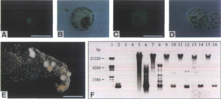
DNA solution stained with FITC-dextran was micro-injected into tobacco {Nicotiana tabacum) mesophyll protoplasts immobilized in a very thin layer of medium solidified either with alginate or with agarose. The injec-tion was aimed at the cytoplasm-rich nuclear region and routinely monitored under fluorescence illumination. Delivered injection solution appeared to diffuse very rapidly through injected cells. The targeted cell compart-ment was generally evenly fluorescent immediately upon delivery of injection solution. Following microinjection, FITC-fluorescence was often confined to the cytoplasm of targeted cells in the nuclear area, along the plasma membrane and in cytoplasmic strands (Fig. IA, B). Alternatively, it appeared to be evenly distributed over the whole cell after the tonoplast was accidentally penet-rated and the injection solution was delivered into the vacuole (Fig. 1C, D). In very rare cases, microinjection
generated a fluorescent vesicle in targeted cells, which increased in diameter during the injection process and could reach about the size of a nucleus (data not shown). FITC-fluorescence was never observed exclusively in the nucleus not even when microinjection was particularly aimed at this compartment and FITC-dextran with a molecular weight of 2 x 10⁶ was injected, which is too
large for diffusion through nuclear pores (Leonetti et al., 1991). Targeting microinjection into the cytoplasm was considered to be essential for successful genetic trans-formation. The microinjection procedure was therefore optimized in order to allow efficient delivery of injection solution into this compartment.
Protoplast were generally injected 24 h after immobil-ization. Cells with most chloroplasts located in the nuclear area represented the best targets for microinjection (Fig. IB, D). Such cells were in a healthy state and about to divide. In addition, they were generally stable enough to stand cytoplasmic injection and had only partially regenerated cell walls that could easily be penetrated with injection capillaries. It was possible to keep embedded cells at the optimal stage for microinjection by incubating them for up to 4 d at 4CC in the dark. Embedding in
medium containing agarose or alginate was apparently equally suitable for target cell immobilization. Alginate embedding, however, was easier and therefore preferen-tially used. Moving the capillary tip over longer distances through both types of gels frequently resulted in clogging and often hindered the impaling of target cells. However, the upper hemisphere of embedded cells was only covered by a very thin gel layer through which coaxial microinjec-tion at an angle of 40° was easily possible. Injecmicroinjec-tion capillaries with an inner tip diameter of 0.5-0.6 ^m were found to be optimal for efficient delivery of injection solution. Sharper needles were frequently clogged after only a few injections. When injection capillaries with a larger tip diameter were used, cells were often hard to impale and burst upon delivery of excessive volumes of injection solution. Highest frequencies of successful cyto-plasmic injections were obtained when a comparatively low injection pressure was continuously applied during the whole injection process. After several injections the needle tips were generally partially clogged and the injec-tion pressure had to be increased.
Using the optimal injection procedure on average about 50% of all injections could be targeted to the cytoplasm of cells that were at the right stage for microinjection. It was possible to inject up to 30 cells with one particular injection capillary and routinely to deliver injection solution into the cytoplasm of 20-40 cells per hour.
Stable transformation following injection of the neo (neomycin phosphotransferase II) gene
Paromomycin-resistant clones were recovered following coinjection of FITC-dextran and pSHI913 containing the
1 2 3 4 5 6 7 8 9 10 11 12 13 14 15 16
I I I I V
V | | I i f
~a !§ 9 • •'»
Fig. 1. (A-D) Photographs taken immediately after rmcroinjection showing the delivery of injection solution into different compartments of targeted cells. (A) FITC-fluorescence confined to the cytoplasm following microinjection into this compartment. Scale bar 50 ^m. (B) The same cell as in (A) under bright field illumination. (C) FITC-fluorescence apparently distributed over the whole cell after injection into the vacuole. Scale bar 50 fim. (D) The same cell as in (C) under bright field illumination. (E) Paromomycin-resistant clones after 4 weeks of selection. Scale bar: 5 mm. (F) Southern blot showing stable integration of the neo gene in plants regenerated from randomly chosen resistant clones. The blot was probed with the neo coding sequence. Lane 1: size marker, reference sizes are indicated on the left; lane 2: 10 pg 0.8 kb Hindlll fragment of pSHI913; lanes 3,4: 10 ^g wild-type (N. tabacum SRI) DNA; lanes 5-16: 10 ^g DNA from transformed plants (even numbers: restricted with Hindlll, odd numbers: non-restricted); lanes 5-12: Four plants regenerated from different clones that were obtained from one particular plate.
neo gene. After about 4 weeks of selection growing clones
could be identified (Fig. IE) and transferred on to regen-eration medium. In order to confirm that the selection was essentially tight, plants were regenerated from 30 randomly chosen clones. All regenerated plants were selfed or backcrbssed and produced seeds. From 26 of the analysed plants kanamycin-resistant offspring was obtained. Genomic DNA isolated from 19 regenerated plants including the ones which exclusively produced kanamycin-sensitive offspring was subjected to Southern analysis. Bands corresponding to the neo coding sequence appeared in lanes with restricted DNA at correct positions as well as in the high molecular weight fraction of non-restricted DNA, proving stable integration of the neo gene (Fig. IF, lanes 5-16) in all analysed plants except for one. In lanes with DNA from untransformed control plants no bands were observed (Fig. IF, lanes 3 and 4). Some of the analysed plants have been regenerated from different resistant calli that were obtained from individual plates. Such plants always showed completely distinct integration patterns proving that they were derived from independent transformation events (Fig. IF, lanes 5-12). When linearized pSHI913 was injected into the cyto-plasm of target cells at a concentration of 50 fig ml"1
together with FITC-dextran, on average 3.5% of the injected cells developed into paromomycin-resistant clones (Tables 1, 2). The plasmid concentration in the
injection solution could be increased from 50 fig ml 1 to 1 mg ml ~l with no effect on the delivery of the solution to target cells. Injecting pSHI913 at a concentration of 1 mgml"1 resulted in an about 3.5 times higher average
percentage of paromomycin-resistant clones per success-fully injected cell (Table 1). Although the incubation of embedded protoplasts at 4°C before microinjection did not affect the overall protoplast plating efficiency (clones per cultured viable protoplast; data not shown), it was found to reduce the average percentage of paromomycin-resistant clones obtained per cell injected into the cyto-plasm (Table 2).
Plating efficiency of injected cells
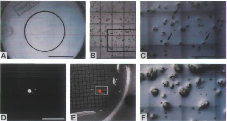
To be able to follow the fate of particular injected cells a simple method was established to record their position in the thin gel layer. A coverslip with an imprinted 0.5 mm grid was embedded in the solid basis medium below the immobilized cells (Fig. 2A). Using a low magnification objective a Polaroid picture of the cells above the grid was taken (Fig. 2B). FITC-dextran was coinjected with 1 mg ml ~l plasmid DNA into the cytoplasm. Targeted cells were marked and numbered on the Polaroid picture. For each cell notes were taken concerning the estimated amount of injected solution as well as the injection pressure used. On average, 23% of the injected cells
1162 Kostetsi.
Table 1. Effect of the plasmid DNA concentration in the injection solution on the percentage of paromomycin resistant clones obtained
per cell injected with pSHI913
Experiment A B C D E F G Total Table 2. Effect of tig ml'1 pSHI913 Experiment A B C D E F Total 50/igmT1 Cytoplasmic injections 50 42 23 26 141 cold-storing Resistant clones 2 3 0 0 5 protoplasts on the
Protoplasts not cold-stored Cytoplasmic injections 77 93 79 59 46 354 Resistant clones 5 1 2 3 1 12 Resistant clones/ cytoplasmic injection 4 7 0 0 3.6 lmgml ' Cytoplasmic injections 110 60 39 45 50 46 28 378
percentage of paromomycin-resistant clones
Resistant clones/ cytoplasmic injection 6.5 1.1 2.5 5.1 2.2 3.4 Resistant clones 7 7 2 11 4 11 4 46 obtained per
Protoplasts cold-stored for 1-4 d Cytoplasmic injections 83 66 97 71 19 336 Resistant clones 1 1 1 1 1 5 Resistant clones/ cytoplasmic injection 6.4 11.7 5.1 24.4 8.0 23.9 14.3 12.2
cell injected with 50
Resistant clones/ cytoplasmic injection 1.2 1.5 1.0 1.4 5.2 1.5
developed within 5-7 d into microcalli (Fig. 2C, F; Table 3). All other targeted cells either collapsed com-pletely during the first hours after microinjection (Fig. 2C, F) or remained apparently alive but were unable to divide. In individual experiments the plating efficiency of injected cells varied over a wide range from only 4% to 63% (Table 3). In the same experiments the plating effi-ciency of non-injected control cells that were selected 1 d after embedding for being at the optimal stage for micro-injection was determined to range from 69% to 100% with an average of 87% (Table 3).
Neither the amount of injection solution delivered into the cytoplasm nor the injection pressure used was found to have an obvious effect on the plating efficiency obtained (data not shown). In a series of experiments cells were impaled with the help of a piezo stepper. The application of this device did not significantly influence the plating efficiency of injected cells either (data not shown).
Using the possibility of recording the position of par-ticular injected cells evidence was generated confirming that cytoplasmic injection is in fact essential for stable transformation. pSHI913 was microinjected at a concen-tration of 1 mg ml"l. All cells which had not been injected
into the cytoplasm, but into the vacuole or into a vesicle were destroyed with a broken injection capillary before selection. In these experiments an average percentage (17.4%) of paromomycin-resistant clones per injected cell was obtained.
Transient expression of the firefly luciferase gene (Luc) in particular injected cells
FITC-dextran and lmgml"1 linearized pAMLuc
containing the firefly luciferase gene (Luc) was injected into cells embedded above a 0.5 mm grid. One day after microinjection Luc expression was assayed in vivo. Bioluminescence of varying intensity emitted from single cells transiently expressing the injected Luc gene was imaged using a sensitive video camera equipped with a macro lens (Fig. 2D). Following superimposition of the bioluminescence image and a corresponding reflected light reference picture the detected light spots could be assigned to particular injected cells (Fig. 2E). Transient Luc expression was exclusively observed in cells that had been injected into the cytoplasm. Bioluminescence emission was never detected following delivery of injection solution
- . ...» -/I - ;
Fig. 2. (A) [protoplasts embedded in a streak of thin alginate-medium on the top of solid basis medium above a coverslip with an imprinted 0.5 mm grid. Scale b a r 10 mm. (B) 3 1/4x4 1/4 inch Polaroid picture showing the position of immobilized protoplasts on the 0.5 mm grid. The picture is reproduced at about half of its original size. (C) and ( F ) are close-ups of the marked area immediately and 5 d after microinjection, respectively. Scale bar: 1 mm. (C) Immobilized protoplasts immediately after microinjection of pAMLuc. Arrows point at the injected cells. Scale bar: 500 jim. (D) Bioluminescence image showing four cells that are transiently expressing the Luc gene 24 h after microinjection. The cells are emitting light of different intensity. Scale bar: 3 mm. (E) Supenmposition of the bioluminescence image (D) on to a corresponding reflected light reference picture. Luminescent regions are marked red. The white rectangle encloses the area shown in (C) and ( F ) . Two of the injected cells shown in (C) are transiently expressing the Luc gene. ( F ) Three of the injected cells shown in (C) have developed into microcalli after 5 d of culture. Two of these microcalli derive from transiently Luc-expressing cells (E).
Table 3. Plating efficiency of injected cells
Experiment Injected cells
Cytoplasmic injections 4 g 12 29 23 26 13 14 19 23 57 46 Microcalli after 5-7 d 2 5 3 to 2 4 1 1 § i 16 2 Plating efficiency 50 63 25 35 9 15 8 50 32 22 28 4 Non-injected Analysed cells 19 16 26 28 19 16 22 '25 24 control cells Microcalli after 5-7 d 15 14 18 25 19 16 21 34 17 Plating efficiency 79 88
m
89 100 100 95 96 71 A B C D E F G H I K L M Total 274 63 23 195 169 87into the vacuole or into a vesicle. As shown in Table 4, 48.9% of the cells that were apparently alive 1 d after microinjection into the cytoplasm transiently expressed the transferred Luc gene.
Cells assayed for Luc expression were subsequently
cultured without selection and developed normally into calli similar to those obtained from control cells that have not been incubated in luciferase substrate solution (data not shown). However, stably Luc-expressing clones were never found when transiently Luc-expressing injected cells
1164 Kost etal.
were mass-cultured together with an excess of non-injected cells and the resulting calli were assayed again after several weeks. Such clones were only obtained by cutting microcalli that had developed from transiently Luc-expressing cells out of the embedding gel after about 5 d and culturing them individually. Following coinjection of pAMLuc and pSHI913 stably Lwc-expressing clones could also be recovered from transiently Lwc-expressing cells by selecting for paromomycin resistance.
Transformation efficiency
In the literature the percentage of stably transformed clones obtained per cell surviving DNA delivery and continuing normal development is generally referred to as the stable transformation efficiency of microinjection. Using the system described here under optimal conditions, on average, 23% of the successfully injected cells developed into microcalli (Table 3) and 12.2% gave rise to paromomycin-resistant stably transformed clones (Table 1). The average efficiency of stable transformation can therefore be calculated to be 53%. Transient Luc expression was detected also in roughly 50% of the successfully injected cells that were alive 1 d after microin-jection (Table 4), indicating that stable integration of the
injected genes occurred in essentially all transiently expressing cells that developed into clones. These calcula-tions are based on results obtained with a different, independent series of experiments. They were substanti-ated by determining the efficiency of transient and stable transformation in individual experiments (Table 5).
Discussion
During the last decade successful gene transfer by microin-jection was demonstrated with different types of plant cells (Neuhaus and Spangenberg, 1990; Simmonds et al, 1992; Lusardi etal, 1994; Neuhaus etal, 1993). However, the considerable potential of this technique for experi-mental and applied plant biology has been only partly exploited to date. Plant cell microinjection is technically difficult and requires a lot of experience. Little reliable information is available on the parameters that are spe-cifically important for DNA microinjection into plant cells, although a considerable amount of work has been dedicated to the optimization of the same technique for animal cells (Capecchi, 1980; Brinster et al, 1985; Proctor, 1992). We have established an efficient system that allows well controlled DNA microinjection into tobacco proto-plasts and subsequent analysis of the resulting transient and stable reporter gene expression. The results obtained in individual experiments can be assessed 24 h after microinjection by non-destructively analysing transient
Luc expression. The system allows effective testing of
factors that are important for successful DNA microinjec-tion into plant cells and can addimicroinjec-tionally be employed by inexperienced workers to obtain expertise in this technique.
The possibility of routinely monitoring DNA delivery into tobacco protoplasts by coinjection of FITC-dextran is an essential feature of the system we have established. Routine microinjection of DNA solution containing a fluorescent dye is novel for plant cells and was found to
Table 4. Percentage of transiently hue-expressing cells per cell apparently surviving the first 24 h following injection ofpAMhuc
Experiment Cytoplasmic
injections
Cells alive after 24 h
Transiently Luc-expressing cells
Transiently Luc-expressing cells/ cell alive after 24 h
A B C D E 52 51 57 46 35 20 13 30 10 15 8 5 19 3 8 40.0 38.5 63.3 30.0 53.3 Total 241
Table 5. Efficiency of transient
Experiment A B C D Cytoplasmic injections 23 8 7 14 and stable 88 43
transformation in individual experiments Cells alive Transiently
after 24 h expressing cells n.d. n.d. 2 6 n.d. n.d. 1 3 Transient transformation efficiency n.d. n.d. 50 50 Microcalli after 5-7 d 5 3 1 3 48.9 Stably expressing clones 2 2 1 1 Stable transformation efficiency 40 66.7 100 33.3 n.d. = not detected.
have a number of important advantages. (1) The general injection procedure could be optimized for efficient deliv-ery of DNA solution and fine tuned for each particular targeted cell. (2) It was possible to identify and replace clogged injection capillaries immediately. (3) The amount of injected DNA solution could be roughly estimated. (4) The targeted compartment could be verified for each injected cell. (5) The number of successfully injected cells and, therefore, the transformation efficiency could be exactly determined. Tobacco protoplasts injected with DNA solution containing 1% FITC-dextran were able to divide normally and to develop into stably transformed microcalli. Similarly, Pepperkok et al. (1988) have found that FITC-dextran injected into animal cells at concentra-tions below 2% does not interfere with cell division.
In animal systems DNA delivery directly into the nucleus of target cells has been reported to be an absolute requirement for efficient stable transformation by micro-injection (Capecchi, 1980; Brinster et al, 1985). DNA microinjection into plant cells was therefore generally also aimed at the nucleus, although the delivery of reporter genes into this compartment has never been routinely confirmed (Crossway et al, 1986; Reich et al., 1986; Schnorf et al, 1991). Microinjection of stained solutions into the nucleus of plant cells has been described to be very difficult (Steinbiss and Stabel, 1983; Aly and Owens, 1987) and was only in rare cases reported to be successful (Aly and Owens, 1987; Schnorf et al, 1991). We have not been able to deliver injection solution containing FITC-dextran directly into the nucleus of tobacco protoplasts. Targeted nuclei were often pushed with the tip of injection capillaries through injected cells without being impaled. They were obviously not anchored stably enough within the cells to allow penetration of the nuclear membranes. However, microinjection generated in rare cases spherical fluorescent vesicles within targeted cells. When such vesicles reached the right size they looked under fluorescence illumination like a successfully injected nucleus.
Using an optimized procedure we have aimed DNA microinjection at the cytoplasm-rich nuclear area of tobacco protoplasts. In about half of the targeted cells the cytoplasm was successfully injected. In most other cases the injection solution was delivered into the vacuole. Since in the highly vacuolated tobacco protoplasts the cytoplasm is only a very thin layer, we found it surprising that the large central vacuole was not targeted more often. The tonoplast is apparently a very flexible mem-brane that is difficult to penetrate with injection capillar-ies. It has generally been presumed that genes injected into the vacuole of plant cells are rapidly degraded and never expressed. Our results have confirmed this assump-tion. In contrast, injection of plasmid DNA into the cytoplasm of target cells resulted in a high efficiency of transient and stable expression of the transferred genes.
Stable transformation efficiencies between 14% and 26% have been reported for microinjection into plant proto-plasts (Crossway et al, 1986; Reich et al, 1986). In our experiments, on average, 53% of the clones derived from cells that had been successfully microinjected into the cytoplasm were stably transformed. In order to obtain such a high transformation efficiency it was essential to inject plasmid DNA at a concentration of 1 mg ml"1. For
microinjection into the nuclei of animal cells and into plant cells injection solutions containing plasmid DNA at 20-1000 times lower concentrations have generally been used to date (Capecchi, 1980; Brinster et al, 1985; Crossway et al, 1986; Reich et al, 1986).
A large number of additional parameters are probably important for efficient stable genomic integration of genes that have been transferred into the cytoplasm of target cells. Comparatively small DNA fragments, for example, are likely to diffuse more efficiently through nuclear pores. We plan to test whether cytoplasmic microinjection of small DNA fragments that contain just the reporter gene coding sequences and the necessary expression signals increases the transformation efficiency. Injection of DNA fragments that are chemically linked to polypeptides with nuclear targeting signals (Howard et al, 1992; Tinland
et al, 1992) might have the same effect.
The overall efficiency of DNA microinjection is deter-mined not only by the efficiency of stable transformation but also by the survival of injected cells. At 23%, the plating efficiency of injected protoplasts we obtained was quite low but well within the range of what has been reported earlier (13-50%; Crossway et al, 1986; Reich
et al, 1986). An important future application of the
microinjection system we have established will be the development of a gentler procedure for efficient DNA delivery into plant cells. Using bevelled injection capillar-ies with tips that are at the same time very sharp and have a comparatively large inner diameter might prove to be an important step towards such a procedure.
The shoot cultures used for protoplast isolation have been kept under strictly stable conditions and protoplast isolation, embedding as well as microinjection were always performed in a consistent manner. However, in individual experiments both the plating efficiency of injected proto-plasts and the transformation efficiency showed large variation. Protoplasts from individual batches can obvi-ously be quite different in their ability to survive microin-jection and to express injected genes. Similar to Aly and Owens (1987) we have found that efficient DNA micro-injection is possible into embedded protoplasts that have been kept at the optimal stage for microinjection for several days, by incubation at 4 °C. This may be conveni-ent for many applications, although we found that cold-storing of protoplasts results in a reduced stable transformation efficiency.
1166 Kost etal.
microinjected cells that were alive after 24 h. Interestingly, essentially all transiently expressing cells that developed into clones apparently stably integrated the injected gene. A similar very high percentage of stable transformed clones obtained per transiently expressing and dividing cell has also been determined after gene transfer by microprojectile bombardment into cells within tobacco leaves (Hunold et al, 1994). We never observed stably transformed clones when transiently Luc-expressing cells were mass-cultured after the LUC-assay together with an excess of untransformed cells. The transiently Luc-expressing cells were apparently over-grown, possibly because their ATP-pools have been reduced as con-sequence of the energy consuming light emission.
The results presented here allow a number of conclu-sions that are of general importance for the transforma-tion of plant cells by DNA microinjectransforma-tion and also by other methods. (1) DNA delivery into the cytoplasm of target cells can result in a high efficiency of transient and stable transformation. (2) Using high plasmid DNA concentrations increases the stable transformation effi-ciency resulting from cytoplasmic microinjection. (3) FITC-dextran introduced into the cytoplasm of plant cells at a concentration high enough to be visible under fluorescence illumination does not hinder cell division or transient expression and stable integration of cotrans-ferred genes. (4) Following cytoplasmic microinjection of plasmid DNA at a high concentration, essentially all cells that transiently express the transferred genes and divide give rise to stably transformed clones. Using the effective microinjection system reported here it should be possible to collect additional interesting information concerning gene transfer to plant cells by microinjection and other techniques.
Acknowledgements
We would like to thank M. Schnorf for very fruitful discussions. We are also grateful to M. Schnorf and N-H. Chua for providing plasmids as well as to Mike Saul for helpful comments on the manuscript and language correction. B.K, was supported by a grant of the Swiss Federal Institute of Technology.
References
Aly MAM, Owens I D . 1987. A simple system for plant cell
microinjection and culture. Plant Cell, Tissue and Organ
Culture 10, 159-74.
Brinster RL, Cben HY, Tnimbauer ME, Yagle MX, Palmiter RD. 1985. Factors affecting the efficiency of introducing
foreign DNA into mice by microinjecting eggs. Proceedings
of the National Academy of Sciences, USA 82, 4438-42.
Cabocbe M. 1980. Nutritional requirement of protoplast-derived
haploid tobacco cells grown at low densities in liquid medium.
Planta 149, 7-18.
Capecchi MR. 1980. High efficiency transformation by direct
microinjection of DNA into cultured mammalian cells. Cell
22, 479-88.
Crossway A, Oakes JV, Irvine JM, Ward B, Knauf VC, Shewmaker CK. 1986. Integration of foreign DNA following
microinjection of tobacco mesophyll protoplasts. Molecular
and General Genetics 202, 179-85.
Holm PB, Knudsen S, Mouritzen P, Negri D, Olsen FL, Roue
C. 1994. Regeneration of fertile barley plants from mechanic-ally isolated protoplasts of the fertilized egg cell. The Plant
Cell 6, 531-43.
Howard EA, Zupan JR, Citovsky V, Zambryski PC. 1992. The
VirD2 protein of A. tumefaciens contains a C-terminal bipartite nuclear localization signal: implications for nuclear uptake of DNA in plant cells. Cell 68, 109-18.
Hunold R, Bronner R, Hahne G. 1994. Early events in
microprojectile bombardment: cell viability and particle location. The Plant Journal 5, 593-604.
Kost B, Schnorf M, Potrykus I, Neuhaus G. 1995.
Non-destructive detection of firefly luciferase (LUC) activity in single plant cells using a cooled, slow-scan CCD camera and an optimized assay. The Plant Journal 8, 155-66.
Kranz E, Lore H. 1993. In vitro fertilization with isolated, single
gametes results in zygotic embryogenesis and fertile maize plants. The Plant Cell 5, 739^16.
Rung S-D, Wu R. 1993. Transgenic plants: engineering and
utilization. San Diego: Academic Press.
Lawrence WA, Davies DR. 1985. A method for the
microinjec-tion and culture of protoplasts at very low densities. Plant
Cell Reports 4, 33-5.
Leonctti JP, Mechti N, Degols G, Gagnor C, Lebleu B. 1991.
Intracellular distribution of microinjected antisense oligonu-cleotides. Proceedings of the National Academy of Sciences,
USA 88, 2702-6.
Lusardi MC, Neuhaus-Url G, Potrykus I, Neuhaus G. 1994. An
approach towards genetically engineered cell fate mapping in maize using the Lc gene as a visible marker: transactivation capacity of Lc vectors in differentiated maize cells and microinjection of Lc vectors into somatic embryos and shoot apical meristems. The Plant Journal 5, 571-82.
Maniatis T, Fritsch EF, Sambrook J. 1982. Molecular cloning:
a laboratory manual. Cold Spring Harbor Laboratory Press. '
Millar AJ, Short SR, Hiratsuka K, Chua N-H, Kay SA. 1992.
Firefly luciferase as a reporter of regulated gene expression in higher plants. Plant Molecular Biology Reporter 10, 324-37.
Murashige T, Skoog F. 1962. A revised medium for rapid
growth and bioassays with tobacco tissue cultures. Physiologia
Plantarum 15, 473-97.
Murray MG, Thompson WF. 1980. Rapid isolation of high
molecular weight plant DNA. Nucleic Acids Research 8, 4321-5.
Neuhaus G, Spangenberg G. 1990. Plant transformation by
microinjection techniques. Physiologia Plantarum 79, 213-17.
Neuhaus G, Bowler C, Kem R, Chua N-H. 1993. Calcium/
calmodulin-dependent and -independent phytochrome signal transduction pathways. Cell 73, 937-52.
Neuhaus-Url G, Neuhaus G. 1993. The use of the non-radioactive
digoxigenin chemiluminescent technology for plant genomic Southern blot hybridization: a comparison with radioactivity.
Transgenic Research 2, 115-20.
Pepperkok R, Schneider C, Philipson L, Ansorge W. 1988. Single
cell assay with an automated capillary microinjection system.
Experimental Cell Research 178, 369-76.
Pinkert CA. 1994. Transgenic animal technology: a laboratory
handbook San Diego: Academic Press.
cells in culture: theory and practice. Methods in Molecular
and Cellular Biology 3, 209-31.
Rekh TJ, Iyer VN, Miki BL. 1986. Efficient transformation of
alfalfa protoplasts by the intranuclear microinjection of Ti plasmids. Bio I Technology 4, 1001-4.
Schnorf M, Nenhaus-Url G, Galli A, Iida S, Potrykns I, Neuhaus
G. 1991. An improved approach for transformation of plant cells by microinjection: molecular and genetic analysis.
Transgenic Research 1, 23-30.
Schnorf M, Potrykns I, Neuhaus G. 1994. Microinjection
technique: routine system for characterization of microcapil-laries by bubble pressure measurement. Experimental Cell
Research 210, 260-76.
Simmonds J, Stewart P, Simmonds D. 1992. Regeneration of
Triticum aestivum apical explants after microinjection of germ
line progenitor cells with DNA. Physiologia Plantarum 85, 197-206.
Steinbiss HH, Stabel P. 1983. Protoplast derived tobacco cells
can survive capillary microinjection of the fluorescent dye lucifer yellow. Protoplasma 116, 223-7.
Tinland B, Koukolikova-Nicola Z, Hall MN, Hohn B. 1992. The
T-DNA-linked VirD2 protein contains two distinct functional nuclear localization signals. Proceedings of the National