A N A T O M I C B A S E S O F M E D I C A L , R A D I O L O G I C A L A N D S U R G I C A L T E C H N I Q U E S
The acetabular blood supply: implications for periacetabular
osteotomies
Received: 22 April 2002 / Accepted: 27 February 2003 / Published online: 16 August 2003 Ó Springer-Verlag 2003
Abstract As the popularity of juxta-acetabular osteot-omies in adults increases, concern arises that such a procedure will potentially cause avascular necrosis of the acetabular fragment. In order to verify the remaining vascularization after a Bernese periacetabular osteoto-my, an injection study with colored latex was performed. The vascularity of the outside of the periacetabular bone was studied in 16 hips after injection of colored latex into the abdominal aorta and the inside in four hips. To confirm the conclusions drawn from the anatomic study, a Bernese periacetabular osteotomy was performed in two additional hips after latex injection. This study demonstrated that through a modified Smith-Peterson approach and with execution of the osteotomies from the inside of the pelvis the acetabular fragment remains vascularized by the supra-acetabular and acetabular branches of the superior gluteal artery, the obturator artery and the inferior gluteal artery. Some uncertainty remains about how much correction is tolerated by the smaller blood vessels.
Vascularisation arte´rielle de l’ace´tabulum: implications dans les oste´otomies pe´ri-ace´tabulaires
Re´sume´ Au fur et a` mesure qu’augmente la popularite´ des oste´otomies juxta-ace´tabulaires chez l’adulte, des inquie´tudes se font jour concernant les risques potentiels de ne´crose avasculaire du fragment ace´tabulaire apre`s une telle intervention. Pour ve´rifier la vascularisation restante apre`s une oste´otomie pe´ri-ace´tabulaire
ber-noise, une e´tude anatomique apre`s injection de latex colore´ a e´te´ re´alise´e. La vascularisation du versant ex-terne du fragment pe´ri-ace´tabulaire a e´te´ e´tudie´e sur 16 hanches apre`s injection de latex colore´ dans l’aorte ab-dominale et celle de son versant interne sur 4 hanches. Pour confirmer les conclusions tire´es du travail anato-mique, une oste´otomie pe´ri-ace´tabulaire bernoise a e´te´ re´alise´e sur deux hanches supple´mentaires apre`s injec-tion de latex. Cette e´tude a montre´ que, par une voie d’abord de Smith-Petersen modifie´e et en re´alisant l’oste´otomie a` partir du versant interne du bassin, le fragment ace´tabulaire restait vascularise´ par les bran-ches supra-ace´tabulaires et ace´tabulaires de l’arte`re glute´ale supe´rieure, par l’arte`re obturatrice et par l’art-e`re glute´ale infe´rieure. Il persiste quelques impre´cisions sur l’importance de la correction que peuvent tole´rer ces petits vaisseaux.
Keywords Acetabulum Æ Vascular anatomy Æ Periacetabular osteotomy Æ Avascular necrosis
Introduction
Avascular necrosis (AVN) of the acetabular fragment is one of the main arguments of opponents of juxta-ace-tabular osteotomy techniques [13, 15]. AVN has been reported with the use of rotational periacetabular oste-otomies in adults [9, 12, 19, 20, 22, 23] and with the Bernese periacetabular osteotomy (PAO) when the inferior cut is intra-articular, interrupting the acetabular branch of the obturator artery [11]. Previous studies have suggested that the only remaining blood supply after a periacetabular osteotomy is the obturator artery [12, 13]. However, there are no published studies on the blood supply following a Bernese periacetabular osteotomy.
The purpose of this study was to examine the blood supply to the acetabulum with particular reference to the Bernese periacetabular osteotomy [6,17, 18].
DOI 10.1007/s00276-003-0149-3
M. Beck Æ M. Leunig Æ T. Ellis Æ J. B. Sledge Æ R. Ganz
Electronic Supplementary Material The french version of this article is available in the form of electronic supplementary material and can be obtained by using the Springer Link server located at http:// dx.doi.org/10.1007/s00276-003-0149-3
M. Beck (&) Æ M. Leunig Æ T. Ellis Æ J. B. Sledge Æ R. Ganz Department of Orthopedic Surgery,
University of Bern, Inselspital, 3010 Bern, Switzerland
E-mail: [email protected] Tel.: +41-31-6322222
Materials and methods
The vascularization of the periacetabular bone was studied in fresh cadavers (age 60–85 years) after injection of approximately 250 ml of green-colored latex into the abdominal aorta. Optimal filling of even very small arteries was obtained if the color of the skin of the toes changed to the color of the injected latex. Hips with previous operations in the area of interest were excluded from the study. Radiographs were not obtained, but from the clinical appearance none of the hips was dysplastic. During the dissections for an anatomic study on the gluteus minimus [1], the importance of the superior gluteal artery for the vascularization of the acetabulum was recognized and the vascular study extended to the inferior and internal parts of the acetabulum. Therefore the anatomy of the superior gluteal artery was studied in 16 hips and in five of these hips additionally the blood supply of the inferior gluteal artery, the obturator artery, and the medial and lateral circumflex arteries and their anastomoses was examined. To complete the understanding of the vascular anatomy, the blood supply from the inside of the pelvis was studied in four additional hips.
After completion of the anatomic study, a Bernese periacetab-ular osteotomy [6,17, 18] was performed in two hips, after injection of the arteries with colored latex, to confirm the results obtained from the anatomic study. After osteotomy of the anterior superior iliac spine, the iliac muscle was stripped off the inside of the pelvis. The direct and reflected tendons of the rectus muscle were divided and the joint capsule prepared. By entering the gap between the capsule and the external obturator muscle, the infra-acetabular notch was palpated and the ischium osteotomized incompletely. After complete osteotomy of the superior pubic ramus, the abductor muscles were tunneled over a width of about 3 cm from the interspinous crest to the sciatic notch to take a retractor for soft tissue protection, and the ilium was osteotomized. From the inside the osteotomy was subsequently completed in the area of the quadrilateral plate. The fragment was mobilized and fixed in a substantial correction (20° abduction, 20° flexion). The hips were dissected and the remaining blood supply to the periacetabular bone was documented.
Results
Superior gluteal artery (SGA)
After division from the superficial branch, the deep branch of the SGA divided into four rami: superior, inferior, supra-acetabular and acetabular (Fig. 1).
The superior ramus (Ø 1.5–2 mm) skirted the supe-rior margin of the gluteus minimus to the antesupe-rior superior iliac spine, where it anastomosed with branches of the deep and superficial circumflex iliac arteries and the iliolumbar artery. The inferior ramus (Ø 1.5–2 mm) ran with the superior gluteal nerve on the lateral aspect of the gluteus minimus to the tensor fasciae latae and anastomosed with the ascending branch of the lateral femoral circumflex artery (LFCA).
The supra-acetabular ramus (Ø 1–1.5 mm) coursed within the muscular body of the gluteus minimus to the acetabular roof, joined the acetabular ramus and continued to the interspinous crest. At the interspinous crest it anastomosed with branches from the iliolumbar artery and from the ascending branch of the LFCA (Fig. 2). The supra-acetabular ramus originated directly from the deep branch in four specimens, from the superior ramus in three specimens, from the inferior
ramus in eight specimens, and it was absent in one specimen.
The acetabular ramus (Ø 1 mm) followed the inferior border of the gluteus minimus to the posterior superior acetabulum and continued about 1.5–2 cm proximal to the rim across the roof of the acetabulum. It anasto-mosed with the supra-acetabular ramus about midway across the acetabular roof. It originated from the deep branch in all specimens.
Inferior gluteal artery (IGA)
The IGA emerged beneath the piriformis muscle and divided into a branch for the sciatic nerve, a branch on the piriformis muscle, and two or three small vessels (Ø 0.5–0.8 mm) to the posterior wall. Four of five hips had anastomoses to the superior gluteal artery on the pos-terior superior roof of the acetabulum. An anastomosis to the posterior branch of the obturator artery in the infra-acetabular notch was found in all specimens.
Medial femoral circumflex artery (MCFA)
There were two branches (Ø 0.5 mm) of the deep branch of the MFCA to the acetabulum, one supplying the anterior inferior joint capsule. The other coursed along
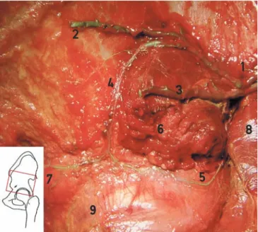
Fig. 1 Lateral view of a left hip after removal of the gluteus medius and parts of the gluteus minimus. In the greater sciatic notch (1) the superior gluteal artery (SGA) divides into superior (2), inferior (3), supra-acetabular (4) and acetabular rami (5). The posterior quadrant of the gluteus minimus (6) was retained to show the intramuscular course of the supra-acetabular ramus, which, after the anastomosis with the acetabular ramus, continued to the anterior inferior iliac spine (7). 8, Piriformis with the tendon divided and turned down; 9, reflected tendon of the rectus femoris muscle
the lateral border of the pectineus muscle, supplied the antero-inferior part of the acetabulum (Fig. 3) and continued to the iliopectineal eminence, where it anas-tomosed with branches from the iliolumbar artery. In two of five hips an anastomosis to the acetabular branch of the obturator artery was present.
Obturator artery (OA)
The OA originated in three hips from the anterior trunk and in one hip from the internal iliac artery at the bifurcation into the anterior and posterior trunk. It coursed inferior to the pelvic brim and supplied three or four branches to the quadrilateral plate and the superior pubic ramus. In one hip, an anastomosis with the infe-rior epigastric artery was present. After the obturator canal, the OA divided into an acetabular branch to the acetabular fossa, and a posterior branch and an anterior branch. The acetabular branch (Ø 1 mm) entered the joint via the incisura acetabuli deep to the transverse ligament. In the acetabular fossa it divided into a branch to the round ligament and three to five branches to the floor of the acetabulum. The posterior branch ran along
the rim of the obturator foramen and gave a branch to the inferior acetabulum, which anastomosed in the infra-acetabular notch with the IGA. The anterior branch supplied the obturator externus muscle and the superior pubic ramus. There was no anastomosis to the SGA anteriorly. In two of five hips an anastomosis with the MFCA was present.
Fourth lumbar artery
The fourth lumbar artery (Ø 1–1.5 mm) branched off the internal iliac artery proximal to the bifurcation of the external and internal iliac arteries. It passed between the psoas and iliacus muscles and divided into two main branches. One branch skirted along the iliac crest and anastomosed anteriorly with branches of the deep iliac circumflex artery. The other branch ramified into several branches on the iliacus muscle, some perforating the muscle and supplying the underlying bone.
Iliolumbar artery
The iliolumbar artery originated from the posterior trunk in three hips and from the OA in one. Besides its
Fig. 3 Anterior view of a right hip. Two branches of the medial femoral circumflex artery (MFCA) (1) are given to the anterior wall. The anastomosis to the inferior ramus of the iliolumbar artery (2) runs medial to the anterior inferior iliac spine (3). 4, Femoral head; 5, detached and reflected joint capsule. The inferior ramus of the iliolumbar artery (2) anastomoses with the supra-acetabular ramus (6) of the SGA in the area of the interspinous crest Fig. 2 Anterolateral view of a right hip. The supra-acetabular
ramus of the SGA (1) runs on the roof of the acetabulum and anastomoses with the ascending branch of the lateral femoral circumflex artery (LFCA) (2) in the area of the anterior inferior iliac spine (3). 4, Reflected tendon of the rectus femoris muscle; 5, joint capsule
spinal branch, it divided into a superficial branch (Ø 1– 1.5 mm) on the peritoneal surface of the iliacus muscle and into a deep branch (Ø 1–1.5 mm) for the vascular-ization of the innominate bone. The deep branch divided into three rami, with the nutrient artery to the innomi-nate bone being the largest (Fig. 4). The superior ramus ran to the anterior superior iliac spine and anastomosed with the SGA and the deep and superficial circumflex iliac artery. The inferior ramus ran along the pelvic brim and continued to the anterior inferior iliac spine and to the iliopectineal eminence. At the anterior inferior iliac spine, the inferior ramus anastomosed with the supra-acetabular ramus of the SGA and with the ascending branch of the LFCA. At the iliopectineal eminence, it met the branch of the MFCA (Fig. 3).
In two of four hips the nutrient artery of the ilium entered the bone 1 cm anterior to the iliosacral joint and lateral of the pelvic brim. In the other two hips the nutrient artery entered the bone about 1 cm medial to the pelvic brim. In one of these hips the iliolumbar artery originated from the OA. The branches to the interspin-ous crest and the iliopectineal eminence (inferior ramus of the iliolumbar artery) were supplied by the OA in these two hips.
Blood supply after periacetabular osteotomy
In both hips the blood supply to the acetabular fragment was preserved by the supra-acetabular and acetabular ramus of the SGA, the acetabular branches of the IGA (Fig. 5) and the OA. The blood supply from the fourth lumbar artery, the iliolumbar artery and from the OA to the innominate bone and the quadrilateral plate was interrupted during the approach for the supra-acetabu-lar and retroacetabusupra-acetabu-lar cuts. During the approach to the joint capsule the direct and reflected head of the rectus femoris muscle are detached, together with the anasto-mosis to the ascending branch of the LFCA. Postero-inferiorly, the anastomosis in the infra-acetabular notch between the IGA and the OA was interrupted by the osteotomy of the ischium. No observations were made about the remaining blood supply from the MFCA to the anterior wall. Thereafter, lateral and anterior cover were both increased by 20°, which resulted in an anterior gap of about 1.5 cm. The previously untouched vessels all remained intact; the acetabular rami of the SGA and IGA became slightly stretched. In both cases, however, the supra-acetabular ramus was protected within the
Fig. 4 Anterior view of the inside of the pelvis in a right hip. The iliolumbar artery (1) arises together with the obturator artery (2) from the posterior trunk. The nutrient artery (3) enters the iliac bone anterior to the pelvic brim. 4, Blood vessel to the iliac muscle; 5, superior ramus to the interspinous crest; 6, inferior ramus to the anterior inferior iliac spine and the iliopectineal eminence
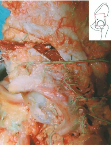
Fig. 5 Acetabular fragment of a left hip in the corrected position with a gap of 1.5–2 cm at the most anterior part of the supra-acetabular osteotomy. The proximal femur has been removed. Remaining blood supply is from the supra-acetabular (1) and acetabular (2) rami of the SGA and acetabular branches (3) of the inferior gluteal artery (IGA)
gluteus minimus, which prevented traction on this vessel.
A schematic representation of the anatomic study is given in Fig. 6.
Discussion
This study was carried out to establish the blood supply to the acetabulum, with respect to the Bernese periace-tabular osteotomy. Early in the course of the dissection, the importance of the SGA and the impact of the sur-gical approaches on its blood supply were recognized. Therefore, the blood supply through the SGA was studied in 16 hips and in five of these also the blood supply to the inferior acetabulum. In only four hips was the intrapelvic blood supply studied. The reason was that the Bernese PAO is currently performed through a modified Smith-Peterson approach, where the soft tis-sues are detached from the inside of the pelvis. As a result the intrapelvic blood supply from the iliolumbar and fourth lumbar arteries is inevitably interrupted and for this study has only lesser interest. The results of the current study are based on the dissection of non-dys-plastic hips. For the majority of dysnon-dys-plastic hips these results also apply. In severe dysplasia with a dislocated
hip an altered course of the blood vessels may be anticipated.
The importance of the SGA for the vascularization of the acetabular roof is known [5, 13, 25]. The presence of a ‘‘supra-acetabular’’ artery was described by von Lanz [14], who based on the work of Bergoin and Louis [2] Letournel and Judet [16] described an ‘‘artery of the roof of the acetabulum’’. However, the division of the deep branch into four rami (superior, inferior, supra-acetab-ular and acetabsupra-acetab-ular) and the importance of both the supra-acetabular and acetabular rami for the vasculari-zation of acetabular roof have not been described pre-viously. The close relationship of the acetabular and supra-acetabular rami to the gluteus minimus and par-ticularly the intramuscular course of the supra-acetab-ular ramus protects these vessels during surgery and displacement of the acetabular fragment. The IGA supplies two or three branches to the posterior wall. Judging from the small diameter of the vessels, the contribution to the acetabular blood supply seems less important; however, it connects the systems of the SGA and the OA. After a PAO, the blood supply through the IGA remained intact (Fig. 5). Based on the anatomic study the impairment of the acetabular blood supply caused by a PAO was anticipated and was confirmed by the subsequent dissection. In the present study a mod-erate to substantial correction with an increase of lateral and anterior cover of 20° each was performed. At this stage, where the acetabular fragments had a gap ante-riorly about 1.5 cm, the acetabular rami of the SGA and IGA were slightly stretched. The supra-acetabular ramus, which runs within the muscle substance of the
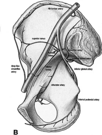
Fig. 6 A Diagram of the blood supply to the external side of acetabulum of a right hip. The contribution of the MFCA and its branches to the anteroinferior acetabulum are omitted. B Intra-pelvic blood supply. The visceral arteries are omitted
gluteus minimus, was well protected and did not seem to be under excessive tension (Fig. 5). But the effect of the correction on the blood vessels was difficult to assess because the latex alters the mechanical properties of the blood vessels. Although even the smaller acetab-ular branches of the SGA and IGA remained intact after the correction, the altered mechanical properties and small number of osteotomies make conclusions about the remaining patency of the blood vessels debatable. Intraoperative assessment of the blood flow by means of a laser Doppler flowmetry revealed only an insignificant change of the blood flow between osteotomy and mobilization of the acetabular fragment [8]. This supports the conclusion that the acetabular blood supply through the major blood vessels is not significantly altered by the displacement during the correction.
The blood supply to the inferior acetabulum through the OA after its emergence from the obturator foramen remained intact as described by others [12, 13]. To protect the OA, the detachment of the iliacus muscle should be strictly subperiosteal and must not extend into the obturator foramen. If the inferior osteotomy runs through the radiological teardrop the acetabular branch is cut, and if in addition the abductor muscu-lature is detached, AVN of the acetabular fragment can occur [11]. The blood supply of the MFCA to the anterior wall corresponds to previous descriptions [10], and due to its course and position it remains intact during a PAO.
The vascular anatomy of the internal iliac artery and its branches is highly variable [7, 13, 24]. This is reflected in our dissections by the varying origins of the OA and the iliolumbar artery. The principal nutrient foramen usually is described anterior to the iliosacral joint and lateral to the pelvic brim [4, 7, 14]. However, in our study in two of four hips the principal nutrient foramen was medial to the pelvic brim. When the entry-point is lateral to the pelvic brim, the artery can usually be visualized during the operative procedure and can be coagulated. However, when it is medial, the artery may not be seen and bleeding from its intraosseous course after the iliac osteotomy can be substantial.
The risk for AVN of the acetabular fragment after juxta-acetabular osteotomies is one of the main argu-ments of opponents of this technique [15]. The risk for AVN was considered unlikely in triple osteotomies in children [3]. However, all reorientation procedures, whether they are close to or distant from the acetabulum and whether they are based on one osteotomy or several cuts, do interrupt the blood supply from the iliolumbar artery to the supra-acetabular bone. Using laser Doppler flowmetry Hempfing et al. [8] have shown that the in-traosseous blood flow decreased by nearly 40% in this area when the vessel entering the iliac bone was coagu-lated after selective exposure. For rotational acetabular osteotomies performed via a lateral or posterolateral approach [12] and for other PAOs, where the outside of the pelvis is stripped down to the hip joint capsule [13],
the blood supply of the acetabular fragment relies solely on the OA, and if the acetabular fragment is too thin AVN of the acetabulum can occur [19, 22, 23].
The modification of the Bernese PAO through a two-incision technique [15] or through an ilioinguinal ap-proach has the advantage of keeping the anastomosis between the supra-acetabular ramus and the ascending branch of the LCFA intact (Fig. 2), but with the two-incision technique the blood supply from the IGA and probably the acetabular ramus of the SGA is inter-rupted. The advantage of the modified Smith-Peterson approach is the exposure of the joint capsule, which allows an anterior capsulotomy for the correction of an anterior femoro-acetabular impingement [21]. In our opinion anterior capsulotomy is mandatory [21] and an important factor for a good result, and outweighs the preservation of the capsular blood supply. However, the blood supply of the hip joint capsule and its contribution to the vascularization of the acetabular fragment is un-known and is the subject of further investigation.
A previous shelf acetabuloplasty or a Chiari osteot-omy compromises the blood supply through the SGA and the blood supply to the acetabular fragment will rely on the OA and on the vessel of the MCFA to the anterior wall (Fig. 3).
Our dissections show that by performing all cuts from the inside of the pelvis [17, 18] the blood supply to the acetabulum is maintained through the OA and through the supra-acetabular and acetabular branches of the SGA. Even tunneling the abductors over a width of 3 cm leaves these vessels intact. This was also supported by the laser Doppler flowmetry, where this step of sur-gery had no effect on the blood flow [8]. We therefore recommend performing periacetabular reorientation procedures, especially the supra-and retroacetabular cuts, from inside the pelvis with a supra-acetabular bone bridge of 2–2.5 cm. This will allow sufficient perfusion of the acetabular fragment.
References
1. Beck M, Sledge JB, Gautier E, Dora CF, Ganz R (2000) The anatomy and function of the gluteus minimus muscle. J Bone Joint Surg Br 82: 358–363
2. Bergoin M, Louis R (1961) Vascularisation de l’os coxal. Travaux de l’Institut d’Anatomie de Marseille 19: 91–95 3. Damsin JP, Lazennec JY, Gonzales M, Gue´rin-Surville H,
Hannoun L (1992) Arterial supply of the acetabulum in the fetus: application to periacetabular surgery in childhood. Surg Radiol Anat 14: 215–221
4. Ebraheim NA, Lu J, Biyani A, Yang H (1997) Anatomic considerations of the principal nutrient foramen and artery on internal surface of the ilium. Surg Radiol Anat 19: 237–239 5. Fischer LP, Noyer D, Gonon GP, Carret JP, Morin A,
Cler-mont A (1977) Vascularisation de l’os coxal. Bull Assoc Anat 61: 343–356
6. Ganz R, Klaue K, Vinh TS, Mast JW (1988) A new periace-tabular osteotomy for the treatment of hip dysplasias. Tech-nique and preliminary results. Clin Orthop 232: 26–36 7. Gray’s anatomy (1995) Gray’s anatomy, 38th edn. Churchill
8. Hempfing A, Leunig M, No¨tzli HP, Beck M, Ganz R (2003) Acetabular blood flow during Bernese periacetabular osteoto-my (PAO). An intraoperative study using laser Doppler flow-metry. Accepted J Orthop Res
9. Hijikata HS, Ogichi H, Macida H, Takagi K, Miyake T, Umehara S (1991) Treatment and prognosis of complication for rotational acetabular osteotomy. Hip Joint 17: 217–220 10. Howe WW, Lacey T, Schwartz RP (1950) A study of the gross
anatomy of the arteries supplying the proximal portion of the femur and the acetabulum. J Bone Joint Surg Am 32: 856–866 11. Hussel JG, Rodriguez JA, Ganz R (1999) Technical compli-cations of the Bernese periacetabular osteotomy. Clin Orthop 363: 81–92
12. Itokazu M, Takahashi K, Matsunaga T, Hayakawa D, Emura S, Isono H, Shoumura S (1997) A study of the arterial supply of the human acetabulum using a corrosion casting method. Clin Anat 10: 77–81
13. Katthagen BD, Spies H, Bachmann G (1995) Arterial vascu-larization of the bony acetabulum. Z Orthop Grenzgeb 133: 7-13
14. von Lanz T, Wachsmuth W (1984) Praktische Anatomie: Becken, vol 2, part 8. Springer, Berlin Heidelberg New York, pp 47–52
15. Lazennec JY, Mora Valladares N, Laudet CG, Barabas D, Ramare S, Hansen S, Gue´rin-Surville H, Saillant G (1998) Anatomic bases of a new technique of juxta-acetabular oste-otomy. Technical principles and performance. Surg Radiol Anat 20: 153–159
16. Letournel E, Judet R (1993) Fractures of the acetabulum, 2nd edn. Springer, Berlin Heidelberg New York, pp 20–22
17. Leunig M, Siebenrock KA, Mahomed MN, Ganz R (1999) Bernese periacetabular osteotomy: technical aspects and clini-cal results. Hip Int 9: 119–126
18. Leunig M, Siebenrock KA, Ganz R (2001) Rationale of peri-acetabular osteotomy and background work. J Bone Joint Surg Am 83: 438–448
19. Matsui M, Masuhara K, Nakata K, Nishii T, Sugano N, Ochi T (1997) Early deterioration after modified rotational acetab-ular osteotomy for the dysplastic hip. J Bone Joint Surg Br 79: 220–224
20. Morishima Y, Yamada H, Morita M, Yoshihara Y, Henmi O, Washimi O, Terada N (2001) Hip-shelf procedure in the treatment of osteonecrosis of the transpositioned acetabulum after rotational acetabular osteotomy. J Orthop Sci 6: 435–438 21. Myers SR, Eijer H, Ganz R (1999) Anterior femoroacetabular impingement after periacetabular osteotomy. Clin Orthop 363: 93–99
22. Ninomiya S, Tagawa H (1984) Rotational acetabular osteoto-my for the dysplastic hip. J Bone Joint Surg Am 66: 430–436 23. Ninomiya S (1989) Rotational acetabular osteotomy for the
severely dysplastic hip in the adolescent and adult. Clin Orthop 247: 213–216
24. Pick JW, Anson BJ, Ashley FL (1942) The origin of the obturator artery. A study of 640 body halves. Am J Anat 70: 317–344
25. Thomas G (1965) Die arterielle Gefa¨ssversorgung des Pfannendachgebietes beim Erwachsenen. Arch Orthop Unfall-chir 58: 300–305