Abstract During sustained, fatiguing maximal volun-tary contraction of muscles of one hand, muscles of the other hand gradually become activated also. Such effort-induced mirror movements indicate a decreased ability of the central nervous system (CNS) to selectively control individual muscles. We studied whether altered transcallosal inhibition (TCI) contributed to this phe-nomenon. TCI was determined in ten healthy subjects by measuring the ipsilateral silent period (iSP) and the con-tralateral silent period (cSP) during a sustained contrac-tion of the abductor digiti minimi, induced by focal uni-hemispheric ipsilateral transcranial magnetic stimulation. Mirror movements occurred in all subjects in response to the effort. There was a bilateral increase in cSPs and a parallel increase in the iSP in the contralateral working muscle. In contrast, the iSP in the mirroring muscle re-mained unchanged, explained by a balance of increased crossed pyramidal inhibition (cSP) and decreased trans-callosal inhibition. In finely tuned unimanual move-ments, mirroring activity of the contralateral hand is sup-pressed by TCI originating in the working hemisphere. During sustained, effortful contractions, the outflow of the contralateral hemisphere is increased due to reduced TCI. Effort-induced mirror contractions are thus the re-sult of disinhibition of contralateral crossed projections rather than disinhibition of ipsilateral uncrossed path-ways.
Keywords Transcallosal inhibition · Mirror movements · Effort · Fatigue · Human
Introduction
Mirror movements are unintended and unnecessary movements that accompany voluntary activity in homol-ogous muscles on the opposite side of the body. They are observed in a variety of hereditary and acquired disor-ders, and may relate to heterogeneous pathophysiologi-cal mechanisms such as coactivation of the contralateral hemisphere (Schott and Wyke 1981; Nelles et al. 1998; Balbi et al. 2000), activation of ipsilateral (uncrossing) projections (Regli et al. 1967; Nirkko et al. 1997; Balbi et al. 2000), or bilateral branching of corticospinal neu-rons (Regli et al. 1967; Woods and Teuber 1978; Schott and Wyke 1981; Farmer et al. 1990; Carr et al. 1993; Nirkko et al. 1997; Nelles et al. 1998; Balbi et al. 2000). In healthy subjects, they occur during early childhood, but gradually disappear thereafter during the maturation of the central nervous system (CNS; Nass 1985; Heinen et al. 1998; Mayston et al. 1999). It is thought that in adults inhibitory connections between the hand areas of the two hemispheres exist to allow for independent hand movements (Geffen et al. 1994).
Mirror movements typically occur in healthy adults only during sustained effortful and fatiguing voluntary contractions (Cernacek 1961; Todor and Lazarus 1986; Dimitrijevic et al. 1992; Armatas et al. 1994). Such ef-fort-induced mirror movements indicate a reduced ability of the CNS to selectively control individual muscles. The mechanisms causing this phenomenon are not clear, but effort-related facilitation or fatigue-induced disinhi-bition from one cortical hemisphere to the other via a callosal route has been suspected (Cernacek 1961; Nass 1985; Todor and Lazarus 1986). Well in line with this, we did not observe effort-induced mirror movements in a multiple sclerosis patient with marked callosal atrophy (K.M. Rösler, unpublished work).
Transcallosal inhibition (TCI) can be assessed by transcranial magnetic stimulation (TMS; Ferbert et al. 1992; Meyer et al. 1995; Rösler et al. 1995). When uni-hemispheric TMS is performed during an ongoing, tonic voluntary contraction, the activity is temporarily sup-Z. Arányi
Department of Neurology,
Semmelweis University Faculty of Medicine, Budapest, Hungary K.M. Rösler (
✉
)Department of Neurology, University of Bern, Inselspital, 3010 Bern, Switzerland
e-mail: kroesler@insel.ch
Tel.: +41-31-6323098, Fax: +41-31-6323011 DOI 10.1007/s00221-002-1101-1
R E S E A R C H A R T I C L E
Zsuzsanna Arányi · Kai M. Rösler
Effort-induced mirror movements
A study of transcallosal inhibition in humans
Received: 29 November 2001 / Accepted: 6 March 2002 / Published online: 30 April 2002 © Springer-Verlag 2002
pressed in muscles of the ipsilateral side of the body. This ipsilateral silent period (iSP) is not found in patients with lesions of the anterior part of the corpus callosum (Meyer et al. 1995), hence a transcallosal route is presumed to ac-count for it. Using this method, absence of TCI has been demonstrated in young children with mirror movements (Heinen et al. 1998; Mayston et al. 1999). The present study was undertaken to analyze TCI during effort-in-duced mirror movements in healthy adults.
Materials and methods
SubjectsTen healthy subjects (six men and four women, aged 28–63 years, mean 36 years) gave informed written consent to participate in the study, which was approved by the local ethics committee. All sub-jects were right-handed and none of them had a history of previ-ous neurological disorders, implanted metal in the eye or in the brain, or a cardiac pacemaker. The subjects were informed that a magnetic stimulation study of central fatigue was performed. They were unaware of the fact that the activity of the contralateral hand (the mirror movement) was the focus of the analysis.
Magnetic stimulation
Transcranial magnetic stimulation was performed by a Magstim 200 stimulator (The Magstim Company, Spring Gardens, Whit-land, UK), using a focal, figure-of-eight coil (outside diameter of each coil, 90 mm). For stimulation, the coil was positioned tan-gential to the skull, centered over the appropriate motor cortex, with the coil current in the intercept flowing anteroposteriorly. For each subject, the coil position for eliciting maximal contralateral responses was determined individually; it was, on average, 6 cm lateral to the vertex and 1 cm anterior to the interaural line.
Electrical and mechanical recordings
Bilateral EMG recordings were made from the abductor digiti min-imi (ADM) muscle by surface electrodes attached in a belly-tendon arrangement. The EMG signal was amplified using a 1902 program-mable signal conditioner (CED, Cambridge, UK) and sampled at 4 kHz by a stand-alone AD converter (MacLab, ADInstruments Pty, Castle Hill, NSW, Australia) connected to a personal computer (Macintosh, Apple Computer Cupertino, Calif., USA). The data were stored on hard disc for later offline analysis. The EMG signals from both ADM muscles were continuously displayed on a screen to monitor the on-going contraction. Fifty-hertz high-pass filtering was needed to eliminate low-frequency artifacts in the EMG traces. To record stimulation responses, epochs of 500 ms duration each were recorded, of which 100 ms preceded the stimulus.
The isometric voluntary contraction force of left little finger abduction was measured by placing the finger on a lever attached to a force transducer (Sensotec Columbus, Ohio, USA). The left forearm and hand were fastened with Velcro straps to the platform holding the force transducer. The force signal was DC-amplified using a Sedia amplifier (Sedia, Givisiez, Switzerland). During the experiments, the force signal was fed into an oscilloscope in front of the subject, to allow visual feedback of the exerted force and an estimate of fatigue.
Experimental protocol
In all experiments, the left ADM was the “working” muscle (i.e., the target muscle of the subjects' effort), and the right ADM was the “mirroring” muscle (consequently, in this paper, the right
hemisphere is referred to as the working hemisphere, and the left hemisphere as the mirroring hemisphere). The left ADM was cho-sen as working muscle because mirror movements are usually greater with left-sided voluntary contractions than with contrac-tions on the right (Todor and Lazarus 1986; Armatas et al. 1994; Liepert et al. 2001). The subjects sat on a chair, with the left hand fixed to the mechanical recording device. The fingers of the right, mirroring hand were taped together to allow isometric conditions as on the left side. The degree of mirror movements was clinically graded according to Nass (1985), using a scale of 1–4, as absent (0), slight (1), mild (2), moderate (3), and marked (4).
Stimulus thresholds were determined with the muscle at rest, defined as the intensity of stimulator output evoking a response in 50% of trials (Rothwell et al. 1999). For all subsequent stimula-tions, the stimulator output was increased to approximately 140% of the threshold. The maximal voluntary contraction force (MVC) was determined in 3 trials.
The preexercise iSP was determined separately on both sides in 10 trials during contractions of 20% MVC of the left ADM (preex-ercise-iSPwork) and of the right ADM (preexercise-iSPmir). Subjects were told to maintain the level of contraction some seconds after the stimulus had been given. The subjects were then asked to per-form a sustained MVC with their left (working) ADM, for as long as possible, with visual feedback of the exerted force, and verbally encouraged by the examiner. When the exerted force had decreased to 50% of MVC, mirror movements in the right (mirroring) ADM were invariably present. At this point, exercise-iSPmirwas recorded after stimulation of the right (working) hemisphere (Fig. 1B). Ten trials were collected by stimulating every 3 s, while the subject continued with the contraction. The postexercise-iSPmir was then measured in 10 trials some 1–5 min after the exercise, to avoid the immediate postexercise facilitation observed with ipsilateral (Brasil-Neto et al. 1999) and contralateral (Samii et al. 1996) brain stimulation. This measurement was done similarly to the preexer-cise measurement, hence the subjects were asked to maintain a muscle contraction of 20% MVC during the stimulus and during a few seconds after the stimulus. The subjects were then given some 5–10 min to recover. After that, the procedure was repeated with left-hemisphere stimulation, to determine the exercise- and post-exercise-iSP in the working muscle (iSPwork; Fig. 1A).
Along with the iSPs, we also recorded the contralateral postex-citatory silent period (cSP) before, during, and after exercise. The cSP is considered an indicator of inhibitory mechanisms related to fatigue during a sustained contraction (McKay et al. 1996; Taylor et al. 1996; Sacco et al. 1997).
Data analysis
The EMG signals were rectified off-line and trials were averaged for each condition. The mean amplitude of the rectified EMG
pre-Fig. 1A, B Schematic view of the tested systems. Thick arrows
ceding the stimulus during 100 ms was defined as the EMG back-ground activity. The iSPs were quantified by the period of relative EMG suppression after the stimulus, i.e., when the EMG activity dropped below the background activity. The onset latency of the iSP, its duration, and the mean amplitude reduction relative to the baseline (depth of inhibition) were measured. The duration of cSPs was similarly measured from individual trials and averaged
later. The depth of the cSP was not separately measured because activity was always reduced to zero (i.e., depth of inhibition = 100%). The Statview version 4.5 software package (1994; Abacus Concepts, Berkeley, Calif.) was employed for statistical analysis. Nonparametric tests were employed to compare values across con-ditions and between sides. The value of P<0.05 was considered statistically significant.
Results
In all subjects, the electromyographic activity in the working muscle declined gradually during the exercise, due to fatigue (Fig. 2). The exercise-iSP was determined when the force of the working muscle had decreased by 50%. At that time, the activity in the mirroring muscle had increased considerably (Fig. 2), reaching 3 or 4 on the clinical scale in all subjects, and a mean EMG ac-tivity of 49% of that on the working side (approximately 25% of MVC). Cocontractions of ipsilateral and contra-lateral heterologous arm muscles were also observed, but they were not as marked as the mirror movement (as ob-served by others: Todor and Lazarus 1986). Most
sub-Fig. 2 Bilateral EMG recordings in the abductor digiti minimi
(ADM) of one subject. Four epochs of 20 s each are shown. Am-plification is the same on both sides (MVC maximal voluntary contraction force)
Fig. 3A–D Duration of contralateral and ipsilateral silent periods,
before, during, and after exercise. Thick arrows depict the pre-sumed path of the inhibitory response for: contralateral silent
peri-od in the working muscle (cSPworkin A), in the mirroring muscle (cSPmir in B), ipsilateral silent period in the working muscle (iSPworkin C), and in the mirroring muscle (iSPmirin D)
jects were not aware of the presence of mirror move-ments and cocontractions. Non of them knew that the un-intended mirroring activity was the target of interest in the study.
Ipsilateral silent period
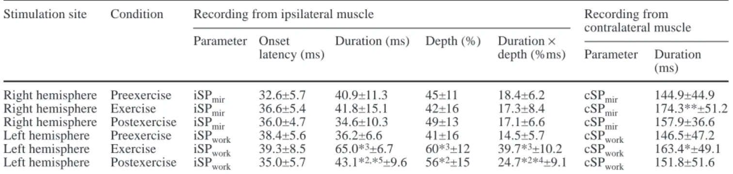
Before the exercise, the iSPs were similar in the mirror-ing and the workmirror-ing muscles (Table 1). Durmirror-ing and after the exercise, the iSPmir did not change, neither in time nor in depth (Fig. 3, Table 1). The iSPworkincreased sig-nificantly in all subjects during exercise (Figs. 3, 4; Table 1); both depth of inhibition and duration increased (Table 1). This increase was observed in all subjects. After exercise, iSPwork recovered partially toward the
preexercise levels (Table 1, Figs. 3, 4). The background activity of the mirroring muscle during exercise-iSP measurement was considerably smaller than that in the working muscle during the exercise-iSP measurements. A relation between background EMG level and the dura-tion or depth of the iSP (before, during, and after exer-cise; left and right side) was not found by linear regres-sion analysis. Thus, the observed iSP changes were inde-pendent of background EMG levels.
An ipsilateral motor-evoked potential was never re-corded (i.e., an excitatory phenomenon preceding the TCI, stemming from uncrossed monosynaptic or oligo-synaptic corticospinal input).
Contralateral silent period
During exercise, the cSP increased significantly in dura-tion on both sides (the working side and the mirroring side; Table 1, Fig. 3). After exercise, the duration de-creased again (Table 1, Fig. 3). In all trials, contralateral motor-evoked potentials preceded the cSP. These re-sponses were not further analyzed.
Discussion
Effort-induced mirror movements are a highly reproduc-ible phenomenon and were observed in all of our sub-jects. When the EMG of the target muscle had decreased to 50% due to fatigue (increasing the perceived effort to sustain the contraction), the contralateral mirroring con-traction was clinically always graded moderate to marked. In parallel, the mirroring muscle's EMG reached 25% of MVC (or approximately 50% of that of the working muscle) on average (Fig. 2). Our hypothesis was that this type of unintended (and usually unnoticed) activity was associated with a decrease in transcallosal inhibition from the working to the opposite hemisphere. We used fatiguing contractions of the left ADM because previous studies had indicated that left-sided voluntary contractions would produce stronger mirror movements than contractions on the right side (Todor and Lazarus 1986; Armatas et al. 1994; Liepert et al. 2001). We
ap-Table 1 Ipsilateral (iSP) and contralateral (cSP) silent period before, during, and after the exercise (mean ± SD)
Stimulation site Condition Recording from ipsilateral muscle Recording from
contralateral muscle Parameter Onset Duration (ms) Depth (%) Duration ×
latency (ms) depth (%ms) Parameter Duration
(ms) Right hemisphere Preexercise iSPmir 32.6±5.7 40.9±11.3 45±11 18.4±6.2 cSPmir 144.9±44.9 Right hemisphere Exercise iSPmir 36.6±5.4 41.8±15.1 42±16 17.3±8.4 cSPmir 174.3**±51.2 Right hemisphere Postexercise iSPmir 36.0±4.7 34.6±10.3 49±13 17.1±6.6 cSPmir 157.9±36.6 Left hemisphere Preexercise iSPwork 38.4±5.6 36.2±6.6 41±16 14.5±5.7 cSPwork 146.5±47.2 Left hemisphere Exercise iSPwork 39.3±8.5 65.0*3±6.7 60*3±12 39.7*3±10.2 cSP
work 163.4*±49.1 Left hemisphere Postexercise iSPwork 35.0±5.7 43.1*2,*5±9.6 56*2±15 24.7*2*4±9.1 cSP
work 151.8±51.6 *1P<0.05; *2P<0.01; *3P<0.005 (Wilcoxon; compared with preexercise); *4P<0.01; *5P<0.005 (Wilcoxon; compared with exercise)
Fig. 4 Original recordings from the working abductor digiti
min-imi muscle in one subject. Each trace is the average of 10 trials. The mean prestimulus EMG level (background EMG) is given by the hatched lines. The ipsilateral silent periods are indicated by the hatched areas. Note the increase in duration and depth of the silent period during exercise, and the partial recovery to preexer-cise values after exerpreexer-cise
plied the paradigm of measuring the ipsilateral silent period (iSP) in response to TMS to quantify TCI. The presence of iSPs after unihemispheric transcranial mag-netic brain stimulation has been linked previously to transcallosal connections in healthy subjects and patients (Ferbert et al. 1992; Meyer et al. 1995). Thus, we expected the iSP to decrease in the mirroring muscle (iSPmir; Fig. 1B).
Our results are well in line with our hypothesis. Dur-ing exercise, inhibition within the crossed pyramidal path increased bilaterally, as demonstrated by increases in cSPmirand cSPwork(Table 1, Fig. 3A, B). Yet, despite this increase, the iSPmir remained unchanged during exercise (Table 1, Fig. 3D). The final path of the iSP measurement is the crossed pyramidal pathway. Our finding of an unchanged iSPmir is thus explained by a balance between increased crossed pyramidal inhibition and decreased transcallosal inhibition. By the same token, the parallel increase in the iSPwork and cSPwork (Fig. 3A, C) suggests unchanged transcallosal activity originating from the mirroring hemisphere. It is notewor-thy that measurements of iSPworkwere always performed after those of iSPmir. Nevertheless, pre- and postexercise results were similar in the two sets of measurements (Fig. 3), arguing against a sequence effect caused by in-sufficient recovery from fatigue. Moreover, persisting fatigue would have resulted in a reduction of iSPwork, which was not observed. Summarized, our results con-form with our hypothesis of a side-specific reduction of TCI from the working to the opposite (mirroring) hemi-sphere, which occurs in association with the presence of effort-induced mirror movements.
It should be noted that ipsilateral excitatory responses (ipsilateral MEPs) were not observed here. They could arise from uncrossed corticoreticulospinal or corticopro-priospinal projections (Ziemann et al. 1999), or from uncrossed, fast-conducting corticomotoneuronal paths (Nirkko et al. 1997). In patients, the presence of such paths is associated with mirror movements (Nirkko et al. 1997), and “unmasking” of uncrossed paths was previ-ously suggested to account for effort-induced mirror movements in healthy subjects (Zülch and Müller 1969; Nass 1985). If, during unimanual tasks, TCI were to ac-count for the suppression of uncrossed paths, then the appearance of mirror movements during effort would im-ply a reduction of TCI from the nonworking to the work-ing hemisphere. This was not observed in the present study (Fig. 3C). Hence, our data do not support a role for uncrossed pathways in effort-induced mirror movements. Lack of transcallosal inhibition from the working to the mirroring hemisphere was previously assumed to account for the physiological mirror movements of young children. In preschool children, TMS yields no iSP (Heinen et al. 1998). In these children, bilateral activation of both motor cortices occurs during unimanual tasks, as shown by bilateral activation of transcortical, cutaneo-muscular long-loop reflexes (LLRs; Mayston et al. 1999). Both the appearance of iSP and reduction of LLRs are paralleled by the disappearance of mirror movements
during maturation (Heinen et al. 1998; Mayston et al. 1999). Thus, in preschool children, mirror movements probably arise from lack of transcallosal inhibition from the working to the mirroring hemisphere; resulting in bi-lateral activation of the crossed pyramidal tract. Our pres-ent data suggest a similar mechanism in adult effort-induced mirror movements. During unimanual move-ments, neuroimaging studies demonstrate bilateral activa-tion of supplementary motor areas (premotor cortex and supplementary motor area; Nirkko et al. 2001). Such bilateral activation would result in mirror movements, but during effortless finely tuned hand movements, mirroring by the contralateral hemisphere is suppressed by TCI to allow for manual side independence (Geffen et al. 1994). Transcallosal inhibition was demonstrated by TMS (Fer-bert et al. 1992; Meyer et al. 1995; Rösler et al. 1995) and is also suggested by neuroimaging studies, where the pri-mary motor area of the mirroring hemisphere is deactivat-ed (Dettmers et al. 1996; Allison et al. 2000; Nirkko et al. 2001). When the effort of the unimanual movement in-creases (e.g., by the wish to overcome the fatigue-induced loss of force), TCI decreases – as shown here – and side independence is lost, resulting in mirror move-ments. The decrease in TCI is well in line with previous studies demonstrating an enhancement of excitability of the nonworking motor cortex during unimanual, high-force (at least 50% of MVC) tonic muscle contractions by use of cortical paired-pulse stimulation (Muellbacher et al. 2000; Liepert et al. 2001). In further accordance with this, an increase in regional cerebral blood flow in the ipsilateral hemisphere was observed with increasingly forceful contractions in one positron emission tomogra-phy study (Dettmers et al. 1996).
The increased duration of the cSPwork during an ef-fortful fatiguing contraction was an expected finding, since it has been demonstrated previously (McKay et al. 1996; Taylor et al. 1996, 2000; Sacco et al. 1997; Taylor and Gandevia 2001). Here, we observed an effort-related increase in the cSP also in the mirroring muscle (Table 1, Fig. 3B). Theoretically, two mechanisms could account for this increased contralateral crossed inhibition: First, inhibitory activity from the working hemisphere could be mediated via a callosal route to the mirroring hemi-sphere. This is unlikely given our result of a decrease in TCI from the working to the mirroring hemisphere. Second, inhibition could be a consequence of the sus-tained muscle contraction of the mirroring muscle, either by refractoriness or fatigue of the involved cells, or through enhanced supraspinal inhibition by a reflex mechanism. The present data allow no judgement about these possibilities. Nevertheless, while inhibitory seg-mental reflexes have been demonstrated during fatigue (Bigland-Ritchie et al. 1986), inhibitory transcortical LLRs have not been described during fatigue. On the contrary, excitatory LLRs increase during fatigue of small hand muscles (Duchateau and Hainaut 1993). Thus, a passive central phenomenon (refractoriness or fatigue of descending systems) in response to the sustained muscle contraction is possibly involved.
The presence of bilateral inhibition of the cortical motor outflow during an effortful and fatiguing contrac-tion is interesting in the light of activacontrac-tion of the supple-mentary motor areas, as suggested here and in neuroim-aging studies (Dettmers et al. 1995, 1996; Nirkko et al. 2001). It is unclear at which level of the motor hierarchy the inhibitory mechanism accounting for the cSP acts. A supraspinal mechanism is suggested by the observations that the latter part of the cSP is not accompanied by a depression of spinal reflexes (Fuhr et al. 1991) and be-cause the cSP is longer than the SP after peripheral nerve stimulation (Wilson et al. 1993). It has previously been proposed that muscle fatigue is associated with both in-creased excitation and inin-creased inhibition within the motor cortex (Taylor et al. 1996). Enhanced descending supraspinal drive could compensate for the loss of exci-tation (or the increase in inhibition) during fatiguing contractions (Duchateau and Hainaut 1993). Our present results are in line with this notion.
Acknowledgements We are grateful to Professor C.W. Hess for
his generous and enthusiastic support. We are also indebted to PD Dr. J. Mathis for help with the laboratory equipment.
References
Allison JD, Meador KJ, Loring DW, Figueroa RE, Wright JC (2000) Functional MRI cerebral activation and deactivation during finger movement. Neurology 54:135–142
Armatas CA, Summers JJ, Bradshaw JL (1994) Mirror movements in normal adult subjects. J Clin Exp Neuropsychol 16:405–413 Balbi P, Trojano L, Ragno M, Perretti A, Santoro L (2000) Pat-terns of motor control reorganization in a patient with mirror movements. Clin Neurophysiol 111:318–325
Bigland-Ritchie B, Dawson NJ, Johansson RS, Lippold OCJ (1986) Reflex origin for the slowing of motoneurone firing rates in fatigue of human voluntary contractions. J Physiol (Lond) 379:451–459
Brasil-Neto JP, Araujo VP, Carneiro CR (1999) Postexercise facil-itation of motor evoked potentials elicited by ipsilateral volun-tary contraction. Muscle Nerve 22:1710–1712
Carr LJ, Harrison LM, Evans AL, Stephens JA (1993) Patterns of central motor reorganization in hemiplegic cerebral palsy. Brain 116:1223–1247
Cernacek J (1961) Contralateral motor irradiation – cerebral domi-nance. Arch Neurol 4:165–172
Dettmers C, Fink GR, Lemon RN, Stephan KM, Passingham RE, Silbersweig D, Holmes A, Ridding MC, Brooks DJ, Frackowiak RS (1995) Relation between cerebral activity and force in the motor areas of the human brain. J Neurophysiol 74:802–815
Dettmers C, Ridding MC, Stephan KM, Lemon RN, Rothwell JC, Frackowiak RS (1996) Comparison of regional cerebral blood flow with transcranial magnetic stimulation at different forces. J Appl Physiol 81:596–603
Dimitrijevic MR, McKay WB, Sarjanovic I, Sherwood AM, Svirtlih L, Vrbova G (1992) Co-activation of ipsi- and contra-lateral muscle groups during contraction of ankle dorsiflexors. J Neurol Sci 109:49–55
Duchateau J, Hainaut K (1993) Behaviour of short and long laten-cy reflexes in fatigued human muscles. J Physiol (Lond) 471:787–799
Farmer SF, Ingram DA, Stephens JA (1990) Mirror movements studied in a patient with Klippel-Feil syndrome. J Physiol (Lond) 428:467–484
Ferbert A, Priori A, Rothwell JC, Day BC, Colebatch JG, Marsden CD (1992) Interhemispheric inhibition of the human motor cortex. J Physiol (Lond) 453:525–546
Fuhr P, Agostino R, Hallett M (1991) Spinal motor neuron excit-ability during the silent period after cortical stimulation. Elec-troencephalogr Clin Neurophysiol 81:257–262
Geffen GM, Jones DL Geffen LB (1994) Interhemispheric control of manual motor activity. Behav Brain Res 64:131–140 Heinen F, Glocker FX, Fietzek U, Meyer BU, Lücking CH,
Korinthenberg R (1998) Absence of transcallosal inhibition following focal magnetic stimulation in preschool children. Ann Neurol 43:608–612
Liepert J, Dettmers C, Terborg C, Weiller C (2001) Inhibition of ipsilateral motor cortex during phasic generation of low force. Clin Neurophysiol 112:114–121
Mayston MJ, Harrison LM, Stephens JA (1999) A neurophysio-logical study of mirror movements in adults and children. Ann Neurol 45:583–594
McKay WB, Stokic DS, Sherwood AM, Vrbova G, Dimitrijevic MR (1996) Effect of fatiguing maximal voluntary contraction on excitatory and inhibitory responses elicited by transcranial magnetic motor cortex stimulation. Muscle Nerve 19:1017– 1024
Meyer B-U, Röricht S, Gräfin von Einsiedel H, Kruggel F, Weindl A (1995) Inhibitory and excitatory interhemispheric transfers between motor cortical areas in normal humans and patients with abnormalities of the corpus callosum. Brain 118:429– 440
Muellbacher W, Facchini S, Boroojerdi B, Hallett M (2000) Changes in motor cortex excitability during ipsilateral hand muscle activation in humans. Clin Neurophysiol 111:344– 349
Nass R (1985) Mirror movement asymmetries in congenital hemiparesis: the inhibition hypothesis revisited. Neurology 35:1059–1062
Nelles G, Cramer SC, Schaechter JD, Kaplan JD, Finklestein SP (1998) Quantitative assessment of mirror movements after stroke. Stroke 29:1182–1187
Nirkko AC, Rösler KM, Ozdoba C, Heid O, Schroth G, Hess CW (1997) Human cortical plasticity: functional recovery with mirror movements. Neurology 48:1090–1093
Nirkko AC, Ozdoba C, Redmond SM, Bürki M, Schroth G, Hess CW, Wiesendanger M (2001) Different ipsilateral representa-tions for distal and proximal movements in the sensorimotor cortex: activation and deactivation patterns. Neuroimage 13:825–835
Regli F, Filippa G, Wiesendanger M (1967) Hereditary mirror movements. Arch Neurol 16:620–623
Rösler KM, Nirkko AC, Hess CW (1995) Electrophysiological as-sessment of functional corpus callosum integrity in internal hydrocephalus. Muscle Nerve 18:787–788
Rothwell JC, Hallett M, Berardelli A, Eisen A, Rossini P, Paulus W (1999) Magnetic stimulation: motor evoked potentials. Electroencephalogr Clin Neurophysiol [Suppl] 52:97–103 Sacco P, Thickbroom GW, Thompson ML, Mastaglia FL (1997)
Changes in corticomotor excitation and inhibition during pro-longed submaximal muscle contractions. Muscle Nerve 20:1158–1166
Samii A, Wassermann EM, Ikoma K, Mercuri B, Hallett M (1996) Characterization of postexercise facilitation and depression of motor evoked potentials to transcranial magnetic stimulation. Neurology 46:1376–1382
Schott GD, Wyke MA (1981) Congenital mirror movements. J Neurol Neurosurg Psychiatry 44:586–599
Taylor JL, Gandevia SC (2001) Transcranial magnetic stimulation and human muscle fatigue. Muscle Nerve 24:18–29
Taylor JL, Butler JE, Allen GM, Gandevia SC (1996) Changes in motor cortical excitability during human muscle fatigue. J Physiol (Lond) 490:519–528
Taylor JL, Butler JE, Gandevia SC (2000) Changes in muscle afferents, motoneurons and motor drive during muscle fatigue. Eur J Appl Physiol 83:106–115
Ziemann U, Ishii K, Borgheresi A, Yaseen Z, Battaglia F, Hallett M, Cincotta M, Wassermann EM (1999) Dissociation of the pathways mediating ipsilateral and contralateral motor-evoked potentials in human hand and arm muscles. J Physiol (Lond) 518:895–906
Zülch KJ, Müller N (1969) Associated movements in man. In: Vinken PJ, Bruyn GW (eds) Handbook of clinical neurology. North-Holland, Amsterdam, pp 404–426
Todor JI, Lazarus JA (1986) Exertion level and the intensity of associated movements. Dev Med Child Neurol 28:205– 212
Wilson SA, Lockwood RJ, Thickbroom GW, Mastaglia FL (1993) The muscle silent period following transcranial magnetic cor-tical stimulation. J Neurol Sci 114:216–222
Woods BT, Teuber HL (1978) Mirror movements after childhood hemiparesis. Neurology 28:1152–1157