HAL Id: hal-02665934
https://hal.inrae.fr/hal-02665934
Submitted on 31 May 2020
HAL is a multi-disciplinary open access
archive for the deposit and dissemination of
sci-entific research documents, whether they are
pub-lished or not. The documents may come from
teaching and research institutions in France or
abroad, or from public or private research centers.
L’archive ouverte pluridisciplinaire HAL, est
destinée au dépôt et à la diffusion de documents
scientifiques de niveau recherche, publiés ou non,
émanant des établissements d’enseignement et de
recherche français ou étrangers, des laboratoires
publics ou privés.
A Rhodococcus qsdA-Encoded Enzyme Defines a Novel
Class of Large-Spectrum Quorum-Quenching Lactonases
Stéphane Uroz, Phil M. Oger, Emilie Chapelle, Marie-Thérèse Adeline, Denis
Faure, Yves Dessaux
To cite this version:
Stéphane Uroz, Phil M. Oger, Emilie Chapelle, Marie-Thérèse Adeline, Denis Faure, et al..
A
Rhodococcus qsdA-Encoded Enzyme Defines a Novel Class of Large-Spectrum Quorum-Quenching
Lactonases. Applied and Environmental Microbiology, American Society for Microbiology, 2008, 74
(5), pp.1357-1366. �10.1128/AEM.02014-07�. �hal-02665934�
A
PPLIED ANDE
NVIRONMENTALM
ICROBIOLOGY, Mar. 2008, p. 1357–1366
Vol. 74, No. 5
0099-2240/08/$08.00
⫹0 doi:10.1128/AEM.02014-07
Copyright © 2008, American Society for Microbiology. All Rights Reserved.
A Rhodococcus qsdA-Encoded Enzyme Defines a Novel Class of
Large-Spectrum Quorum-Quenching Lactonases
䌤
†
Ste
´phane Uroz,
1,2‡ Phil M. Oger,
2* Emilie Chapelle,
1Marie-The
´re
`se Adeline,
3Denis Faure,
1and Yves Dessaux
1Interactions Plantes et Microorganismes de la Rhizosphe
`re, Institut des Sciences du Ve
´ge
´tal, CNRS, Avenue de la Terrasse,
91198 Gif-sur-Yvette Cedex,
1Laboratoire de Sciences de la Terre, UMR CNRS 5570, Ecole Normale Supe
´rieure, 46,
Alle
´e d’Italie, 69364 Lyon Cedex,
2and Institut de Chimie des Substances Naturelles, CNRS, Avenue de
la Terrasse, 91198 Gif-sur-Yvette Cedex,
3France
Received 3 September 2007/Accepted 22 December 2007
A gene involved in N-acyl homoserine lactone (N-AHSL) degradation was identified by screening a genomic
library of Rhodococcus erythropolis strain W2. This gene, named qsdA (for quorum-sensing signal degradation),
encodes an N-AHSL lactonase unrelated to the two previously characterized N-AHSL-degrading enzymes, i.e.,
the lactonase AiiA and the amidohydrolase AiiD. QsdA is related to phosphotriesterases and constitutes the
reference of a novel class of N-AHSL degradation enzymes. It confers the ability to inactivate N-AHSLs with
an acyl chain ranging from C
6to C
14, with or without substitution at carbon 3. Screening of a collection of 15
Rhodococcus strains and strains closely related to this genus clearly highlighted the relationship between the
ability to degrade N-AHSLs and the presence of the qsdA gene in Rhodococcus. Bacteria harboring the qsdA gene
interfere very efficiently with quorum-sensing-regulated functions, demonstrating that qsdA is a valuable tool
for developing quorum-quenching procedures.
Gram-negative bacteria couple gene expression to
popula-tion density by a regulatory mechanism named quorum sensing
(QS). QS relies upon the production and the perception of one
or more signal molecules by the bacterial population. An
im-portant class of these signals is the N-acyl homoserine lactone
(N-AHSL) class (9). Molecules belonging to this class exhibit a
conserved structure, with a backbone composed of a
homo-serine lactone (HSL) N-linked to an acyl chain via an amide
bond. Variation in N-acyl chain length and the oxidation status
of N-AHSLs provide for specificity of the signal. Of particular
interest is the finding that QS regulates pathogenicity, or
pathogenicity-related functions, in bacteria of medical or
en-vironmental importance (15, 32, 50).
If QS and N-AHSLs are important components of the
strat-egy of adaptation by bacteria to their biotic environment,
es-pecially a plant surface, one might suspect that the eukaryotic
hosts and competing bacteria might have developed strategies
to interfere with this communication system. Indeed, QS
inhi-bition was reported through the production of antagonists (10)
or the production of N-AHSL degradation enzymes by plants
(5), animals (2, 34), and a wide range of bacterial genera (14,
16, 18–20, 30, 31, 44, 49). In spite of the large diversity of
degrading bacteria, only two families of
N-AHSL-inactivating enzymes (N-AHSLases) have been described to
date: the AiiA-like N-AHSL lactonases (6, 19, 49) and the
AiiD-like N-AHSL amidohydrolases (14, 20, 30). Whatever
their physiological role, N-AHSLases have been used
effi-ciently to interfere with the expression of QS-regulated
func-tions in bacteria (6, 19, 20, 35, 42). This strategy has been
termed quorum quenching (QQ). It proved to be a valuable
trail toward definition of novel biocontrol agents such as
nat-ural isolates degrading N-AHSLs (8, 23, 44). QQ occurs in
natural environments as indicated by the coexistence of
N-AHSL-producing and -degrading strains in biofilms (14) or
in the rhizosphere (4). Among the species harboring an
N-AHSLase activity, Rhodococcus erythropolis is remarkable
because it is the only bacterium in which three enzymatic activities
directed at N-AHSLs have been characterized: an oxidoreductase
activity, which reduces 3-oxo-N-AHSLs to their hydroxylated
equivalents (43); an amidohydrolase (43); and a lactonase (29).
The marked R. erythropolis QQ capabilities suggest that it might
be used in biocontrol protocols, especially since it is a natural
inhabitant of soils worldwide (44).
In this study we report the isolation of one of the three genes
encoding N-AHSLase activities from R. erythropolis strain W2.
We show that this gene encodes a phosphotriesterase
(PTE)-like broad-spectrum N-AHSL lactonase, which is found only in
the Rhodococcus genus and solely in strains capable of
degrad-ing N-AHSLs.
MATERIALS AND METHODS
Strains, media, growth conditions, and chemicals.Bacterial strains and plas-mids are listed in Table 1. All strains were grown at 25°C, with the exception of the Chromobacterium violaceum biosensor and Escherichia coli, which were grown at 30°C and 37°C, respectively. Unless otherwise stated, LBm medium was buffered at pH 6.5 to prevent spontaneous lactonolysis of N-AHSLs and used as the rich medium (44). All media were solidified with 16 g/liter of agar. Antibiotics were used at the following final concentrations (ing/ml; see Table 1 for specific strain requirements): ampicillin, 100; gentamicin, 25; tetracycline, 10; rifampin, 100; and streptomycin, 250. X-Gal (5-bromo-4-chloro-3-indolyl--D -galactopyr-anoside) and IPTG (isopropyl--D-thiogalactopyranoside) were included in
me-* Corresponding author. Mailing address: Laboratoire de Sciences
de la Terre, UMR CNRS 5570, Ecole Normale Supe
´rieure, 46, Alle
´e
d’Italie, 69364 Lyon Cedex, France. Phone: (33) 4 72 72 87 92. Fax:
(33) 4 72 72 86 77. E-mail: [email protected].
† Supplemental material for this article may be found at http://aem
.asm.org/.
‡ Present address: Interactions Arbres Microorganismes, INRA,
54280 Champenoux, France.
䌤
Published ahead of print on 11 January 2008.
dia at 40g/ml and 100 g/ml, respectively. Hexanoyl-HSL (C6-HSL) and its
3-oxo derivative were purchased from Sigma. The other N-AHSLs were gener-ous gifts from S. R. Chhabra and P. Williams (University of Nottingham, United Kingdom).
Detection of N-AHSLs.N-AHSLs were separated and visualized on thin-layer chromatography (TLC) plates as described previously (37). Saturated short-chain N-AHSLs were detected by TLC using the Chromobacterium violaceum
biosensor CV026 (22). Oxo and hydroxy derivatives and long-chain N-AHSLs were detected using the Agrobacterium biosensor NTL4(pZLR4) (21).
Detection of N-AHSL degradation products.The N-AHSL degradation assays were performed as described earlier (45), using actively growing DH5␣ cells expressing or not the qsdA gene in pH 6.5 buffered LB medium. The incubation time was set to 24 h, because this allowed work at low concentration and thus reduction of the interference of the medium with the data collected. Under these
TABLE 1. Bacterial strains and plasmids
Strain or plasmid Characteristics or synonym Source or reference
Strains tested for N-AHSL degradation and
presence of the qsdA gene
Actinoplanes utahensis NRRL 12052
Source of aculeacin A deacylase
39
Corynebacterium glutamicum ATCC 14752
Glutamine-producing strain
38
Escherichia coli
⌲-12
Lab collection
Flavobacterium sp. strain ATCC 27551
Parathion-degrading strain
24
Gordonia alkalivorans strain 98
Petroleum-degrading strain
Lab collection
Microbacterium sp. strain POLB142
Mannopine-degrading rhizospheric strain; soil isolate
27
Mycobacterium sp. strain Tn20
Drought-resistant Sahara desert isolate
This study
Nocardia asteroides CECT 3051
ATCC 19247; type strain
CECT
aPseudonocardia autotrophica CECT 3044
ATCC 13181; type strain
47
Ralstonia solanacearum GMI 1000
Type strain
Lab collection
Rhodococcus erythropolis
W2
N-AHSL-degrading isolate from soil
44
DCL14
Limonene-degrading strain
46
SQ1
ATCC 4277-1; R. erythropolis type strain
33
Mic1
Mycorhizosphere isolate
Lab collection
MP50
Aromatic-nitrile-degrading strain
41
CECT 3008
ATCC 11048
CECT
Rhodococcus fascians D188
Fasciation-causing agent on dicotyledons
3
Rhodococcus opacus HL PM-1
2,4,6-Trinitrophenol-degrading strain
25
Rhodococcus rhodochrous CECT 3042
ATCC 6846
CECT
Rhodococcus corynebacteroides
CECT 420
ATCC 14898; soil isolate
CECT
Rhodococcus sp. strain RHA1
Polychlorinated biphenyl-degrading strain
Lab collection
N-AHSL biosensors and
N-AHSL-producing strains used for QQ assays
Agrobacterium tumefaciens NTL4(pZLR4)
Hydroxy, oxo, and long-chain N-AHSL sensor strain
21
Chromobacterium violaceum CV026
Reduced N-AHSL sensor strain
22
Pectobacterium carotovorum Pcc797
Soft-rot-causing agent
Lab collection
Pseudomonas fluorescens 1855-344
Rhizospheric strain
Lab collection
Agrobacterium tumefaciens
15955tra
cOctopine-type strain, constitutive for Ti plasmid conjugal transfer
Lab collection
C58C1RS
Ti plasmid-cured C58 derivative resistant to rifampin and streptomycin
Lab collection
Escherichia coli strains used for cloning
DH5
␣
Lab collection
VCS257
Stratagene
Plasmids
pUC19
Amp
rLab collection
pGEM-T
Amp
rPromega
pUC1318::Gm
Amp
rGm
r40
pME6032
Tc
r12
pCP13/B
Tc
r; cosmid vector
Lab collection
pSU16
Tc
r; cosmid clone conferring N-AHSL degradation capability
This study
pSU16-11
pUC19 with a 3.2-kb EcoRI fragment from pSU16 conferring N-AHSL
degradation
This study
pSU16-12
pUC19 with a 3.5-kb BamHI fragment from pSU16 conferring N-AHSL
degradation
This study
pSU40
pME6032 harboring the 3.2-kb EcoRI fragment from pSU16-11
This study
pSU8-1
pGEM-T with a 1.8-kb PCR fragment containing orf1 and qsdA
This study
pSU8-1
⌬SalI
pSU8-1 with a SalI deletion in orf1
This study
pSU8-1::Gm
pSU8-1 with insertion of a Gm
rcassette at the AgeI site of qsdA
This study
pSU8-1
⌬SalI::Gm
pSU8-1
⌬SalI with insertion of a Gm
rcassette at the AgeI site of qsdA
This study
pSU-qsdA
W2qsdA ORF amplified and cloned into pET16b
This study
pSUreg1
Upstream regulatory region of qsdA amplified and cloned into pGEM-T
This study
conditions, a fraction of ca. 25% of the initial amount of C6-HSL is
spontane-ously converted into C6-HS by chemical lactonolysis in the presence or absence
of E. coli cells. The enzymatic degradation of N-AHSLs can proceed through two different routes (Fig. 1), which lead to the formation of either N-AHS (lactonoly-sis) (Fig. 1, top) or HSL and an alkyl chain (amidoly(lactonoly-sis) (Fig. 1, bottom). The presence of HSL in the incubation medium was determined after trapping of the free amine with dansyl chloride as described earlier (48). The formation of the ring-opened derivative of N-AHSLs following lactone hydrolysis was inves-tigated using high-pressure liquid chromatography (HPLC) on a Waters chro-matograph equipped with a Waters separation module 2659 and an Atlantis reverse-phase column (4.6 by 150 mm; 5m) coupled to an electrospray ioniza-tion-mass spectrometry detector (Waters Micromass ZQ 200). Retention times and mass spectra were determined for individual molecules in solution as stan-dards. Thirty microliters of the degradation assay sample was injected and eluted with water–0.1% formic acid (solvent A) and acetonitrile–0.1% formic acid (solvent B) with the following elution sequence: 100% solvent A for 5 min, a linear gradient to reach 20% solvent B in 5 min, and 80% solvent A and 20% solvent B for 10 min. Between samples, the column was rinsed by applying 100% B solvent (3 min). The column was then reequilibrated with 100% solvent A for 7 min. The specific fragments expected to appear in the mass spectra of C6-HSL
and C6-HS are all present; fragment 200 is characteristic for C6-HSL, while
fragment 218 is characteristic for C6-HS. Fragment 102 corresponds to HSL and
therefore appears in the C6-HSL spectrum, while fragment 120 is characteristic
for HS and appears in the C6-HS spectrum (28).
Library construction.As strain W2 is especially difficult to lyse by conventional methods, genomic DNA preparation was based on the method of Eulberg et al. (7) with the following modifications. Strain W2 was grown overnight in 50 ml of
LB medium buffered at pH 8.0 with glycine (3%, wt/vol). Prior to DNA extrac-tion, cells were treated for 2 hours with ampicillin and erythromycin (100g/ml, final concentration) to disturb cell wall synthesis and favor the subsequent lysis of the cells. A genomic library of strain W2 was constructed by partially digesting 100g of total genomic DNA with Bsp143I (Sau3AI) and ligating the fragments in BamHI-linearized cosmid vector pCP13/B as described by Hayman and Far-rand (11). The ligation products were packaged using the Gigapack III Gold packaging kit (Stratagene, La Jolla, CA) as recommended by the manufacturer and recovered by transfection into E. coli VCS257. The W2 genomic library generated in this study contained over 4,000 clones, with an average insert size of 23 kb and fewer than 1% empty clones. This corresponds to a theoretical coverage of 15 times of Rhodococcus genome (1).
Screening of the genomic library for N-AHSL degradation ability.A total of 2,880 library clones were screened directly in their E. coli strain VCS257 host by use of a modification of the microplate N-AHSL degradation assay described by Uroz et al. (44), using C. violaceum CV026 as an indicator strain. Clones were first grown in presence of antibiotics and then subcultured in medium devoid of antibiotics but supplemented with 25M of C6-HSL. Cosmid clones were
con-sidered to confer the ability to degrade N-AHSLs only if a total disappearance of the C6-HSL was observed after a 24-h period. The ability of the clones to
effectively degrade the N-AHSL was confirmed by separating the degradation products by TLC and detecting the presence of N-AHSL by the appropriate biosensor. This allows the detection of false-positive degradation due to the presence of compounds inhibiting N-AHSL detection or the growth of the biosensor.
DNA sequence analysis. Sequence analysis was performed with ORF FINDER. Nucleotide and amino acid sequence comparisons were made using
FIG. 1. Scheme for identification of N-AHSL-degradation products. N-AHSL inactivation in bacteria proceeds through two known pathways:
lactonolysis (top) or amide hydrolysis (bottom). N-AHSL lactonolysis is a reversible reaction which yields the corresponding N-AHS, which can
be separated by HPLC and identified by mass spectrometry (MS). Amide hydrolysis is an irreversible reaction which yields HSL and the
corresponding acyl side chain. HSL can be detected by HPLC after trapping the free amine with dansyl chloride.
the BLAST protocol. Multiple alignments were performed using the Pileup subroutine of the GCG package (version 10; GCG Inc., Madison, WI).
Subcloning of the qsdA region.To identify the gene coding for the N-AHSL degradation activity, the 3.2-kb EcoRI fragment was subcloned as shown in Fig. 2 using the primers QS (5⬘-ATGAGTTCAGTACAAACCGTTCGTG-3⬘), QR (5⬘-TCAGCTCTCGAAGTACCGACGTGGG-3⬘), and IR (5⬘-TCACCATTTT TCAACGGCCG-3⬘) and available restriction sites. The qsdA gene was disrupted by insertion of a gentamicin resistance gene from pUC1318::Gm, cloned as an XmaI fragment at the unique AgeI site of the qsdA gene. Southern hybridization was performed at 62°C with a qsdA probe amplified with the QS/QR primer pair and digoxigenin labeled according to the manufacturers’ instructions (Roche/ Boehringer Mannheim).
QQ assays.The ability of the cloned qsdA genes to interfere with the expres-sion of QS-regulated functions was tested in heterologous expresexpres-sion systems. Plasmid pSU40 (qsdAW2cloned into pME6032) was transferred to E. coli strain
DH5␣, Pseudomonas fluorescens strain 1855-344, and the octopine-type conjugal transfer constitutive strain Agrobacterium tumefaciens 15955trac
to yield DH5␣(pSU40), 1855-344(pSU40), and 15955trac (pSU40), respectively. The
ability of these strains to interfere with QS-regulated functions was first evalu-ated using the streak assay described by Molina et al. (23), using C. violaceum CV026 as an indicator strain. The ability to interfere with virulence in Erwinia
carotovora was tested in the potato tuber assay as described by Uroz et al. (44).
Additionally, the ability of the qsdA locus to interfere intracellularly with QS was tested in Agrobacterium tumefaciens. Ti plasmid conjugation assays were per-formed essentially as described by Oger et al. (26), using strain 15955trac
as a transfer constitutive donor and C58C1RS as a recipient. In all experiments, strains harboring the empty vector pME6032 were used as negative control.
Nucleotide sequence accession numbers.The qsdA locus sequence has been deposited at GenBank under accession number AY541692, and the qsdA allele sequences have been deposited under accession numbers EF218062, EF218066, EF218065, and EF589962 (R. erythropolis strains SQ1, MP50 CECT 3008, and Mic1, respectively), EF218064 (R. corynebacteroides CECT 420), and EF218063 (R. rhodochrous strain CECT 3042).
RESULTS AND DISCUSSION
Identification of the Rhodococcus erythropolis gene involved
in N-AHSL degradation.
Genes involved in QS signal
degra-dation were isolated from a genomic library of R. erythropolis
strain W2 on the basis of the ability of specific cosmids to
confer C
6-HSL degradation capability upon the E. coli host
cells. Degradation was clearly evidenced for 20 out of the 2,880
clones screened. Based on their restriction patterns, the 20
cosmids fell into four groups, which all shared single common
EcoRI and BamHI fragments of ca. 3.2 and 3.5 kb,
respec-tively. The cosmid pSU16, which harbors the smallest insert,
was chosen for further studies. DNA fragments resulting from
BamHI or EcoRI restrictions of pSU16 DNA were shotgun
subcloned into pUC19. Only two sets of clones, containing
either a 3.5-kb BamHI (pSU16-12) (not shown) or a 3.2-kb
EcoRI (pSU16-11) (Fig. 2) fragment conferred upon their E.
coli host the ability to degrade N-AHSLs with acyl chains
ranging from C
6- to C
12-HSL, independently of the
substitu-tion at carbon 3. HPLC analyses of the growth media of these
clones confirmed the disappearance of the N-AHSL (data not
shown).
The sequence of the EcoRI fragment of pSU16-11 (3,152 bp;
GenBank accession number AY541692) contains two
incom-plete open reading frames (ORFs) and two comincom-plete ORFs,
which were named orf1 and qsdA (for QS signal degradation)
(Fig. 2). The first incomplete ORF (to the right in Fig. 2) shows
similarities to alleles of the gntR/fadR family of transcriptional
regulators and is most closely related to that of Pseudomonas
aeruginosa strain PAO1 (PA1627) (identity, 40%; similarity,
58%). The first complete ORF, orf1, could encode a serine-rich
protein of 172 amino acids. It does not show any significant
homology with peptide sequences available in the databases,
suggesting that it might be a pseudogene. The second complete
ORF, qsdA, could encode a protein of 323 amino acids related
to members of the PTE superfamily, which is found in a wide
range of organisms from bacteria to eukarya. PTEs are zinc
metalloenzymes which were initially identified as efficiently
catalyzing the hydrolysis of a variety of organophosphorus
compounds (13), but a growing number of PTE homologues
which also show amidohydrolase or lactonase activities have
been characterized (36). Finally, the protein encoded by the
incomplete orf3 shows 48% identity with proteins annotated as
acyl coenzyme A synthetases (AMP-forming)/AMP-acid
li-gases II of Ralstonia metallidurans and other enzymes related
to the lipid metabolism and transport. Acyl coenzyme A
syn-thetases are involved in both the synthesis and turnover of fatty
acids in bacteria.
Because the ORFs qsdA and orf1 were the only
uninter-rupted ORFs present in pSU16-11, they were likely to
deter-mine the C
6-HSL degradation ability conferred by that clone.
To confirm this hypothesis, various constructions were
gener-ated (Fig. 2). As expected, the constructions lacking one or
both partial ORFs (e.g., pSU8-1 and pSUqsdA
W2) still
con-ferred N-AHSL-degrading capabilities upon E. coli.
Con-versely, constructions lacking qsdA (pSU reg1) or harboring a
disrupted qsdA gene (pSU8-1::Gm) did not confer N-AHSL
degradation ability upon their host. From these results, it is
clear that qsdA is necessary and sufficient to code for N-AHSL
degradation in the original pSU16 cosmid.
qsdA codes for a PTE-like N-AHSL lactonase.
The
identifi-cation of the degradation products of N-AHSLs was
per-formed by a combination of HPLC and mass spectrometry
analyses to detect the presence of HSL or N-AHS generated by
amidolysis or lactonolysis, respectively (Fig. 1). The
spontane-ous degradation was evaluated in control experiments that
used uninoculated LB medium or medium inoculated with E.
coli DH5
␣ or DH5␣ with the empty cloning vector. Figure 3A
presents results obtained for strain DH5
␣(pME6032). In each
FIG. 2. Identification of the qsdA gene. The genetic organization of
the qsdA locus derived from the complete sequence of the 3.2-kb
EcoRI fragment conferring C
6-HSL degradation upon its host is shown
at the top. Broken arrows symbolize the primers used for subcloning
and their orientations. Restriction sites also used for subcloning are
shown. See Table 1 for a description of the plasmids. Plasmid
con-structions used to identify the qsdA gene were assayed for their ability
(
⫹) or inability (⫺) to confer N-AHSL degradation upon their host
against a set of N-AHSLs, including C
6-HSL, O-C
6-HSL, C
7-HSL,
C
8-HSL, O-C
8-HSL, O-C
10-HSL, C
12-HSL, O-C
12-HSL, and O-C
14-HSL. Each construct gave identical degradation results regardless of
the N-AHSL present in the medium.
experiment, two peaks were visible on the HPLC spectra after
a 24-h incubation. Their position at 15.8 and 21 min as well as
mass spectra correspond to C
6-HS and C
6-HSL, respectively.
The presence of C
6-HS in the control experiments results from
the spontaneous lactonolysis of C
6-HSL in aqueous medium.
Each control condition yielded the same amount of
spontane-ous lactonolysis (ca. 25%; data not shown), showing that DH5
␣
does not itself facilitate the degradation of N-AHSLs. At the
end of experiments performed with E. coli DH5
␣(pSU40)
ex-pressing qsdA, no N-AHSL could be detected (Fig. 3B),
indi-cating that a complete conversion of the initial C
6-HSL
oc-curred. Attempts to detect the presence of HSL, which would
indicate a cleavage of the N-AHSL molecules by an
amidohy-drolase, failed (data not shown). At the same time, the
pres-ence of C
6-HS was clearly visible (Fig. 3). Thus, qsdA must
code for an N-AHSL lactonase activity, the presence of which
in Rhodococcus has been recently identified by Park and
col-leagues (29).
QsdA clearly belongs to the PTE family of zinc-dependent
metalloproteins (Fig. 4). It is totally unrelated to the known
bacterial N-AHSL lactonases, which belong to the
Zn-depen-dent glyoxylase family, or to the known N-AHSL
amidohydro-lases, which belong to the cluster of
-lactam acylases. Thus,
the present study extends the number of families of
N-AHSL-degrading enzymes of bacterial origin to three and to include
the PTE family. PTEs were first described for their ability to
cleave the phosphotriester bond in phosphotriesters but were
later shown to be promiscuous enzymes, harboring lactonase
FIG. 3. QsdA lactonase activity. E. coli strains DH5
␣(pME6032) and DH5␣(pSU40) were incubated in pH 6.5 buffered LBm medium with 50
M C
6-HSL for 24 h. The medium was analyzed at 0 and 24 h by HPLC-mass spectrometry. Under the experimental conditions used, C
6-HS
(molecular weight, 217) and C
6-HSL (molecular weight, 199) had retention times of 15.8 and 21 min, respectively, and mass spectra were composed
of the following main fragments: m/z
⫽ 218, 200, and 120 for C
6-HS, and m/z
⫽ 200, 102, and 99 for C
6-HSL. (A) Spontaneous degradation of
C
6-HSL in aqueous medium. (B) DH5
␣(pSU40) after 24 h of incubation. A single peak at a retention time of 15.8 min is visible on the HPLC
spectrum, which is identified as C
6-HS. C
6-HSL has completely disappeared from the medium. The formation of C
6-HS, correlated with the
absence of HSL in the medium, is indicative of a lactonase activity.
or amidohydrolase activities (36). QsdA, however, does not
confer the ability to degrade the prototypic phosphotriester
methyl parathion, nor does the prototype PTE gene, opd, code
for N-AHSL degradation (data not shown). Although QsdA
exhibits the canonical structure of PTEs, the sequence of its
metal binding site, which is central to the enzyme activity by
fixing two molecules of zinc and forming the catalytic pocket in
which the substrate inserts, differs from the consensus
se-quence of PTEs at 3 of 12 positions (CD1 and CD2 domains in
Fig. 4). In contrast, the sole PTE/QsdA homologue known
from Rhodococcus, i.e., the TIG:13193 gene from strain RHA1
(Rsp_RHA1 in Fig. 4), as well as the closest homologues of
QsdA, i.e., the PTE alleles of several Mycobacterium strains
and other actinobacteria (Mtu_CDC1551 in Fig. 4), all present
the canonical signature of the PTEs, including the 12
con-served amino acids from the zinc binding domain.
Interest-FIG. 4. Alignment of selected PTEs from bacterial and eukaryotic origins. In addition to QsdA from R. erythropolis strain W2 (Rer_W2), PTE
homologues used in this alignment include a putative QsdA homologue from Rhodococcus corynebacterioides CECT 420 (Rco_420, this study);
putative PTEs from Rhodococcus sp. strain RHA1 (Rsp_RHA1, accession no. TIG:13193), Agrobacterium tumefaciens strain C58 (Atu_C58,
accession no. AAL43882), Pseudomonas syringae pv. Syringae strain B728a (Psy_B728a, accession no. YP_233869), Shigella flexneri strain 2457T
(Sfl_2457T, accession no. AAP19319), Mycobacterium tuberculosis strain CDC1551 (Mtu_CDC1551, accession no. AAK44461), and Escherichia coli
K-12 (Eco_K12, accession no. AAC76404); PTEs from Chryseobacterium balustinum strain BC9 (Cba_BC9, accession no. CAD19996); OPD from
Flavobacterium sp. strain ATCC 27551 (Fsp_ATCC27551, accession no. CAD13181); and PTER from Homo sapiens sapiens (Hsa, accession no.
Q96BW5) and from Mus musculus (Mmu, accession no. Q60866). The positions of the essential amino acids residues of the catalytic site (A), the
substrate binding site (S), and the dimerization domains (H) are shown above the alignment. CD1 and CD2, zinc binding conserved domains 1
and 2. The 27 residues conserved among known PTE sequences are shown in reverse font. QsdA-specific amino acid substitutions which are not
observed in other PTEs (including alleles not shown in the figure) are highlighted by a gray background.
ingly, strain RHA1 and Mycobacterium sp. strain Tn20 are both
unable to degrade N-AHSLs. In addition, when cloned and
expressed in E. coli, the TIG:13193 ORF from RHA1 does not
confer N-AHSL degradation capabilities to its host. Thus, the
lack of N-AHSL degradation in strain RHA1 is due not to a
lack of expression of the protein from its native promoter but
to the lack of N-AHSL lactonase activity of the TIG:13193
protein, which will consequently be referred to not as a QsdA
but as a classical PTE homologue. QsdA is therefore the first
prokaryotic member of the PTE family with demonstrated
N-AHSL lactonase activity.
QsdA is a Rhodococcus-specific PTE-like N-AHSL lactonase.
The wide occurrence of PTE homologues in species closely
related to R. erythropolis and the specific signature and activity
of QsdA prompted us to determine the phylogenetic
distribu-tion of this gene in the actinobacterial cluster. We screened a
collection of related strains belonging to the Rhodococcus
ge-nus, i.e., R. corynebacteroides (one strain), R. erythropolis (five
strains), R. fascians (one strain), R. rhodochrous (one strain),
and R. opacus (one strain), for their ability to inactivate
N-AHSLs and the presence of the qsdA gene. The ability to
degrade N-AHSLs appeared ubiquitous among strains of R.
erythropolis, since all assayed isolates (5/5) degraded a range of
N-AHSLs identical to that previously reported for strain W2
(acyl chain length from C
6to C
14, with or without substitution
on C3 [see Table S1 in the supplemental material]). N-AHSL
degradation was observed in three of the five Rhodococcus
species tested, i.e., R. erythropolis, R. rhodochrous, and R.
corynebacteroides, but not in R. fascians and R. opacus,
al-though these species are more closely related to R. erythropolis
than are R. rhodochrous and R. corynebacteroides (Fig. 5), and
not in the polychlorinated biphenyl-degrading Rhodococcus sp.
strain RHA1. The degradation ability was also tested in
mem-bers of genera closely related to Rhodococcus, such as
Pseudo-nocardia autotrophica (strain CECT 3044), Corynebacterium
glutamicum (strain ATCC14752), Gordonia alkalivorans (strain
98), Microbacterium sp. (strain POLB142), Mycobacterium sp.
(strain Tn20), Nocardia asteroides (strain CECT 3051),
Actino-planes utahensis (strain NRRL 12052), and Flavobacterium sp.
(strain ATCC 27551). None of these assayed strains degraded
N-AHSLs.
The qsdA gene was detected by Southern hybridization,
un-der low-stringency conditions using the cloned qsdA gene from
strain W2 as a probe, in all but one of the strains able to
degrade N-AHSLs, including R. corynebacteroides, R.
eryth-ropolis, and R. rhodochrous. Southern hybridization performed
FIG. 5. Phylogenetic distribution of N-AHSL degradation in Rhodococcus. The ability to degrade N-AHSLs among bacteria is highlighted on
a phylogenetic tree based on the 16S rRNA gene. Strains shown in bold were tested in the present study. N-AHSL degradation data from the
literature are reported for all other strains. Wherever possible the nature of the enzymatic activity is noted: A, N-AHSL amidohydrolase; L,
N-AHSL lactonase; O, N-AHSL oxidoreductase; u, undetermined. The number of identified activities is also reported (e.g., A, A indicates two
amidohydrolases). The presence (
⫹) or absence (⫺) of a qsdA homologue was determined by hybridization with a qsdA probe. The
N-AHSL-degrading strains belonging to the Rhodococcus genus are boxed. Agrobacterium tumefaciens strain C58 harbors two N-AHSL lactonases (AttM and
AiiB), and R. erythropolis strain W2 harbors at least three N-AHSL modification/degradation enzymes: an amidohydrolase (A), an oxidoreductase
(O), and a lactonase (L). NT, not tested.
on genomic DNA restricted with EcoRI, BamHI, and HindIII
confirmed that the gene is present as a single copy in the
genomes of these strains. The only R. erythropolis strain that
did not hybridize with the qsdA probe was strain DCL14 (46).
This strain is indistinguishable from strain W2 in terms of 16S
rRNA gene sequence and ability to inactivate QS molecules.
This result is, however, consistent with the observation by Uroz
et al. (43), who demonstrated the occurrence of N-AHSLase
activities other than lactonase in R. erythropolis strains W2 and
DCL14. The qsdA gene could not be detected in any of the
Rhodococcus strains unable to degrade N-AHSLs or in the
strains from the related genera.
A total of six qsdA alleles (in addition to qsdA from strain
W2) were cloned into pGEM-T Easy following PCR
amplifi-cation from the genomic DNAs of the four R. erythropolis
clones, as well as the R. rhodochrous and R. corynebacteroides
strains showing a positive hybridization signal in Southern
analysis. All alleles conferred the ability to degrade N-AHSLs
upon E. coli DH5
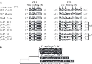
␣. The deduced QsdA peptides fell into two
homology groups, which were termed A1 and A2 (Fig. 6A).
Sequences (DNA and protein) were almost identical within
each group and were ca. 88% identical and 93% similar at the
protein level between groups A1 and A2. The conserved zinc
binding domains CD1 and CD2 of the QsdA alleles of clusters
A1 and A2 diverge at one position (residue 167) (Fig. 6A).
Meanwhile, domains CD1 and CD2 of allele clusters A1 and
A2 also diverge from the consensus PTE domains at two or
three positions (QsdA residues 19, 167, 169), suggesting that
the six alleles might derive from the same PTE ancestor.
How-ever, the qsdA allele phylogeny is not congruent with the
Rhodococcus 16S rRNA gene phylogeny (Fig. 6B).
Further-more, the Rhodococcus species harboring these alleles do not
appear to form a clade within the Rhodococcus genus (Fig. 5).
This suggests that qsdA is a Rhodococcus-specific N-AHSL
lactonase that evolved in this genus and was transferred
hori-zontally at several points during the speciation of Rhodococcus.
qsdA confers QS quenching abilities in heterologous
sys-tems.
We tested the ability of all the cloned qsdA alleles,
including qsdA
W2as well as qsdA
420, qsdA
SQ1, qsdA
3008,
qsdA
MP50, qsdA
Mic1and qsdA
3042, to interfere with the
expres-sion of QS-regulated functions, using three different quenching
assays in which qsdA is expressed in heterologous systems.
All alleles conferred upon their host the ability to inactivate
the same range of N-AHSLs as the wild-type strain W2 (i.e.,
N-AHSLs with or without substitution on carbon 3 and with an
acyl chain ranging from 6 to 14 carbons [data not shown]),
regardless of the plasmid vector used to express the gene. They
were able to quench the synthesis of violacein by C. violaceum
CV026 grown in the presence of C
6-HSL with the same
effi-ciency as the wild-type R. erythropolis strain W2 (data not
shown).
When expressed in P. fluorescens strain 1855-344, qsdA
W2conferred quenching capabilities closely matching that of the
wild-type R. erythropolis strain W2 (Table 2). Strains
1855-344(pSU40) and 1855-344(pME6032) were inoculated
inde-pendently at various ratios with the potato soft rot pathogen P.
carotovorum strain PCC797 on potato tubers. In the absence of
the quencher, the maceration zones averaged 2.8 cm on the
potato tubers. Conversely, in the presence of the quenching
strain expressing qsdA
W2, a clear reduction of symptom
sever-ity was visible, since no maceration occurred at quencher/
pathogen ratios of 10:1 and 1:1 whatever the concentration of
pathogen used for the assay (10
5or 10
6cells ml
⫺1).
When expressed in the Agrobacterium tumefaciens conjugal
transfer constitutive strain 15955tra
c, qsdA
W2
prevented the
accumulation in the medium of the N-AHSLs normally
pro-duced by this strain (data not shown). In addition, the qsdA
W2gene conferred upon this strain the ability to degrade N-AHSLs
provided exogenously (data not shown). Furthermore, the
ex-pression of the qsdA
W2gene totally abolished Ti plasmid
con-FIG. 6. Alignment of the conserved zinc biding domains of QsdA
homologues. (A) OPD F.asp, canonical PTE sequence from
Flavobac-terium sp. strain ATCC 27551; RHA1 R.sp, deduced peptide sequence
from the PTE homologue from Rhodococcus sp. strain RHA1 encoded
by gene TIG:13193; PHP M.sme, PTE homologue from Mycobacterium
smegmatis; consensus PTE, the 12 conserved amino acids from the
PTE zinc binding domains CD1 and CD2 are indicated (#).
Noncon-served positions are indicated in lowercase letters. qsdA-deduced
pro-tein sequences used in this alignment include those from Rhodococcus
corynebacteroides strain CECT 420 (QsdA_420), Rhodococcus
rhodo-chrous strain CECT 3042 (QsdA_3042), and Rhodococcus erythropolis
strains SQ1 (QsdA_SQ1), CECT 3008 (QsdA_3008), W2 (QsdA_W2),
Mic1 (QsdA_Mic1), and MP50 (QsdA_MP50). Divergences from the
consensus PTE zinc domain sequence are underlined in the QsdA
sequences. Two divergent alleles of QsdA (A1 and A2) are found in
Rhodococcus. (B) Taxonomic relationship between strains harboring
allele A1 (black background) and strains harboring allele A2 (white
background) based on a 16S rRNA gene phylogeny.
TABLE 2. QQ capabilities of P. fluorescens strain 1855-344
harboring qsdA
P. carotovorum PCC 797 concn (cells ml⫺1)a Pathogen/ quencher ratioAvg extent of maceration zone (cm)⫾ SE with P. fluorescens 1855-344 carryingb: pME6032 pSU40
10
51:10
2.6
⫾ 0.5
NM
1:1
2.8
⫾ 0.5
NM
10:1
2.5
⫾ 0.6
2.2
⫾ 0.8
10
61:10
2.8
⫾ 0.4
NM
1:1
2.8
⫾ 0.5
NM
10:1
2.8
⫾ 0.5
2.8
⫾ 0.8
aConcentration of the P. carotovorum cell suspension used for inoculation of potato tubers.
b
jugal transfer by this strain. Indeed, 15955tra
c(pME6032)
transferred its Ti plasmid to the recipient strain C58C1RS at
high frequency (1.7 10
⫺2transconjugants per donor cells),
while 15955tra
c(pSU40) no longer conjugated its Ti plasmid to
detectable levels; i.e., transfer frequencies were below the
de-tection level of 5
⫻ 10
⫺8transconjugants per donor cell.
Therefore, the qsdA-based N-AHSL-turnover was sufficient to
prevent the accumulation of the typical A. tumefaciens QS
signal molecules in the growth medium and the QS regulation
of Ti plasmid conjugal transfer.
Due to the worldwide distribution of this genus in the soil
and to the marked quenching abilities of the wild-type strains
and cloned qsdA genes, Rhodococcus isolates and derivatives
harboring this gene could become interesting natural
biocon-trol agents directed toward QS-regulated traits in plant
patho-gens.
Role of qsdA in Rhodococcus.
The above observations raise
questions regarding the role of qsdA in Rhodococcus. The
analysis of the neighboring sequences (encoding an acyl
coen-zyme A synthetase and a FadR peptide analogous to a fatty
acid biosynthesis regulatory protein) suggests a possible
in-volvement in fatty acid metabolism. The observation that even
closely related clones, such as R. erythropolis strain DCL14, or
species, such as R. opacus and R. fascians, do not possess the
qsdA gene or a N-AHSLase activity supports the view that the
function encoded by qsdA may be either nonessential or
re-dundant. This view is also supported by the observation that a
W2 qsdA mutant (harboring a disrupted qsdA gene) has growth
properties and N-AHSL degradation ability similar to those of
the wild-type parent (data not shown). In such a light, the
hypothesis suggested by Kaufmann et al. (17) that
3-oxo-do-decanoyl-HSL and spontaneous reorganization products could
play a role as antibiotics targeting specifically gram-positive
bacteria is a very tempting alternative explanation accounting
for the presence of qsdA within the Rhodococcus genus. Strain
W2 and related N-AHSL degraders therefore appear very well
equipped to resist N-AHSLs produced by gram-negative soil
competitors, with a complete degradative arsenal composed of
at least two N-AHSL degradation activities, including a
lacto-nase and an amidohydrolase, and an additional N-AHSL
mod-ification activity (29, 43). The putative toxicity of N-AHSLs on
Rhodococcus remains to be demonstrated, since it has not been
observed in preliminary experiments performed with the
wild-type strains used in the present work.
ACKNOWLEDGMENTS
This work was made possible by grants from the European Union
5th Framework Program EcoSafe (QLK3-2000-01759) to Y.D., from
the French Ministe
`re de la Recherche et de la Technologie GEOMEX
program to P.M.O., and from the French Ministe
`re de la Recherche et
de la Technologie to S.U., all of which are gratefully acknowledged.
We thank Claudine Elmerich and Carmela Giglionne (CNRS,
Gif-sur-Yvette), Annie Chaboud (IBCP-Lyon), Mohammed Bendahmane
(ENS-Lyon), Robert van der Geize (University of Groningen), and
Paul Williams (University of Nottingham) for helpful discussions. We
especially acknowledge all the colleagues who provided the strains
tested for N-AHSL degradation in this study.
REFERENCES
1. Bigey, F., G. Janbon, A. Arnaud, and P. Galzy. 1995. Sizing of the
Rhodo-coccus sp. R312 genome by pulsed-field gel electrophoresis. Localization of
genes involved in nitrile degradation. Antonie Leeuwenhoek 68:173–179.
2. Chun, K. C., E. A. Ozer, M. J. Welsh, J. Zabner, and E. P. Greenberg. 2004. Inactivation of a Pseudomonas aeruginosa quorum-sensing signal by human airway epithelia. Proc. Natl. Acad. Sci. USA 101:3587–3590.
3. Crespi, M., D. Vereecke, W. Temmerman, M. Van Montagu, and J. Desomer. 1994. The fas operon of Rhodococcus fascians encodes new genes required for efficient fasciation of host plants. J. Bacteriol. 176:2492–2501. 4. D’Angelo-Picard, C., D. Faure, I. Penot, and Y. Dessaux. 2005. Diversity of
N-acyl homoserine lactone-producing and -degrading bacteria in soil and tobacco rhizosphere. Environ. Microbiol. 7:1796–1808.
5. Delalande, L., D. Faure, A. Raffoux, S. Uroz, C. D’Angelo-Picard, M. Elasri,
A. Carlier, R. Berruyer, A. Petit, P. Williams, and Y. Dessaux.2005. Plant-, temperature- and pH-dependent stability of hexanoyl-homoserine lactone, a mediator of quorum-sensing regulation. FEMS Microbiol. Ecol. 52:13–20. 6. Dong, Y. H., J. L. Xu, X. Z. Li, and L. H. Zhang. 2000. AiiA, an enzyme that
inactivates the acylhomoserine lactone quorum-sensing signal and attenuates the virulence of Erwinia carotovora. Proc. Natl. Acad. Sci. USA 97:3526– 3531.
7. Eulberg, D., L. A. Golovleva, and M. Schlomann. 1997. Characterization of catechol catabolic genes from Rhodococcus erythropolis 1CP. J. Bacteriol.
179:370–381.
8. Fray, R. G. 2002. Altering plant-microbe interaction through artificially ma-nipulating bacterial quorum sensing. Ann. Bot. (London) 89:245–253. 9. Fuqua, C., M. R. Parsek, and E. P. Greenberg. 2001. Regulation of gene
expression by cell-to-cell communication: acyl-homoserine lactone quorum sensing. Annu. Rev. Genet. 35:439–468.
10. Givskov, M., R. de Nys, M. Manefield, L. Gram, R. Maximilien, L. Eberl, S.
Molin, P. D. Steinberg, and S. Kjelleberg.1996. Eukaryotic interference with homoserine lactone-mediated prokaryotic signalling. J. Bacteriol. 178:6618– 6622.
11. Hayman, G. T., and S. K. Farrand. 1988. Characterization and mapping of the agrocinopine-agrocin 84 locus on the nopaline Ti plasmid pTiC58. J. Bacteriol. 170:1759–1767.
12. Heeb, S., Y. Itoh, T. Nishijyo, U. Schnider, C. Keel, J. Wade, U. Walsh, F.
O’Gara, and D. Haas.2000. Small, stable shuttle vectors based on the minimal pVS1 replicon for use in gram-negative, plant-associated bacteria. Mol. Plant-Microbe Interact. 13:232–237.
13. Hong, S. B., and F. M. Raushel. 1996. Metal-substrate interactions facilitate the catalytic activity of the bacterial phosphotriesterase. Biochemistry 35: 10904–10912.
14. Huang, J. J., J. Han, L. Zhang, and J. R. Leadbetter. 2003. Utilization of acyl-homoserine lactone quorum signals for growth by a soil pseudomonad and Pseudomonas aeruginosa PAO1. Appl. Environ. Microbiol. 69:5941– 5949.
15. Jones, S., B. Yu, N. J. Bainton, M. Birdsall, B. W. Bycroft, S. R. Chhabra,
A. J. Cox, P. Golby, P. J. Reeves, S. Stephens, et al.1993. The lux autoinducer regulates the production of exoenzyme virulence determinants in Erwinia
carotovora and Pseudomonas aeruginosa. EMBO J. 12:2477–2482.
16. Kang, B. R., J. H. Lee, S. J. Ko, Y. H. Lee, J. S. Cha, B. H. Cho, and Y. C.
Kim.2004. Degradation of acyl-homoserine lactone molecules by
Acineto-bacter sp. strain C1010. Can. J. Microbiol. 50:935–941.
17. Kaufmann, G. F., R. Sartorio, S.-H. Lee, C. J. Rogers, M. M. Meijler, J. A.
Moss, B. Clapham, A. P. Brogan, T. J. Dickerson, and K. D. Janda.2005. Revisiting quorum sensing: discovery of additional chemical and biological functions for 3-oxo-N-acylhomoserine lactones. Proc. Natl. Acad. Sci. USA
102:309–314.
18. Leadbetter, J. R., and E. P. Greenberg. 2000. Metabolism of acyl-homoserine lactone quorum-sensing signals by Variovorax paradoxus. J. Bacteriol. 182: 6921–6926.
19. Lee, S. J., S. Y. Park, J. J. Lee, D. Y. Yum, B. T. Koo, and J. K. Lee. 2002. Genes encoding the N-acyl homoserine lactone-degrading enzyme are wide-spread in many subspecies of Bacillus thuringiensis. Appl. Environ. Microbiol.
68:3919–3924.
20. Lin, Y. H., J. L. Xu, J. Hu, L. H. Wang, S. L. Ong, J. R. Leadbetter, and L. H.
Zhang.2003. Acyl-homoserine lactone acylase from Ralstonia strain XJ12B represents a novel and potent class of quorum-quenching enzymes. Mol. Microbiol. 47:849–860.
21. Luo, Z. Q., S. Su, and S. K. Farrand. 2003. In situ activation of the quorum-sensing transcription factor TraR by cognate and noncognate acyl-homo-serine lactone ligands: kinetics and consequences. J. Bacteriol. 185:5665– 5672.
22. McClean, K. H., M. K. Winson, L. Fish, A. Taylor, S. R. Chhabra, M.
Camara, M. Daykin, J. H. Lamb, S. Swift, B. W. Bycroft, G. S. Stewart, and P. Williams.1997. Quorum sensing and Chromobacterium violaceum: exploi-tation of violacein production and inhibition for the detection of N-acylhomo-serine lactones. Microbiology 143:3703–3711.
23. Molina, L., F. Constantinescu, L. Michel, C. Reimmann, B. Duly, and G.
De´fago.2003. Degradation of pathogen quorum-sensing molecules by soil bacteria: a preventive and curative biological control mechanism. FEMS Microbiol. Ecol. 45:71–81.
24. Mulbry, W. W., and J. S. Karns. 1989. Parathion hydrolase specified by the
Flavobacterium opd gene: relationship between the gene and protein. J.
Bacteriol. 171:6740–6746.
25. Nga, D. P., J. Altenbuchner, and G. S. Heiss. 2004. NpdR, a repressor involved in 2,4,6-trinitrophenol degradation in Rhodococcus opacus HL PM-1. J. Bacteriol. 186:98–103.
26. Oger, P., K. S. Kim, R. L. Sackett, K. R. Piper, and S. K. Farrand. 1998. Octopine-type Ti plasmids code for a mannopine-inducible dominant-nega-tive allele of traR, the quorum-sensing activator that regulates Ti plasmid conjugal transfer. Mol. Microbiol. 27:277–288.
27. Oger, P. M., H. Mansouri, X. Nesme, and Y. Dessaux. 2004. Engineering root exudation of Lotus toward the production of two novel carbon com-pounds leads to the selection of distinct microbial populations in the rhizo-sphere. Microb. Ecol. 47:96–103.
28. Ortori, C. A., S. Atkinson, S. R. Chhabra, M. Camara, P. Williams, and D. A.
Barrett.2007. Comprehensive profiling of N-acylhomoserine lactones pro-duced by Yersinia pseudotuberculosis using liquid chromatography coupled to hybrid quadrupole-linear ion trap mass spectrometry. Anal. Bioanal. Chem.
387:497–511.
29. Park, S. Y., B. J. Hwang, M. H. Shin, J. A. Kim, H. K. Kim, and J. K. Lee. 2006. N-acyl homoserine lactone producing Rhodococcus spp. with different AHL-degrading activities. FEMS Microbiol. Letters 261:102–108. 30. Park, S. Y., H. O. Kang, H. S. Jang, J. K. Lee, B. T. Koo, and D. Y. Yum.
2005. Identification of extracellular N-acylhomoserine lactone acylase from a
Streptomyces sp. and its application to quorum quenching. Appl. Environ.
Microbiol. 71:2632–2641.
31. Park, S. Y., S. J. Lee, T. K. Oh, J. W. Oh, B. T. Koo, D. Y. Yum, and J. K.
Lee.2003. AhlD, an N-acylhomoserine lactonase in Arthrobacter sp., and predicted homologues in other bacteria. Microbiology 149:1541–1550. 32. Piper, K. R., S. Beck von Bodman, and S. K. Farrand. 1993. Conjugation
factor of Agrobacterium tumefaciens regulates Ti plasmid transfer by autoin-duction. Nature 362:448–450.
33. Powell, J. A., and J. A. Archer. 1998. Molecular characterisation of a
Rhodo-coccus ohp operon. Antonie Leeuwenhoek 74:175–188.
34. Rasmussen, T. B., and M. Givskov. 2006. Quorum sensing inhibitors: a bargain of effects. Microbiology 152:895–904.
35. Reimmann, C., N. Ginet, L. Michel, C. Keel, P. Michaux, V. Krishnapillai,
M. Zala, K. Heurlier, K. Triandafillu, H. Harms, G. Defago, and D. Haas.
2002. Genetically programmed autoinducer destruction reduces virulence gene expression and swarming motility in Pseudomonas aeruginosa PAO1. Microbiology 148:923–932.
36. Roodveldt, C., and D. S. Tawfik. 2005. Shared promiscuous activities and evolutionary features in various members of the amidohydrolase superfam-ily. Biochemistry 44:12728–12736.
37. Shaw, P. D., G. Ping, S. L. Daly, C. Cha, J. E. Cronan, Jr., K. L. Rinehart,
and S. K. Farrand.1997. Detecting and characterizing N-acyl-homoserine lactone signal molecules by thin-layer chromatography. Proc. Natl. Acad. Sci. USA 94:6036–6041.
38. Simic, P., H. Sahm, and L. Eggeling. 2001. L-Threonine export: use of
peptides to identify a new translocator from Corynebacterium glutamicum. J. Bacteriol. 183:5317–5324.
39. Takeshima, H., J. Inokoshi, Y. Takada, H. Tanaka, and S. Omura. 1989. A deacylation enzyme for aculeacin A, a neutral lipopeptide antibiotic, from
Actinoplanes utahensis: purification and characterization. J. Biochem. (Tokyo)
105:606–610.
40. Tichi, M. A., and F. R. Tabita. 2002. Metabolic signals that lead to control of
cbb gene expression in Rhodobacter capsulatus. J. Bacteriol. 184:1905–1915.
41. Trott, S., S. Burger, C. Calaminus, and A. Stolz. 2002. Cloning and heterologous expression of an enantioselective amidase from Rhodococcus
erythropolis strain MP50. Appl. Environ. Microbiol. 68:3279–3286.
42. Ulrich, R. L. 2004. Quorum quenching: enzymatic disruption of N-acylho-moserine lactone-mediated bacterial communication in Burkholderia
thailan-densis. Appl. Environ. Microbiol. 70:6173–6180.
43. Uroz, S., S. R. Chhabra, M. Ca`mara, P. Wiliams, P. M. Oger, and Y. Dessaux.2005. N-Acylhomoserine lactone quorum-sensing molecules are modified and degraded by Rhodococcus erythropolis W2 by both amidolytic and novel oxidoreductase activities. Microbiology 151:3313–3322. 44. Uroz, S., C. D’Angelo-Picard, A. Carlier, M. Elasri, C. Sicot, A. Petit, P.
Oger, D. Faure, and Y. Dessaux.2003. Novel bacteria degrading N-acylho-moserine lactones and their use as quenchers of quorum-sensing-regulated functions of plant-pathogenic bacteria. Microbiology 149:1981–1989. 45. Uroz, S., P. Oger, S. R. Chhabra, M. Camara, P. Williams, and Y. Dessaux.
2007. N-acyl homoserine lactones are degraded via an amidolytic activity in
Comamonas sp strain D1. Arch. Microbiol. 187:249–256.
46. Van der Werf, M. J., H. J. Swarts, and J. A. de Bont. 1999. Rhodococcus
erythropolis DCL14 contains a novel degradation pathway for limonene.
Appl. Environ. Microbiol. 65:2092–2102.
47. Warwick, S., T. Bowen, H. McVeigh, and T. M. Embley. 1994. A phylogenetic analysis of the family Pseudonocardiaceae and the genera Actinokineospora and Saccharothrix with 16S rRNA sequences and a proposal to combine the genera Amycolata and Pseudonocardia in an emended genus Pseudonocardia. Int. J. Syst. Bacteriol. 44:293–299.
48. Yates, E. A., B. Philipp, C. Buckley, S. Atkinson, S. R. Chhabra, R. E.
Sockett, M. Goldner, Y. Dessaux, M. Camara, H. Smith, and P. Williams.
2002. N-Acylhomoserine lactones undergo lactonolysis in a pH-, tempera-ture-, and acyl chain length-dependent manner during growth of Yersinia
pseudotuberculosis and Pseudomonas aeruginosa. Infect. Immun. 70:5635–
5646.
49. Zhang, H. B., L. H. Wang, and L. H. Zhang. 2002. Genetic control of quorum-sensing signal turnover in Agrobacterium tumefaciens. Proc. Natl. Acad. Sci. USA 99:4638–4643.
50. Zhang, L., P. J. Murphy, A. Kerr, and M. E. Tate. 1993. Agrobacterium conjugation and gene regulation by N-acyl-L-homoserine lactones. Nature