HAL Id: hal-01449376
https://hal.sorbonne-universite.fr/hal-01449376
Submitted on 30 Jan 2017
HAL is a multi-disciplinary open access
archive for the deposit and dissemination of
sci-entific research documents, whether they are
pub-lished or not. The documents may come from
teaching and research institutions in France or
abroad, or from public or private research centers.
L’archive ouverte pluridisciplinaire HAL, est
destinée au dépôt et à la diffusion de documents
scientifiques de niveau recherche, publiés ou non,
émanant des établissements d’enseignement et de
recherche français ou étrangers, des laboratoires
publics ou privés.
Distributed under a Creative Commons Attribution| 4.0 International License
sub-functionalization of patterning and homeostatic roles
João E. Carvalho, Maria Theodosiou, Jie Chen, Pascale Chevret, Susana
Alvarez, Angel R. de Lera, Vincent Laudet, Jenifer C. Croce, Michael
Schubert
To cite this version:
João E. Carvalho, Maria Theodosiou, Jie Chen, Pascale Chevret, Susana Alvarez, et al..
Lineage-specific duplication of amphioxus retinoic acid degrading enzymes (CYP26) resulted in
sub-functionalization of patterning and homeostatic roles. BMC Evolutionary Biology, BioMed Central,
2017, 17 (1), pp.24. �10.1186/s12862-016-0863-1�. �hal-01449376�
R E S E A R C H A R T I C L E
Open Access
Lineage-specific duplication of amphioxus
retinoic acid degrading enzymes (CYP26)
resulted in sub-functionalization of
patterning and homeostatic roles
João E. Carvalho
1, Maria Theodosiou
2, Jie Chen
2,5, Pascale Chevret
3, Susana Alvarez
4, Angel R. De Lera
4,
Vincent Laudet
2,6, Jenifer C. Croce
1and Michael Schubert
1*Abstract
Background: During embryogenesis, tight regulation of retinoic acid (RA) availability is fundamental for normal development. In parallel to RA synthesis, a negative feedback loop controlled by RA catabolizing enzymes of the cytochrome P450 subfamily 26 (CYP26) is crucial. In vertebrates, the functions of the three CYP26 enzymes (CYP26A1, CYP26B1, and CYP26C1) have been well characterized. By contrast, outside vertebrates, little is known about CYP26 complements and their biological roles. In an effort to characterize the evolutionary diversification of RA catabolism, we studied the CYP26 genes of the cephalochordate amphioxus (Branchiostoma lanceolatum), a basal chordate with a vertebrate-like genome that has not undergone the massive, large-scale duplications of vertebrates.
Results: In the present study, we found that amphioxus also possess three CYP26 genes (CYP26-1, CYP26-2, and CYP26-3) that are clustered in the genome and originated by lineage-specific duplication. The amphioxus CYP26 cluster thus represents a useful model to assess adaptive evolutionary changes of the RA signaling system following gene duplication. The characterization of amphioxus CYP26 expression, function, and regulation by RA signaling demonstrated that, despite the independent origins of CYP26 duplicates in amphioxus and vertebrates, they convergently assume two main roles during development: RA-dependent patterning and protection against fluctuations of RA levels. Our analysis suggested that in amphioxus RA-dependent patterning is sustained by CYP26-2, while RA homeostasis is mediated by CYP26-1 and CYP26-3. Furthermore, comparisons of the regulatory regions of CYP26 genes of different bilaterian animals indicated that a CYP26-driven negative feedback system was present in the last common ancestor of deuterostomes, but not in that of bilaterians.
Conclusions: Altogether, this work reveals the evolutionary origins of the RA-dependent regulation of CYP26 genes and highlights convergent functions for CYP26 enzymes that originated by independent duplication events, hence establishing a novel selective mechanism for the genomic retention of gene duplicates.
Keywords: Branchiostoma lanceolatum, Cephalochordate, Duplication-Degeneration-Complementation (DDC) model, Lancelet, Retinoic acid (RA) signaling
* Correspondence:[email protected] 1
Sorbonne Universités, UPMC Université Paris 06, CNRS, Laboratoire de Biologie du Développement de Villefranche-sur-Mer, Observatoire Océanologique de Villefranche-sur-Mer, 181 Chemin du Lazaret, 06230 Villefranche-sur-Mer, France
Full list of author information is available at the end of the article
© The Author(s). 2017 Open Access This article is distributed under the terms of the Creative Commons Attribution 4.0 International License (http://creativecommons.org/licenses/by/4.0/), which permits unrestricted use, distribution, and reproduction in any medium, provided you give appropriate credit to the original author(s) and the source, provide a link to the Creative Commons license, and indicate if changes were made. The Creative Commons Public Domain Dedication waiver (http://creativecommons.org/publicdomain/zero/1.0/) applies to the data made available in this article, unless otherwise stated.
Background
During animal development, the vitamin A-derived mor-phogen retinoic acid (RA) mediates a number of crucial functions, including, for example, early embryonic pat-terning and organogenesis, by acting on different cellular processes ranging from proliferation to cell death [1–7]. In vertebrates, normal development requires a very tightly controlled balance of the total amount of avail-able RA, which is maintained through positive and nega-tive feedback loops associated, respecnega-tively, with RA production (chiefly by RALDH1, 2, and 3, for retinalde-hyde dehydrogenase 1, 2, and 3) and RA degradation (chiefly by CYP26A1, B1, and C1, for cytochrome P450 subfamily 26A1, B1, and C1) [8–12]. The biological re-sponse to endogenous RA, in turn, is mediated by het-erodimers of two nuclear receptors, the retinoic acid receptor (RAR) and the retinoid X receptor (RXR), with the expression levels of RAR in particular being tightly linked to the availability of RA [1, 3, 4]. RAR/RXR het-erodimers directly exert their transcriptional function by binding to RA response elements (RAREs) in the regula-tory regions of RA target genes [13]. A typical RARE is composed of two direct repeats (DRs) corresponding to a conserved nucleotide sequence [(A/G)G(G/T)TCA] separated by a spacer composed of one, two or five nu-cleotides (corresponding to, respectively, DR1, DR2, and DR5 elements) [13–15]. Upon RA binding, RAR/RXR heterodimers generally function as ligand-activated tran-scription factors, but can also mediate RA-dependent re-pression of target genes in a context-specific manner, the exact molecular modalities of which still remain to be established [16].
During vertebrate development, the RA degrading en-zymes of the CYP26 subfamily play critical roles in the formation of an anterior-posterior (A-P) RA gradient as well as in the compensation of RA level fluctuations by oxidizing RA into biologically inactive compounds [17]. They are thus characterized by dynamic, yet highly spe-cific, developmental expression patterns in vertebrates [18], with CYP26A1, for example, being expressed in the anterior ectoderm in the early embryo and subsequently becoming localized, amongst other tissues, to the hind-brain, the pharyngeal arches, and the tail bud. Similarly, both CYP26B1 and CYP26C1 are detectable in specific rhombomeres of the hindbrain and in pharyngeal arches as well as in fin and limb buds of the developing embryo [18]. Concomitantly, the loss of CYP26 function has been associated both with A-P patterning defects, most prom-inently in the developing central nervous system (CNS) and the mesoderm, and an increased sensitivity to RA teratogenicity [19]. For instance, CYP26A1 knockout mice are characterized by a posteriorization of the hindbrain and the vertebral column, and CYP26B1 genetic ablation leads to craniofacial and limb malformations [20].
Interestingly, while the loss of CYP26C1 alone does not result in overt anatomical abnormalities [21], the com-bined removal of CYP26C1 with either CYP26A1 or CYP26B1 induces phenotypes that are more severe than those aforementioned, thereby suggesting that CYP26C1 plays an important cooperative role in the CYP26-mediated control of endogenous RA levels during verte-brate development [19, 21].
In line with this cooperative action of CYP26 enzymes, the vertebrate RA signaling system in general is charac-terized by complex feedback mechanisms that are mediated, either indirectly or directly, by RAR/RXR-dependent signaling. As an example of an indirect regu-lation, it has been shown that, in the vertebrate trunk, RA, generated by RALDH activity, represses and con-fines FGF8 expression to rostral and caudal domains (i.e. to the heart- and tail bud-associated progenitor fields) [16]. This action is mediated by RAR/RXR heterodimers binding to a repressive DR2 RARE located upstream of the FGF8 gene [22, 23] that, in turn, activates CYP26 pression both anteriorly and posteriorly to limit the ex-tent of RA activity [16, 24, 25]. In addition, the expression of CYP26A1 and CYP26B1 have been shown to be dependent on RA activity, thereby generating a CYP26-controlled negative feedback loop in RA sensitive tissues to reduce the overall amount of available RA [9, 18, 26, 27]. For CYP26A1, this regulation is directly me-diated by RAR/RXR heterodimers binding to DR5 RAREs in the promoter region, while for CYP26B1 this control seems to be indirect [26, 27]. Note further that the vertebrate CYP26C1 gene is likely to contribute dif-ferently than its paralogs to this negative feedback sys-tem, as CYP26C1 expression is actually downregulated following RA stimulation [9].
The intricate molecular mechanisms controlling the ca-tabolism of endogenous RA during vertebrate development likely arose at the base of this lineage following the whole genome duplication (WGD) events that took place during early vertebrate diversification [28, 29]. Therefore, the evo-lutionary elaboration of the RA signaling system in general seems to be tightly linked to the duplication of RA metab-olism genes. The so-called DDC model (for Duplication-Degeneration-Complementation) predicts three possible outcomes following duplication of a gene: non-functionalization (i.e. the loss of one of the duplicates), neo-functionalization (i.e. one of the copies retains the ancestral role, while the other duplicate assumes a novel functional-ity) or sub-functionalization (i.e. both duplicates assume a part of the function of the single ancestral gene) [30, 31]. While the model predicts that the most likely outcome fol-lowing duplication of a gene is the loss of one of the dupli-cates (i.e. non-functionalization), very clear examples for the neo-functionalization and the sub-functionalization of duplicated genes remain scarce [32, 33].
In order to develop a credible scenario for the evolu-tionary diversification of the vertebrate RA system and investigate the implications of the DDC model in the duplication of RA metabolism genes, we decided to study the function and regulation of RA degradation during embryonic development of the cephalochordate amphioxus (Branchiostoma lanceolatum). Due to its phylogenetic position at the base of chordates, amphi-oxus is a very useful model to characterize chordate-and vertebrate-specific innovations, both on a logical and a genomic level. For instance, at the morpho-logical level, amphioxus and vertebrates share a dorsal CNS, a postanal tail as well as pharyngeal gill slits [34, 35], while, conversely, amphioxus lacks some vertebrate-specific characters, such as definitive neural crest and placodes as well as a cartilaginous or bony skeleton [34, 35]. Furthermore, amphioxus is a basal chordate that did not undergo WGD [36, 37] and that possesses a vertebrate-like RA signaling pathway [29, 36]. Thus, while RA signaling in vertebrates is generally controlled by three RARs (RARα, RARβ, RARγ) and three RXRs (RXRα, RXRβ, RXRγ) that form a multitude of different heterodimers, the amphioxus genome contains only one RAR and one RXR gene [38]. Nevertheless, administra-tion of exogenous RA during amphioxus gastrulaadministra-tion leads, as observed in vertebrates, to the posteriorization of the amphioxus CNS and endoderm, hence preventing, for example, the formation of mouth and gill slits [38– 43]. These regionalization defects are further associated with a deregulation of RAR and Hox gene expression, which have been shown to be direct targets of RA sig-naling in amphioxus, as they are in vertebrates [44].
In amphioxus, three CYP26 genes (CYP26-1, CYP26-2, and CYP26-3) have been reported, which are clustered together in the genome and have possibly emerged from a lineage-specific duplication [29]. This CYP26 locus of-fers a rare, if not unique, opportunity to investigate the adaptive changes following lineage-specific duplication that led to the retention of three CYP26 genes in the genome. The results from our analyses thus show that the three amphioxus CYP26 genes arose by lineage-specific tandem duplication of a single, ancestral CYP26 gene. They further provide evidence that these three genes assume two main functions during amphioxus de-velopment, as they do in vertebrates, i.e. patterning of the embryo and protection against RA level fluctuations. These two roles have been sub-functionalized in amphi-oxus with CYP26-2 mediating RA-dependent develop-mental patterning and CYP26-1 and CYP26-3 assuming the protection of the embryo from RA teratogenesis. Moreover, the presence of functional RAREs in the amphioxus CYP26 cluster indicates that RA degradation is regulated in cephalochordates like in vertebrates, i.e. directly by RAR/RXR heterodimers, hence establishing a
negative RA feedback system. Comparative genomic analyses of CYP26 regulatory regions from different bila-terian animals further suggest that this CYP26-dependent negative RA feedback system is not unique to chordates, but probably arose earlier in animal evolution and was already present in the last common ancestor of all deuterostomes, but not in that of all bilaterians. The adaptive advantages of an elaborate CYP26-driven RA degradation system are discussed. In sum, the evolution-ary history of amphioxus CYP26 genes provides an ex-cellent example for the sub-functionalization of two distinct developmental functions and a paradigm for un-derstanding the selective mechanisms acting on dupli-cated genes and leading to their retention in the genome.
Results
CYP26 genes were duplicated independently several times in bilaterian evolution
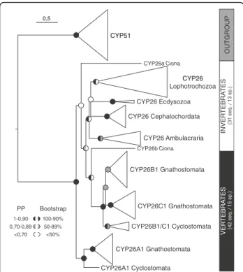
Previous analyses have reported three CYP26 genes in the Florida amphioxus, Branchiostoma floridae, and have suggested that they likely originated by lineage-specific duplication from a single ancestral CYP26 gene [29, 36]. Here, we have identified and cloned three CYP26 genes from the European amphioxus, Branchiostoma lanceola-tum. To further assess the phylogenetic relationships of the amphioxus CYP26 genes relative to each other and to other members of the CYP26 subfamily, thereby dis-tinguishing between orthologous and paralogous CYP26 genes, we first carried out phylogenetic analyses using as outgroup the CYP51 genes, which constitute the CYP subfamily that is most closely related to the CYP26 genes [45]. For this phylogenetic tree reconstruction, we used all CYP26 sequences from 15 vertebrates and 13 in-vertebrates, including three cephalochordate species (B. lanceolatum, B. floridae, and B. belcheri). Of note, while we successfully identified genes encoding CYP26 in the genomes of priapulids, brachiopods, mollusks, annelids, sea urchins, hemichordates, cephalochordates, ascidian tunicates, and vertebrates, we were unable to do so in those of nematodes and arthropods (as previously re-ported [2, 46]).
Within vertebrates, we found three CYP26 paralogs in both cyclostomes (CYP26A1, CYP26B1/C1a, and CYP26B1/C1b) and gnathostomes (CYP26A1, CYP26B1, and CYP26C1). Multiple CYP26 paralogs were also iden-tified in most invertebrates species studied (two in Capi-tella teleta, Ciona intestinalis, Lottia gigantea, Priapulus caudatus, and Saccoglossus kowalevskii, three in B. lan-ceolatum, B. floridae, B. belcheri, Crassostrea gigas, and Ptychodera flava, and four in Lingula anatina), with the notable exceptions of the cephalopod Octopus bimacu-loidesand the sea urchin Strongylocentrotus purpuratus,
each of which possesses only a single CYP26 gene (Additional file 1).
The results of the phylogenetic analysis (Fig. 1 and Additional file 2), obtained with both the Bayesian Infer-ence (BI) and the Maximum Likelihood (ML) methods, suggested an early phylogenetic separation of the vertebrate CYP26A1 sequences from the vertebrate CYP26B1/C1 sequences. Vertebrate CYP26A1 and CYP26B1/C1 thus formed two independent clades within the CYP26 subfamily, both of which being strongly supported: 0,91/96 (posterior probability/boot-strap percentage) for CYP26A1 and 1/99 for CYP26B1/ C1. Within these two vertebrate CYP26 clades, the cyclostome sequences consistently branched at the base: Lethenteron japonicum CYP26A1at the base of the ver-tebrate CYP26A1 (0,96/98) and CYP26B1/C1a and CYP26B1/C1bfrom L. japonicum and Petromyzon mari-nus at the base of the vertebrate CYP26B1/C1 (0,7/58). Within the vertebrate CYP26B1/C1 clade, the associ-ation of the gnathostome CYP26B1 sequences (0,73/58) was less robustly supported than that of the
gnathostome CYP26C1 sequences (1/95), which might be related to the presence of chondrichthyan-specific CYP26B1duplicates (CYP26B1 and CYP26B2 from Cal-lorhinchus milii and Leucoraja erinacea) disrupting the base of the CYP26B1 branch. Of note, while our analysis revealed the general presence of CYP26A1, CYP26B1, and CYP26C1 paralogs in chondrichthyans, we were un-able to identify a CYP26A1 gene in C. milii and a CYP26C1gene in L. erinacea. Altogether, these data sug-gest that the diversification of vertebrate CYP26 genes was a highly complex process, involving WGD, lineage-specific duplications as well as secondary gene losses.
Outside vertebrates, the CYP26 sequences from ecdy-sozoans, ambulacrarians, and cephalochordates always grouped together with very strong support values: the ecdysozoan P. caudatus (1/100), the ambulacrarians P. flavaand S. kowalevskii (1/99), and the cephalochordates B. lanceolatum, B. floridae and B. belcheri (1/99). Col-lectively, these results suggest that, within each of these invertebrate groups, the CYP26 gene complement origi-nated independently by linage-specific duplication. In contrast, the two sequences from the tunicate C. intesti-nalis did neither associate with each other, nor reliably with one of the major CYP26 clades in the tree. It is therefore impossible to comment on the nature and ori-gin of the CYP26 duplication in this animal. Similarly, the reconstruction of the evolutionary history of lopho-trochozoan CYP26 genes is complicated by the lack of phylogenetic resolution between the sequences from the five analyzed lophotrochozoan species, which formed an unresolved polytomy in our analysis. Nonetheless, there is evidence for lineage-specific duplications of CYP26 genes in the brachiopod L. anatina, which possesses four CYP26 paralogs that established two distinct clades in the tree, one very strongly (1/100) and one very weakly (0,8/–) supported. Furthermore, two of the three CYP26 sequences from the oyster C. gigas are grouped within in a single clade, but the support for this associ-ation is very weak (0,95/–). Future studies will thus have to address the processes underlying the evolution of lophotrochozoan CYP26 genes.
To gain further insights into the diversification of CYP26 genes in different animal lineages, we next con-ducted a phylogenetic dating analysis (Additional file 3). This survey indicated that the CYP26 genes of the ecdy-sozan P. caudatus were likely duplicated independently at the end of the Ordovician (about 446 Mya). Similarly, within the ambulacrarians, we found evidence for linage-specific duplications in hemichordates, which likely oc-curred during two different periods: the early Carbon-iferous for S. kowalevskii (about 342 Mya) and the middle Triassic for P. flava (about 240 Mya). Finally, in chordate lineages, CYP26 genes have also likely been du-plicated independently in cephalochordates, tunicates,
0,5 PP 1-0,90 0,70-0,89 <0,70 Bootstrap 100-90% <50% 50-89% CYP51 CYP26 Lophotrochozoa CYP26 Ecdysozoa CYP26 Cephalochordata CYP26 Ambulacraria CYP26B1 Gnathostomata CYP26C1 Gnathostomata CYP26B1/C1 Cyclostomata CYP26A1 Gnathostomata CYP26b Ciona CYP26a Ciona CYP26A1 Cyclostomata VER TEBRA TES (42 seq. / 15 sp.) INVER TEBRA TES (31 seq. / 13 sp.) OUTGROUP
Fig. 1 Phylogenetic analysis of the CYP26 subfamily. Diagrammatic summary of Bayesian Inference (BI) and Maximum Likelihood (ML) analyses of the phylogenetic relationships within the CYP26 subfamily, with CYP51 used as outgroup. The detailed tree is shown in Additional file 2 and sequence information is given in Additional file 1. Branch lengths are representative of the amino acid substitution rate and branch support is indicated at each major node as posterior probabilities (PP) for the BI tree and as bootstrap percentages for the ML tree. Furthermore, the total number of sequences (seq.) and species (sp.) is provided for the CYP26 subfamily
and vertebrates, with the three cephalochordate genes resulting from an initial duplication in the early Carbon-iferous (about 358 Mya), followed by a subsequent dupli-cation during the middle Permian (about 295 Mya). In contrast, while it is difficult to conclude on the timing of the duplication giving rise to the two CYP26 genes in the ascidian C. intestinalis, the evolution of the verte-brate CYP26 complement is complex and implies a series of duplications, including an ancient split into CYP26A1and CYP26B1/C1 and the subsequent diversi-fication of CYP26A1, CYP26B1, and CYP26C1 during the Cambrian period (about 548 to 510 Mya). It should be added that, consistent with the results of the phylo-genetic tree, the dating analysis did not yield reliable in-formation on the timing of the duplications of lophotrochozoan CYP26 genes.
CYP26-2 expression is suggestive of a function in developmental patterning of theB. lanceolatum embryo
In vertebrates, CYP26A1, CYP26B1, and CYP26C1 have very distinct expression patterns with several key do-mains being conserved between different species [6, 18]. Thus, we next assessed the temporal and spatial
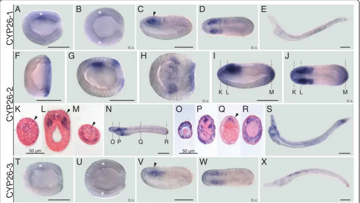
distribution of the three cephalochordate-specific dupli-cates in the amphioxus B. lanceolatum by in situ hybridization (ISH). The ISH results revealed that the expression profiles of CYP26-1 and CYP26-3 are gener-ally quite similar (Fig. 2a-e, t-x). For both genes, no sig-nal was detectable by ISH from fertilization through mid gastrulation. Expression of both CYP26-1 and CYP26-3 is first identifiable at late gastrula stages as a weak signal in the lateral anterior mesoderm (Fig. 2a, b, t and u). As development proceeds, this domain becomes associated with the most anterior somites at mid neurula stage (Fig. 2c, d, v and w). At this stage, 1 and CYP26-3 are also discreetly and transiently expressed in the an-terior central nervous system (CNS), at the level of the first somite (Fig. 2c and v). Expression of both genes re-mains very weak and chiefly associated with mesodermal tissues during subsequent developmental stages (Fig. 2e and x): while the CYP26-1 signal is most evident in cen-tral and posterior regions of the larva (Fig. 2e), CYP26-3 is mainly detectable in central and more anterior larval territories (Fig. 2x).
In contrast to CYP26-1 and CYP26-3, CYP26-2 has a much more complex developmental expression profile
d.v. d.v. d.v. d.v. d.v. d.v.
A
B
C
D
E
T
U
V
W
X
F
G
H I
K L M O P Q RJ
K
L
M
N
O
P
Q
R
S
K L M 50 µm 50 µmCYP26-1
CYP26-3
CYP26-2
*
*
*
*
*
*
Fig. 2 Developmental expression patterns of amphioxus CYP26 genes. Whole mount in situ hybridization experiments were carried out for CYP26-1 (a-e), CYP26-2 (f-s), and CYP26-3 (t-x). Amphioxus (Branchiostoma lanceolatum) embryos and larvae are shown as lateral views with anterior to the left and the dorsal side up, excepting for (b, d, h, j, u, w), which are dorsal views (d.v.) with anterior to the left, and for (k-m) and (o-r), which are cross-sections viewed from the front. Cross-sections in (k-m) are through the embryo shown in (i, j) at the levels indicated by the dashed lines and cross-sections in (o-r) are through the embryo shown in (n) at the levels indicated by the dashed lines. White asterisks in (a, b, t, u) highlight inconspicuous early expression domains of CYP26-1 and CYP26-3. Arrowheads in (c, v) indicate central nervous system expression and in (k-m, q) ectodermal signal. Developmental stages shown are: early gastrula (f), late gastrula (a, b, g, h, t, u), mid neurula (c, d, i-m, v, w), very early larva (n-r), early (60 h) larva (e, s, x). Scale bars are 100μm for the whole mounts and 50 μm for the cross-sections
during B. lanceolatum embryonic and early larval devel-opment. Expression of CYP26-2 is first detectable by ISH at the mid gastrula stage with the signal being local-ized globally around the blastopore (Fig. 2f ). The blastopore-associated signal subsequently weakens and, at the late gastrula stage, an additional expression do-main appears in a region corresponding to presumptive lateral mesoderm and anterior neuroectoderm (Fig. 2g and h). By the mid neurula stage, the mesodermal signal has been expanded into the two anterior-most somite pairs (Fig. 2i, j and l). At this stage, CYP26-2 is further still detectable in the anterior neuroectodem (Fig. 2i, j and l). Additionally, the gene is now expressed in the ectoderm, most conspicuously in the anterior and pos-terior tips of the embryo (Fig. 2i, j and k-m), as well as in the anterior- and posterior-most endoderm (Fig. 2i, j and m). At the mid neurula stage, the blastopore-associated signal becomes perceivable in the newly formed tail bud (Fig. 2f-j and m). In very early larvae, just before the opening of the larval mouth, expression of CYP26-2 is detectable anteriorly in all germ layers, i.e. the ectoderm, the mesoderm, the endoderm as well as in the CNS, with the signal being least noticeable in the endoderm (Fig. 2n-p). Furthermore, in both ectoderm and CNS, individual cells are labeled along the A-P body axis (Fig. 2n and q) and, at the posterior end of the em-bryo, the tail ectoderm strongly expresses CYP26-2 (Fig. 2n and r). In the amphioxus larva, the overall do-mains of CYP26-2 expression are maintained, with con-spicuous labeling anteriorly and posteriorly and a weaker signal in the center (Fig. 2s). In sum, while CYP26-1 and CYP26-3 expression is very discreet and chiefly limited to the mesoderm, that of CYP26-2 is de-tectable in all germ layers and dynamically changes in space and time throughout development.
CYP26 acts as a fine regulator of RA levels in the patterning of theB. lanceolatum larval tail fin
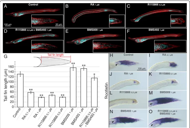
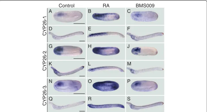
Disruption of CYP26 activity causes very severe defects during vertebrate development [18]. To determine the role of CYP26 enzymes in the amphioxus embryo, we subsequently disrupted endogenous RA degradation dur-ing amphioxus development by treatments with the CYP26-specific inhibitor R115866 [47, 48]. For compari-sons, the R115866 treatments were carried out in paral-lel to treatments with RA or with two different RAR antagonists (BMS009 and BMS493). The capacity of R115866 to inhibit endogenous RA degradation was verified by double treatments of R115866 and BMS493. The results obtained from these different pharmaco-logical treatments of B. lanceolatum embryos are in large agreement between each other and with previous studies that characterized the roles of RA signaling in amphioxus endoderm specification and pharyngeal
patterning [38–43]. Thus, while the downregulation of RA signaling activity by RAR antagonists (1μM of either BMS009 or BMS493) resulted in an enlarged pharynx and an expansion of pharyngeal structures (Fig. 3e and f ), the upregulation of endogenous RA signaling by 1 μM RA led to a shortening of the pharynx and the malformation of pharyngeal structures (such as the mouth and the gill slits) (Fig. 3b). Consistently, the local upregulation of RA signaling by 0,5μM R115866 yielded similar results (Fig. 3c), and co-treatments of 0,5 μM R115866 and 1μM BMS493 led to an attenuation of the severe phenotype induced by 0,5 μM R115866 alone with at least a partial recovery of pharynx formation and patterning (Fig. 3d).
Importantly, these pharmacology-based experiments revealed a previously undescribed role of RA signaling in developmental patterning of the amphioxus larval tail fin, a finding that is consistent with localized expression of CYP26-2 in the amphioxus tail fin ectoderm. At 60 h of development, amphioxus larvae are characterized by an ectodermal tail fin that is pointy in shape and on average 130,2μm long (Fig. 3a and g). When amphioxus embryos were treated with RA at the gastrula stage, the resulting tail fins of 60-h larvae are round and signifi-cantly shorter. These effects were observable with ex-ogenous treatments of both 0,1 μM and 1 μM RA (Fig. 3b and g). Similar results were obtained with R115866 treatments at 0,1 μM and 0,5 μM (Fig. 3c and g). Conversely, RAR antagonist treatments, with either BMS009 or BMS493, had the inverse effect: the tail fin becomes pointier in shape and is slightly elongated (Fig. 3e-g). Co-treatment of 0,5 μM R115866 with 1 μM BMS493 led to a partial rescue of the tail fin phenotype with an almost normal shape and a slight reduction of the overall length (Fig. 3d and g).
It has previously been shown that the amphioxus larval tail fin is composed of columnar epidermal cells that con-tain a large ciliary rootlet [49, 50] and that RA signaling promotes tail regression in late, pre-metamorphic B. flori-daelarvae by downregulating the gene encoding the main component of the ciliary rootlet: the protein Rootletin [51]. However, the regulation of Rootletin expression by RA signaling in the tail fin of early B. floridae larvae has not yet been reported. Given the effects on early tail fin formation we observed in B. lanceolatum in response to the alteration of endogenous RA signaling levels, we de-cided to investigate the patterns of Rootletin expression in 60-h B. lanceolatum larvae following the pharmacological treatment regimes detailed above. As previously described for late B. floridae larvae [51], Rootletin is expressed in the basal compartment of the columnar tail fin cells in 60-h B. lanceolatumlarvae (Fig. 3h). At this developmental stage, the gene is further detectable in a small number of lateral ectodermal cells (Fig. 3h).
While treatment with 0,1μM RA resulted in a marked reduction of Rootletin expression concomitant with an apical compaction of the columnar tail fin cells (Fig. 3i), 1 μM RA very strongly restricted the Rootletin expres-sion domain (Fig. 3j). The effect on tail fin development of either 0,1 μM or 0,5 μM of the CYP26 inhibitor R115866 was similar to that of exogenous RA and the R115866-treated larvae were thus generally characterized by a significant reduction of Rootletin expression (Fig. 3k and l). The RAR antagonists BMS009 and BMS493 led to an apical expansion of Rootletin expression as well as to an increase of the overall length of the tail fin (Fig. 3m and n). Furthermore, the RAR antagonist treatments in-duced Rootletin expression in additional lateral ectoder-mal cells not directly associated with the tail fin (Fig. 3m and n). Intriguingly, while co-treatments of 0,5 μM R115866 and 1μM BMS493 restored a shortened tail fin
with almost normal shape, expression of Rootletin remained expanded into the apical territory of the tail fin and detectable in lateral ectodermal cells (Fig. 3o). Altogether, these observations suggest that the formation of the ectodermal tail fin in B. lanceolatum is dependent of RA signaling, with CYP26-dependent degradation playing an important role in fine tuning of endogenous RA signaling levels to ensure proper tail fin outgrowth.
CYP26-1 and CYP26-3 are highly responsive to RA and specifically upregulated to avoid teratogenic effects of RA
In vertebrates, CYP26 enzymes are known to function locally to reduce RA levels hence protecting target tis-sues from RA teratogenesis. In this context, the regula-tion of CYP26 activity is mediated, at least in part, by RA signaling, which very dynamically up- or down-regulates CYP26 expression in a tissue-dependent
H
J
N
I
K
M
O
Rootletin A R l o r t n o C 0,1 µM RA 1 µM R115866 0,1 µM BMS009 1 µM BMS493 1 µM R115866 0,5 µM + BMS493 1 µM R115866 0,5 µML
A
Control RA 1 µMB
C
D
E
F
R115866 0,5 µM R115866 0,5 µM + BMS493 1 µM BMS009 1 µM BMS493 1 µM Control RA 1 µM R115866 0,5 µM R115866 0,1 µM BMS0 09 1 µM BMS49 3 1 µM R115866 0,5 µM + BMS493 1 µ MG
Tail fin length100 µm 50 µm
100 µm
RA 0,1 µM
T
ail fin length (
µm )160 140 120 100 80 60 40 20 0
Fig. 3 Effects of retinoic acid (RA) signaling alterations on the development of amphioxus. (a-f) Early amphioxus (Branchiostoma lanceolatum) larvae at 60 h of development with anterior to the left and the dorsal side up are shown as maximal projections of confocal microscopy scans displaying auto-fluorescence (in red) and Hoechst nuclear staining (in cyan). Magnifications of the tail region are boxed, with anterior to the left and the dorsal side up. Scale bars are 100μm for the larvae and 50 μm for the magnified tail fins. (g) Tail fin length measurements following pharmacological treatments. The length of the tail fin of B. lanceolatum larvae at 60 h of development was measured, as indicated in the sche-matics. The graph shows the tail fin lengths ± standard deviation, with n = 15, for the different treatment conditions indicated. Tail fin lengths that are significantly different from the DMSO control are indicated as“*”, for a cutoff value of p = 0,005, and as “**”, for a cutoff value of p = 0,001. (h-o) Rootletin expression in the tail fin of early B. lanceolatum larvae at 60 h of development, with anterior to the left and the dorsal side up. Arrow-heads in (h, m-o) indicate expression in lateral ectodermal cells. Pharmacological treatments are as indicated. Scale bars are 100μm
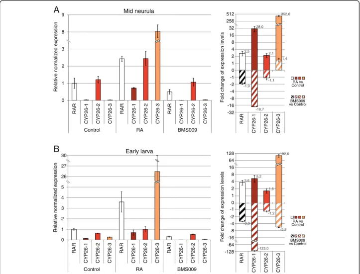
context [18]. To obtain insights into the regulation of CYP26genes by RA signaling in B. lanceolatum, we per-formed quantitative real time PCR (qPCR) analyses on amphioxus embryos at two developmental stages (mid neurula and early larva) that have been treated, at the early gastrula stage, with either 1μM RA or 1 μM of the RAR antagonist BMS009. The qPCR experiments assessed the changes in relative expression of the three B. lanceolatum CYP26genes normalized by RAR expres-sion in control embryos (Fig. 4a and b).
Through these analyses, we found that the expression levels of CYP26-1 and CYP26-3 in control embryos were very low when compared to CYP26-2 (about 45,0 to 50,0 times less in mid neurulae and 2,5 to 5,0 times less in early larvae) (Fig. 4a and b). RA treatments very significantly increased the expression of both CYP26-1 and CYP26-3 at the mid neurula stage (an average of 28
and 352,6 fold, respectively), while CYP26-2 levels in-creased merely by about 2,1 fold (Fig. 4a). In contrast, when embryos were treated with the RAR antagonist BMS009, CYP26-1 levels decreased by an average of 18,7 fold in mid neurulae, while the overall transcrip-tion of CYP26-2 and CYP26-3 remained relatively un-changed when compared to controls (a decrease of about 1,1 fold for CYP26-2 and an increase of about 1,4 fold for CYP26-3) (Fig. 4a). In early larvae, RA treat-ment also significantly increased the expression levels of CYP26-1 and CYP26-3 (by an average of 5,2 and 102,6 fold, respectively), while that of CYP26-2 in-creased only by about 1,6 fold (Fig. 4b). The RAR an-tagonist BMS009 had the opposite effect and strongly decreased CYP26-1 levels by about 123,0 fold and CYP26-3 levels by an average of 5,8 fold (Fig. 4b). In contrast, CYP26-2 expression dropped by only about
0 1 2 3 8 9 RAR
CYP26-1 CYP26-2 CYP26-3
RAR
CYP26-1 CYP26-2 CYP26-3
RAR
CYP26-1 CYP26-2 CYP26-3
Control RA BMS009
Relative normalized expression
A
B
Early larva 1 2 4 8 16 32 256 512 0 -1 -16 -32 -8 -4 -2 2,5 28,0 2,1 352,6 -1,9 -18,7 -1,1 1,4 RARCYP26-1 CYP26-2 CYP26-3 RA vs Control
BMS009 vs Control
Fold change of expression levels
Mid neurula
Relative normalized expression
0 1 2 3 4 5 26 27 30 RAR
CYP26-1 CYP26-2 CYP26-3
RAR
CYP26-1 CYP26-2 CYP26-3
RAR
CYP26-1 CYP26-2 CYP26-3
Control RA BMS009 0 -1 1 2 4 8 16 64 128 -2 -64 -128 -16 -8 -4 3,6 5,2 1,6 102,6 -3,3 -123,0 -1,2 -5,8 RAR
CYP26-1 CYP26-2 CYP26-3 RA vs Control
BMS009 vs Control
Fold change of expression levels
Fig. 4 Quantitative changes of amphioxus CYP26 expression in response to retinoic acid (RA) signaling alterations. Expression of CYP26-1, CYP26-2, and CYP26-3 in amphioxus (Branchiostoma lanceolatum) was assessed by quantitative real time PCR (qPCR) at two developmental stages (mid neurula at 20 h of development and early larva at 48 h of development). RAR expression was also established and used as a normalized positive control. (a) Relative normalized expression (left panel) and fold change of expression levels (right panel) at the mid neurula stage, using a Log2
1,2 fold (Fig. 4b). These results indicate that, although CYP26-2 is the amphioxus CYP26 gene most strongly and broadly expressed during embryogenesis, CYP26-1 and CYP26-3 are much more reactive to alterations of endogenous RA signaling levels.
Following this quantification, we next assessed, by ISH, the developmental expression profiles of amphioxus CYP26 genes upon RA signaling-altering pharmaco-logical treatments. Using the same developmental stages as for the qPCR analyses (mid neurulae and early larvae), we found that the upregulation of CYP26-1 expression upon RA treatment was not uniform (Fig. 5a-f ). At the mid neurula stage, CYP26-1 was most strongly induced in the anterior half of the embryo in all tissue layers (i.e. in CNS, ectoderm, mesoderm, and endoderm), all along the CNS as well as in the posterior tip of the embryo in all tissue layers excepting the mesoderm (Fig. 5a and b). In early larvae, the gene was upregulated in all tissue layers of the anterior half of the animal, but largely un-detectable posteriorly, except in a few cells in the ecto-derm (Fig. 5d and e). As expected from the qPCR experiments, expression of CYP26-1 in embryos and lar-vae treated with the RAR antagonist BMS009 was very inconspicuous, but, after an extended coloration step, was nonetheless detectable in mesodermal tissues (Fig. 5c and f ).
Treatment effects were similar, but not identical, for CYP26-2. At the mid neurula stage, RA strongly induced CYP26-2anteriorly and posteriorly in all tissue layers as well as all along the CNS and ectoderm (Fig. 5h). How-ever, following RA treatment, CYP26-2 expression was more conspicuous anteriorly and posteriorly in the em-bryo, when compared to that of CYP26-1. In contrast, in early larvae the effects of RA treatments were less pro-nounced for 2 than for 1. Chiefly, CYP26-2 expression expanded slightly in the mesoderm and ectoderm in the anterior half of the animal (Fig. 5k and l). Of note, the reduction of CYP26-2 expression in the anterior endoderm of RA-treated larvae was due to the absence of pharyngeal structures normally expressing the gene [43]. Treatments with the RAR antagonist BMS009 generally weakened the CYP26-2 signal, most noticeably in the CNS, the mesoderm, and both the an-terior and posan-terior ectoderm (Fig. 5j and m).
Finally, the expression of CYP26-3 was also very strongly expanded by RA at mid neurula and early larva stages, even more strongly than that of CYP26-1 with a general expansion of the staining observed throughout the embryo at both stages (Fig. 5n and o). Following RA treatment, expression of the gene was induced anteriorly and posteriorly in all tissue layers, most conspicuously in the center of the embryo in the CNS, ectoderm, and
BMS009
A
R
l
o
r
t
n
o
C
C
B
A
Q
R
S
N
O
P
K
L
M
G
H
J
D
E
F
CYP26-1
CYP26-3
CYP26-2
Fig. 5 Spatial changes of amphioxus CYP26 expression in response to retinoic acid (RA) signaling alterations. Whole mount in situ hybridization experiments were carried out for CYP26-1 (a-f), CYP26-2 (g-m), and CYP26-3 (n-s) on amphioxus (Branchiostoma lanceolatum) embryos treated at the late blastula stage with either DMSO (as control), all-trans RA (1μM) or RAR antagonist BMS009 (1 μM). The BMS009-treated embryo in (c) and larva in (f) were subjected to an extended coloration period. Mid neurula embryos at 20 h of development (a-c, g-j, n-p) and early larvae at 48 h of development (d-f, k-m, q-s) are shown as lateral views with anterior to the left and the dorsal side up. Scale bars are 100μm
mesoderm (Fig. 5n and o). By the early larval stage, CYP26-3remained generally upregulated throughout the animal (Fig. 5q and r), which contrasts with a more re-stricted distribution of CYP26-1 and CYP26-2 transcripts at this stage of development in response to RA treat-ments (Fig. 5e and l). In embryos and larvae treated with the RAR antagonist BMS009, the CYP26-3 signal was very weak and chiefly limited to mesodermal tissues (Fig. 5p and s). Altogether, these results show that treat-ments with RA and RAR antagonist induce similar, but not identical, tissue-specific responses of amphioxus CYP26-1, CYP26-2, and CYP26-3, thereby supporting the notion that each one of the three CYP26 genes is re-quired for a distinctive set of developmental functions.
RA regulatesCYP26-1, CYP26-2, and CYP26-3 directly and via evolutionary conserved functional RAREs present in theCYP26 cluster
To assess, whether the observed effects of RA and RAR antagonist on amphioxus CYP26 expression were medi-ated directly by RAR/RXR heterodimers, we first carried out pharmacological treatments in the presence of puro-mycin, a compound that efficiently blocks de novo pro-tein synthesis in developing amphioxus [44]. Amphioxus mid neurulae were hence treated with puromycin for 5 min prior to adding RA or the RAR antagonist BMS009. The embryos were then sampled 1 h later and, following RNA extraction, the expression levels of CYP26-1, CYP26-2, and CYP26-3 were determined by qPCR (Fig. 6). The results showed that, in the presence of puromycin, RA treatments significantly upregulated expression of both CYP26-1 and CYP26-3 (by about 5,8
fold and 5,9 fold, respectively), while that of CYP26-2 in-creased more modestly, by about 1,6 fold (Fig. 6). The RAR antagonist BMS009 had the inverse effect, reducing CYP26-1 and CYP26-3 levels by about 1,8 fold and 2,9 fold, respectively. In contrast, CYP26-2 expression stayed relatively stable, decreasing only by about 1,1 fold (Fig. 6). Altogether, these results are consistent with the effects of RA pharmacology on the expression of CYP26 genes described above. Furthermore, they indicate that the re-sponses to RA and RAR antagonist treatments of all three amphioxus CYP26 genes do not require de novo protein synthesis, thereby implying that they must be mediated directly by RAR/RXR protein heterodimers present in the target tissues.
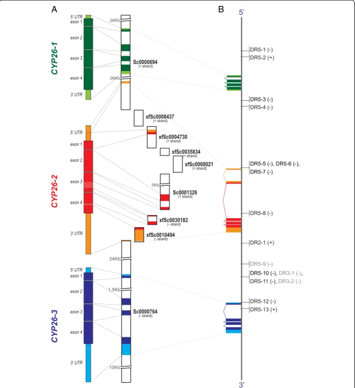
In order to obtain deeper insights into the mecha-nisms of the direct regulation of CYP26-1, CYP26-2, and CYP26-3 by RA signaling we then investigated the gen-omic environment of CYP26 genes in the three amphi-oxus species with available genomes: B. floridae [36, 37], B. belcheri [52], and B. lanceolatum [53]. Our analyses allowed us to reconstruct the entire genomic clusters for the three amphioxus CYP26 genes, previously described only for B. floridae [29] (Fig. 7). Although variable in size (about 85 Kbp in B. floridae, 56 Kbp in B. belcheri, and 79 Kbp in B. lanceolatum from the start codon of CYP26-1 to the stop codon of CYP26-3), the order of the three CYP26 genes within the continuous clusters is conserved between the three amphioxus species, lending further support to the notion that these genes originated by tandem duplications from a single ancestral CYP26 gene at the base of the cephalochordates. Making use of the complete sequences of the three amphioxus CYP26
0 1 2 3
CYP26-1 CYP26-2 CYP26-3 CY
P2 6-1 CYP 26-2 CYP26-3 CY P26-1 CYP26-2 CYP26-3 Control RA BMS009
Relative normalized expression
Mid neurula Puromycin-treated 0 -1 1 2 3 4 5 6 7 -2 -3 5,8 1,6 5,9 -1,8 -1,1 -2,9 CY P26-2 C YP 2 6-3 C YP 26 -1 RA vs Control BMS009 vs Control
Fold change of expression levels
Fig. 6 Dynamics of amphioxus CYP26 gene expression following protein synthesis inhibition and retinoid treatments. Expression of CYP26-1, CYP26-2, and CYP26-3 was assessed in amphioxus (Branchiostoma lanceolatum) by quantitative real time PCR (qPCR) at the mid neurula stage (at 20 h of development) following protein synthesis inhibition by puromycin treatment (200μg/ml) at 19 h of development and subsequent treatment with DMSO (as control), all-trans RA (1μM) or RAR antagonist BMS009 (1 μM) for 1 h. Relative normalized expression (left panel) and fold change of expression levels (right panel) relative to the controls are shown
A
B
Fig. 7 Organization and distribution of conserved retinoic acid response elements (RAREs) in the amphioxus CYP26 cluster. (a) Schematic representation of the experimentally verified amphioxus (Branchiostoma lanceolatum) CYP26-1, CYP26-2, and CYP26-3 gene sequences, including the 5′ and 3′ untranslated regions (UTRs) and the exons of the coding region, as well as of the corresponding scaffolds of the B. lanceolatum genome that cover the complete CYP26 cluster and thus also include intronic and intergenic regions. The names of the B. lanceolatum genome scaffolds are indicated, as is their orientation relative to the CYP26 cluster (+/− strand). (b) Distribution of conserved amphioxus RAREs in the B. lanceolatum CYP26 cluster. The orientation of each RARE is indicated relative to the CYP26 cluster (+/−). RAREs recognized and bound by the B. lanceolatum RAR/RXR heterodimer in vitro are indicated in black, RAREs that do not associate with the B. lanceolatum RAR/RXR heterodimer in vitro are shown in grey. The RARE sequences are given in Additional file 4 and the in vitro assays are shown in Fig. 8
clusters, we further searched systematically for con-served RAREs in the vicinity of the CYP26-1, CYP26-2, and CYP26-3 genes. This in silico survey included a gen-omic region encompassing 20 Kbp upstream of the CYP26-1 start codon and 20 Kbp downstream of CYP26-3 stop codon (Fig. 7). In total, we identified 38 candidate RAREs in B. floridae, 30 in B. belcheri, and 21 in B. lanceolatum. Of these, 16 RAREs were conserved between all three amphioxus species, both in terms of DR motif sequences and their relative position within the CYP26 cluster (Fig. 7b and Additional file 4).
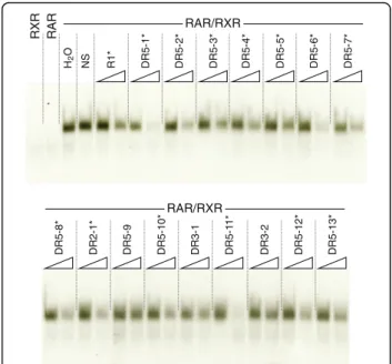
These conserved amphioxus CYP26 RAREs, i.e. 13 DR5, 1 DR2, and 2 DR3 elements, were subsequently used in EMSA analyses to investigate their capacity, in vitro, to interact with the B. lanceolatum RAR/RXR het-erodimer. Note that, even though DR3 elements are not considered as common RAREs, they were included in this survey. The results showed that the DR2 element and 12 of the 13 DR5 elements can be recognized and bound by the B. lanceolatum RAR/RXR heterodimer (Figs. 7b and 8), indicating that most of these RAREs might be functional in vivo. Furthermore, the consensus signature of the in vitro validated amphioxus DR5 ele-ments [(A/G)G(G/T)T(C/G)A NNN(A/G)(A/C/G) (A/ G)G(G/T)(T/A)CA] is very similar to the classical verte-brate DR5 signature [(A/G)G(G/T)TCA (N)5(A/G)G(G/
T)TCA] (Additional file 4), suggesting that, as in verte-brates [54, 55], amphioxus CYP26 genes are regulated directly by RA signaling via RAREs located in close vicinity of the open reading frames (ORFs).
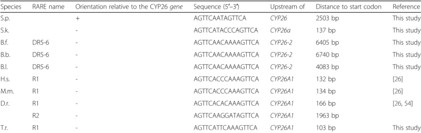
Furthermore, comparisons of the conserved and vali-dated amphioxus CYP26 RAREs with functional RAREs associated with vertebrate CYP26 genes revealed that the amphioxus DR5-6 sequence located upstream of CYP26-2 (Fig. 7b and Additional file 4) is identical in sequence to a DR5 RARE located upstream of vertebrate CYP26A1genes [AGTTCA (N)5AGTTCA] [26, 54]. To
verify, whether this DR5 RARE motif is a chordate innovation or an ancestral signature of bilaterian CYP26 genes, we screened the genomic regions surrounding CYP26 genes in the annelid C. teleta, the mollusk L. gigantea, the echinoderm S. purpuratus, the hemichord-ate S. kowalevskii, and three vertebrhemichord-ates (Takifugu rubripes, Mus musculus, and Homo sapiens). While no DR5 RARE with a similar motif could be identified in the annelid C. teleta and the mollusk L. gigantea, the conserved DR5 RARE was recovered in all three verte-brate species as well as in the hemichordate S. kowalevs-kii. Intriguingly, in the echinoderm S. purpuratus, instead of a DR5 RARE, we found a DR2 RARE with two similar DR sequences [AGTTCA] in an inverse orientation relative to the conserved DR5 RAREs in other species (Table 1). Of note, the conserved amphi-oxus DR5 RARE is located significantly further away
from the CYP26 start codon than in the other studied species. Although the biological significance of this find-ing still remains to be explored, these results nonetheless suggest that the direct control of CYP26 expression by RA signaling, mediated at least in part by a conserved DR5 RARE, is an ancestral feature that was most likely absent in the last common ancestor of all bilaterians and that was thus only subsequently acquired at the base of the deuterostomes.
Discussion
CYP26 genes and their evolutionary involvement in RA-dependent A-P patterning in chordates
In this study, we assessed the developmental expression patterns and the biological functions of the three CYP26 duplicates from the European amphioxus B. lanceolatum. Globally, our results suggest a sub-functionalization of these genes. Of the three genes, CYP26-1 and CYP26-3
DR5-9 DR5-10* DR5-8* DR2-1* DR3-1 DR5-1 1* DR3-2 DR5-12* DR5-13* RAR/RXR R1* DR5-1* DR5-2* DR5-3* DR5-4* DR5-5* DR5-6* DR5-7* NS HO RAR RXR RAR/RXR 2
Fig. 8 In vitro validation of putative retinoic acid response elements (RAREs) in the amphioxus CYP26 cluster. Using in vitro synthesized Branchiostoma lanceolatum RAR and RXR proteins, the binding of the cognate RAR/RXR heterodimer to putative RAREs identified in the amphioxus CYP26 cluster was assessed. The mouse CYP26A1 DR5 sequence (R1) [26] (Table 1) was used as32P-radiolabeled double-stranded probe. The RXR and RAR lanes respectively contained only in vitro synthesized RXR or RAR proteins, together with the radiolabeled probe. The H20 (water) lane contained radiolabeled probe as well as
RAR and RXR proteins. The NS (specific) lane contained a non-specific and unlabeled double-stranded DR element in 100-fold molar excess relative to the radiolabeled probe as well as RAR and RXR pro-tein. All other lanes contained one of the 16 putative amphioxus DR el-ements (for nomenclature explanations see Fig. 7b) at 10-fold (left lane) or 100-fold (right lane) molar excess, along with RAR and RXR protein and the radiolabeled mouse R1 element, to test the binding specificity of the putative amphioxus RAREs. A black asterisk indicates that a given DR element can be recognized by the B. lanceolatum RAR/RXR heterodi-mer, as it outcompetes binding to the mouse R1 element
are characterized by weak and disperse expression pat-terns, while CYP26-2 displays a dynamic, tissue-specific pattern along the A-P axis. Considering that A-P pattern-ing in early stages of amphioxus development is dependent on RA signaling [42, 44], the expression profile of CYP26-2, at very distinct positions along the A-P axis of the gastrula and early neurula, is suggestive of a func-tional role for this gene in this RA-dependent patterning
process. This notion is further supported by the results obtained by pharmacological inhibition of CYP26 action during gastrulation, which yielded embryos and larvae that resemble those treated with exogenous RA, displaying se-vere A-P patterning defects [43]. Thus, we propose that CYP26 enzymes assume two main functions during amphioxus development (Fig. 9), which are equivalent to those observed in vertebrates: (1) the mediation of
RA-Table 1 Conserved retinoic acid response elements (RAREs) with the characteristic sequence signature AGTTCA(N)5AGTTCA
identified in the vicinity of bilaterian CYP26 genes
Species RARE name Orientation relative to the CYP26 gene Sequence (5′–3′) Upstream of Distance to start codon Reference
S.p. + AGTTCAATAGTTCA CYP26 2503 bp This study
S.k. - AGTTCATACCCAGTTCA CYP26a 137 bp This study
B.f. DR5-6 - AGTTCAACAAAAGTTCA CYP26-2 6405 bp This study
B.b. DR5-6 - AGTTCAACAAAAGTTCA CYP26-2 6740 bp This study
B.l. DR5-6 - AGTTCAACAAAAGTTCA CYP26-2 4083 bp This study
H.s. R1 - AGTTCACCCAAAGTTCA CYP26A1 132 bp [26]
M.m. R1 - AGTTCACCCAAAGTTCA CYP26A1 134 bp [26]
D.r. R1 - AGTTCACACAAAGTTCA CYP26A1 166 bp [26,54]
R2 - AGTTCAAGGATAGTTCA CYP26A1 1963 bp
T.r. R1 - AGTTCATTCAAAGTTCA CYP26A1 103 bp This study
Species name abbreviations: B.b. Branchiostoma belcheri, B.f. Branchiostoma floridae, B.l. Branchiostoma lanceolatum, D.r. Danio rerio, H.s. Homo sapiens, M.m. Mus musculus, S.k. Saccoglossus kowalevskii, S.p. Strongylocentrotus purpuratus, T.r. Takifugu rubripes
CYP26-1 CYP26-2 CYP26-3
5’ 3’ RA: ) l a m r o n ( ) l a m r o n ( (high) RA: (high) Active RAR/RXR: Active RAR/RXR: Developmental patterning
Protection from teratogenic RA
levels
RAREs
RAREs RAREs
CYP26-1 CYP26-2 CYP26-3
5’ 3’
RAREs
Fig. 9 Model for regulation and function of CYP26 genes in amphioxus. The position of the functionally validated retinoic acid response elements (RAREs) within the amphioxus CYP26 cluster is indicated relative to the start codons of the three amphioxus CYP26 genes (CYP26-1, CYP26-2, and CYP26-3). The relative expression of each of the genes is indicated during normal development (normal levels of RA and active RAR/RXR receptor heterodimers) as well as during exposure to RA (high levels of RA and active RAR/RXR receptor heterodimers), hence highlighting the dominance of CYP26-2 during normal development and the importance of CYP26-1 and CYP26-3 for protection against RA teratogenesis
dependent developmental patterning and (2) the protec-tion against fluctuaprotec-tions of RA levels. Due to its conspicu-ous and tissue-specific expression during development, we hypothesize that the former is mainly assumed by CYP26-2, while the latter is dependent on the activity of CYP26-1 and CYP26-3.
During early development, the expression domains of CYP26-2along the A-P axis are inversely correlated with those of Hox1 and HNF3-1, both of which are direct tar-gets of RA signaling in amphioxus [44, 56]. The poster-ior expression limit of CYP26-2 in anterposter-ior tissues thus abuts the anterior border of both the Hox1 and HNF3-1 domains [44]. Concomitantly, the blastopore-associated expression of CYP26-2 seems to delineate the posterior limits of both Hox1 and HNF3-1 [44, 56]. Given that RAR expression is ubiquitous during gastrulation [38], the presence of CYP26-2 in defined domains along the A-P axis might thus be crucial for the creation of RA sinks to subdivide the developing embryo into zones with and without active RA signaling. This is reminis-cent of the conserved expression and function of CYP26A1 during vertebrate gastrulation: CYP26A1 is expressed in the anterior neural ectoderm and functions to establish A-P boundaries in the developing CNS [18]. It does so by establishing an anterior sink for RA pro-duced posteriorly in the paraxial mesoderm, hence creat-ing a RA gradient along the A-P axis of the neuroectoderm [57, 58].
By the mid neurula stage, the CYP26-2 signal is present in the entire anterior CNS, with a posterior limit at the boundary between the first and second somite. This CYP26 expression is hence limited to a region homologous to the vertebrate forebrain and midbrain [59], which is considerably different from what is ob-served in the vertebrate CNS, where CYP26 genes are expressed in the developing hindbrain and are funda-mental for its patterning along the A-P axis [18, 19]. In the amphioxus hindbrain homolog, RA signaling medi-ated by RAR/RXR has been shown to confer regional identity along the A-P axis by controlling the collinear expression of Hox genes [42]. However, our work sug-gests that, in contrast to the situation in vertebrates, this process does not involve the deployment of CYP26 genes in the amphioxus hindbrain homolog. Thus, while the early role for CYP26 in neural patterning was probably already present in the last common ancestor of amphi-oxus and vertebrates, a CYP26-dependent mechanism for subsequent hindbrain regionalization probably evolved in vertebrates, following the vertebrate-specific CYP26duplications.
Following neurulation, the three amphioxus CYP26 genes are expressed in anterior mesoderm, most notice-ably in the anterior-most somites. This is intriguing, as to date there is no convincing evidence for a
requirement of RA signaling in amphioxus mesoderm development [41, 42, 60] and the inhibition of CYP26 function by pharmacological treatments does not yield a mesodermal phenotype [43]. These observations raise an important question: why are CYP26 genes expressed in the anterior amphioxus mesoderm, as they are in verte-brate head mesoderm [25], if they are not required for A-P patterning of this tissue? We speculate that the con-spicuous expression of CYP26 in the anterior somites is an amphioxus innovation and is thus not comparable to the role of CYP26 in the vertebrate head mesoderm. In-stead, the CYP26 genes might function in the anterior amphioxus mesoderm to establish a buffer zone between the CNS dorsally and the pharynx ventrally, which re-quire distinct RA signaling cues for A-P regionalization [41, 42]. Although further analyses aiming, for example, at the visualization of in vivo RA signaling levels [58] will be required to test this hypothesis, it would none-theless be interesting to assess, whether this mechanism for separating two gradient-based A-P patterning sys-tems is being used more widely during development of small-sized embryos.
In the course of development, the blastopore-associated expression of amphioxus CYP26-2 becomes incorporated into the tail bud, a structure that, from the neurula stage on, plays a central role in posterior elong-ation of the embryo and larva and that is further charac-terized by the expression of tissue-specific marker genes, such as Wnt3 [61]. In vertebrates, both CYP26A1 and Wnt3aare also co-expressed in the developing tail bud, and CYP26A1 null mutants are characterized by a trun-cated tail and a spatial expression of Wnt3a that is ab-normally restricted towards the midline of the posterior neural plate [62]. These observations suggest that, in the vertebrate tail bud, CYP26A1 functions to keep RA levels low to create a permissive environment required for the posterior elongation of the embryo [16, 63]. In contrast, in amphioxus, treatments with CYP26 inhibi-tor, exogenous RA or the RAR antagonist BMS009 do not affect expression of Wnt3 in the tail bud [41]. Im-portantly, these pharmacological treatments, albeit af-fecting tail fin development, also do not impact posterior elongation of the developing embryo [39, 60]. Together, these findings support the notion that CYP26 function, and more generally the RA signaling system, is not required for tail bud-driven body extension in amphioxus and that this role for RA likely evolved in the vertebrate lineage.
CYP26 activity is required for the development of the amphioxus tail fin
Previous studies in the Florida amphioxus, B. floridae, have shown that the administration of excess RA during gastrulation results in a small anus [39] and that the
continuous administration of RA to B. floridae larvae leads to closure of the anus and regression of the tail fin [51]. Our data on the European amphioxus, B. lanceola-tum, are consistent with these previous findings and fur-ther suggest that RA signaling is required for proper tail fin outgrowth. Given that CYP26-2 is the only B. lanceo-latum CYP26gene expressed in the posterior ectoderm, it is very likely responsible for fine-tuning endogenous RA signaling levels in this territory.
The amphioxus tail fin is established by columnar epi-dermal cells that contain a large ciliary rootlet [49, 50] and it has previously been shown that RA promotes the downregulation of a major component of this structure, the protein Rootletin [51]. This downregulation in turn is likely responsible for the induction of tail fin regres-sion upon RA treatments in B. floridae larvae [51]. Our results of RA and CYP26 inhibitor treatments in B. lan-ceolatum are generally consistent with these previous observations, although we identified a major difference in the developmental timing of the involvement of RA signaling in tail fin outgrowth between the two amphi-oxus species. This fact is exemplified by the experimen-tal setups required to obtain tail fin phenotypes. While in B. floridae RA-dependent tail fin malformations can only be obtained by continuous treatment of pre-metamorphic larvae with 6–8 gill slits [51], changes in tail fin morphology can be induced much earlier during B. lanceolatumdevelopment by treating in the course of gastrulation. This suggests that, albeit required for tail fin outgrowth in both amphioxus species, the attenu-ation of high RA signaling levels in the posterior ecto-derm might take place earlier in B. lanceolatum than in B. floridae development. It is possible that this develop-mental difference is mediated by a delayed activation of the expression of CYP26-2 in B. floridae relative to B. lanceolatum, although additional work is required to support this claim. Altogether, these results indicate that amphioxus tail fin development requires low levels of RA signaling, which are maintained by CYP26 activity. When compared to the known functions of CYP26 in vertebrates, where cells expressing CYP26 enzymes are said to be effectively devoid of RA [63], amphioxus tail fin outgrowth might represent a rare example of a devel-opmental process, where CYP26 is deployed to merely reduce, and not to completely eliminate, RA signaling levels.
AmphioxusCYP26 genes are highly responsive RA signaling targets
In vertebrates, it has been established that, in RA-sensitive tissues, RA induces CYP26 to generate a nega-tive feedback loop that reduces the overall amount of available RA [18]. In mice, for example, expression of both CYP26A1 and CYP26B1 is upregulated upon RA
treatment [9] and, at least for CYP26A1, this regulation is directly mediated by RAR/RXR binding to RAREs in the vicinity of the CYP26A1 gene [26]. In contrast, the vertebrate CYP26C1 gene is likely not contributing to this RA-dependent negative feedback system, as its ex-pression is actually downregulated upon RA stimulation [9]. The data presented here suggest that expression of all three amphioxus CYP26 genes, which are lineage-specific duplicates and located in a single cluster in the genome, is positively regulated by RA signaling. Accord-ingly, the transcriptional regulation of B. lanceolatum CYP26-1, CYP26-2, and CYP26-3 is in all likelihood dir-ectly mediated by RAR/RXR heterodimers, given that one DR2-type sequence and 12 DR5-type elements within the CYP26 cluster are recognized and bound by the B. lanceolatum RAR/RXR heterodimer in vitro. Of these 13 in vitro validated RAREs, four DR5 elements are located around the CYP26-1 coding sequence, four DR5 and one DR2 are found in proximity of CYP26-2, and four DR5 elements are associated with the CYP26-3 gene. Although each of the three amphioxus CYP26 genes are likely to be directly regulated by RA signaling, the specific arrangement of RAREs relative to the CYP26 ORFs nonetheless suggests that the expression of each of the three genes is regulated independently by different sets of RAREs. Although this fundamental difference in the regulation of amphioxus and vertebrate CYP26 genes remains to be demonstrated mechanistically in vivo, it is nonetheless tempting to speculate that alterations in the regulation by RA have been key for redefining the func-tions of the different CYP26 genes following their dupli-cation, hence leading to their genomic retention by sub-functionalization.
At least some of the RAREs within the amphioxus CYP26cluster might serve as hubs for long-range regu-lation of gene expression from shared RAREs. Although in both amphioxus and vertebrates it has previously been shown that functional RAREs are generally located in the proximity of genes directly regulated by RA sig-naling [16, 64], long-range regulatory mechanisms of RA signaling have, for example, been implicated in the con-trol of the rostral expansion of posterior Hoxb genes during mouse CNS development [65]. The in vivo valid-ation of the RAREs located within the amphioxus CYP26 cluster will shed light on their contribution to short- and/or long-range transcriptional control mecha-nisms exerted by RA signaling. Our in silico analyses further revealed the presence, within the amphioxus CYP26 cluster, of two DR3 elements in close proximity of one of the in vitro validated DR5 elements. Although DR3 elements are generally not recognized by RAR/RXR heterodimers, they are bound by other nuclear receptors, such as vitamin D receptors [66]. The presence of DR3 elements within the amphioxus CYP26 cluster thus hints
at the possibility that additional nuclear receptors are in-volved in the regulation of amphioxus CYP26 genes.
Interestingly, the consensus sequence of the in vitro validated amphioxus DR5 RAREs is identical to the clas-sical vertebrate DR5 sequence consensus [(A/G)G(G/ T)TCA (N)5(A/G)G(G/T)TCA] [67], suggesting that the
DNA binding properties of amphioxus and vertebrate RAR/RXR heterodimers are highly conserved. Along these lines, we found a similar, conserved DR5 RARE in the regulatory region of a hemichordate CYP26 gene as well as an equivalent DR2 RARE close to the CYP26 gene of a sea urchin. Whether these elements are recog-nized by RAR/RXR heterodimers in these two species is currently unknown. Their presence is nevertheless highly suggestive of a biological function [13], which thus indi-cates that direct regulation of CYP26 genes by RA sig-naling is an ancestral feature that was already present in the last common ancestor of all deuterostomes. In con-trast, the lack of conserved RAREs in the regulatory re-gions of the CYP26 genes of lophotrochozoans, which generally encode RAR and RXR genes in their genomes [2], support the notion that the direct regulation of CYP26transcription by RA signaling is not an ancestral feature of bilaterian animals. This hypothesis is further strengthened by the fact that RARs of gastropod mol-lusks are unable to bind RA and hence to activate tran-scription in its presence [68, 69].
The evolutionary history ofCYP26 genes
In the animal kingdom, the basic molecular components of the RA machinery have previously been described in a wide variety of bilaterian animals, including both pro-tostomes and deuterostomes [2, 45]. Genes encoding the RA degrading enzyme CYP26 have, for example, been identified in the genomes of priapulids, brachiopods, mollusks, annelids, sea urchins, hemichordates, cephalo-chordates, ascidian tunicates, and vertebrates, but not in those of nematodes and arthropods, suggesting a sec-ondary loss of CYP26 subfamily genes in these two ani-mal lineages [2, 46]. We also found evidence for possible secondary losses of CYP26 genes in different vertebrates, including the lamprey P. marinus (lacking CYP26A1), the chimera C. milii (lacking CYP26A1), the skate L. eri-nacea (lacking CYP26C1), the coelacanth Latimeria chalumnae(lacking CYP26C1), and the opossum Mono-delphis domestica (lacking CYP26B1) (Additional files 1 and 2). Additional sequence information will be required to validate the absence of these genes from their respect-ive genomes and to assess, at which point in evolution the confirmed gene losses occurred.
Our data further indicate that the evolutionary diversifi-cation of the vertebrate CYP26 genes was a highly com-plex process. Given the presence of at least one CYP26A1 and two CYP26B1/C1 genes in lampreys, the last common
ancestor of cyclostomes and gnathostomes probably already possessed at least two CYP26 genes. Intriguingly, in the lamprey L. japonicum, CYP26A1 and one of the two CYP26B1/C1, CYP26B1/C1a, are physically linked in a tandem cluster in the genome (on scaffold 19) (Additional file 1), just like CYP26A1 and CYP26C1 in gnathostome genomes [70]. This tandem cluster thus very likely origi-nated before the cyclostome-gnathostome split, probably by a tandem duplication event predating the vertebrate-specific WGD. The two linked CYP26 genes were then duplicated during the first WGD, which took place in the basal vertebrate lineage before the cyclostome-gnathostome split [71–73]. The subsequent loss of one of the duplicated CYP26A1 genes yielded the CYP26 comple-ment of extant lampreys, such as L. japonicum: one CYP26A1 and two CYP26B1/C1 genes. In the gnathos-tome lineage, additional duplications [71–73] resulted in the diversification of CYP26B1 and CYP26C1 genes from the ancestral CYP26B1/C1. Although requiring additional scrutiny, this proposed scenario for vertebrate CYP26 di-versification suggests that, if two rounds of WGD oc-curred in the course of vertebrate evolution, the first likely took place before and the second after the cyclostome-gnathostome split [71, 72]. Alternatively, a single, ancient WGD might have occurred before the cyclostome-gnathostome split and was followed by independent seg-mental duplications in the cyclostome and gnathostome lineages [73]. Interestingly, in teleost fish, which under-went an additional round of WGD [74], there are also only three CYP26 genes, one member of each gnathostome paralogy group (CYP26A1, CYP26B1, and CYP26C1), and synteny analyses have revealed that the teleost-specific CYP26 duplicates have been non-functionalized in the course of evolution, in accordance with the DDC model [30, 31, 70].
Our phylogenetic analyses also provided evidence that CYP26 genes underwent multiple duplication events, not only in vertebrates, but also in invertebrates, such as cepha-lochordates, ascidian tunicates, hemichordates, mollusks, annelids, brachiopods, and priapulids. Together, these re-sults suggest that the ancestral bilaterian possessed a single CYP26gene that was subjected to independent duplication events in different animal lineages. By correlating the tim-ing of the lineage-specific duplications of invertebrate CYP26 genes with the geological timescale, we observed that the duplication events fall within three distinct time periods, i.e. the late Ordovician, the early Carboniferous, and the middle Permian/middle Triassic. Thus, the priapu-lid CYP26 duplication took place during the late Ordovi-cian, the first cephalochordate duplication and that identified in the hemichordate S. kowalevskii in the early Carboniferous, and the second cephalochordate duplication and that in the hemichordate P.flava in the middle Per-mian/middle Triassic. During the late Ordovician, a period