HAL Id: hal-02279611
https://hal-amu.archives-ouvertes.fr/hal-02279611
Submitted on 5 Sep 2019
HAL is a multi-disciplinary open access
archive for the deposit and dissemination of sci-entific research documents, whether they are pub-lished or not. The documents may come from teaching and research institutions in France or abroad, or from public or private research centers.
L’archive ouverte pluridisciplinaire HAL, est destinée au dépôt et à la diffusion de documents scientifiques de niveau recherche, publiés ou non, émanant des établissements d’enseignement et de recherche français ou étrangers, des laboratoires publics ou privés.
canettii by high-throughput biochemical profiling
Ahmed Loukil, Feriel Bouzid, Djaltou Aboubaker Osman, Michel Drancourt
To cite this version:
Ahmed Loukil, Feriel Bouzid, Djaltou Aboubaker Osman, Michel Drancourt. Decrypting the envi-ronmental sources of Mycobacterium canettii by high-throughput biochemical profiling. PLoS ONE, Public Library of Science, 2019, 14 (9), pp.e0222078. �10.1371/journal.pone.0222078�. �hal-02279611�
RESEARCH ARTICLE
Decrypting the environmental sources of
Mycobacterium canettii by high-throughput
biochemical profiling
Ahmed Loukil1☯, Fe´riel Bouzid1,2☯, Djaltou Aboubaker Osman3, Michel DrancourtID1,4*
1 Aix-Marseille Univ., IRD, MEPHI, IHU Me´diterrane´ e-Infection, Marseille, France, 2 Universite´ de Gafsa, Faculte´ des Sciences de Gafsa, Gafsa, Tunisia, 3 Institut de Recherche Me´dicinale, Centre d’Etudes et de Recherche de Djibouti (CERD), Djibouti, Re´publique de Djibouti, 4 IHU Me´diterrane´e Infection, Marseille, France
☯These authors contributed equally to this work.
Abstract
Mycobacterium canettii is a smooth bacillus related to the Mycobacterium tuberculosis com-plex. It causes lymph nodes and pulmonary tuberculosis in patients living in countries of the Horn of Africa, including Djibouti. The environmental reservoirs of M. canettii are still unknown. We aimed to further decrypt these potential reservoirs by using an original approach of High-Throughput Carbon and Azote Substrate Profiling. The Biolog Phenotype profiling was performed on six clinical strains of M. canettii and one M. tuberculosis strain was used as a positive control. The experiments were duplicated and authenticated by neg-ative controls. While M. tuberculosis metabolized 22/190 (11%) carbon substrates and 3/95 (3%) nitrogen substrates, 17/190 (8.9%) carbon substrates and three nitrogen substrates were metabolized by the six M. canettii strains forming the so-called corebiologome. A total at 16 carbon substrates and three nitrogen substrates were metabolized in common by M. tuberculosis and the six M. canettii strains. Moreover, at least one M. canettii strain metabo-lized 36/190 (19%) carbon substrates and 3/95 (3%) nitrogen substrates for a total of 39/285 (13%) substrates. Classifying these carbon and nitrogen substrates into ten potential envi-ronmental sources (plants, fruits and vegetables, bacteria, algae, fungi, nematodes, mol-lusks, mammals, insects and inanimate environment) significantly associated carbon and nitrogen substrates metabolized by at least one M. canettii strain with plants (p = 0.006). These results suggest that some plants endemic in the Horn of Africa may serve as ecologi-cal niches for M. canettii. Further ethnobotaniecologi-cal studies will indicate plant usages by loecologi-cal populations, then guiding field microbiological investigations in order to prove the definite environmental reservoirs of this opportunistic tuberculous pathogen.
Introduction
Mycobacterium canettii is a smooth tubercle bacillus isolated by G. Canetti in 1969 and belong-ing to theMycobacterium tuberculosis complex [1]. About one hundred cases only have been
a1111111111 a1111111111 a1111111111 a1111111111 a1111111111 OPEN ACCESS
Citation: Loukil A, Bouzid F, Osman DA, Drancourt M (2019) Decrypting the environmental sources of
Mycobacterium canettii by high-throughput
biochemical profiling. PLoS ONE 14(9): e0222078. https://doi.org/10.1371/journal.pone.0222078 Editor: Lanbo Shi, New Jersey Medical School, Rutgers University, UNITED STATES Received: June 11, 2019 Accepted: August 21, 2019 Published: September 3, 2019
Copyright:© 2019 Loukil et al. This is an open access article distributed under the terms of the Creative Commons Attribution License, which permits unrestricted use, distribution, and reproduction in any medium, provided the original author and source are credited.
Data Availability Statement: All relevant data are within the paper.
Funding: This work was funded by Agence Nationale de la Recherche (grant FEDER PA 0000320 PRIMI) to Prof Michel DRANCOURT. The funders had no role in study design, data collection and analysis, decision to publish, or preparation of the manuscript.
Competing interests: The authors have declared that no competing interests exist.
reported in the literature, and most of these patients have been infected in the Horn of Africa and particularly in Djibouti [2,3]. Recently, we confirmed thatM. canettii was still circulating in the Horn of Africa by isolatingM. canettii strains from patients presenting with pulmonary tuberculosis in Djibouti [4]. Initial molecular analysis of a large collection ofM. canettii iso-lates revealed a high genetic diversity with traces of intraspecies horizontal gene transfer [5]. Moreover, whole genome sequencing analysis of five representative isolates showed thatM. canettii was the M. tuberculosis complex species most closely related to the last common pro-genitor of this complex [6].
M. canettii is acknowledged as a M. tuberculosis complex species with an environmental res-ervoir, and this hypothesis is mainly based on the observation of the absence of human-to-human transmission [7]. However, the putative environmental sources of infection and reser-voirs remain completely elusive [3,7]. A mouse model using an oral route forM. canettii inoc-ulation has demonstrated the potential of ingestedM. canettii bacilli to relocate to lungs and other organs [8]. This experimental data supported that people could get infected with envi-ronmentalM. canettii by ingesting M. canettii-contaminated drinks or foodstuffs.
In a previous study, we used the Biolog Phenotype profiling (Biolog Inc., Hayward, CA) to characterize the metabolic profile of another environmental, non-tuberculous mycobacterium, Mycobacterium ulcerans [9]. In that study, we definedM. ulcerans sole-carbon-source utiliza-tion profile and we found this approach particularly conclusive for the quest of environmental sources ofM. ulcerans. Therefore, we embarked in using high-throughput carbon and nitrogen substrate profiling ofM. canettii to help in the quest of its potential sources and reservoirs.
Materials and methods
Bacterial strains
M. canettii CIP 140010059T,M. canettii DJ480, M. canettii DJ734, M. canettii DJ514, M. canet-tii DJ517 and M. canetcanet-tii DJ613 were isolated from clinical sources in Djibouti in 2016 [4] and M. tuberculosis Beijing family was used as a positive control [10]. They were grown at 37˚C in Middlebrook 7H10 agar medium (Becton Dickinson, Le Pont de Claix, France) supplemented with 10% oleic acid-albumin-dextrose-catalase (OADC) (Becton Dickinson) and 0.5% glycerol in a biosafety level 3 laboratory.
Biolog phenotype profiling
The Biolog Phenotype profiling (Biolog Inc.) was performed using PM1, PM2A and PM3B biolog 96-well microplates giving 190 carbon sources and 95 nitrogen sources, as previously described [9–11]. All experiments were conducted in duplicate using two independent plates during the same day for each strain. For each mycobacterial strain, colonies were removed from the Middlebrook 7H10 agar plate using a sterile swab already dipped in 0.1% Tween 80 solution, then rubbed and grinded against the wall of a dry glass tube. Colonies were starved in phosphate buffered saline (PBS) for 24 hours at 25˚C in order to minimize the background in the control well without substrate sources and to prevent color development in wells with sub-strate sources. A suspension made in the inoculating fluid IF-0a GN/GP (Biolog Inc.) was vig-orously vortexed in the presence of glass beads to declump mycobacterial cells to obtain a uniform suspension. The turbidity of the suspension was adjusted to 81% transmittance. The PM-additive solutions for each plate were made according to the manufacturer’s instructions (Table 1). A mixture solution specific for each plate was prepared by mixing 1.76 mL of the mycobacterial suspension with 22.24 mL of specific inoculating fluid (Table 2) for PM1 and PM2A plates. Then, 3.52 mL of cell suspension was mixed with 44.48 mL of inoculating fluid for the PM3B plate (Table 2). Finally, the PM plates were inoculated with 100μL of the specific
mixture solution and incubated in the OmniLog (Biolog, Inc.) system at 37˚C for seven days [10]. The principle of this system is based on a redox reaction from which mycobacteria reduce a tetrazolium dye to a purple color substrate. The variable level of purple color depends upon the conditions in each PM well and indicates the metabolically active state in some wells but not others. Using the OmniLog PM Software, the color change was read and recorded every 15 minutes giving a dye reduction value. The set of values obtained of each well provides kinetic information as Area Under the Curve (AUC). For each PM plate, non-inoculated wells con-taining only additive solutions were used as negative controls. Then, the reproducibility of the results was assessed using the OmniLog PM Software by superposing the AUCs obtained for the homologous wells of the duplicate. Then, the “mode average” calculated average AUC for the two homologous wells. The AUC of the negative controls were compared to values of posi-tive and negaposi-tive wells to set up a threshold. Wells were considered as having a moderately positive growth when the AUC of the well was equal to or lower than 1.25 times the AUC of the negative control and a highly positive growth when the AUC of the well was equal to or higher than 1.50 times the AUC value of the negative control [9].
Environmental sources of metabolized substrates
Based on our previously reported study [9], we sorted all the substrates present in the PM1, PM2 and PM3B plates into 10 categories of potential environmental sources including plants, fruits and vegetables, bacteria, algae, fungi, nematodes, mollusks, mammals, insects and inani-mate environment. We compared the proportion of each category to substrates metabolized by at least one of the sixM. canettii strains versus substrates not metabolized by any of the six M. canettii strains using Chi-square test (R-studio) (https://www.rstudio.com).
Results
Substrates metabolized by
M. canettii strains
All the experiments for the sixM. canettii strains and M. tuberculosis were duplicated. In addi-tion, negative wells (containing cell-free reagents) presented very low background and flat
Table 1. Composition and preparation of 12x PM additive solutions.
Ingredient 1x conc. 120x conc. Grams /100 mL PM 1 & 2 (mL) PM 3 (mL) MgCl2, 6H2O 2 mM 240 mM 4.88 10 10 CaCl2, 2H2O 1 mM 120 mM 1.76 Tween 80 0.01% 1.2% 1.2 10 10 D-glucose 5 mM 600 mM 10.8 - 10 Sterile water 80 70 Total 100 100 https://doi.org/10.1371/journal.pone.0222078.t001
Table 2. Recipe for 1x PM inoculating fluids from stock solutions for two plates of PM1, PM2 or PM3 (100μl / well). PM stock solution PM (mL) IF-0a GN/GP (1.2x) 20 PM additive (12x) 2 Dye mix G (100x) 0.24 Cell suspension (13.64x) 1.76 Total 24 https://doi.org/10.1371/journal.pone.0222078.t002
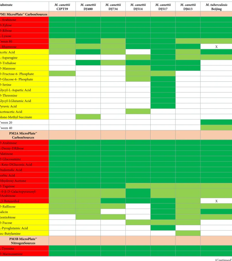
lines in all the PMs plates consistent with the absence of metabolic activity. For the positive controlM. tuberculosis, 22/190 (11%) carbon substrates and 3/95 (3%) nitrogen substrates were metabolized (Table 3). ForM. canettii, 17 carbon substrates and three nitrogen substrates were metabolized by the sixM. canettii strains under investigation, forming the so-called core-biologome (Table 3). In addition, 36/190 (19%) carbon substrates and 3/95 (3%) nitrogen sub-strates for a total of 39/285 (13%) subsub-strates here investigated were metabolized by at least one of the sixM. canettii strains.
ComparingM. canettii with M. tuberculosis, 16 carbon substrates and three nitrogen sub-strates were metabolized in common byM. tuberculosis and the six M.canettii strains (Table 3). Further, the sixteen carbon substrates metabolized by at least 1/6M. canettii strains and not metabolized byM. tuberculosis included L-rhamnose, trehalose, mannose, D-fructose-6-phosphate, 2,3-butanediol, D-fucose, D-glucose-6-phosphate, D-serine, glycyl-L-aspartic acid, D-threonine, glycyl-l-glutamic acid, pyruvic acid, acetoacetic acid, mono methyl-succinate, L-pyroglutamic acid and sec-butylamine. No difference was observed for nitrogen substrates. Among these 16 substrates, 2,3-butanediol and L-rhamnose were metabolized by the sixM. canettii strains and not by M. tuberculosis.
Environmental sources of metabolized substrates
All the 190 carbon substrates and 95 nitrogen substrates included in the PM1, PM2 and PM3B plates were classified into ten potential environmental sources. In a first step, comparison of these ten categories to carbon substrates metabolized by at least one of the six testedM. canettii strains versus non-metabolized carbon substrates yielded a significant association betweenM. canettii metabolized carbon sources and plants (p-value = 0.015). Further associations did not reach the 0.05 level of significance, i.e. algae (p = 0.062), mollusks (p = 0.072), insects
(p = 0.096), inanimate environment (p = 0.328), fruits and vegetables (p = 0.399), bacteria (p = 0.577), fungi (p = 0.651), mammals (p = 0.7) and nematodes (p = 0.754). In a second step, comparing categories of potential sources to the carbon and nitrogen substrates metabolized or not by at least oneM. canettii strain confirmed a significant association with plants (p = 0.006). While the associations with insects (p = 0.051) were marginally significant, the associations with algae (p = 0.072), fungi (p = 0.1525), mollusks (p = 0.355), inanimate envi-ronment (p = 0.4165), nematodes (p = 0.5072), fruits and vegetables (p = 0.5176), bacteria (p = 0.783) and mammals (p = 0.8668) were not statistically significant.
Discussion
We are reporting on the high throughput biochemical profiling ofM. canettii using the Omni-Log1 system (Biolog Inc.). This approach has already been reported for some mycobacteria of medical interest, includingM. tuberculosis, Mycobacterium bovis and M. bovis BCG [10], as well as forM. ulcerans and Mycobacterium marinum [9]. Also, the environmental Mycobacte-rium smegmatis has been investigated by using phenotypic PM1, PM3 and PM5 plates to test carbon and nitrogen sources and nutrient supplements [12]. In our study and previously reported studies, data were validated by the negativity of negative controls and the reproduc-ibility of results over two wells per substrate. The protocol used in our study was adapted from the protocol used for studyingM. smegmatis [12] whereas extensive modifications in preparing the inoculum for PM plates have been made over a previously reported protocol for studying M. tuberculosis [10]. In this report, the authors chose to investigate aM. tuberculosis Beijing clinical isolate instead of theM. tuberculosis H37Rv investigated by Biolog in a previously reported work [10]. Indeed,M. tuberculosis H37Rv is now cultured for 115 years [13] for approximately more than 50 passages in the authors’ laboratory; with a demonstrated genomic
Table 3. Carbone and nitrogen substrates metabolized by sixM. canettii strains compared with one M. tuberculosis strain Beijing on Biolog PM1, PM2 and PM3B plates. Substrate M. canettii CIPT59 M. canettii DJ480 M. canettii DJ734 M. canettii DJ514 M. canettii DJ517 M. canettii DJ613 M. tuberculosis Beijing PM1 MicroPlate™ CarbonSources L-Arabinose D-Xylose D-Ribose L-Lyxose Tween 80 L-Rhamnose X Acetic Acid L-Asparagine D-Trehalose D-Mannose D-Fructose-6- Phosphate D-Glucose-6- Phosphate D-Serine Glycyl-L-Aspartic Acid D-Threonine Glycyl-LGlutamic Acid Pyruvic Acid Acetoacetic Acid Mono Methyl Succinate Tween 20 Tween 40 PM2A MicroPlate™ CarbonSources D-Arabinose 2-Deoxy-DRibose Palatinose D-Glucosamine 5-Keto-DGluconic Acid Oxalomalic Acid Sorbic Acid Dihydroxy Acetone D-Tagatose 3-0-β-D-Galactopyranosyl-DArabinose 2,3-Butanediol X D-Raffinose Salicin Gentiobiose D-Fucose L-Pyroglutamic Acid Sec-Butylamine PM3B MicroPlate™ NitrogenSources L-Tyrosine D-Mannosamine (Continued )
derive [14] that renders these pluralM. tuberculosis H37Rv strains unrepresentative of the M. tuberculosis strains causing modern tuberculosis. Therefore, the results we obtained with M. tuberculosis used as a positive control must be tempered due to differences in methodology [10].
We observed that theM. canettii corebiologome (substrates metabolized by all the six M. canettii strains under investigation) was limited to only 17/190 (9%) carbon substrates and 3/ 95 (3%) nitrogen substrates. Searching for the potential sources common to these substrates highlighted plants as potential sources, but not fruits or vegetables. Our interpretation of these observations is thatM. canettii may reside in close association with non-fruit bearing plants. This interpretation is reinforced by evidence for a potential relationship betweenM. canettii and cellulose, the major constituent of plant cell walls. Indeed, an unexpected conservation of cellulase-encoding genes has been found in the genome ofM. canettii including the Cel6 gene, which encodes a fully active cellulase [15].
The unique geographical specificity ofM. canettii strains, which, apart from the three initial (lost) strains reported by G. Canetti himself, have all been isolated from patients exposed to one of the regions of the Horn of Africa [3], led us to the hypothesis thatM. canettii lives in close association with one or several plants specifically endemic to this part of the world (Table 4). Moreover, populations must be in close contact with these potential plant reservoirs, either by using these plants for tissue preparation, medicinal and recreational activities or feed-ing. The fact thatM. canettii is temperature-sensitive and specifically provokes lymph node infection along the digestive tract of patients [3] furthermore narrows the possibility to alimen-tary contacts with uncooked or poorly cooked plants [16]. Moreover, in this study, no signifi-cant association was found betweenM. canettii and sources related to aquatic environment (algae and mollusks), confirming that the potential plant reservoirs ofM. canettii should be located in the arid environment in the Horn of Africa.
In this short list of plants,Catha edulis (known as khat) is fulfilling all these criteria as potential source forM. canettii. Khat, cultivated in Ethiopia and Eritrae, is then imported and circulating in Djibouti, Yemen and Somalia. Khat is specifically consumed as a stimulant or as a medicinal plant by local populations including children younger than 10 years in the Horn of Africa [17]. Interestingly, rhamnose, that we found to be one of the two substrates specifi-cally metabolized byM. canettii but not by closely related M. tuberculosis, was detected in the leaves of khat [18].
Conclusions
The original approach combining experimental study with a keyword study has been effi-ciently used to detect some ecological niches ofM. ulcerans, a nontuberculous environmental mycobacterium [9]. Applying the very same methodology toM. canettii was also efficient in the present study, pointing towards some plants as potential ecological niches for this environ-mental tuberculous mycobacterium. Capitalizing on this new knowledge, further
Table 3. (Continued) Substrate M. canettii CIPT59 M. canettii DJ480 M. canettii DJ734 M. canettii DJ514 M. canettii DJ517 M. canettii DJ613 M. tuberculosis Beijing Alloxan
Substrates metabolized by all testedM. canettii strains forming corbiologome are shaded red. Substrates metabolized by at least one M. canettii strains are shaded yellow.
Highly positive wells are shaded dark green. Moderately positive wells are shaded light green. X, substrates which are not metabolized byM. tuberculosis and
metabolized by all testedM. canettii.
Table 4. List of plants endemic in countries of the Horn of Africa.
Country Endemic plants Use by local inhabitants References
Republic of Djibouti
Teucrium spicatum [19]
Phagnalon lavranosii [20]
Cynoglossopsis somalensis [21]
Matthiola puntensis Flowering plants [21]
Livistona carinensis Leaves are used to cover theroofs [22,24]
Euphorbia godana [24]
Euphorbia amicorum [25]
Aloe ericahenriettae [24,26]
Aloe
mcloughliniiChristian
Medecine: Laxative, soaking, crushed leaves or branches or stems in water for 12 h and the water is taken orally [21,27] Caralluma mireillae [28] Polygala goudahensis [21] Taverniera oligantha [21,29] Volutaria djiboutensis [30] Aloe djiboutiensis [26] Echidnopsis hirsuta [31,32]
Aponogeton nudifloris Food: Tuber consumption [30]
Kalanchoe elliptica [30]
Farsetia longistyla [33]
Hildebrandtia somalensis [34–36] Ethiopia Cordeauxia edulis Food: The seeds are eaten dried, boiled, roasted or raw. Drinks: People made a tea out of the leaves
Medicine:C. edulis can regulate gastric secretion. A study showed that the consumption of the plant
enhances the production of erythrocytes and is therefore used as a remedy for anemia.
[29]
Aloe mcloughliniiChristian
[37]
Catha edulis Euphoric plant, recreational use [38]
Echidnopsis hirsuta [32,35]
Rhus glutinosa ssp abyssinica
Species subject to strong animal pressure particularly when annual grasses disappear from pastures [19]
Buxus hildebrandtii Used for the construction of traditional huts [19,21]
Farsetia longistyla [33]
Hildebrandtia somalensis The twigs are burned and the fumes are used by Borana women to purify and perfume their bodies and their clothes
[34–36]
Commiphora guidottii Source of sweet myrrh, use of the fragrant resin [39,40]
Echinops kebericho
Mesfin
Aromatic plant, medicinal plant with antimicrobial activities, treats fever, headache, stomachache, and cough
[41]
Thymus schimperi Aromatic plant, medicinal plant with antimicrobial activities [41]
Thymus serrulatus Aromatic plant [41]
Lippia adoensis Aromatic plant [41]
Aframomum corrorima Aromatic plant [41]
Somalia Cyclamen somalense Use of the fragrant resin [35]
Cordeauxia edulis Food: The seeds are eaten dried, boiled, roasted or raw. Drinks: People made a tea out of the leaves. Medicine:C. edulis can regulate gastric secretion. A study showed that the consumption of the plant
enhances the production of erythrocytes and is therefore used as a remedy for anemia.
[29]
Livistona carinensis Leaves are used to produce mats and baskets [22,23]
Euphorbia amicorum [25]
Dirachma somalensis [25]
Commiphora guidottii Source of sweet myrrh, use of the fragrant resin [39,40]
Echidnopsis hirsuta [32,35]
(Continued )
ethnobotanical studies will indicate plant usages by local populations, guiding field microbio-logical investigations in order to disclose one or several sources of infection for populations. In particular, plants could be screened by using PCR-based methods for the detection of DNA sequences specific forM. canettii, selecting plants to be further cultured. If these investigations confirm any association betweenM. canettii and khat or other plants, infection with M. canet-tii should be included in the very short list of phytonoses which are human infections trans-mitted by plants [42].
Acknowledgments
The authors acknowledge the technical contribution of Muriel Militello, the contribution of Olga Cusack for manuscript editing and Magdalen Lardière for English reviewing.
Author Contributions
Data curation: Ahmed Loukil, Fe´riel Bouzid. Formal analysis: Ahmed Loukil.
Writing – original draft: Ahmed Loukil, Fe´riel Bouzid, Djaltou Aboubaker Osman, Michel
Drancourt.
References
1. Van Soolingen D, Hoogenboezem T, De Haas PEW, Hermans PWM, Koedam MA, Teppema KS, et al. A Novel Pathogenic Taxon of the Mycobacterium tuberculosis Complex, Canetti: Characterization of an Exceptional Isolate from Africa. Int J Syst Bacteriol. 1997; 47: 1236–1245.https://doi.org/10.1099/ 00207713-47-4-1236PMID:9336935
2. Boyer-Cazajous G, Martinaud C, De´han C, Hassan MO, Gaas Y, Chenilleau-Vidal M-C, et al. High prev-alence of multidrug resistant tuberculosis in Djibouti: a retrospective study. J Infect Dev Ctries. 2014; 8. https://doi.org/10.3855/jidc.3837PMID:24518635
3. Aboubaker Osman D, Bouzid F, Canaan S, Drancourt M. Smooth Tubercle Bacilli: Neglected Opportu-nistic Tropical Pathogens. Front Public Health. 2016; 3.https://doi.org/10.3389/fpubh.2015.00283 PMID:26793699
4. Bouzid F, Astier H, Osman DA, Javelle E, Hassan MO, Simon F, et al. Extended spectrum of antibiotic susceptibility for tuberculosis, Djibouti. Int J Antimicrob Agents. 2018; 51: 235–238.https://doi.org/10. 1016/j.ijantimicag.2017.07.007PMID:28711677
5. Fabre M, Koeck J-L, Le Fleche P, Simon F, Herve V, Vergnaud G, et al. High Genetic Diversity Revealed by Variable-Number Tandem Repeat Genotyping and Analysis of hsp65 Gene Polymorphism in a Large Collection of “Mycobacterium canettii” Strains Indicates that the M. tuberculosis Complex Is a Recently Emerged Clone of “M. canettii.” J Clin Microbiol. 2004; 42: 3248–3255.https://doi.org/10. 1128/JCM.42.7.3248-3255.2004PMID:15243089
6. Supply P, Marceau M, Mangenot S, Roche D, Rouanet C, Khanna V, et al. Genomic analysis of smooth tubercle bacilli provides insights into ancestry and pathoadaptation of Mycobacterium tuberculosis. Nat Genet. 2013; 45: 172–179.https://doi.org/10.1038/ng.2517PMID:23291586
Table 4. (Continued)
Country Endemic plants Use by local inhabitants References
Buxus hildebrandtii Used for the construction of traditional huts [19,21]
Kalanchoe elliptica [19]
Farsetia longistyla [33]
Hildebrandtia somalensis [34–36] The names of plants common to the countries of the Horn of Africa are shaded green.
7. Koeck J-L, Fabre M, Simon F, Daffe´ M, Garnotel e´., Matan AB, et al. Clinical characteristics of the smooth tubercle bacilli ‘Mycobacterium canettii’ infection suggest the existence of an environmental res-ervoir. Clin Microbiol Infect. 2011; 17: 1013–1019.https://doi.org/10.1111/j.1469-0691.2010.03347.x PMID:20831613
8. Bouzid F, Bre´geon F, Lepidi H, Donoghue HD, Minnikin DE, Drancourt M. Ready Experimental Translo-cation of Mycobacterium canettii Yields Pulmonary Tuberculosis. Ehrt S, editor. Infect Immun. 2017; 85. https://doi.org/10.1128/IAI.00507-17PMID:28923895
9. Zingue D, Bouam A, Militello M, Drancourt M. High-Throughput Carbon Substrate Profiling of
Mycobac-terium ulcerans Suggests Potential Environmental Reservoirs. Small PLC, editor. PLoS Negl Trop Dis.
2017; 11: e0005303.https://doi.org/10.1371/journal.pntd.0005303PMID:28095422
10. Khatri B, Fielder M, Jones G, Newell W, Abu-Oun M, Wheeler PR. High Throughput Phenotypic Analy-sis of Mycobacterium tuberculoAnaly-sis and Mycobacterium bovis Strains’ Metabolism Using Biolog Pheno-type Microarrays. Manganelli R, editor. PLoS ONE. 2013; 8: e52673.https://doi.org/10.1371/journal. pone.0052673PMID:23326347
11. Bochner BR. Global phenotypic characterization of bacteria. FEMS Microbiol Rev. 2009; 33: 191–205. https://doi.org/10.1111/j.1574-6976.2008.00149.xPMID:19054113
12. Baloni P, Padiadpu J, Singh A, Gupta KR, Chandra N. Identifying feasible metabolic routes in
Mycobac-terium smegmatis and possible alterations under diverse nutrient conditions. BMC Microbiol. 2014;14.
https://doi.org/10.1186/1471-2180-14-14PMID:24467879
13. Kubica GP, Kim TH, Dunbar FP. Designation of strain H37Rv as the neotype of Mycobacterium
tuber-culosis. Int J Syst Bacteriol. 1972; 22: 99–106.
14. Ioerger TR, Feng Y, Ganesula K, Chen X, Dobos KM, Fortune S, et al. Variation among Genome Sequences of H37Rv Strains of Mycobacterium tuberculosis from Multiple Laboratories. J Bacteriol. 2010; 192: 3645–3653.https://doi.org/10.1128/JB.00166-10PMID:20472797
15. Mba Medie F, Ben Salah I, Drancourt M, Henrissat B. Paradoxical conservation of a set of three cellu-lose-targeting genes in Mycobacterium tuberculosis complex organisms. Microbiology. 2010; 156: 1468–1475.https://doi.org/10.1099/mic.0.037812-0PMID:20150238
16. Aboubaker Osman D, Garnotel E, Drancourt M. Dry-heat inactivation of “Mycobacterium canettii.” BMC Res Notes. 2017;10.https://doi.org/10.1186/s13104-016-2357-z
17. Kandela P. SANA’A Women’s rights, a tourist boom, and the power of khat in Yemen. The Lancet. 2000; 355: 1437.https://doi.org/10.1016/S0140-6736(05)74640-7
18. Gelle´rt M, Szendrei K, Reisch J. Dihydromyricetin 3-O-rhamnoside from leaves of Catha edulis. Phyto-chemistry. 1981; 20: 1759–1760.https://doi.org/10.1016/S0031-9422(00)98579-0
19. Gouvernement de Djibouti. Monographie nationale de la diversite´ biologique de Djibouti [Internet]. 2000 [cited 19 May 2019]. Available from:http://www.environnement.dj/wp-content/uploads/2018/03/ Monographie-Nationale-de-la-Diversit%C3%A9-Biologique.pdf
20. Roskov Y, Kunze T, Paglinawan L, Orrell T, Nicolson D, Culham A, et al. Species 2000 & ITIS Cata-logue of Life, 2013 Annual Checklist [Internet]. Apr 2013 [cited 05 May 2019]. Available from:http:// www.catalogueoflife.org/annual-checklist/2013/
21. Audru J, Ce´sar J, Lebrun J-P. Les plantes vasculaires de la Re´ publique de Djibouti. Flore illustre´e [Inter-net]. CIRAD-EMVT; 1994. Available from:http://agritrop.cirad.fr/312163/
22. Dowe JL. A Taxonomic Account of Livistona R.Br. (Arecaceae). 2009; 160.
23. Govaerts R, Dransfield J. World checklist of palms. Kew: Royal Botanic Gardens; 2005.
24. Govaerts R. World checklist of selected plant families published update. Kew: Royal Botanic Gardens; 2011.
25. Govaerts R, Frodin DG, Radcliffe-Smith A, Carter S. World checklist and bibliography of Euphorbiaceae (with Pandaceae) [Internet]. Royal Botanic Gardens, Kew; 2000. Available from:http://agris.fao.org/ agris-search/search.do?recordID=US201300050879
26. Mccoy T. Aloe djiboutiensis and Aloe ericahenriettae two new species from Djibouti: And the mystery of
Aloe eumassawana’s natural habitat solved. Cactus Succul J. 2007; 79: 269–273.https://doi.org/10. 2985/0007-9367(2007)79[269:ADAAET]2.0.CO;2
27. Hassan-Abdallah A, Merito A, Hassan S, Aboubaker D, Djama M, Asfaw Z, et al. Medicinal plants and their uses by the people in the Region of Randa, Djibouti. J Ethnopharmacol. 2013; 148: 701–713. https://doi.org/10.1016/j.jep.2013.05.033PMID:23707214
28. Govaerts R. World checklist of seed plants 3,2,a 3,2,a. Antwerp: MIM bvba; 1999.
29. Lock JM. Legumes of Africa: a check-list. Kew: Royal Bot. Gardens; 1989.
30. Roskov Y, Abucay L, Orrell T, Nicolson D, Bailly N, Kirk PM, et al. Species 2000 & ITIS Catalogue of Life. 2017, Annual Checklist. Digital resource atwww.catalogueoflife.org/annual-checklist/2017. Spe-cies 2000: Naturalis, Leiden, the Netherlands. ISSN 2405-884X. 2018.
31. Musselman LJ. Flora of Somalia. Econ Bot. 2007; 61: 200–200.https://doi.org/10.1663/0013-0001 (2007)61[200a:FOSV]2.0.CO;2
32. Hedberg I, editor. Apiaceae to Dipsacaceae. Addis Ababa: National Herbarium [u.a.]; 2003.
33. Edwards S, Tadesse M, Demissew S, Hedberg I. Magnoliaceae to flacourtiaceae. Flora Ethiop Eritrea. 2000; 2: 532.
34. Hedberg I, Kelbessa E, Edwards S, Demissew S, Persson E. Flora of Ethiopia and Eritrea, Volume 5:
Gentianaceae to Cyclocheilaceae. 2006.
35. Thulin M. Flora of Somalia: Volume 3. Royal Botanic Gardens; 2006.
36. Demissew S. The genus Hildebrandtia (Convolvulaceae) from mainland Africa and Arabia. Kew Bull. 1996; 525–541.
37. Demissew S, Nordal I. Aloes and other Lilies of Ethiopia and Eritrea. Shama books; 2010.
38. Hedberg I, Edwards S. Flora of Ethiopia, vol. 3. Addis Ababa, Ethiopia: National Herbarium, Biology Dept., Science Faculty, Addis Ababa University; Uppsala, Sweden: Dept. of Systematic Botany, Upp-sala University; 1989.
39. Thulin M, Lock JM, editors. Flora of Somalia. Vol. 2: Angiospermae (Tiliaceae—Apiaceae). Kew: Royal Botanic Gardens; 1999.
40. Andersson M, Bergendorff O, Shan R, Zygmunt P, Sterner O. Minor Components with Smooth Muscle Relaxing Properties from Scented Myrrh (Commiphora guidotti). Planta Med. 1997; 63: 251–254. https://doi.org/10.1055/s-2006-957665PMID:9225607
41. Demissew S. A Description of Some Essential Oil Bearing Plants in Ethiopia and Their Indigenous Uses. J Essent Oil Res. 1993; 5: 465–479.https://doi.org/10.1080/10412905.1993.9698266
42. Riet F de SJ van der. Diseases of plants transmissible between plants and man (Phytonoses) exist–fol-low-up paper. Med Hypotheses. 2000; 54: 310–311.https://doi.org/10.1054/mehy.1999.0703PMID: 10790767