Open Access
Review
SF-1 a key player in the development and differentiation of
steroidogenic tissues
Pierre Val, Anne-Marie Lefrançois-Martinez*, Georges Veyssière and
Antoine Martinez
Address: UMR CNRS 6547, Physiologie Comparée et Endocrinologie Moléculaire, Université Blaise Pascal, Clermont II, Complexe Universitaire des Cézeaux, 24 avenue des Landais, 63177 Aubiere Cedex, France
Email: Pierre Val - [email protected]; Anne-Marie Lefrançois-Martinez* - [email protected]; Georges Veyssière - [email protected]; Antoine Martinez - [email protected]
* Corresponding author
Abstract
Since its discovery in the early 1990s, the orphan nuclear receptor SF-1 has been attributed a central role in the development and differentiation of steroidogenic tissues. SF-1 controls the expression of all the steroidogenic enzymes and cholesterol transporters required for steroidogenesis as well as the expression of steroidogenesis-stimulating hormones and their cognate receptors. SF-1 is also an essential regulator of genes involved in the sex determination cascade. The study of SF-1 null mice and of human mutants has been of great value to demonstrate the essential role of this factor in vivo, although the complete adrenal and gonadal agenesis in knock-out animals has impeded studies of its function as a transcriptional regulator. In particular, the role of SF-1 in the hormonal responsiveness of steroidogenic genes promoters is still a subject of debate. This extensive review takes into account recent data obtained from SF-1 haploinsufficient mice, pituitary-specific knock-outs and from transgenic mice experiments carried out with SF-1 target gene promoters. It also summarizes the pros and cons regarding the presumed role of SF-1 in cAMP signalling.
Review
SF-1 general characteristics A steroidogenic-tissue enriched factor
While trying to uncover the molecular mechanisms con-trolling steroidogenic genes expression, two independent research teams identified an AGGTCA (Ad4) motif in the promoter region of these genes that was able to bind a putative member of the nuclear receptor superfamily [1,2]. This 53 kDa protein called Ad4BP (Adrenal 4 Bind-ing Protein) or SF-1 (Steroidogenic Factor 1) is encoded by a cDNA that was succesively cloned by the two teams [1,3] and is specifically expressed in steroidogenic tissues [1]. SF-1 shows high homology with the drosophila Ftz-F1
transcription factor, which controls fushi tarazu homeotic gene expression [4]. The gene encoding SF-1, which is conserved amongst metazoans, [5,6], was called Ftz-f1. It encodes four different proteins ELP1, ELP2, ELP3 and SF-1 by using alternative promoters and splicing. Although ELPs are likely to play a repressive role on certain nuclear receptors, [7,8], study of mice that were selectively ablated for SF-1 has shown that it was the key activator of ster-oidogenic endocrine function [9].
SF-1/Ad4BP: A nuclear receptor
Nuclear receptors usually display common features such as a DNA-binding domain (DBD), a ligand binding Published: 18 September 2003
Nuclear Receptor 2003, 1:8
Received: 11 August 2003 Accepted: 18 September 2003 This article is available from: http://www.nuclear-receptor.com/content/1/1/8
© 2003 Val et al; licensee BioMed Central Ltd. This is an Open Access article: verbatim copying and redistribution of this article are permitted in all media for any purpose, provided this notice is preserved along with the article's original URL.
SF-1, an orphan nuclear receptor
Figure 1
SF-1, an orphan nuclear receptor. A- Canonical nuclear receptor and SF-1 structure comparison. AF1 : activation function
1; DBD : DNA binding domain; LBD : ligand binding domain; AF2 : activation function 2. SF-1 does not harbor a classical AF-1 domain. B- SF-1 functional domains. Zn I and Zn II : zinc fingers of the DBD; Ftz-F1 : Ftz-F1 box ; NLS : nuclear localization sig-nal ; P-rich : proline-rich region ; FP : functiosig-nal region encompassing the Ftz-F1 box and the proline-rich region ; pAF : proxi-mal activation function ; pRD : proxiproxi-mal repression domain ; H1 : helix 1 of the LBD ; dRD : distal repression domain; AF2 AH : activation function 2 activation hexamer; S203 * serine at position 203, implicated in SF-1 responsiveness to the MAPK path-way. Factors interacting with SF-1 that have allowed delineation of the functional domains are shown. Their interaction sites are figured by the grey bars.
domain (LBD) and two activation domains, amino-termi-nal 1 (activation function 1) and carboxytermiamino-termi-nal AF-2, whose activity is normaly dependent on the presence of a ligand [10] (figure 1A). In all the species studied so far, SF-1 harbors a classical DBD characterized by two Cys2 -Cys2 zinc fingers in the N-terminal region [11]. However, as opposed to a majority of nuclear receptors, SF-1 binds DNA as a monomer, in a manner reminiscent of NGFI-B (Nur 77), ROR or LRH-1/CPF binding [10,12,13]. This binding is stabilized by an A box or Ftz-F1 box which rec-ognizes nucleotides flanking the AGGTCA nuclear recep-tor core binding sequence on its 5' side (figure 1B). This protein domain defines the binding specificity of a nuclear receptor, as a function of its responsive element [12,14]. The role of A box in SF-1 activity is illustrated by a human mutation that results in sex reversal and adrenal failure [15]. Nuclear receptors usually shuttle from the cytoplasm where they bind their cognate ligands to the nucleus where they activate transcription. This shuttling is dependent on nuclear localisation signals (NLS). One NLS is present on SF-1, downstream of the DBD (amino acids 89 à 101). It is required for transcriptional activity [14]. Although SF-1 harbors a putative LBD highly con-served across species, it is classified as an orphan receptor because no bona fide SF-1 ligand was identified so far [16,17]. Recent experiments show that SF-1 LBD helices 1 and 12 can adopt an active conformation independently of a ligand, in response to phosphorylation of a serine res-idue at position 203 [18]. A conserved AF-2 domain that recruits coactivators, is present in SF-1. It is necessary but not sufficient for SF-1 transcativating activity [14,19–21]. In a majority of nuclear receptors, the AF-2 domain coop-erates with the constitutive amino-terminal AF-1 domain in order to activate transcription. Such N to C interactions are essential for ligand-activated nuclear receptors func-tion such as PPARγ [22] or AR [23,24]. SF-1 amino-termi-nal region upstream of the DBD is very short and does not possess a classical AF-1. In fact SF-1 AF-2 cooperates with two activating domains downstream of the DBD. The proximal activation domain (pAF: proximal activation function) overlapping the hinge region and helix H1 of the putative LBD (amino acids 187–245) is required for maximal SF-1 activity with the coactivator SRC-1. It har-bors a serine residue at 203, the phosphorylation of which is essential for SF-1 activity [18,20,25]. The FP region (amino acids 78 to 172), composed of the Ftz-F1 box and of a proline-rich region, interacts with c-jun and TFIIB for maximal activity [14].
SF-1 expression sites
As expected from its function as an essential regulator of steroidogenesis, SF-1 is expressed in the testes and ovaries as early as their anlages appear (figure 2). In the ovary SF-1 expression rapidly decreases during development and increases after birth [26–28]. Though the placenta is a
major steroid producing tissue during pregnancy, it only shows slight SF-1 expression, detected by RT-PCR [29], but not by in situ hybridisation [30] nor by northern-blot [31]. Moreover, SF-1 knock-out mice do not show placen-tal development nor placenplacen-tal steroid synthesis defects [29]. Ben-Zimra et al. have confirmed that SF-1 is dispen-sable in the placenta by showing that AP2 can substitute for SF-1 to induce P450scc expression in this tissue [32]. SF-1 expression has also been detected in the pituitary anlage, in the precursors of gonadotrope cells that control reproductive function and in the ventro-medial hypotha-lamus (VMH) which regulates the gonadotrope axis as well as some aspects of metabolism [33–35]. At last, SF-1 has also been detected in skin [36] where it is associated to steroidogenic enzymes [37–39] and in the spleen [31,40].
SF-1 developmental expression pattern Adrenals and gonads
SF-1 expression during development has been studied by
in situ hybridization, immunohistochemistry and more
recently with a transgenic mice line expressing the GFP reporter gene under the control of SF-1 regulatory regions. Gonads and adrenals are derived from a common precur-sor, the adrenogenital primordium (AGP), located between the coelomic epithelium of the urogenital ridge and dorsal aorta. AGP is evidenced as early as 11.5 days of embryonic development in rat [28] and as early as E9 (embryonic day 9) in mouse [26] (figure 2). In mouse, adrenals and gonads anlages progressively individualize from E9.5 to E10.5 and are perfectly distinct at E13. Pri-mordial germ cells reach the sexually undetermined gonadal anlage by E10. After E11.5-E12, the bipotent gonad differentiates in testis (when Sry is expressed) or in ovary. Adrenal primordium is colonized at E11.5 by nerve cells that will later form the medulla. The cortex and medulla are perfectly distinct at around E16-E16.5 [26,28]. Functional zonation of the cortex is then estab-lished progressively. P450 aldosterone synthase express-ing cells are found scattered throughout the cortex at E16 in rat and localize to the gland periphery by E19. Mean-while, P450-11β expressing cells prolipherate and localize in the future zonae fasciculata and reticularis [41]. SF-1 is detected at E9 in the AGP in mouse (E11.5 in rat). At the time when gonadal and adrenal primordia individ-ualize SF-1 is expressed in both cell populations and remains expressed at the same level throughout adrenal development. When cortex and medulla separate, SF-1 expression localizes to the cortical regions [26,28]. After E18.5 and until 6 dpp (days post partum), SF-1 is hardly detectable in mouse adrenals, and reaches fœtal accumulation levels at 10 dpp [42]. This can be correlated
to the postnatal stress hypo-responsive period, whose bio-chemical basis is still unclear [43].
In the sexually undetermined gonad, SF-1 expression is high until E12.5. It disappears in the ovary until E18.5 and increases thereafter [26]. In female rat maximum accumulation is observed after 7 dpp concomitantly with increases in the number of steroidogenic theca cells [27]. In the testis, SF-1 expression remains elevated during the whole gestation and localizes in both steroidogenic Ley-dig cells and Sertoli cells that surround seminiferous tubules at E15 [26]. In rat testis, expression is maximal at around 7 dpp and then decreases through to adulthood when SF-1 accumulation is markedly reduced in Sertoli cells [27]. Data regarding SF-1 expression in human ster-oidogenic tissues are incomplete. Gonadal primordium appears in the urogenital ridge at around 32 dpo (days post ovulation), concomitantly with SF-1 expression, and
is sexually determined at 44 dpo, when SF-1 expression markedly decreases in the ovary [44,45]. Adrenal develop-ment in primates largely differs from what is observed in rodents. During the major part of in utero life, adrenals are composed of a large fœtal zone (80 to 90% of total vol-ume) characterized by strong outgrowth and high steroid producing capacities (mainly DHEA-S). This zone is sur-rounded by a transitional zone, which produces cortisol and that will later become the zona fasciculata. In turn, transitional zone is itself surrounded by the definitive zone, the embryonic equivalent to the zona glomerulosa. After birth, fœtal zone regresses and the cortex acquires its definitive structure [46]. SF-1 is expressed as soon as adre-nal differentiates at 33 dpc (days post coïtum), at a higher level than what is observed in undetermined gonads. SF-1 expression persists throughout fœtal development and extends from foetal zone to definitive zone of the cortex [47].
Adrenals and gonads development – SF-1 expression patterns
Figure 2
Adrenals and gonads development – SF-1 expression patterns. The key events of steroidogenic tissues development
in mice are shown. Molecular players in male sex (SRY, SOX9, SF-1, Dax-1) and female sex determination (Wnt4, Dax-1) are shown. Dax-1 function in sex determination is still unclear. Abreviations : E, embryonic day; dpc, days post coïtum ; dpp, days post partum; dpo, days post ovulation. SF-1 levels of expression are schematically presented below the time scale.
Hypothalamus and pituitary
In adult mice, SF-1 is expressed in the ventro-medial hypothalamus [34,35,48,49] and in the pituitary [33]. During development, SF-1 is first expressed in the dien-cephalon at E12.5, and in the forming hypothalamus between E14.5 and E17 [26]. It is also expressed in the pituitary at E13.5-E14.5 where it preceds the transcripts for LHβ and FSHβ [33]. In adults, expression is restricted to gonadotrope cells that produce LH [33,34]. Although the VMH is implicated in the regulation of sexual func-tions, there is no dimorphism of SF-1 expression in this tissue in adult mice [35].
SF-1 knock-out Gonads and adrenals
The SF-1 and ELP isoforms were knocked-out in mice by three teams simultaneously [29,34,50]. Homozygous knock-out offsprings are observed in normal proportions at birth, but die by eight days because of acute glucocorti-coid and mineralocortiglucocorti-coid deficiency. Corticosterone concentrations are very low and correlate with a marked increase in plasma ACTH, confirming primary adrenal failure and normal function of the pituitary corticotropes, in the absence of negative feedback by glucocorticoids [9,50]. Corticosteroid injections allow survival of knock-out mice [9]. Homozygous knock-knock-outs also show female external genitalia, regardless of their genetic sex. Internal observation of SF-1 ablated mice shows a complete lack of adrenal glands and gonads and a persistence of Müllerian ducts, regardless of the genetic sex [29,50]. Absence of tes-tes in homozygous mutant male mice precludes MIS (müllerian inhibiting substance) and androgen produc-tion, which probably accounts for the observed pheno-types. It is noteworthy that the abnormalities observed at birth are not observed during precocious development. Indeed, at E10.5, when sex determination has not yet occured, the bipotential gonad is normal and colonized by primordial germ cells in knock-out animals. However at E12-E12.5, when sex determination normally occurs, mutant mice gonads regress by apoptosis. The adrenal pri-mordium also forms and progressively regresses at E11.5 [50]. Selective ablation of SF-1 but not of the ELP1-3 tran-scripts resulted in the same phenotypes, demonstrating the essential role of SF-1 in their etiology [9].
Altogether, these results show that SF-1 is indeed required for differentiation and maintenance of the primordia for adrenals and gonads, but that its presence is not required for their early formation. The mechanisms underlying the complete degeneration of the primordia are largely unknown.
VMH and pituitary
SF-1 is normally expressed in the VMH and pituitary. The histological appearance and physiology of both tissues
have been studied in knock-out animals. SF-1 -/- adult mice neither express LHβ nor FSHβ, two markers of pitui-tary gonadotrope cells, and do not show the characteristic structures of the VMH [33–35,51,52]. These are present at E17 but progressively regress thereafter, an observation reminiscent of the situation in the gonads and adrenals [35]. The cause of the absence of gonadotropins in gona-dotrope cells is not clear. Absence of GnRH in SF-1 -/-mice is not probable, as hypothalamic GnRH-producing neurons are present and normally deliver it to the medial eminence of the pituitary [35,53]. Pituitary-specific abla-tion of SF-1 confirms the pituitary origin of the absence of gonadotropins. Indeed these animals show the same gonadotrope defects though their VMH is perfectly nor-mal [53–55]. A direct effect of SF-1 in the pituitary is thus probable, especially when considering that it is able to control the in vitro expression of LH, FSH and GnRH receptor [56–63]. However, although GnRH receptor is not detected in knock-out mice [33,53], supra-physiologic doses of GnRH are able to induce LH and FSH expression through an unknown SF-1-independent mechanism [35,53].
Primary consequences of SF-1 ablation at the level of pitu-itary and hypothalamus have secondary consequences in their target organs. Indeed, pituitary-specific knock-out mice show marked hypogonadism which is characterized by a 95% decrease in male and female gonad mass and absence of sexual maturation, resulting in sterility [35,53,54]. However, hypoplastic gonads have normal morphology of immature gonads, indicating that their precocious determination and differentiation normally occur. This phenotype, which is equivalent to the pheno-type of gonadotropes-ablated mice [64], can be reversed by PMSG injection, indicating that this is indeed gonado-tropins absence which is responsible for gonadal hypopla-sia in pituitary-specific knock-outs [53]. This is confirmed by observation of an attenuated gonadal phenotype in mice with a pituitary-specific SF-1 hypomorphic allele [55].
Lesions of the ventro-medial hypothalamus obtained by stereotaxic surgery induce hyperphagia and obesity, sug-gesting that the VMH could participate to satiety and feed-ing control [65]. It is noteworthy that SF-1 -/- mice that are maintained by adrenal transplantation, develop marked obesity after 8 weeks. Around 6 months, their body mass is nearly twice as wild type and this is correlated to a marked increase in adipose tissue. Although the molecu-lar basis for such a phenotype is not known, obesity seems to be correlated to a decrease in daily exercise observed as early as 7 weeks [52]. These results confirm that the VMH may be implicated in the etiology of certain obesity phe-nomenons, and suggest that SF-1 may have broader roles in metabolism control.
Collectively, these data indicate that SF-1 is required for the differentiation of gonadotrope cells of the pituitary and of the ventro-medial hypothalamus. Its presence does not seem to be required for formation of the VMH of which the anlage is present in knock-out mice, but may participate to its maintenance during developement.
Spleen
The discovery that SF-1 is expressed in the spleen raises the question of its potential role in non-steroidogenic periph-eral tissues. SF-1 ablation induces defects in the establish-ment of splancnic vascularization as well as defects in erythropoïesis, although most hematopoïetic cell lines are correctly matured and differentiated [40]. Whether SF-1 could be implicated in the vascularization of other tissues such as the adrenals or gonads is an interesting question. A defect in the formation of the endothelium could at least in part, be responsible for the involution of gonadal and adrenal primordia in knock-out mice. It is notewor-thy that a steroidogenic tissue-specific endothelial growth factor - EG-VEGF - has recently been identified [66]. A potential implication of SF-1 in EG-VEGF or EG-VEGF receptor expression has not been studied so far.
Human mutations of SF-1 : A comparison with murine phenotypes
Three SF-1 mutations have been described in humans so far. Heterozygous G35E mutation which is located in the first zinc finger of the DBD induces complete sex reversal of the 46XY affected individual, which is associated to major adrenal insufficiency [67]. Heterozygous R255L mutation, located in the hinge region, induces bilateral adrenal agenesis, but does not impair ovarian develop-ment of the 46XX patient [68]. Homozygous R92Q muta-tion which alters the A box of SF-1 is responsible for right adrenal agenesis, left adrenal hypoplasia and sex reversal. Both the parents and sister of the affected individual are heterozygous for the mutation and do not show striking phenotypes [15]. All these mutations affect SF-1 binding and transactivating capacities without altering its accumu-lation. Mutations that result in a phenotype when hetero-zygous, completely abrogate SF-1 transactivating capacity in cell transfection [67,68]. As the resulting proteins have no dominant-negative effect, the drastic phenotypes observed in heterozygotes, suggest that SF-1 works as a dosage-sensitive protein in vivo [67,68]. This is consistent with the absence of phenotype of heterozygotes bearing the R92Q mutation which only slightly alters SF-1 trans-activating properties in vitro [15]. These results prompted investigators to study the effects of SF-1 haploinsuffi-ciency in mice. Although not as obvious as in humans, heterozygous SF-1 knock-out results in adrenal hypopla-sia in both male and female mice at E15.5. After birth, adrenal hypoplasia is associated to zona fasciculata hyper-trophy as well as a marked decrease in corticosterone
response to stress or feeding, although basal corticoster-one is unchanged. It's noteworthy that conserved basal levels are correlated to increased StAR and MC2R tran-scripts, indicating that factors other than SF-1 can regulate StAR and MC2R transcription, at least in haploinsufficient mice [69]. Collectively, these results show that SF-1 acts as a dosage-sensitive factor for the proper differentiation and function of the adrenal cortex in both mouse and human. It is more difficult to put together human and murine gonadal phenotypes resulting from SF-1 mutation. Mutant 46XY heterozygous (G35E) or homozygous (R92Q) individuals show complete sex reversal character-ized by poorly differentiated hypoplastic gonads and Müllerian duct persistence [15,67]. These observations confirm the essential role of SF-1 for testes differentiation and Müllerian duct regression. On the contrary, a 46XX heterozygous woman bearing the R255L mutation does not show significant ovarian dysfunction. The hetero-zygous state of the mutation may account for the absence of ovarian phenotype. However, the complete adrenal agenesis observed in this patient argues against such an explanation [68]. LRH-1 is a close homologue to SF-1 which is overexpressed in the liver where it participates to bile acids synthesis control by binding to SFRE-like sequences [70]. It is important to consider that LRH-1 is highly expressed in the ovaries of humans and rodents where it may control the promoter activity of steroid hydroxylase genes [71–73] and to a much lesser extent in the adrenal cortex [42,73]. The high level of LRH-1 expres-sion in the ovaries may thus account for redundancy, masking the effect of SF-1 mutation. However, as hetero-zygous SF-1 mutant mice show non-characterized ovarian hypoplasia, species-specific mechanisms in ovarian differ-entiation may be envisaged [69]. Though, one must keep in mind that SF-1 null mice do not express SF-1 protein whereas human patients still express the mutant version that may contribute to the displacement of subtle equilib-riums. At last, another possible explanation is that pitui-tary gonadotrope function is not affected in humans whereas it is abolished in mice. The strong ovarian pheno-type in mice would then result from a combination of central and primary defects. Gonad-specific SF-1 knock-out may provide important information regarding this issue.
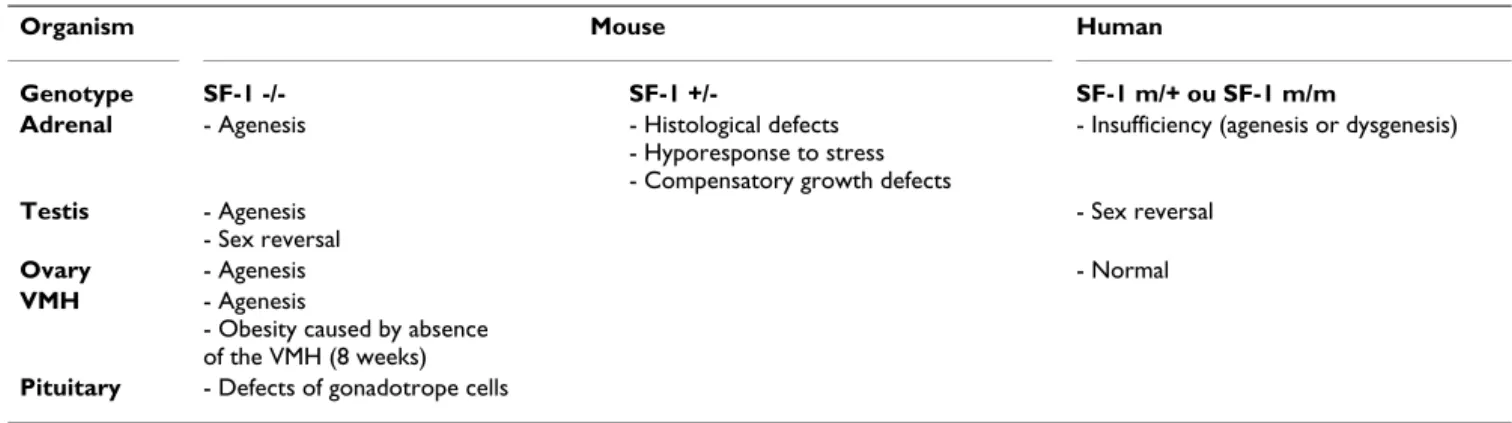
Despite interspecies differences, observations in SF-1 null mice and human mutants clearly show a key role for SF-1 in the development and / or differentiation of steroidog-enic tissues and their central regulators (hypothalamus and pituitary) (table 1). Tissue and cell-specific SF-1 knock-outs should be valuable models to differentiate between primary effects of SF-1 ablation in steroidogenic tissues and effects resulting from central nervous system defects.
A role for SF-1 in cell differenciation and proliferation?
All the data presented above do not allow to distinguish between SF-1 implication in cell fate determination / dif-ferentiation and proliferative effects. ES cells stably expressing SF-1 are able to morphologically differentiate into steroidogenic cells that express the rate-limiting P450scc enzyme. P450scc expression and subsequent pro-gesterone production are stimulated by cAMP. This activa-tion is prevented by cycloheximide, indicating that it requires de novo synthesis of factors that are absent from unstimulated cells. At last, P450scc induction is inde-pendent of SF-1 transactivating properties as measured by activation of a reporter gene, under the control of a SF-1 responsive element. This suggests that SF-1 is required for differentiation of ES cells and for the expression of factors that are implicated in cAMP responsiveness, although it doesn't directly participate in the latter [74].
After unilateral adrenalectomy, the remaining adrenal is able to compensate for adrenal defect by engaging in hypertrophy and hyperplasia (compensatory growth), through a regulatory loop implicating the VMH. Beusch-lein et al. have shown that in SF-1 +/- mice, there was no compensatory growth in the remaining adrenal following unilateral adrenalectomy, indicating that SF-1 was proba-bly required for hyperplasia and hypertrophy to occur [75]. This phenotype is correlated to absence of overex-pression of AsP, an adrenal-specific protease which cleaves pro-γ-melanotropin into an adrenal mitogenic peptide [76], and of PCNA, a cell proliferation marker [77], in response to unilateral adrenalectomy. This shows that beyond its role in cell differentiation, SF-1 may also be crucial for cell proliferation in steroidogenic tissues.
SF-1 target genes
Genes implicated in steroidogenesis
SF-1 was initially identified for its capacity to interact with and activate the promoters of the steroidogenic enzymes P450SCC, CYP11B1 and CYP21 [1,2], through the
con-sensus AGGTCA sequence. In the last ten years, transient transfection studies have shown that SF-1 participates to the expression of all steroidogenic enzymes in the adrenal cortex and gonads (table 2). In any case except for CYP11B2 which synthesizes aldosterone in the zona glomerulosa [78], SF-1 activates basal expression of these genes. Because of its tissue-restricted pattern of expres-sion, SF-1 is obviously a key factor in the tissue-restricted expression of steroidogenic enzymes. Its implication in their cAMP responsiveness is still a subject of debate. At least, cAMP-sensitive promoter regions often overlap with SF-1 responsive elements the mutation of which alters cAMP responsiveness [47,78–86]. These experiments indicate that SF-1 may be required to mediate cAMP responsivenes, but they do not show that it is sufficient for this process. Indeed, inside P450scc promoter, a proximal SF-1 responsive element (-40) is solely required for basal activity, whereas a distal site (-1600) is required for hor-monal sensitivity in vivo [79]. If SF-1, was intrinsically a mediator of cAMP signaling, it would be difficult to understand how it would behave differently from one site to another. This underlines the essential role of the DNA context of the SF-1 responsive element in its ability to transduce activation of the cAMP pathway to promoters. Besides steroidogenic genes, SF-1 is also able to modulate cholesterol delivery to steroidogenic reactions, by control-ling expression of the HDL-receptor SR-BI, of intracellular cholesterol transporter SCP2 [87], and of StAR which transfers cholesterol from the outer to the inner mito-chondrial membrane [88]. SF-1 can also stimulate de novo cholesterol synthesis in steroidogenic tissues through acti-vation of HMG-CoA synthase, irrespective of intracellular cholesterol concentrations [89]. SF-1 also controls expres-sion of the receptors for ACTH and FSH, the hormones that stimulate adrenal and gonadal steroidogenesis, respectively. Our group has also demonstrated that SF-1 could also control akr1-b7 expression in vitro and in vivo. This gene whose expression is controlled by ACTH in the
Table 1: Phenotypes resulting from genetic ablation or mutations of the orphan nuclear receptor SF-1.
Organism Mouse Human
Genotype SF-1 -/- SF-1 +/- SF-1 m/+ ou SF-1 m/m
Adrenal - Agenesis - Histological defects
- Hyporesponse to stress - Compensatory growth defects
- Insufficiency (agenesis or dysgenesis)
Testis - Agenesis - Sex reversal
- Sex reversal
Ovary - Agenesis - Normal
VMH - Agenesis
- Obesity caused by absence of the VMH (8 weeks)
adrenal cortex, encodes a protein which detoxifies isoc-aproaldehyde, produced by the cleavage of cholesterol through action of P450scc [42,90–93]. It is noteworthy that a new class of genes coding proteins able to reduce toxic side-effects of steroidogenic reactions, is emerging. Indeed, expression of superoxyde-dismutase 2 (SOD2), which protects adrenal cells against free radicals generated during steroidogenesis, is also controlled by ACTH [94]. However Chinn and colleagues have not studied the role of SF-1 in the control of SOD2 expression in adrenocorti-cal cells. Altogether, these results confirm the major role of SF-1 as a key activator of steroidogenesis. However, most of these studies rely upon transient transfections in heterologous cells that do not necessarily represent a good
model enough for studying the role of SF-1 on gene tran-scription. As SF-1 null mice completely lack steroidogenic tissues, the easiest alternative to study these mechanisms in a physiological context, is to generate transgenic mice harboring wild-type or mutant constructs of the promoter being studied.
Genes of the central nervous system
Analyses of SF-1 null mice have shown the key role of this factor for the establishment of the hypothalamus-pitui-tary-adrenal and hypothalamus-pituitary-gonadal axes. Neuronal NO synthase participates to the secretion of GnRH, the neuropeptide responsible for the synthesis and secretion of LH and FSH by pituitary [95]. Although SF-1
Table 2: A summary of SF-1 target genes. (After Hammer and Ingraham, Ref. [11], updated) The cell types where the studies were conducted are presented. Studies in vivo using transgenic approaches are specified. Basal: SF-1 is required for basal promoter activity of the gene being studied. Induction: SF-1 participates to the cAMP responsiveness of the gene. A different induction signal is stated. N.D. SF-1 implication in the cAMP responsiveness was not determined. Lower case letters before the names of the promoters correspond to the species that were studied (h: human; r: rat; b: bovine; m: mouse).
Gene promoter Cell type /Transgenic technique Basal activity/induction by cAMP Bibliography
hP450scc Additive transgenesis, H-295 basal + induction [79,80]
rP450scc MA-10, rat granulosa basal [138,188]
bP450scc Y1, bovine luteal cells, COS1 basal + induction [162]
h3β-HSD type II H295, HeLa basal + N.D. [189]
mCYP21 Y1 basal + N.D. [2,190]
hCYP11B1 Y1, H295 basal + induction [78,81]
bCYP11B1 Y1, I-10 (Leydig) basal + induction. [191,192]
hCYP17 H295 basal + induction [47,82,183,185]
bCYP17 Y1, COS-1 basal + induction [83,84]
rCYP19 R2C, H450, Y1 basal + induction [86,164,193,194]
hCYP19 Bovine luteal cells basal + induction [85]
h/mCYP11B2 H295 inhibition by SF-1 [78]
AKR1-B7 Additive transgenesis, HeLa, CV-1 basal [42,90–92]
HDL-R/SR-BI Y1, HTB9 basal + induction [195–198]
SCP2 Y1, HTB9 basal + induction [199]
hStAR Y1, BeWo, H295 basal + induction [200–203]
mStAR Y1, MA-10 basal [112]
rStAR Y1, HTB9 (human bladder carcinoma) basal + induction [204]
bStAR HeLa basal + induction [205]
HMG-CoA reductase CV-1, MA-10 basal + N.D. [89]
hMC2R H295-R basal + induction [206,207]
mMC2R Y1 basal + N.D. [208]
mFSH-R HEK-293 basal [60]
rFSH-R JEG-3 basal + inhibition of PKA induction [209]
rLH-R Granulosa basal inhibition [210]
R-GNRH αT3-1 basal [61,62,211,212]
LHβ Additive transgenesis, LβT2, αT3-1, basal + induction by GnRH in association with Sp1, EGR-1 and Ptx1
[58,156,157,213,214]
hαGSU LβT2 gonadotropes basal + induction by GnRH [215]
Neuronal mNOS αT3-1, NIH3T3 basal [96]
b-ocytocin TM4 basal [216,217]
PRL-R MLTC (Leydig) basal [218]
MIS Knock-in mice, HeLa, JEG-3 basal [104,107,108]
DAX-1 Additive transgenesis, H295, JEG-3 basal [187,219–221]
RLF/INSL3 MA-10, HeLa, mLTC-1, HEK293 basal [109,110]
knock-out mice do not show obvious GnRH secretion defect [35,53], SF-1 is able to control neuronal NO syn-thase gene transcription in vitro [96]. This raises the ques-tion of a more general role of NO in the etiology of the phenotypes in null mice. Pituitary glycoproteins LH and FSH are composed of a common subunit, αGSU, which is associated to a β chain, specific to LH or FSH. Gonadotro-pins production and secretion is controlled by GnRH which activates Ca2+ and PKC pathways through its
recep-tor [97]. In vitro, SF-1 controls expression of the GnRH receptor, αGSU and LHβ, but not of FSHβ. All these pro-teins are absent from SF-1 -/- mice pituitaries suggesting that SF-1 participates to their expression in vivo [53,54]. However, null mice treatment with supra-physiological GnRH doses induces LH and FSH expression in the absence of detectable amounts of GnRH receptor [35,53]. Although the question of GnRH action in SF-1 null mice is as yet unresolved, these surprising results shed light on transcription factors other than SF-1, that may participate to LH and FSH expression in vivo.
Genes implicated in sexual differentiation and sex determination
In mammals, male sex determination is triggered by the Y chromosome-borne SRY gene. SRY activation in turn trig-gers expression of SOX9 which stimulates MIS transcrip-tion in Sertoli cells. This hormone from the TGF-β family, is responsible for the regression of Müllerian ducts that normally form oviducts, uterus and the upper third of the vagina in females [98].
SF-1 expression in the urogenital ridge at E9.0 [26] pre-cedes expression of SRY in pre-sertoli cells at E10.5 in mouse [99,100]. SF-1 has recently been shown to regulate human and porcine SRY expression. One SF-1 responsive site at -327 is required for a modest activation of human SRY promoter [101] whereas two sites are essential for porcine SRY promoter activity in porcine genital ridge cells [102]. Although the physiological relevance of such an observation is not established yet, treatment of NT2/ D1 embryonic carcinoma cells with cAMP induces SF-1 phosphorylation that prevents its binding to the SRY pro-moter, thus downregulating human SRY expression [101]. Nonetheless, it is noteworthy that residual SRY expression is observed in SF-1 knock-out mice [103], suggesting that SF-1 may not be essential for triggering SRY expression. SF-1 and MIS are coexpressed in developing Sertoli cells [104], and SF-1, in association with SOX9 [105], GATA-4 [106] and WT-1 [107] is able to stimulate MIS promoter activity in transient transfections (figure 3). Male SF-1 null mice show Müllerian duct persistence [29,34,50]. How-ever, the absence of gonads in these animals does not allow a conclusion on a direct effect of SF-1 to be drawn. This difficulty was overcome by mutating the proximal SF-1 responsive element of the MIS promoter by
homolo-gous recombination with the endogenous MIS locus. This mutation reduces MIS accumulation albeit to an extent which is insufficient to prevent Müllerian duct regression, whereas when the SOX9 binding site is mutated by the same procedure, there is Müllerian duct persistence in male mice (figure 3). These results suggest that SOX9 trig-gers MIS expression whereas SF-1 acts as a quantitative regulator [108].
Gonadal descent is a process associated to male sexual dif-ferentiation which is dependent on regression of the cra-nial suspensory ligament and proliferation of the gubernaculum specifically in males. The knock-out of the Insl3/RLF gene, which codes a protein produced by Leydig cells, results in a defect in testes descent. It is noteworthy that SF-1, at least in vitro, is able to control RLF expression through three responsive-elements [109,110]. Confirma-tion of the role of SF-1 in testis descent via RLF should come from observation of testes position in SF-1 +/- mice, or of gubernaculum proliferation in null mice.
SF-1 target genes : unanswered questions
The number of SF-1 potential target genes is rapidly grow-ing. However few studies are based on the in vivo demon-stration that SF-1 responsive elements are indeed required for putative target genes expression. Analysis of putative target genes in SF-1 haplo-insufficient mice may confirm the role of SF-1 in their expression. However, compensa-tory mechanisms as those observed for StAR expression in SF-1 +/- mice, may mask the effects of SF-1 dosage reduc-tion [69].
The presence of gonadal and adrenal anlages in SF-1 knock-out mice and their rapid disappearance during development suggests that SF-1 regulates genes that are implicated in cell survival and/or proliferation. The majority of known SF-1 target genes is responsible for the maintenance of differentiated function rather than sur-vival of steroidogenic tissues. Degeneration of steroidog-enic tissues in null-mice is due to apoptosis [50]. It was recently shown that glucocorticoids can protect glandular tissues such as ovarian follicular cells against apoptosis although they have a pro-apoptotic effect on hemat-opoïetic cells [111]. As SF-1 controls the expression of StAR [112] and P450scc [79], two enzymes that are indis-pensable for glucocorticoids production, apoptosis in SF-1 null-mice may be due to glucocorticoids defects. In fact, this is rather unlikely because StAR [113] or P450scc [114] genetic ablation results in histological defects of the adrenals that are linked to progressive cholesterol accu-mulation, but does not result in adrenal regression during development.
POMC is the pituitary peptide that is cleaved to produce ACTH, pro-γ-MSH and β-LPH. Interestingly, POMC
knock-out mice show defective adrenal development [115]. The role of ACTH in trophic and mitogenic stimu-lation of the adrenal cortex is still a subject of intense debate [76,116,117]. The N-POMC(1–52) peptide, derived by cleavage of pro-γ-MSH shows highly mitogenic activity on adrenocortical cells. AsP, the adrenal-specific protease which is responsible for its cleavage, and its cog-nate receptor have been cloned recently. These are specif-ically expressed in the outermost regions of the cortex and the protease is required for Y1 cells growth [76,118]. An important question is now to determine whether SF-1 reg-ulates AsP and its receptor expression in the adrenal cortex.
Control of SF-1 expression and activity
SF-1 activity on its target genes must be tightly controlled. This can be achieved by ligands or cofactors (coactivators/ corepressors or transcription factors) or directly by modu-lating expression of the nuclear receptor. In numerous steroidogenic genes promoters, cAMP-responsive regions overlap with SF-1 responsive elements. This chapter will particularly address the complex issue of cAMP signalling transduction through SF-1.
SF-1: still an orphan?
During the last decade, numerous orphan nuclear recep-tors have been cloned without any known ligand. Some, like LXR, FXR, PXR or CAR, have since been attributed a ligand, whereas others like Nur77/NGFI-B, Nurr-1, LRH-1 or DAX-1, have no known ligand so far and seem to be activated by other mechanisms [5,10]. 25-hydroxycholes-terol, an hydroxylated cholesterol derivative is able to acti-vate CYP21 promoter transcription in a SF-1 dependent manner in heterologous CV-1 cells, indicating that it could be a SF-1 endogenous ligand [119]. Nonetheless, in steroidogenic MA-10 cells that naturally express SF-1, both exogenous and endogenous 25-hydroxycholesterol are unable to stimulate endogenous P450scc expression as well as six SF-1-responsive reporter genes [16]. Collec-tively, these results tend to prove that 25-hydroxycholes-terol is not a bona fide ligand for SF-1 in steroidogenic cells. This is confirmed by the ability of SF-1 LBD to adopt an active conformation independently of any ligand [18].
Factors controlling SF-1 transcription
A small 90 bp SF-1 proximal promoter is specifically expressed in adrenocortical cells in transient transfections. This region encompasses an E-box (-87/-82), a Sp1 binding site (-30/-24) and a CAT box that binds CBF (-68/ -59) [120,121] (figure 4). If the role of the latter is not MIS promoter
Figure 3
MIS promoter. Binding sites for the transcription factors required for proper MIS expression are shown. Their position in
human MIS promoter is indicated (hMIS). WT1 and Dax-1 are able to respectively stimulate or repress MIS promoter activity by interacting with SF-1 bound to its proximal responsive element. Effect of the mutation of the proximal SFRE or SOX9 responsive element introduced by knock-in in mice on Müllerian ducts regression is indicated. Abreviations : Sox BS, SOX fac-tors binding site; SFRE, SF-1 responsive element.
clearly demonstrated, transcriptional control of SF-1 expression at least requires the E-box which is conserved in human and which is functional in both steroidogenic and non-steroidogenic cells [120,122–124]. This element is able to bind the ubiquitous USF factor contained within pituitary αT3-1 cells, steroidogenic Y1 and JEG-3 cells, as well as CV1 and HeLa cells [123]. None of these interac-tions however, can account for tissue-restricted SF-1 expression. SF-1 itself is able to bind a site which is present in its own first intron in rat and human genes (+156/ +163) [124,125] (figure 4). Whereas Nomura et al., [125] have shown the role of this sequence for the expression of a reporter gene in Y1 cells, or in response to SF-1 overex-pression in heterologous CV-1 cells, Woodson et al., using the rat gene and Oba et al., using the human gene, were unable to obtain the same results in either steroidogenic or non-steroidogenic cells [120,124]. However, it is worth considering that some sequences contained within the first intron might be required for SF-1 expression, though their specificity is not yet established [124]. This is con-firmed by the use of different lengths of SF-1 regulatory regions and intragenic sequences in transgenic mice [49,126]. SF-1 is expressed in the urogenital ridge as early as E9.5 at similar levels in males and females. When sex determination occurs between E10.5 and E12.5, SF-1 expression strongly decreases in the ovary until E18.5, whereas it remains elevated in the testis. After birth, expression rises in the ovary although it is reduced in adult testis [26,27]. Pod-1/Capsulin is a transcription fac-tor of the b-HLH family which is able to heterodimerize with other factors of the family by binding to E-boxes (fig-ure 4). It participates to kidney and lung differentiation [127] and displays a sexually dimorphic pattern of expres-sion in the gonad, which is reminescent of SF-1 gonadal expression. However, Pod-1 is expressed in gonadal regions where SF-1 is not expressed (i.e. coelomic epithe-lial cells, peritubular myoid cells and epitheepithe-lial-like cells). In fact, it seems that Pod-1 may act as a repressor of SF-1 promoter activity through interaction with the previously described E-box [128]. Sox9 and SF-1 are colocalized in somatic cells of the testis and follow parallel expression patterns during development [129]. They both participate to transcriptional activation of the MIS promoter in males [105]. Recent results show that Sox9 is able to induce SF-1 expression in heterologous cells and that a Sox9 binding site (-110/-104) is required for SF-1 expression in Sertoli and Y1 cells [130] (figure 4). GATA-4 is also dimorphi-cally expressed in the gonad. Whereas its expression is high in the undetermined gonad it decreases as ovary dif-ferentiation starts, although it is maintained in the testis. This dimorphism may participate to the control of MIS expression [131], but GATA-4 is also able to moderately activate SF-1 expression via a conserved site at -177/-172 (figure 4). This activation is dependent on the cell type and seems to be essentially restricted to Sertoli cells where
SF-1 participates to MIS expression [132]. Although most elements required for SF-1 expression in steroidogenic tis-sues are likely to be localized in the 5' flanking regions and first intron of the gene, a GFP/SF-1 fusion, the expres-sion of which is directed by a 50 kb BAC comprised of SF-1 promoter, first exon and first intron, is not expressed in pituitary gonadotropes [49]. Although a single transgenic line was studied so far, this indicates that further down-stream sequences might be required for pituitary expres-sion. However, there is no mention of pituitary expression in the work of Zubair et al., who used SF-1 regulatory regions extending from the first intron to the seventh exon [126]. Altogether, these data allow a better understanding of SF-1 tissue-specific expression especially in the gonads but do not establish a link between SF-1 transcription and the cAMP signalling pathway.
Is SF-1 accumulation altered by cAMP pathway stimulations? In vivo, SF-1 protein accumulation in the adrenals and
gonads is unchanged by a four weeks hypophysectomy in rats [133]. However an eighty hours treatment of mice with dexamethasone, induces a marked reduction of SF-1 mRNA accumulation in the adrenals [134], suggesting that compensatory mechanisms may have masked the effects of long term hypophysectomy on SF-1 accumula-tion. Nonetheless, lipopolysaccharide treatment which induces increases in circulating ACTH concentrations, has no effect on SF-1 accumulation [134]. The problem is far more complex in cell culture systems. Whereas numerous papers show that SF-1 mRNA accumulation is unchanged in Y1, MA-10, theca or bovine granulosa cells in response to forskolin or PKA catalytic subunit overexpression [135–138], one paper describes a slight increase in bovine adrenocortical cells treated with ACTH [139]. At last, in human granulosa cells [140] or in forskolin-treated Y1 cells, SF-1 protein accumulation increases independently of an increase in its mRNA accumulation [137]. Based on this observation, Aesoy et al. have proposed a post-trans-lational model in which PKA could stabilize SF-1 protein [137]. However, this was only demonstrated in a heterol-ogous cell system overexpressing both SF-1 and the PKA catalytic subunit. Interestingly, we have not been able to obtain similar results with both Y1 and ATC-1 [141] adrenocortical cell cultures treated with forskolin or ACTH, respectively. Our unpublished data rather suggest that SF-1 accumulates into cell nucleus in response to cAMP, without a concomitant increase in overall protein accumulation (Bruno Ragazzon, personal communica-tion). This increase in SF-1 nuclear accumulation correlates with increased SF-1 binding in gel shift experi-ments (Christelle Aigueperse, personal communication). Whatever the changes in SF-1 expression may be, simple modulations of SF-1 accumulation are unlikely to account for some SFREs being implicated in cAMP-responsiveness while others only support basal promoter activity in vivo
[42,79]. This differential activity may be achieved by more subtle mechanisms that control SF-1 activity, such as interaction with other transcription factors and / or cofactors.
SF-1 cofactors
Ligand-dependent nuclear receptors activate their target genes transcription through interactions with coactivators and/ or corepressors that link receptors to the transcrip-tion machinery. Accordingly, SF-1 harbors an AF2 activa-tion domain in its LBD (figure 1B). This motif (LLIEML, consensus LLXXL) is necessary but not sufficient for trans-activation [14,19–21] which also depends on two amino-terminal regions of the protein, the FP region [14] and a proximal activation domain [20,25]. SF-1 proteins bear-ing mutations in their AF2 domain have dominant nega-tive properties on CYP17 promoter activation by PKA in Y1 cells. This suggests that SF-1 AF2 is implicated in the transduction of the cAMP signal [83]. As ligand-activated nuclear receptors, SF-1 interacts with numerous coactiva-tors such as SRC1 [19,20], RIP140 [142], PNRC and PNRC2 [143,144], hMBF1 [145], TIF2 [25], p/CIP [146] and GCN5 [147]. These interactions, independent of an exogenous ligand, are dependent on AF-2 and for some of them, on proximal interaction domain integrity. Most of these interactions are mediated by LXXLL motifs found on coactivators. PNRC and PNRC2 are quite unique in that they interact with SF-1 and other nuclear receptors through SH3 proline-rich motifs [143,144]. None of these coactivators is specific for SF-1 and none of them shows a steroidogenic tissue-restricted pattern of expression. Nonetheless, two of them may be required for integration
of the cAMP signalling (figure 5). TIF2 and p/CIP interact with SF-1 through the AF-2 and proximal activation domain [25,146]. Overexpression of TIF2 or p/CIP in het-erologous or Y1 cells, stimulates transcription of a reporter gene driven by four copies of a SF-1 responsive-sequence of the bovine CYP17 promoter. However, whereas p/CIP increases sensitivity to PKA overexpression in the presence of SF-1, overexpression of the catalytic subunit of the PKA inhibits potentiation of SF-1 activity by TIF2, through a decrease in TIF2 protein accumulation [146]. In a more physiological system, one might imagine that on the sites where it participates to cAMP-responsive-ness, SF-1 would preferentially associate to p/CIP, whereas where it participates to basal promoter activity, 1 would rather associate to TIF2. What could allow SF-1 to choose between those two coactivators? Although the sequence of the SF-1 responsive element may participate to this choice, it is possible that adjacent transcription factors, endowed with cAMP sensing capacity, may mod-ulate cofactors recruitment by SF-1. This would allow modulation of SF-1 activity in response to extra-cellular signals.
DP103 is a DEAD-box protein which is highly expressed in steroidogenic tissues (table 3). Although it has intrinsic RNA helicase properties as other proteins of its family [148], DP103 physically interacts with SF-1 through a newly described repressive domain, located in the vicinity of the proximal activation domain [149]. DP103 harbors a C-terminal repression domain that by itself, represses SF-1 activity. DP103 repressive function is independent of its helicase activity and can decrease P450scc and P450c21 Schematic representation of SF-1 regulatory regions
Figure 4
Schematic representation of SF-1 regulatory regions. A summary of the experimental results obtained with SF-1
regu-latory regions in humans, rats and mice is presented. Cis elements are conserved across the three species. Numbering may vary from one species to another. Pod-1/Capsulin and USF bind to the same response element. USF activates SF-1 transcrip-tion whereas Pod-1 is likely to repress it. Abreviatranscrip-tions : GATA, GATA proteins response element; Sox BS, Sox proteins bind-ing site; CAT, CAAT box; Sp1, Sp1 response element; Inr, initiator; SFRE, SF-1 responsive element; USF, upstream stimulatory factor; CBF, CAT box binding factor.
promoter activity, inducing a significant reduction in pro-gesterone production by Y1 cells [148]. One interesting question is now to analyze whether DP103 is implicated in the cAMP responsiveness of steroidogenic enzymes genes.
Transcription factors associated with SF-1
Apart from interaction with bona fide cofactors, SF-1 also interacts with numerous transcription factors (table 3) that modulate its activity either by binding to adjacent DNA sequences or by interacting with SF-1 without
bind-ing to DNA [106,107,150–152]. The regions of interaction between SF-1 and other transcription factors are rather broadly delineated. These can overlap with pre-viously described regions such as AF-2 or the proximal activation domain [14,160] but also extend to the DBD [107,153] and LBD [107,154] or distal [152] and proxi-mal [148] repression domains. These interactions result in variable responses depending on the promoter or cell type under experiment.
Presumed effect of the coactivators p/CIP and TIF2 on SF-1 transactivation
Figure 5
Presumed effect of the coactivators p/CIP and TIF2 on SF-1 transactivation. (After Borud et al., Ref. [188]). In the
absence of PKA, p/CIP and TIF2 potentiate SF-1 transcriptional activity. When SF-1 is associated to p/CIP, overexpression of PKA induces a marked increase in SF-1 transcriptional activity. On the contrary, PKA overexpression prevents TIF2-dependent potentiation of SF-1 activity. The decrease of TIF2 protein accumulation caused by PKA overexpression may explain these observations. SFRE : SF-1 responsive element.
A first class of factors, illustrated by the studies on MIS and LH-β promoters, encompasses proteins that will restrict SF-1 target genes expression in a very narrow region of the organism in a particular context. Indeed, the combinatory interactions between SF-1, WT-1 [107], GATA-4 [106,150], SOX-9 [105,108] and DAX-1 [107] allow MIS expression specifically by Sertoli cells of the male gonad, just before regression of the Müllerian ducts (figure 3). Also, this is an interaction between Egr-1, Ptx1 and SF-1 that allows expression of the LH-β subunit in pituitary gonadotrope cells in response to GnRH stimulation [155– 159].
Another group of factors is more implicated in SF-1 target genes response to external stimuli. Indeed, SF-1 interaction with TReP132 [160,161], Sp1 [153,162] or CREB [86,163,164] allows recruitment of the CBP/p300 coactivator which interconnects multiple cell signalling pathways [165]. This may participate to the cAMP respon-siveness of some SF-1 target genes.
At last, a third interaction group allows repression of SF-1 activated genes. DAX-1 is the paradigm of such repressors. It encodes a peculiar nuclear receptor devoid of a classical DNA binding domain, which is replaced by three-and-a half repeats of an alanine and glycine-rich motif [166,167]. DAX-1 overexpression subsequent to Xp21 duplication, induces male to female sex reversal in
humans [168,169], a phenotype that can be mimicked by Dax-1 overexpression in transgenic mice with a poor Sry allele [170]. DAX-1 mutations in humans are responsible for adrenal hypoplasia congenita as well as hypogonadotrophic hypogonadism suggesting that DAX-1 may perform similar functions as DAX-1 [DAX-17DAX-1]. Indeed, SF-1 and Dax-SF-1 are coexpressed in steroidogenic tissues as well as in the VMH and pituitary, early in development [47,172,173]. However, when overexpressed in Y1 adren-ocortical cells, DAX-1 markedly impairs steroidogenic output and transcriptionaly represses the promoters of SF-1 target genes such as StAR, P450scc, 3β-HSD [SF-174,SF-175] and akr1-b7 [92]. Essentially two mechanisms have been proposed for DAX-1 mediated transcriptional repression, although they are probably not mutually exclusive. DAX-1 can repress SF-DAX-1 target genes expression either by bind-ing DNA to hairpin-like structures [176], or by physically interacting with SF-1, independently of the DNA context [107]. This interaction implies a carboxy-terminal repres-sion domain (437 to 447) and a proximal interaction domain of SF-1 (226 to 230) [152] (figure 1) that contact amino-terminal LXXLL domains of DAX-1 [151,177]. This physical interaction allows the recruitment of the core-pressors NcoR [152] and Alien [178] to SF-1-responsive genes promoters. The physiological relevance of such a negative functional interaction between Dax-1 and SF-1 is as yet unclear as Dax-1 knock-out in mice does not lead to marked adrenal defects [179]. However, it is noteworthy
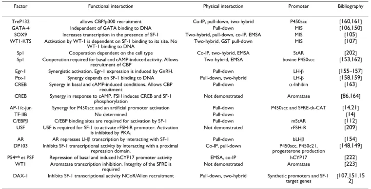
Table 3: Transcription factors or cofactors that interact with SF-1. For each interacting factor, the resulting functional interaction, the experimental approach used to evidence the physical interaction and the model promoter are indicated.
Factor Functional interaction Physical interaction Promoter Bibliography
TreP132 allows CBP/p300 recruitment Co-IP, pull-down, two-hybrid P450scc [160,161]
GATA-4 Independent of GATA binding to DNA Pull-down MIS [106,150]
SOX9 Increases transcription in the presence of SF-1 Two-hybrid, pull-down, co-IP, EMSA MIS [105]
WT1-KTS Activation by WT-1 is dependent on SF-1 binding to its site. No
WT-1 binding to DNA Two-hybrid, GST pull-down MIS
[107]
Sp1 Cooperation dependent on the cell type Co-IP, two-hybrid, EMSA StAR [202]
Sp1 Cooperation required for basal and cAMP-induced activity. Allows recruitment of CBP
Two-hybrid, EMSA bovine P450scc [153,162]
Egr-1 Synergistic activation. Egr-1 expression is induced by GnRH. Pull-down LH-β [155–157]
Ptx-1 Synergy depends on SF-1 binding to DNA Pull-down, two-hybrid LH-β [158,159]
CREB Synergy in basal and cAMP-induced conditions. Allows CBP recuitment
Pull-down α-Inhibin [163]
CREB Synergy in response to cAMP. FSH induces CREB and SF-1 phosphorylation
Not demonstrated Aromatase [86,164]
AP-1/c-jun Synergy for P450scc and an artificial promoter activation Pull-down P450scc and SFRE-tk-CAT [14,21]
TF-IIB No determined Pull-down [14]
C/EBPβ C/EBP binding sites are required for activation by SF-1 Pull-down mStAR [112]
USF USF is required for SF-1 to activate rFSH-R promoter. Activation
is inhibited by PKA. Not demonstrated rFSH-R
[209]
AR AR represses LHβ transcription by interacting with SF-1 Pull-down bLHβ [154]
DP103 Inhibits SF-1 transcriptional activity by interacting with a proximal repression domain.
Co-IP, pull-down P450scc, P450c21, progesterone production
[148,149]
P54nrb et PSF Repression of basal and induced hCYP17 promoter activity EMSA, co-IP hCYP17 [222]
WT1 Aromatase transcription inhibition. Integrity of the SFRE is
required Not demonstrated Aromatase
[223] DAX-1 Inhibits SF-1 transcriptional activity NCoR/Alien recruitment Pull-down, two-hybrid Synthetic promoters and SF-1
target genes
[107,151,15 2]
that Dax-1 ablation in SF-1 haploinsufficient mice allows a reversion of the histological and functional adrenal defects, suggesting that the two receptors interact in vivo [180]. Furthermore, we have been able to observe Dax-1 downregulation in either ACTH-treated mice or adreno-cortical ATC-1 cells that naturally express Dax-1, indicat-ing that this receptor may be implicated in hormonal responsiveness in vivo (B. Ragazzon, personal communication).
An unexpected repressor of SF-1 activity is the androgen receptor. The increase in plasmatic LH concentrations which is induced by GnRH leads to increased sex steroid production by the gonads. In turn, sex steroids exert a negative feedback on GnRH and LH synthesis (figure 6). Recently, Jorgensen and Nilson, have elegantly demon-strated that androgen receptor was able to repress LH-β promoter transactivation by interacting with SF-1 LBD [154]. Under low GnRH conditions, AR blocks the func-tional interaction between SF-1 and Ptx1/Egr-1. When GnRH concentrations in pituitary gonadotropes increase, Egr-1 expression is induced [156] and allows recruitment of Ptx-1 as well as AR displacement, resulting in the for-mation of an activating complex composed of SF-1, Egr-1 and Ptx1. When circulating androgens increase, activated AR displaces Egr-1 and Ptx1, thus repressing LH-β tran-scription in response to SF-1 [154]. This model (figure 6) perfectly illustrates the complex interactions that are required fo SF-1 target genes activation. It is likely that such mechanisms may participate to the control of SF-1 target genes expression in response to cAMP increases.
Post-translationnal SF-1 alterations
Although it is clear that SF-1 transcriptional activity requires complex interactions with numerous cofactors, the mechanisms underlying the recruitment of these part-ners are still unclear. Because PKA is implicated in the stimulation of most of SF-1 target genes expression, its role in SF-1 activity has been extensively investigated. In vivo, SF-1 is phosphorylated in response to granulosa cells stimulation by FSH [86]. In vitro, PKA can phosphorylate 1 LBD and N-terminal region [101,135]. Although SF-1 seems to be implicated in the cAMP responsiveness of the rat CYP17 gene, its in vitro phosphorylation by PKA decreases its DNA binding capacity, an observation which is not compatible with its activating role [101,135]. In heterologous cell systems SF-1 is phosphorylated on ser-ine 203 by the MAPK ERK2, regardless of the presence of cAMP. This residue is situated in the proximal activation domain, the integrity of which is essential for SF-1 activity. Serine 203 phosphorylation is required for SF-1 transcriptional activity and allows the recruitment of two cofactors, the coactivator GRIP1/TIF2 and the corepressor SMRT [25]. Recently, Desclozeaux et al., have shown that SF-1 hinge and helix 1 of the LBD were able to set helices
2 to 12 of the LBD in an active conformation reminiscent of ligand-activated nuclear receptors, albeit in the absence of a ligand. Helices 2 to 12 recruitment is enhanced by MAPK stimulation and decreased by mutating serine 203, or in the presence of MKP-1, a MAPK specific phos-phatase. At last, serine 203 phosphorylation stabilizes SF-1 LBD, an observation reminiscent of ligand-activated nuclear receptors [18]. This shows that apart from being implicated in cofactor recruitment [25] serine 203 phos-phorylation also structures and stabilizes SF-1 LBD. How-ever, these experiments do not establish a link between cAMP stimulation and SF-1 activity. This may depend on activation of the MAPK pathway by PKA [181]. Indeed, cAMP-induced StAR transcription is dependent on activa-tion of the MPAK pathway in Y1 and MA-10 cells. This activation induces SF-1 phosphorylation in vivo in Y1 cells, resulting in an increase in SF-1 binding in EMSA [182]. However, cAMP-induced P450scc expression does not seem to depend on MAPK activation in the same experiments [182]. Furthermore, cAMP pathway stimula-tion in H295 cells results in a decrease in SF-1 overall phosphorylation [183], indicating that PKA/MAPK crossovers may be gene and cell-specific. Thus in H295 cells, MAPK inhibition by the specific inhibitor PD98059 does not reduce, but on the contrary, stimulates hCYP17 transcription [183]. Indeed, StAR and hCYP17 cAMP-stimulated transcription in H295 cells is dependent on protein phosphatase activities [183,184]. In H295 cells, MKP-1 that can be phosphorylated by PKA in vitro, is overexpressed in response to cAMP stimulation. MKP-1 overexpression leads to an increase in hCYP17 expression, whereas MKP-1 inhibition by an antisense RNA prevents hCYP17 induction by cAMP. Whether MKP-1 is directly implicated in SF-1 activity is still unclear. However, it is noteworthy that phosphatase inhibitors decrease SF-1 transcriptionnal activity on the hCYP17 promoter, indi-cating that at least one phosphatase is required for pro-moter activation by SF-1 [185].
Altogether, these results seem rather contradictory. Although it is probable that the MAPK pathway influences SF-1 activity, it is still unclear whether this implicates direct phosphorylation of SF-1 by a MAPK. Also, it seems that such a mechanism would not necessarily apply to all cell types or promoters. One pitfall of these experiments is that the MAPK pathway is highly sensitive to extracellular changes and temporal variations in the experimental set-ting. Completely different experimental conditions may thus account for the contradictory observations. At last, future experiments may distinguish between overall SF-1 phosphorylation and phosphorylation on specific resi-dues such as serine 203. When these mechanisms are decyphered, it will be interesting to study the effect of one particular phosphorylation or dephosphorylation on the
recruitment of cofactors such as p/CIP versus TIF2 in response to cAMP stimulation.
Another emerging activation control mechanism for nuclear receptors is their acetylation [186]. SF-1 is acetylated in vivo in a heterologous cell system and inter-A model for LHβ promoter repression by a physical interaction between inter-AR and SF-1
Figure 6
A model for LHβ promoter repression by a physical interaction between AR and SF-1. (After Jorgensen and
Nil-son, Ref. [201]) In the presence of elevated androgen concentrations, AR (androgen receptor) interacts with SF-1 on LHβ pro-moter, preventing interaction of Egr-1 with its response elements. LHβ is then turned off. A GnRH pulse (resulting from a reduction in circulating androgen concentrations), favours Egr-1 transcription, increasing its accumulation. In turn, Egr-1 dis-places AR from SF-1 and favours an active setting of transcription factors on LHβ promoter. LHβ transcription is then trig-gered. The resulting LH protein production in turn stimulates androgens production by the gonads.
acts with the acetyl-transferase GCN5, which can acetylate SF-1 in vitro. Although SF-1-dependent activation of a reporter gene may depend on SF-1 acetylation in the pres-ence of GCN5, experimental results obtained by mutating the potential acetylation sites or by the use of the deacety-lase inhibitor trichostatin A, are contradictory [147]. If more substantial data is obtained regarding the role of acetylation on SF-1 activity, it will be interesting to study the implication of certain histone acetyl transferase (HAT) coactivators such as CBP/p300, in SF-1 acetylation in vivo.
Conclusions
A key role for SF-1, in the differentiation and the mainte-nance of the differentiated function of the gonads, adrenals and particular regions of the pituitary and hypothalamus is now clearly established (table 1). Its targets implicated in the maintenance of the differentiated function of steroidogenic tissues and in sex determination are now, at least in part, identified. Tissue-specific SF-1 knock-out at late developmental stages or at adulthood (when differentiated function is established) may confirm the results of transient transfections and may allow the identification of new SF-1 targets.
Although tremendous progress has been accomplished since SF-1 cloning, major questions remain unanswered. Indeed, genes whose down-regulation (or up-regulation) in SF-1 null mice may account for the regression of gonadal, adrenal and VMH anlages are still unidentified. Another important issue is the identification of the mech-anisms that allow activation of this orphan nuclear recep-tor at certain stages during embryonic and post-natal development or in response to external stimuli such as increased cAMP concentrations evoked by trophic hormones in their target tissues. Recent results of our group show that a SF-1 binding site is dispensable at birth for akr1-b7 promoter activity but is required 20 days later [42]. On the contrary, Hoyle et al., show that a SF-1 bind-ing site is required for early developmental expression of DAX-1, but is dispensable after birth [187]. Forthcoming studies will require a careful evaluation of the distinct roles of SF-1 during early development and after birth.
Competing interests
None declared.
Authors' Contributions
P.V. wrote this article as part of is thesis manuscript. A.M. and A-M. L-M. are P.V. PhD supervisors. They helped him with writing this manuscript.
G.V. is the head of the PCEM team.
References
1. Morohashi K, Honda S, Inomata Y, Handa H and Omura T: A
com-mon trans-acting factor, Ad4-binding protein, to the pro-moters of steroidogenic P-450s. J Biol Chem 1992, 267:17913-9.
2. Rice DA, Mouw AR, Bogerd AM and Parker KL: A shared
pro-moter element regulates the expression of three steroidog-enic enzymes. Mol Endocrinol 1991, 5:1552-61.
3. Honda S, Morohashi K, Nomura M, Takeya H, Kitajima M and Omura T: Ad4BP regulating steroidogenic P-450 gene is a member
of steroid hormone receptor superfamily. J Biol Chem 1993, 268:7494-502.
4. Lavorgna G, Karim FD, Thummel CS and Wu C: Potential role for
a FTZ-F1 steroid receptor superfamily member in the con-trol of Drosophila metamorphosis. Proc Natl Acad Sci U S A 1993, 90:3004-8.
5. Laudet V: Evolution of the nuclear receptor superfamily: early
diversification from an ancestral orphan receptor. J Mol Endocrinol 1997, 19:207-26.
6. De Mendonca RL, Bouton D, Bertin B, Escriva H, Noel C, Vanacker JM, Cornette J, Laudet V and Pierce RJ: A functionally conserved
member of the FTZ-F1 nuclear receptor family from Schis-tosoma mansoni. Eur J Biochem 2002, 269:5700-11.
7. Kotomura N, Ninomiya Y, Umesono K and Niwa O:
Transcrip-tional regulation by competition between ELP isoforms and nuclear receptors. Biochem Biophys Res Commun 1997, 230:407-12.
8. Ninomiya Y, Okada M, Kotomura N, Suzuki K, Tsukiyama T and Niwa O: Genomic organization and isoforms of the mouse ELP
gene. J Biochem (Tokyo) 1995, 118:380-9.
9. Luo X, Ikeda Y, Schlosser DA and Parker KL: Steroidogenic factor
1 is the essential transcript of the mouse Ftz-F1 gene. Mol Endocrinol 1995, 9:1233-9.
10. Mangelsdorf DJ, Thummel C, Beato M, Herrlich P, Schutz G, Umes-ono K, Blumberg B, Kastner P, Mark M and Chambon P et al.: The
nuclear receptor superfamily: the second decade. Cell 1995, 83:835-9.
11. Hammer GD and Ingraham HA: Steroidogenic factor-1: its role
in endocrine organ development and differentiation. Front Neuroendocrinol 1999, 20:199-223.
12. Wilson TE, Fahrner TJ and Milbrandt J: The orphan receptors
NGFI-B and steroidogenic factor 1 establish monomer bind-ing as a third paradigm of nuclear receptor-DNA interaction. Mol Cell Biol 1993, 13:5794-804.
13. Nitta M, Ku S, Brown C, Okamoto AY and Shan B: CPF: an orphan
nuclear receptor that regulates liver-specific expression of the human cholesterol 7alpha-hydroxylase gene. Proc Natl Acad Sci U S A 1999, 96:6660-5.
14. Li LA, Chiang EF, Chen JC, Hsu NC, Chen YJ and Chung BC:
Func-tion of steroidogenic factor 1 domains in nuclear localiza-tion, transactivalocaliza-tion, and interaction with transcription factor TFIIB and c-Jun. Mol Endocrinol 1999, 13:1588-98.
15. Achermann JC, Ozisik G, Ito M, Orun UA, Harmanci K, Gurakan B and Jameson JL: Gonadal determination and adrenal
develop-ment are regulated by the orphan nuclear receptor ster-oidogenic factor-1, in a dose-dependent manner. J Clin Endocrinol Metab 2002, 87:1829-33.
16. Mellon SH and Bair SR: 25-Hydroxycholesterol is not a ligand
for the orphan nuclear receptor steroidogenic factor-1 (SF-1). Endocrinology 1998, 139:3026-9.
17. Giguere V: Orphan nuclear receptors: from gene to function.
Endocr Rev 1999, 20:689-725.
18. Desclozeaux M, Krylova IN, Horn F, Fletterick RJ and Ingraham HA:
Phosphorylation and intramolecular stabilization of the lig-and binding domain in the nuclear receptor steroidogenic factor 1. Mol Cell Biol 2002, 22:7193-203.
19. Ito M, Yu RN and Jameson JL: Steroidogenic factor-1 contains a
carboxy-terminal transcriptional activation domain that interacts with steroid receptor coactivator-1. Mol Endocrinol
1998, 12:290-301.
20. Crawford PA, Polish JA, Ganpule G and Sadovsky Y: The activation
function-2 hexamer of steroidogenic factor-1 is required, but not sufficient for potentiation by SRC-1. Mol Endocrinol 1997, 11:1626-35.
21. Li LA, Lala D and Chung BC: Function of steroidogenic factor 1
(SF1) ligand-binding domain in gene activation and interac-tion with AP1. Biochem Biophys Res Commun 1998, 250:318-20.
![Table 2: A summary of SF-1 target genes. (After Hammer and Ingraham, Ref. [11], updated) The cell types where the studies were conducted are presented](https://thumb-eu.123doks.com/thumbv2/123doknet/14051204.460166/8.918.86.835.207.811/table-summary-hammer-ingraham-updated-studies-conducted-presented.webp)