Publisher’s version / Version de l'éditeur:
Vous avez des questions? Nous pouvons vous aider. Pour communiquer directement avec un auteur, consultez la première page de la revue dans laquelle son article a été publié afin de trouver ses coordonnées. Si vous n’arrivez pas à les repérer, communiquez avec nous à [email protected].
Questions? Contact the NRC Publications Archive team at
[email protected]. If you wish to email the authors directly, please see the first page of the publication for their contact information.
https://publications-cnrc.canada.ca/fra/droits
L’accès à ce site Web et l’utilisation de son contenu sont assujettis aux conditions présentées dans le site LISEZ CES CONDITIONS ATTENTIVEMENT AVANT D’UTILISER CE SITE WEB.
Optics Express, 28, 8, pp. 11267-11279, 2020-04-01
READ THESE TERMS AND CONDITIONS CAREFULLY BEFORE USING THIS WEBSITE. https://nrc-publications.canada.ca/eng/copyright
NRC Publications Archive Record / Notice des Archives des publications du CNRC :
https://nrc-publications.canada.ca/eng/view/object/?id=4bb29577-6f8c-4c8f-98c9-92d4b1a5b724
https://publications-cnrc.canada.ca/fra/voir/objet/?id=4bb29577-6f8c-4c8f-98c9-92d4b1a5b724
This publication could be one of several versions: author’s original, accepted manuscript or the publisher’s version. / La version de cette publication peut être l’une des suivantes : la version prépublication de l’auteur, la version acceptée du manuscrit ou la version de l’éditeur.For the publisher’s version, please access the DOI link below./ Pour consulter la version de l’éditeur, utilisez le lien DOI ci-dessous.
https://doi.org/10.1364/OE.389215
Access and use of this website and the material on it are subject to the Terms and Conditions set forth at
Multiphoton laser-induced confined chemical changes in polymer films
Kallepalli, Deepak L. N.; Godfrey, Alan T. K.; Walia, Jaspreet; Variola, Fabio;
Staudte, André; Zhang, Chunmei; Jakubek, Zygmunt J.; Corkum, P. B.
Multiphoton laser-induced confined chemical
changes in polymer films
D
EEPAKL. N. K
ALLEPALLI,
1A
LANT. K. G
ODFREY,
1J
ASPREETW
ALIA,
2F
ABIOV
ARIOLA,
3A
NDRÉS
TAUDTE,
1C
HUNMEIZ
HANG,
1Z
YGMUNTJ. J
AKUBEK,
4 ANDP. B. C
ORKUM1,*1Joint Attosecond Science Laboratory, University of Ottawa and National Research Council of Canada, 100 Sussex Dr., Ottawa K1N 5A2, Canada
2Center for Research in Photonics, School of Electrical Engineering and Computer Science, University of Ottawa, Ottawa K1N 6N5, Canada
3Department of Mechanical Engineering, 161 Louis Pasteur, Colonel By Hall, University of Ottawa, Ottawa K1N 6N5, Canada
4Metrology Research Center, National Research Council Canada, 100 Sussex Dr., Ottawa, K1N 5A2, Canada
Abstract: We report ultrafast-laser-induced photochemical, structural, and morphological changes in a polyimide film irradiated at the polymer-glass interface in back-incident geometry. Back-illumination creates locally hot material at the interface leading to a confined photochemical change at the interface and a morphological change through a blister formation. The laser-induced photochemical changes in polyimide resulted in new absorption and luminescence properties in the visible region. The laser-treated polyimide exhibited photoluminescence anisotropy resulting from formation of ordered polymer upon irradiation by linearly polarized ultrashort laser pulses. Confocal fluorescence microscopy resulted in similar observations to the bulk. Reflection-absorption infrared spectroscopy and X-ray photoelectron spectroscopy together indicated confinement of laser-induced chemical changes at the interface.
© 2020 Optical Society of America under the terms of theOSA Open Access Publishing Agreement
1. Introduction
Back-illumination of a low bandgap polymer through a high bandgap glass substrate with an ultrafast laser pulse leads to energy deposition at the interface and thereby creates hot material confined at the substrate-polymer interface. The deposited energy results in a localized expanding plasma which induces a protruding blister on the film’s surface and a photochemical change confined near the interface [1–7]. The photochemical changes induced by the laser pulse are limited to its penetration depth. In contrast, front illumination creates surface voids and cracks [8,9]. Such high intensities and extreme temperature will result in chemical changes in the polymer such as bond scission followed by rearrangement. The photochemical modification of the polymer results in changes in light absorption and luminescence properties. One potential application of laser-induced fluorescence is 3D optical data storage, where data can be read with laser-induced fluorescence at multiple grey levels [10–13]. We expect the laser-induced fluorescence intensity to scale with absorbed energy [10,12,13].
We investigate the chemical changes induced by an ultrafast laser through back-illumination. We demonstrate the confinement of photochemical changes by imaging the cross-sections of irradiated regions using a focused ion beam (FIB) technique. We also report the nature of chemical changes using fluorescence, confocal micro-Raman (and fluorescence), reflection-absorption Infrared (RAIRS), and X-ray photoelectron spectroscopy (XPS) techniques. This study is related to our work on ultrafast laser-induced blister formation in polyimide films [14].
#389215 https://doi.org/10.1364/OE.389215 Journal © 2020 Received 27 Jan 2020; revised 12 Mar 2020; accepted 23 Mar 2020; published 1 Apr 2020
2. Experimental
We prepared polyimide films on #1.5 Fisherbrand borosilicate glass coverslips. The coverslips were rinsed with acetone, isopropanol, and deionized water to remove any contaminants and dried on a hotplate. Polyimide films were made using PI-2525 and PI-2555 precursors from HD Microsystems following the recommended spin curves and baking conditions. We used a Ti:sapphire laser (Coherent model RegA 9040) producing a near Gaussian pulse in space and time with a duration of ∼ 50-fs duration at a central wavelength of 800 nm. The laser pulse energy was controlled using a motorized half-wave plate followed by a polarizing beam-splitter cube. The laser beam was focused using microscope objectives (10x 0.2 NA, 20x 0.4 NA) mounted into a vertical motor stage (PI M-112, Germany) with a travel range of 25 mm for adjusting the placement of the focal spot on the sample. Since polyimide is transparent for 800 nm (1.55 eV) wavelength, the modification is due to a nonlinear absorption mechanism with a minimum of two photons.
Above the vertical stage, polyimide-on-glass coverslips were mounted onto a 5-axis piezo nano-precision stage (PI, Germany) assembled on top of a micro-precision horizontal XY stage (MICOS MS-4, USA). The nano-precision stage was used for fine adjustment of the focal position. The laser was focused through the glass substrate onto the polymer-glass interface ("back-incidence" geometry). A dichroic mirror was used before the microscope objective in a coaxial geometry, allowing a small portion of the focused laser light to back-reflect from the sample, re-collimate through the objective, and travel to an imaging line for in-situ laser spot monitoring. Coupling in white light and changing the position of the objective also allows for in-situ white-light microscopy. We used a white-light microscope to find the optimal position of the laser focus on the sample, by firing pulses with energies near the damage threshold while adjusting the position of the focal spot. The details on the characterization techniques used in our study are given in AppendixA.
3. Results and discussion
It is known that the maximum laser intensity transmitted to the far side of glass substrate is self-limited by the nonlinear interactions within the glass [15,16]. The intensity leaving the material is, at maximum, equal to the absorption threshold. In our experiments, the substrate is borosilicate glass, whose absorption threshold is ∼ 1.3 ×10¹³W/cm2[17]. The actual intensity
delivered to polyimide (our case study) is the lower of the incident intensity or ∼ 1.3 ×10¹³
W/cm2. In our experiments, the transmission losses of the microscope objective are taken into
account. The volume of blisters increases with the pulse energy delivered to the polymer (after transmission and substrate losses are corrected for) [14].
We characterized the surface topography using atomic force microscopy (AFM) before and after irradiation experiments. Since FIB characterization requires metal coating, we carried out the FIB analysis after completion of all spectroscopy analysis. The following subsections are organized by the characterization techniques and spectroscopy tools employed in our investigation.
3.1. Focused ion beam diagnostics
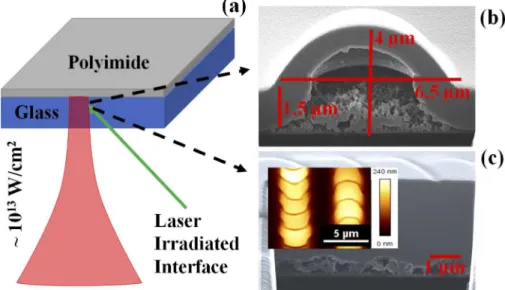
First, we fabricated individual blisters at 320 nJ energy (vacuum intensity ∼ 1.4 ×10¹⁴W/cm2) using a 20x 0.4 Numerical Aperture (NA) microscope objective yielding a focal spot of 3 µm diameter. We focused an ultrafast laser pulse at the glass-polymer interface as shown in schematic Fig.1(a). The polymer nonlinearly absorbs the laser energy, assuming a constant specific heat (a very coarse assumption), the polymer temperature rapidly reaches ∼ 10,000 K. At this temperature, the polymer locally melts and vaporizes forming a protruding blister as shown in Fig.1(b). For our calculations, we used a simple thermodynamic model considering an ultrashort laser pulse with 50 fs duration and a peak intensity of ∼ 1013W/cm2focused to a 3
µm spot. Since, we used a Gaussian pulse, the temperature induced by the pulse is not uniform, leading to vaporization of material in the center and melting on the edges [14].
Fig. 1. (color online) (a) A schematic showing the interaction of a single laser pulse (a central wavelength of 800 nm with peak intensity ∼ 1013W/cm2) focused at the glass-polymer interface as shown by the green arrow. This results in formation of a confined plasma leading to chemical change at the interface (Fig.1(b)) and a blister. (b) FIB image showing interior of a 1.5 µm polyimide blister fabricated at 320 nJ pulse energy using 20x 0.4 NA objective (vacuum intensity ∼ 1.4 ×10¹⁴W/cm2). The height and diameter of the blister are approximately ∼ 2.5 µm and 6.5 µm, respectively. With our 0.4 NA objective, we do not observe rupture even with 650 nJ incident energy (vacuum intensity ∼ 7 ×10¹³ W/cm2). (c) FIB image of laser-treated polyimide fabricated at a pulse energy of 650 nJ with an effective no.of 2-shots per site using a 10x 0.2 NA objective. Inset shows an AFM image of the same. Oriented crescent contours are seen in both AFM and FIB images. Blisters are approximately ∼ 240 nm in height and ∼ 4 µm in diameter.
We used focused gallium (Ga) and helium (He) ion beams to dissect and image the interior of blister as shown in Fig.1(b). The dissected blister showed formation of a hollow bubble underneath the chemically intact surface. Further, the FIB image showed an embedded layer (marked in Fig.1(a)) underneath the intact surface with a contrast from the surrounding unmodified polymer. The thin embedded layer underwent chemical transformation due to nonlinear absorption of 800 nm light. The penetration depth for photochemical modification via nonlinear absorption at laser wavelength of 800 nm is far less than 0.73 µm reported for single-photon modification at 400 nm [1]. However, at the intensities that we use in this experiment, high order absorption will dominate [14].
From the FIB image, it is evident that the ultrafast laser creates a confined chemical change followed by a morphological change (blister) at the glass-polymer interface. We also observed solidified molten material underneath the blister, some of which settled on the underlying glass (as shown in Fig.1(b)); both with same phase contrast. Since the local temperature generated in polyimide exceeds the melting point, the interaction region melts, cools and re-solidifies. The chemical properties of these melted regions are completely different from pristine polyimide. Though similar experiments involving blister formation for thin films of polyimide and titanium using 355 nm and 800 nm excitation wavelengths have been reported, these experiments lack the direct evidence of confined photochemical modification followed by deformation of the intact
polymer caused by a bubble underneath [1,7,18,19]. We report direct experimental evidence for confinement of chemical changes only at the interface using FIB diagnostic. It is not reported by others involving blister-actuated and dynamic release layer Laser-induced forward transfer (LIFT) experiments [1–4,18,20–31].
To determine the nature of the chemical changes at the glass-polymer interface, we created blister patterns on 3.5 µm thick polyimide films. The patterned surface areas of polyimide were matched with excitation beam sizes (∼ cm2) in a conventional fluorimeter and RAIRS to obtain a reasonable signal for spectroscopy analysis (later sections). Polyimide film was patterned using laser pulse energy of 650 nJ with a 10x (0.2 NA) microscope objective at scan speed of 4 mm/s (vacuum intensity ∼ 7 ×10¹³W/cm2) . The repetition rate of the laser was 2 kHz with a
line spacing (on the sample) of 6 µm. The effective no. of shots per focal spot is 2 with these conditions.
Figure1(c) shows an FIB image of a dissected overlapped blisters with effective no. of 2-shots per focal spot. The interior portions contained a series of hollow regions with crescent orientation along the scan direction. Inset shows AFM image of blistered surface. The AFM image of the blistered surface clearly shows oriented crescent contours of blisters in the writing direction of laser beam. When a laser pulse is incident, it imprints a circular contour. Since the sample effectively has 2 shots for each focal spot, the second shot imprints its contour on half of the modified region affected by the first shot while the sample is under motion.
In addition to FIB, we also carried out indirect measurements to study the extent (depth) of photochemical modification using reflection-absorption infrared spectroscopy (RAIRS) and X-ray Photoelectron (XPS) spectroscopy [32,33]. We did not coat polyimide thin films on a metal substrate as required and hence, the IR radiation interacted with the entire film and reflected by the glass substrate. Because of this, we could observe the photochemical changes which occurred beneath the film. The details of the RAIRS spectra recorded are given in AppendixB. We also carried out XPS characterization on these samples. X-rays are confined to the surface of material, with a sampling depth as small as 7.5 nm [32]. We did not observe any changes with XPS because the surface was intact in our case. Together, these spectroscopy techniques indicated the confinement of chemical modification at the polymer-glass interface, which coincided with observations from FIB diagnostics. We have also used other polymers such as polymethyl methacrylate (PMMA) and observed similar photochemical and morphological changes.
3.2. Photo chemistry: bulk fluorescence
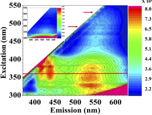
We performed fluorescence (excitation-emission) measurements to study chemical changes induced by an ultrafast intense laser pulse. We recorded fluorescence (emission) spectra for pristine and laser-modified polyimide (as shown in Fig.2). Fluorescence spectra were acquired with 300-550 nm excitation in steps of 5 nm. These spectra showed maximum fluorescence intensity in the blue and green regions for the laser-modified polyimide and almost negligible fluorescence in the visible spectrum for pristine polyimide, except fluorescence [34,35] from the polymer backbone (inset of Fig.2). The fluorescence intensities for pristine and laser-treated polyimide samples were compared and scaled. Their relative intensities are shown in the color scale.
To comprehend the nature of fluorescence, we compared the fluorescence from laser-altered polyimide with carbon dots [36]. Laser-treated polyimide showed fluorescence bands at 547 nm (at 425 nm excitation wavelength), 416 nm, and 436 nm (both at 350 nm excitation wavelength), which are consistent with violet, blue, and green carbon dots. We compared the fluorescence spectra with carbon dots because polymers are carbon rich materials and can be transformed into carbon clusters/carbon dust upon laser irradiation [13,37–43]. It is possible that the observed spectrum could be caused by either carbon dots and/or polymer aggregates resulted from polymer chain scission followed by rearrangement [44].
Fig. 2. (color online) Fluorescence spectra of laser-modified polyimide. Inset shows the background fluorescence of pristine polyimide. The fluorescence intensity of laser-modified polyimide is scaled relative to the pristine material and is shown in the color scale. The maximum fluorescence was obtained at 360 nm excitation shown with a red line. Laser-modified polyimide showed fluorescence bands at 552 nm and 572 nm at excitation wavelengths of 490 nm and 530 nm. Both excitation wavelengths are marked with red arrows. Not shown, we have also compared the spectrum of laser-modified borosilicate glass with the laser-modified polyimide to rule out its contribution.
We also studied the fluorescence properties of laser-modified polyimide above 425 nm excitation. We chose two excitation wavelengths at 490 nm and 530 nm (shown by red arrows in Fig.2) to compare the fluorescence with confocal fluorescence measurements in section3.4. When corrected for detector sensitivity, the fluorescence spectra in Fig.2at both the excitation wavelengths (490 nm and 530 nm) match the confocal fluorescence measurements in Fig.4. Both measurements reveal that the peak shifts relative to their pristine counterparts indicating a distorted polymer matrix.
When a fs laser pulse is focused at the glass-polyimide interface, it forms a hot plasma. When the plasma expands outwards, a temperature gradient is formed. It is possible that the material in the focal volume is vaporized and collects on the interface, resulting in carbon dots. The material that is not vaporized, but melted due to the temperature gradient, forms an underside layer of polyimide as seen in FIB image (Fig.1(b)). The heated, non-vaporized polymer remains attached to the expanding, unmodified polyimide material resulting in a distorted matrix.
Since the experiments involved high peak intensities in the substrate, fluorescence from laser-modified glass should be investigated to rule out its contribution. For this, we recorded fluorescence spectra for laser-modified borosilicate glass under similar excitation conditions. The recorded spectra were completely different from those of laser-modified polyimide which confirm that significant modification was only done to the polymer. However, we could not obtain fluorescent images of these modified regions using the fluorimeter.
3.3. Nature of fluorescence: polarization anisotropy studies
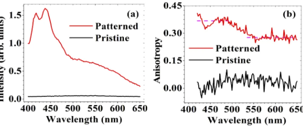
Several carbon-rich materials including carbon dots, polymers, and biological macromolecules have shown polarization anisotropy of fluorescence [35,36,45]. In these studies, linearly polarized light preferentially excites molecules or particles with transition dipole moments parallel to the polarization, leading to polarized emission. Thus, amorphous polymers tend to show less anisotropy, since molecular chains are randomly distributed. In the past, it was shown that a linearly polarized femtosecond laser beam can align polymer chains inducing formation of ordered aggregates, for example via π-π stacking [46–48] in spin-cast films. Fig.3(a) shows the fluorescence spectra for pristine and laser-treated polyimide samples at 360 nm excitation, where both samples have significant absorption. The laser-induced fluorescence for the patterned sample had two partially resolved bands and a shoulder with an intensity pattern resembling a spectrum of H-type aggregates. Spectral characteristics of ordered polymer aggregates are well described by HJ-aggregate model developed by Spano et al. [49–51]. In short, for J-type aggregates or a single planarized chain, the highest energy (shortest wavelength) fluorescence peak, I00peak is the strongest, while for H-type aggregates such as π-π stacked multiple chains
the I00peak is only weakly allowed and the I01peak carries the most intensity. Pristine polyimide
spectrum did not show any ordered aggregate signature due to a random distrbution of polymer chains.
Fig. 3. (color online) (a) Fluorescence spectra of pristine (black) and laser-modified polyimide (red) excited at 360 nm. (b) Polarization anisotropy of fluorescence for pristine (black) and laser-modified polyimide (red). Laser-modified polyimide showed anisotropy around 0.4 in the blue region and 0.3 in the green region. Both are indicated with dashed lines.
To confirm the formation of HJ-aggregates, we performed studies on polarization anisotropy of fluorescence. Molecules aligned along a given linear polarization are excited most strongly and the maximum fluorescence signal is obtained with a parallel analyzer. We acquired fluorescence spectra using 360 nm excitation for four combinations of parallel and perpendicular polarizer-analyzer orientations. Polarization anisotropy is determined from these spectra using the equation (1).
r = IVV− GIVH IVV + 2GIVH
(1) The fluorescence intensities are represented by a letter I followed by two subscripts. The first subscript denotes the excitation polarization and the second represents the fluorescence polarization. H and V represent horizontal and vertical polarization components. G is an instrumental factor related to the preference of the emission optics for the vertical polarization (IHV) to the horizontal polarization (IHH) and r is fluorescence anisotropy [52]. Fig.3(b) shows polarization anisotropy curves for pristine (black) and laser-patterned polyimide (red) samples.
We observed maximum fluorescence when the laser-irradiated sample was oriented so that the polarization of the writing laser was perpendicular to the excitation polarization of the fluorimeter. In these experiments, induced polarization anisotropy of fluorescence could be understood in two ways; (i) an ultrafast laser induces dipole moments parallel or perpendicular to its polarization depending upon net dipole moment along or perpendicular to a backbone axis which exerts a force that aligns molecules [53], and/or (ii) the laser selectively breaks molecular chains aligned with its polarization direction. Considering the peak intensity of the pulses used (∼ 1014W/cm2, far above the damage threshold of 1.3 × 10¹³W/cm2), the latter case is likely, though it has not been reported. In either case, the overall fluorescence is from specific molecular chains only.
3.4. Photo chemistry on micron scale
We collected Raman spectra from a confocal micro-Raman instrument to study photochemical modification on the scale of individual blisters. The black curve in Fig.4(a) shows a characteristic Raman spectrum for pristine polyimide excited with a 532 nm continuous wave (CW) laser. Six main peaks are easily resolved. Spectral signatures appearing at 1776 cm−1corresponds to C=O stretching, and the two peaks at 1620 cm−1and 1666 cm−1correspond to C=C stretching in ring and double ring structures respectively. The peaks at 1419 cm−1, 1377 cm−1, and 1131 cm−1 represent C-N, C-C stretches, and a ring breathing mode [54,55].
A Raman scattering spectrum was recorded from a single blister fabricated using an ultrafast laser at 258 nJ energy with a 0.4 NA objective. Considering the substrate-mediated absorption that onsets for I > 1012W/cm2, the remaining intensity Iabs ≈ 1013W/cm2 is delivered to the polymer. Blisters exhibited strong photoluminescence (fluorescence) as can be seen in Fig.4(a) (increased background for red curve), making detection of Raman-scattered photons difficult. We attempted to record Raman signals at other excitation wavelengths but could not succeed due to overwhelming background fluorescence. In the literature, intensity ratios between D (disorder ≈ 1350 cm−1) and G (graphite ≈ 1580 cm−1) bands are used to indicate carbonization due to ultrafast laser treatment [56]. In our case, we could not compare these intensities accurately since these bands were masked by strong background fluorescence.
To compare ultrafast-laser-induced photoluminescence with standard CW laser treatment, pristine polyimide was irradiated through a NA = 0.9 lens by 32 mW of CW excitation from a 532 nm laser for 60 s, and a Raman spectrum of the region was recorded (blue curve in Fig.4(a)). The CW laser (I = 8 ×10⁶W/cm2) modification did not show any fluorescence, but showed D and G bands indicating carbonization of polyimide [50]. The nature of photochemical modification with ultrafast laser pulses is distinct from that of CW lasers.
Since we observed photoluminescence masking the Raman signal, we acquired fluorescence spectra to compare with our earlier bulk measurements reported in Section2. We recorded fluorescence at four different excitation wavelengths (488 nm, 532 nm, 632 nm, and 785 nm) for pristine and blistered polyimide using a confocal micro-Raman instrument. Point-wise fluorescence spectra were integrated over individual blisters. Fig.4(b) shows integrated fluorescence spectra for polyimide blisters and pristine polyimide at two different excitation wavelengths (488 nm and 532 nm). Polyimide blisters have shown significant increases in fluorescence intensity. Also, fluorescence intensity decreased with increasing excitation wavelength, indicating that the ultrafast laser-induced defect states were formed in the UV region. This agrees with trend seen in bulk fluorescence measurements shown in Fig.2. Fig.4(c) shows fluorescence maps of a single blister for each excitation wavelength.
We noticed that the fluorescence intensity was always lower at the center than at the edges of a blister. This may be due to (i) the intensity of the Gaussian pulse at the center is maximum, inducing maximum pressure at the center resulting in deposition of modified material at the edges and/or (ii) the local temperature in the material at the center of Gaussian pulse results in more vapor formation. This leaves less modified material at the center than surroundings. In
Fig. 4. (color online) (a) Raman spectra for pristine (black), an individual polyimide blister fabricated at 258 nJ pulse energy (red) using 0.4 NA (vacuum intensity ∼ 1.1 ×10¹⁴W/cm2) , and a CW laser-modified polyimide (blue). Raman excitation wavelength was fixed at 532 nm. (b) Integrated photoluminescence of a blister at different excitations. As marked by red arrows in Fig.2, the fluorescence spectra close to the excitation wavelengths of 490 nm and 530 nm are compared. (c)Fluorescence from blisters at various excitation wavelengths with 1 mW power. The fluorescence at 488 nm (i) and 532 nm (ii) excitations showed an order of magnitude (105counts) more than that of fluorescence at excitation wavelengths of 632 nm (iii) and 785 nm (iv).
either case, the local fluorescence at the center is different from its surroundings due to the extent of laser-induced pressure or temperature.
4. Conclusion
Illumination of a polymer through a glass substrate creates a thin and modified polymer layer. Confined beneath the surface, chemical changes to the polymer are inevitable. We have studied these changes using different spectroscopic methods. To observe physical changes, we have focused an ion beam to cut open and then image a blister created by a 320 nJ (vacuum intensity ∼ 1.4 ×10¹⁴W/cm2) , 45-fs pulse focused to a full width at half maximum diameter of 6.5 µm. While some of the pulse energy is deposited in the substrate [14], a great deal is used to create
the hollow region that we observe, partially filled with debris, which localized at the interface. We also see evidence of modified polymer at the hollow region-polymer interface.
To observe chemical changes, we have measured bulk and locally excited fluorescence (using confocal micro-Raman) from the modified polymer. We see fluorescence signals that were not previously present. These changes are evidence of chemical changes to the material. By imaging the fluorescence we find that it comes mainly from the periphery of the bubble suggesting that it arises from material that has been liquefied or vaporized and then deposited at the outer edges of the blister. We also observe that the fluorescence is polarized and depends on the relative polarization of the writing and exciting beams.
The nonuniform chemical modification of this study is not a limitation to laser-induced forwards transfer (LIFT). We have demonstrated that the chemical changes induced by an ultrafast laser are confined to glass-polymer interface. The thrust generated in the mechanical deformation of the film deposits the material. Our study suggests femtosecond pulse irradiation of polymers through a glass substrate could be also important for applications in (i) contaminant-free LIFT, since heat and chemical changes are localized far from the polymer-vacuum interface, (ii) the nonuniformity may be helpful for producing high-quality micro lenses through the refractive index change confined to the interface and/or morphological change, (iii) data storage since laser-induced photochemical changes and anisotropy allow high storage capacity, and (iv) generating high harmonics in gases and solids since ionizing materials are the source of high-harmonic radiation [57,58].
Appendix A: characterization details
The interior modification confined to the interface was studied using focused ion beam (FIB) technique. Zeiss’s ORION NanoFab multi-column (GFIS, and Gallium-FIB) Helium Ion Microscope (HIM) and Focused Ion Beam (Gallium- FIB) were used to study the blistered samples. Gallium ions were used to dissect the blisters and Helium ions were used to image. Prior to performing dissection, the samples were coated with 30 nm Aluminium to protect from damage due to ion beam irradiation. Steady state and polarization-dependent fluorescence measurements were all performed with a Horiba Jobin Yvon Fluorolog Tau-3 Lifetime System. PI-2525 thin films were used for fluorescence measurements.
Raman spectroscopic data was recorded using a Witec Alpha a300 system (confocal micro-Raman) in the back scattering configuration using a 600 groove/mm grating at a resolution of 4.6 cm−1. An excitation laser operating at a wavelength of 532 nm, with a total power of 1 mW focussed through a 20x 0.4 N.A. objective, was used to excite Raman scattering in the polyimide samples. Each spectrum was recorded using 10 accumulations and an integration time of 15 s. Photoluminescence (fluorescence) imaging was performed using the same confocal micro-Raman instrument, with excitation wavelengths of 488 nm, 532 nm, 632 nm, and 785 nm. Images were acquired using a 100x 0.9 NA objective. To maximize the detectable spectral range, a 300 groove/mm grating was used, providing a spectral resolution of 0.27 nm. Images with dimensions of 15 µm by 15 µm were realized by recording 30 spectra per line and 30 lines per image. Each fluorescence spectrum was recorded using a single accumulation with an integration time of 0.5 s.
All AFM images were taken using the Nanowizard II BioAFM (Bruker, JPK Instruments, Berlin, Germany) mounted on an Olympus IX81 inverted microscope, operating in contact mode. Silicon nitride cantilevers (DNP-S, Veeco, CA) were used in contact mode imaging. Reflection-absorption infrared spectroscopy (RAIRS) measurements were performed on a Thermo Nicolet Nexus 870 FTIR spectrometer with the incident light beam hitting the surface of the sample at 80◦. Each spectrum is the average result of 256 accumulations recorded at a spectral resolution of 0.4 cm−1, taken from a region approximately 0.5 cm2in size. A clean borosilicate
covering the 4000 cm−1to 400 cm−1spectral range. XPS measurements were performed using Al Kα as an excitation source with energy of 1,486 eV. Both confocal micro-Raman and RAIRS measurements were carried out on PI-2555 thin films.
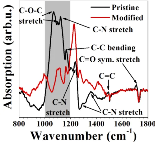
Appendix B: reflection-absorption infrared spectroscopy (RAIRS) studies We used the laser-treated polyimide sample (used in fluorescence studies) for comparison with pristine polyimide in RAIRS studies. Fig.5shows the RAIRS spectra for pristine (black) and laser-modified polyimide (red). The absorbed IR intensity is plotted against the Y-axis in arbitrary units as the IR spectrum for pristine polyimide was vertically shifted for comparison. A clear change in the shape of the RAIRS spectrum for laser-modified sample was observed. Since these experiments were carried out on two different samples, there could be variations in thicknesses of coverslips and polyimide thin films. Hence their intensities cannot be directly compared. However, the relative intensities for each of these vibrational modes in both the spectra can be compared with respect to a fixed vibrational mode. Absorption spectra were normalized to account for different film thicknesses.
Fig. 5. (color online) RAIRS spectra of pristine (black) and laser-modified (red) polyimide.
We assigned bands for pristine polyimide from the Refs. [59–61]. Bands close to 1068 cm−1, 1167 cm−1, and 1236 cm−1were assigned to C-O-C stretching, C-C bending, and C-N stretching [59]. Bands at 1358 cm−1, 1493 cm−1, and 1711 cm−1are assigned to C-N stretching of the aromatic ring, C=C stretching, and C=O symmetric stretching [60]. The bands close to 1099 cm−1 and 1122 cm−1 correspond to C-N imide and 1296 cm−1 correspond to C-N stretching modes [61]. These bands correspond to vibrational modes of different groups of polyimide.
From Fig.5, the pristine polyimide had less absorption in the region above 1200 cm−1compared to laser-modified polyimide. However, this trend was different in the highlighted region shown in the plot in between 1000-1200 cm−1. There are a few changes in peak positions of C-N stretching in the region 1300-1400 cm−1.
The vibrational modes for laser-modified polyimide shifted to 1097 cm−1, 1226 cm−1, and 1411 cm−1 compared to the corresponding vibrational modes of pristine polyimide at 1068 cm−1, 1236 cm−1, and 1358 cm−1, respectively. These studies together with relative intensity differences between laser-modified and pristine polyimide indicate that the laser-modified polyimide underwent photochemical and structural changes. These results agreed with earlier reports on UV exposed polyimide [61]. Oxygen containing groups on polymer surfaces such as C-O, C=O, and O-C=O are responsible for surface hydrophobicity and hydrophilicity of polymers [62] (though the role of surface roughness cannot be ignored). In our case, the absorption of C-O-C stretching (highlighted region) for laser-modified polyimide decreased indicating that the laser-treated polyimide transformed to hydrophobic. We performed hydrophobicity tests on these laser-modified polyimide surfaces and found that the results were in agreement with RAIRS studies reported earlier [62].
Funding
Natural Sciences and Engineering Research Council of Canada (Engage EGP 523138- 18, Discovery RGPIN-2019-04603); Ontario Centres of Excellence (VIP1 Program 29119); National Research Council Canada (Ideation Project A1-015007).
Acknowledgments
All authors acknowledge financial support from Natural Sciences and Engineering Research Council of Canada (NSERC), Ontario Centres of Excellence (OCE). Alan T. K. Godfrey acknowledges financial support from NSERC’s Postgraduate Scholarship - Doctoral and University of Ottawa’s Scholarship. Zygmunt J. Jakubek and Paul B Corkum acknowledge NRC Canada Ideation project (A1-015007) on Hybrid crystalline films for polymer electronics applications. We acknowledge Maohui Chen for training in atomic force microscopy, Choloong Hahn for FIB measurements, Tony Olivieri for training related to polyimide film fabrication, and Alexander Sander for acquiring XPS spectra.
Disclosures
The authors declare no conflicts of interest. References
1. N. T. Kattamis, P. E. Purnick, R. Weiss, and C. B. Arnold, “Thick film laser induced forward transfer for deposition of thermally and mechanically sensitive materials,”Appl. Phys. Lett.91(17), 171120 (2007).
2. M. S. Brown, N. T. Kattamis, and C. B. Arnold, “Time-resolved study of polyimide absorption layers for blister-actuated laser-induced forward transfer,”J. Appl. Phys.107(8), 083103 (2010).
3. M. S. Brown, C. F. Brasz, Y. Ventikos, and C. B. Arnold, “Impulsively actuated jets from thin liquid films for high-resolution printing applications,”J. Fluid Mech.709, 341–370 (2012).
4. N. T. Kattamis, M. S. Brown, and C. B. Arnold, “Finite element analysis of blister formation in laser-induced forward transfer,”J. Mater. Res.26(18), 2438–2449 (2011).
5. J. P. McDonald, V. R. Mistry, K. E. Ray, S. M. Yalisove, J. A. Nees, and N. R. Moody, “Femtosecond-laser-induced delamination and blister formation in thermal oxide films on silicon (100),”Appl. Phys. Lett.88(15), 153121 (2006).
6. J. R. Serrano and D. G. Cahill, “Laser-Induced Blistering of Thin SiO2 on Si,”Microscale Thermophys. Eng.9(2),
155–164 (2005).
7. N. T. Goodfriend, S. V. Starinskiy, O. A. Nerushev, N. M. Bulgakova, A. V. Bulgakov, and E. E. B. Campbell, “Laser pulse duration dependence of blister formation on back-radiated Ti thin films for BB-LIFT,”Appl. Phys. A122(3),
154 (2016).
8. M. Forster, W. Kautek, N. Faure, E. Audouard, and R. Stoian, “Periodic nanoscale structures on polyimide surfaces generated by temporally tailored femtosecond laser pulses,”Phys. Chem. Chem. Phys.13(9), 4155–4158 (2011).
9. C. De Marco, S. M. Eaton, R. Suriano, S. Turri, M. Levi, R. Ramponi, G. Cerullo, and R. Osellame, “Surface Properties of Femtosecond Laser Ablated PMMA,”ACS Appl. Mater. & Interfaces2(8), 2377–2384 (2010).
10. D. L. N. Kallepalli, A. M. Alshehri, D. T. Marquez, L. Andrzejewski, J. C. Scaiano, and R. Bhardwaj, “Ultra-high density optical data storage in common transparent plastics,”Sci. Rep.6(1), 26163 (2016).
11. D. L. N. Kallepalli, R. Kuladeep, S. V. Rao, and D. N. Rao, “Luminescent microstructures in bulk and thin films of PMMA, PDMS, PVA, and PS fabricated using femtosecond direct writing technique,”Chem. Phys. Lett.503(1-3),
57–60 (2011).
12. Z. Nie, H. Lee, H. Yoo, Y. Lee, Y. Kim, K.-S. Lim, and M. Lee, “Multilayered optical bit memory with a high signal-to-noise ratio in fluorescent polymethylmethacrylate,”Appl. Phys. Lett.94(11), 111912 (2009).
13. A. M. Alshehri, K. L. N. Deepak, D. T. Marquez, S. Desgreniers, and V. R. Bhardwaj, “Localized nanoclusters formation in PDMS upon irradiation with femtosecond laser,”Opt. Mater. Express5(4), 858–869 (2015).
14. A. T. K. Godfrey, D. L. N. Kallepalli, J. Ratte, and P. B. Corkum, Ultrafast laser-induced blister formation in polyimide films (unpublished data).
15. D. M. Rayner, A. Naumov, and P. B. Corkum, “Ultrashort pulse non-linear optical absorption in transparent media,”
Opt. Express13(9), 3208–3217 (2005).
16. L. Mercadier, D. M. Rayner, and P. B. Corkum, “Control of Femtosecond Laser Ablation of Thin Films from a Dielectric Surface by Nonlinear Interaction with the Substrate,”Phys. Rev. Appl.2(3), 034001 (2014).
17. A. Ben-Yakar and R. L. Byer, “Femtosecond laser ablation properties of borosilicate glass,”J. Appl. Phys.96(9),
5316–5323 (2004).
18. C. B. Arnold, P. Serra, and A. Piqué, “Laser Direct-Write Techniques for Printing of Complex Materials,”MRS Bull. 32(1), 23–31 (2007).
19. G. Heise, M. Domke, J. Konrad, S. Sarrach, J. Sotrop, and H. P. Huber, “Laser lift-off initiated by direct induced ablation of different metal thin films with ultra-short laser pulses,”J. Phys. D: Appl. Phys.45(31), 315303 (2012).
20. P. Delaporte and A.-P. Alloncle, “Laser-induced forward transfer: A high resolution additive manufacturing technology,”Opt. & Laser Technol.78, 33–41 (2016).
21. N. R. Schiele, D. T. Corr, Y. Huang, N. A. Raof, Y. Xie, and D. B. Chrisey, “Laser-based direct-write techniques for cell printing,”Biofabrication2(3), 032001 (2010).
22. G. Jing, Y. Wang, T. Zhou, S. F. Perry, M. T. Grimes, and S. Tatic-Lucic, “Cell patterning using molecular vapor deposition of self-assembled monolayers and lift-off technique,”Acta Biomater.7(3), 1094–1103 (2011).
23. M. Gruene, A. Deiwick, L. Koch, S. Schlie, C. Unger, N. Hofmann, I. Bernemann, B. Glasmacher, and B. Chichkov, “Laser Printing of Stem Cells for Biofabrication of Scaffold-Free Autologous Grafts,”Tissue Eng. Part C: Methods 17(1), 79–87 (2011).
24. L. Koch, S. Kuhn, H. Sorg, M. Gruene, S. Schlie, R. Gaebel, B. Polchow, K. Reimers, S. Stoelting, N. Ma, P. M. Vogt, G. Steinhoff, and B. Chichkov, “Laser Printing of Skin Cells and Human Stem Cells,”Tissue Eng. Part C: Methods16(5), 847–854 (2010).
25. A. Palla-Papavlu, V. Dinca, C. Luculescu, J. Shaw-Stewart, M. Nagel, T. Lippert, and M. Dinescu, “Laser induced forward transfer of soft materials,”J. Opt.12(12), 124014 (2010).
26. P. Serra, M. Colina, J. M. Fernández-Pradas, L. Sevilla, and J. L. Morenza, “Preparation of functional DNA microarrays through laser-induced forward transfer,”Appl. Phys. Lett.85(9), 1639–1641 (2004).
27. B. Hopp, T. Smausz, Z. Antal, N. Kresz, Z. Bor, and D. Chrisey, “Absorbing film assisted laser induced forward transfer of fungi (Trichoderma conidia),”J. Appl. Phys.96(6), 3478–3481 (2004).
28. J. Barron, P. Wu, H. Ladouceur, and B. Ringeisen, “Biological Laser Printing: A Novel Technique for Creating Heterogeneous 3-dimensional Cell Patterns,”Biomed. Microdevices6(2), 139–147 (2004).
29. J. Xu, J. Liu, D. Cui, M. Gerhold, A. Y. Wang, M. Nagel, and T. K. Lippert, “Laser-assisted forward transfer of multi-spectral nanocrystal quantum dot emitters,”Nanotechnology18(2), 025403 (2007).
30. R. Fardel, M. Nagel, F. Nüesch, T. Lippert, and A. Wokaun, “Fabrication of organic light-emitting diode pixels by laser-assisted forward transfer,”Appl. Phys. Lett.91(6), 061103 (2007).
31. D. P. Banks, K. Kaur, R. Gazia, R. Fardel, M. Nagel, T. Lippert, and R. W. Eason, “Triazene photopolymer dynamic release layer-assisted femtosecond laser-induced forward transfer with an active carrier substrate,”EPL83(3), 38003
(2008).
32. J. Yarwood, “Fourier Transform Infrared Reflection Spectroscopy for Surface Analysis,” Anal. Proc. 30, 6 (1993). 33. C. M. Chan and L.-T. Weng, “Surface Characterization of Polymer Blends by XPS and ToF-SIMS,”Materials9(8),
655 (2016).
34. K. Kanosue, R. Augulis, D. Peckus, R. Karpicz, T. Tamulevicius, S. Tamulevicius, V. Gulbinas, and S. Ando, “Polyimide and Imide Compound Exhibiting Bright Red Fluorescence with Very Large Stokes Shifts via Excited-State Intramolecular Proton Transfer II. Ultrafast Proton Transfer Dynamics in the Excited State,”Macromolecules49(5),
1848–1857 (2016).
35. E. D. Wachsman and C. W. Frank, “Effect of cure history on the morphology of polyimide: Fluorescence spectroscopy as a method for determining the degree of cure,”Polymer29(7), 1191–1197 (1988).
36. M. O. Dekaliuk, O. Viagin, Y. V. Malyukin, and A. P. Demchenko, “Fluorescent carbon nanomaterials: "quantum dots" or nanoclusters?”Phys. Chem. Chem. Phys.16(30), 16075–16084 (2014).
37. S. Hayashi, M. Kataoka, and K. Yamamoto, “Photoluminescence Spectra of Carbon Clusters Embedded in SiO2,”
38. W. Krätschmer, K. Fostiropoulos, and D. R. Huffman, “The infrared and ultraviolet absorption spectra of laboratory-produced carbon dust: evidence for the presence of the C60 molecule,”Chem. Phys. Lett.170(2-3), 167–170
(1990).
39. D. Fink, W. H. Chung, R. Klett, A. Schmoldt, J. Cardoso, R. Montiel, M. H. Vazquez, L. Wang, F. Hosoi, H. Omichi, and P. Goppelt-Langer, “Carbonaceous clusters in irradiated polymers as revealed by UV-Vis spectrometry,”Radiat. Eff. Defects Solids133(3), 193–208 (1995).
40. D. Fink, R. Klett, L. T. Chadderton, J. Cardoso, R. Montiel, H. Vazquez, and A. A. Karanovich, “Carbonaceous clusters in irradiated polymers as revealed by small angle X-ray scattering and ESR,”Nucl. Instruments Methods Phys. Res. Sect. B: Beam Interactions with Mater. Atoms111(3-4), 303–314 (1996).
41. S. Gupta, D. Choudhary, and A. Sarma, “Study of carbonaceous clusters in irradiated polycarbonate with UV-vis spectroscopy,”J. Polym. Sci. Part B: Polym. Phys.38(12), 1589–1594 (2000).
42. E. Despagnet-Ayoub, W. W. Kramer, W. Sattler, A. Sattler, P. J. LaBeaume, J. W. Thackeray, J. F. Cameron, T. Cardolaccia, A. A. Rachford, J. R. Winkler, and H. B. Gray, “Triphenylsulfonium topophotochemistry,”Photochem. Photobiol. Sci.17(1), 27–34 (2018).
43. U. Mahilny, A. Trofimova, S. Nazarov, A. Tolstik, R. Heintzmann, and E. Tolstik, “Highly concentrated phenan-threnequinone polymethylmethacrylate composite for thick reflection holograms recording at 532 nm,”Opt. Mater. Express6(11), 3427–3437 (2016).
44. T. Wang, A. Wang, R. Wang, Z. Liu, Y. Sun, G. Shan, Y. Chen, and Y. Liu, “Carbon dots with molecular fluorescence and their application as a "turn-off" fluorescent probe for ferricyanide detection,”Sci. Rep.9(1), 10723 (2019).
45. D. M. Jameson and J. A. Ross, “Fluorescence Polarization/Anisotropy in Diagnostics and Imaging,”Chem. Rev. 110(5), 2685–2708 (2010).
46. S. Chae, K. H. Jo, S. W. Lee, H.-S. Keum, H. J. Kim, J. Choi, and H. H. Lee, “Selective Chain Alignment of Conducting Polymer Blend Films by an Ultrafast Laser,”Macromol. Chem. Phys.217(4), 537–542 (2016).
47. A. Moliton and R. C. Hiorns, “Review of electronic and optical properties of semiconducting π-conjugated polymers: applications in optoelectronics,”Polym. Int.53(10), 1397–1412 (2004).
48. Y. Martinez-Rubi, Z. J. Jakubek, M. B. Jakubinek, K. S. Kim, F. Cheng, M. Couillard, C. Kingston, and B. Simard, “Self-Assembly and Visualization of Poly(3-hexyl-thiophene) Chain Alignment along Boron Nitride Nanotubes,”J. Phys. Chem. C119(47), 26605–26610 (2015).
49. H. Yamagata and F. C. Spano, “Interplay between intrachain and interchain interactions in semiconducting polymer assemblies: The HJ-aggregate model,”J. Chem. Phys.136(18), 184901 (2012).
50. F. C. Spano and C. Silva, “H- and J-Aggregate Behavior in Polymeric Semiconductors,”Annu. Rev. Phys. Chem. 65(1), 477–500 (2014).
51. T. Eder, T. Stangl, M. Gmelch, K. Remmerssen, D. Laux, S. Höger, J. M. Lupton, and J. Vogelsang, “Switching between H- and J-type electronic coupling in single conjugated polymer aggregates,”Nat. Commun.8(1), 1641
(2017).
52. J. R. Lakowicz and B. R. Masters, “Principles of Fluorescence Spectroscopy, Third Edition,”J. Biomed. Opt.13(2),
029901 (2008).
53. M. Nishikawa, B. Taheri, and J. L. West, “Mechanism of unidirectional liquid-crystal alignment on polyimides with linearly polarized ultraviolet light exposure,”Appl. Phys. Lett.72(19), 2403–2405 (1998).
54. X. J. Gu, “Raman spectroscopy and the effects of ultraviolet irradiation on polyimide film,”Appl. Phys. Lett.62(13),
1568–1570 (1993).
55. A. K. Shukla, V. M. Yadav, A. Kumar, I. A. Palani, and A. Manivannan, “Investigations on effect of laser-induced self-assembled patterning on optical properties of flexible polyimide substrates for solar cell applications,”J. Phys. D: Appl. Phys.51(4), 045502 (2018).
56. C. Cheng, S. Wang, J. Wu, Y. Yu, R. Li, S. Eda, J. Chen, G. Feng, B. Lawrie, and A. Hu, “Bisphenol A Sensors on Polyimide Fabricated by Laser Direct Writing for Onsite River Water Monitoring at Attomolar Concentration,”ACS Appl. Mater. & Interfaces8(28), 17784–17792 (2016).
57. M. Hentschel, R. Kienberger, C. Spielmann, G. A. Reider, N. Milosevic, T. Brabec, P. Corkum, U. Heinzmann, M. Drescher, and F. Krausz, “Attosecond metrology,”Nature414(6863), 509–513 (2001).
58. G. Vampa, T. J. Hammond, N. Thiré, B. E. Schmidt, F. Légaré, C. R. McDonald, T. Brabec, and P. B. Corkum, “Linking high harmonics from gases and solids,”Nature522(7557), 462–464 (2015).
59. M. Garg and J. K. Quamara, “FTIR analysis of high energy heavy ion irradiated kapton-H polyimide,” (2007). 60. Y.-K. Xu, M.-S. Zhan, and K. Wang, “Structure and properties of polyimide films during a far-infrared-induced
imidization process,”J. Polym. Sci. Part B: Polym. Phys.42(13), 2490–2501 (2004).
61. K. P. Adhi, R. L. Owings, T. A. Railkar, W. D. Brown, and A. P. Malshe, “Chemical modifications in femtosecond ultraviolet (248 nm) excimer laser radiation-processed polyimide,”Appl. Surf. Sci.225(1-4), 324–331 (2004).
62. C. Qu, J. Hu, X. Liu, Z. Li, and Y. Ding, “Morphology and Mechanical Properties of Polyimide Films: The Effects of UV Irradiation on Microscale Surface,”Mater. (Basel)10(11), 1329 (2017).