establishes ER redox homeostasis
The MIT Faculty has made this article openly available.
Please share
how this access benefits you. Your story matters.
Citation
Kim, S. et al. “Balanced Ero1 Activation and Inactivation Establishes
ER Redox Homeostasis.” The Journal of Cell Biology 196.6 (2012):
713–725. Web. 4 May 2012.
As Published
http://dx.doi.org/10.1083/jcb.201110090
Publisher
Rockefeller University Press, The
Version
Final published version
Citable link
http://hdl.handle.net/1721.1/70516
Terms of Use
Creative Commons Attribution-Noncommercial-Share Alike 3.0
Unported
The Rockefeller University Press $30.00
Correspondence to Chris A. Kaiser: ckaiser@mit.edu
C.S. Sevier’s present address is Dept. of Molecular Medicine, Cornell University, Ithaca, NY 14853.
Abbreviations used in this paper: BSO, buthionine sulfoximine; CPY, carboxy-peptidase Y; FAD, flavin adenine dinucleotide; MBP, maltose binding protein; PDI, protein disulfide isomerase; SMM, supplemented minimal medium; TEV, Tobacco etch virus.
Introduction
A key step in the maturation of many secreted proteins and extra cellular domains of membrane proteins is the formation of di sulfide bonds in the ER. Disulfide bonds, which stabilize native and functional conformations of proteins, are formed by pairing and oxidation of cysteines during the initial folding process in the ER. In Saccharomyces cerevisiae, most biosynthetic disul fide bonds are formed by dithiol/disulfide transfer reactions with the oxidized form of protein disulfide isomerase (PDI), Pdi1p (Sevier and Kaiser, 2002; Wilkinson and Gilbert, 2004). Pdi1p, in turn, requires other oxidizing molecules to be recycled because Pdi1p cannot generate disulfide bonds by itself. We and other colleagues identified the major disulfidegenerating flavoenzyme Ero1p in S. cerevisiae (Frand and Kaiser, 1998; Pollard et al., 1998). In both yeast and mammalian cells, Ero1 directly transfers disulfide bonds to PDI (Frand and Kaiser, 2000; Mezghrani et al., 2001). The oxidative capacity of the yeast ER depends primarily on the activity of Ero1p: a temperaturesensitive
ero1-1 mutation decreases the resistance of yeast to the reducing agent DTT and induces the unfolded protein response with ac cumulation of secretory proteins, and, after prolonged incubation
at the restrictive temperature, a strain with this mutation loses viability (Frand and Kaiser, 1998, 1999; Cuozzo and Kaiser, 1999; Tu and Weissman, 2002), and overexpression of ERO1 from a multicopy plasmid increases the oxidizing capacity of yeast cells, as shown by their increased resistance to DTT (Frand and Kaiser, 1998).
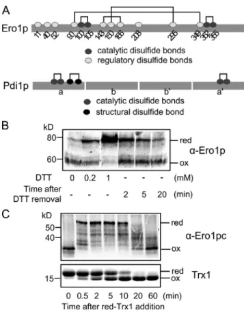
The catalytic cycle of Ero1p depends on a relay of disulfide bonds from the conserved activesite cysteine pair, C352C355, which is proximal to bound flavin adenine dinucleotide (FAD) to the second shuttle cysteine pair, C100C105, which is respon sible for direct disulfide transfer to Pdi1p (Frand and Kaiser, 2000; Gross et al., 2004; Sevier and Kaiser, 2006b). In addition to these two catalytic cysteine pairs, Ero1p contains three cyste ine pairs (C90C349, C143C166, and C150C295) that form regulatory bonds (Sevier et al., 2007). Several lines of evidence indicate that Ero1p is inactive with regulatory bonds formed, whereas it is active with regulatory cysteines in the reduced state. Ero1p is converted from an oxidized (inactive) state to the reduced (active) state in the presence of reduced thioredoxin1 (Trx1) as a substrate; once Trx1 is fully oxidized through the action of active Ero1p, Ero1p returns to the oxidized (inactive) form (Sevier et al., 2007). A double mutant, Ero1pC150AC295A, which cannot form the crucial C150C295 regulatory bond,
T
he endoplasmic reticulum (ER) provides an environ-ment optimized for oxidative protein folding through the action of Ero1p, which generates disulfide bonds, and Pdi1p, which receives disulfide bonds from Ero1p and transfers them to substrate proteins. Feedback regula-tion of Ero1p through reducregula-tion and oxidaregula-tion of regulatory bonds within Ero1p is essential for maintaining the proper redox balance in the ER. In this paper, we show that Pdi1p is the key regulator of Ero1p activity. Reduced Pdi1p resulted in the activation of Ero1p by direct reduction of Ero1pregulatory bonds. Conversely, upon depletion of thiol sub-strates and accumulation of oxidized Pdi1p, Ero1p was inactivated by both autonomous oxidation and Pdi1p-mediated oxidation of Ero1p regulatory bonds. Pdi1p re-sponded to the availability of free thiols and the relative levels of reduced and oxidized glutathione in the ER to control Ero1p activity and ensure that cells generate the minimum number of disulfide bonds needed for efficient oxidative protein folding.
Balanced Ero1 activation and inactivation
establishes ER redox homeostasis
Sunghwan Kim, Dionisia P. Sideris, Carolyn S. Sevier, and Chris A. Kaiser
Department of Biology, Massachusetts Institute of Technology, Cambridge, MA 02139
© 2012 Kim et al. This article is distributed under the terms of an Attribution–Noncommercial– Share Alike–No Mirror Sites license for the first six months after the publication date (see http://www.rupress.org/terms). After six months it is available under a Creative Commons License (Attribution–Noncommercial–Share Alike 3.0 Unported license, as described at http://creativecommons.org/licenses/by-nc-sa/3.0/).
THE
JOURNAL
OF
CELL
BIOLOGY
on April 18, 2012 jcb.rupress.org Downloaded from http://jcb.rupress.org/content/suppl/2012/04/02/jcb.201110090.DC2.html http://jcb.rupress.org/content/suppl/2012/03/07/jcb.201110090.DC1.html Supplemental Material can be found at:et al., 2008). We have reconstituted this coupled reaction in vitro and show that it recapitulates properties of the ER in vivo; as long as an excess of reduced glutathione is available, Pdi1p is maintained in a state enabled to activate Ero1p, which in turn provides oxidizing equivalents to oxidize glutathione.
Results
Ero1p is capable of autonomous inactivation
Because the regulatory bonds link distant sites of the Ero1p polypeptide chain, the presence or absence of these bonds can be readily discerned by mobility differences on nonreducing SDSPAGE (Fig. 1 A). Activity of Ero1p can be inferred from the redox state of the regulatory bonds. In live yeast cells, Ero1p is converted to the reduced (active) form in cells treated with the reducing agent DTT and then returns to the oxidized (inactive) form after DTT removal (Fig. 1 B). Activation of Ero1p by re duction of the regulatory bonds and subsequent inactivation by reformation of the regulatory bonds could also be readily observed in an in vitro assay containing recombinant Ero1p (Ero1pc, 56–424, with an Nterminal (1–55) and a Cterminal (425–563) fragment removed) and an excess of reduced Trx1, exhibits increased Ero1p activity in vitro and in vivo (Sevier
et al., 2007).
A byproduct of Ero1p activity is the generation of hydro gen peroxide as a result of a twoelectron reduction of oxygen per disulfide generated. Thus, although Ero1p activity is essential for biosynthetic disulfide bond formation, uncontrolled Ero1p activity could produce too much reactive oxygen species that would be detrimental to the cell (Gross et al., 2006). Indeed, overexpression of the hyperactive Ero1pC150AC295A mutant inhibits cell growth, highlighting the physiological importance of the regulatory bonds in keeping Ero1p activity in check. Mammalian Ero1 has been shown to modulate its activity through a similar mechanism involving regulatory bonds, show ing that autoregulation in response to the redox environment is a general property of Ero1 (AppenzellerHerzog et al., 2008; Baker et al., 2008). An additional layer of protection against reactive oxygen accumulation in the ER of mammalian cells is provided by an ER resident, peroxiredoxin, which reduces hydrogen peroxide generated by Ero1 to limit peroxide accumulation (Tavender et al., 2008, 2010; Zito et al., 2010). An obvious sequence homolog to peroxiredoxin is absent in yeast; whether additional pathways functionally similar to the peroxiredoxin system exist in the ER of fungi remains to be explored.
The precise mechanism by which the regulatory bonds of Ero1 are reduced and then reoxidized will be crucial to under stand how Ero1p activity is controlled and which features of the redox environment of the ER are sensed by Ero1p. Pdi1p is the most abundant oxidoreductase of the yeast ER and is the pre ferred physiological substrate for oxidation by Ero1p (Nørgaard et al., 2001; Vitu et al., 2010); therefore, Pdi1p is likely to play an important part in regulating Ero1p. In the mammalian ER, the disulfide relay between Ero1 and PDI is regulated at least in part by the availability of PDI (AppenzellerHerzog et al., 2010; Inaba et al., 2010). Interaction between Ero1 and PDI has been shown to be facilitated by a protruding hairpin in Ero1 and the b domain of PDI, which facilitates the inter conversion between the Ox1 and Ox2 redoxactive forms of Ero1 (Masui et al., 2011). Differences between Ero1 and Ero1p, including the lack of a hairpin in Ero1p and the prefer ence of Ero1p for the a domain of Pdi1p (versus the preference of Ero1 for the a domain), suggest that the yeast and mam malian Ero1PDI systems may operate differently.
In vitro assays showing activation of Ero1p have typically used the nonphysiological substrate Trx1, which has a single CxxCcontaining domain with a highly reducing reduction po tential rather than the physiological substrate Pdi1p because re duced Trx1 is more effective than Pdi1p at activating Ero1p by reduction of regulatory bonds (Sevier et al., 2007; Baker et al., 2008; Vitu et al., 2010). Here, we examine the role of Pdi1p in the reduction and reoxidation of the Ero1p regulatory bonds to reveal how the activity of Ero1p is set in a living cell. We have also sought to understand the role of glutathione in this process as the most abundant redox buffer of the ER. Although glutathione itself does not activate Ero1p nor is glutathione efficiently used as a substrate for Ero1p (Tu et al., 2000; Sevier and Kaiser, 2006b), reduced glutathione can stimulate Ero1p activity when used in conjunction with Pdi1p as a substrate (Sevier et al., 2007; Baker
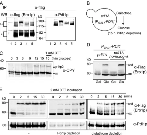
Figure 1. Ero1p regulation by reduction or oxidation of its regulatory bonds. (A) Disulfide connectivity of Ero1p and Pdi1p. In Ero1p, dark gray and light gray circles indicate catalytic and regulatory cysteines, respec-tively. Pdi1p has four thioredoxin domains, a, b, b, and a. In Pdi1p, dark gray and black circles indicate catalytic and structural cysteines, respectively. (B) Ero1p cycles between oxidized (ox) and reduced (red) regulatory states in vivo according to the redox environment. Cells were incubated with 0, 0.2, or 1 mM DTT for 30 min. DTT was removed by placing cells in fresh medium. (C) Ero1p regulation in vitro. 1 µM Ero1pc was incubated with 50 µM reduced Trx1.
on April 18, 2012
jcb.rupress.org
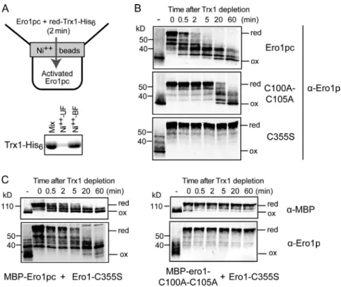
and oxidized forms likely correspond to partially oxidized states of Ero1pc. Parallel reactions using Ero1pc mutants lack ing the shuttle disulfide bond (Ero1pcC100AC105A) or the catalytic disulfide bond (Ero1pcC355S) in the absence of Trx1 exhibited greatly slowed or no autonomous reoxidation of Ero1pc, respectively (Fig. 2 B, middle and bottom). In addition, when the reoxidation reaction was performed in the absence of O2 as an electron acceptor (Fig. S1 A, right), Ero1pc exhibited
slowed autonomous reoxidation. These observations show that Ero1pc is capable of autonomous oxidation to form regulatory bonds and that this oxidation has catalytic requirements similar to biosynthetic disulfide bond formation.
We considered the possibility that autonomous oxidation of the Ero1p regulatory bonds was promoted by the hydrogen peroxide generated by Ero1pc before removal of Trx1. There fore, we examined the effects of hydrogen peroxide. Addition of catalase to the reaction, which should convert any hydrogen peroxide into water and oxygen (Fig. S1 B), did not signifi cantly slow autonomous oxidation of the Ero1p regulatory bonds (Fig. S1 A, middle). Conversely, addition of 100 µM hydro gen peroxide, the maximum concentration that could be obtained from complete oxidation of 100 µM Trx1, did not enhance the oxidation of Ero1pcC100AC105A (Fig. S1 C).
Autonomous oxidation of Ero1p occurs via an intermolecular reaction
Autonomous oxidation of Ero1p could conceivably occur by intramolecular (in cis) or intermolecular (in trans) disulfide bond transfer. In the crystal structure of Ero1pc (Gross et al., 2004), one of the regulatory bonds, C90C349, is close enough to the catalytic site to be oxidized in cis, whereas the other two regula tory bonds, C150C295 and C143C166, are located >20 Å away from the catalytic site, suggesting that the oxidation of these serving as an effective substrate for Ero1p (Fig. 1 C). First,
Ero1pc is inactive with the regulatory bonds in a fully oxidized state. In 0.5 min after addition of reduced Trx1, the regulatory bonds of Ero1pc become reduced, and Ero1pc is thus activated for the oxidation of Trx1. As a consequence of Ero1pc, after 60 min, all of the Trx1 is oxidized, and Ero1pc returns to the oxidized inactive form (Fig. 1 C).
Activation of Ero1pc by reduction of the regulatory bonds initiates on addition of reduced Trx1, indicating that the regu latory bonds can be reduced by direct dithiol/disulfide ex change with reduced Trx1. For the end phase of the reaction in which most of the Trx1 is oxidized and the regulatory bonds of Ero1pc become reoxidized, we wished to know whether reoxi dation of the regulatory bonds occurred by direct oxidation by oxidized Trx1 or instead by autonomous reoxidation by Ero1p when all of the reduced Trx1 had been consumed. To distin guish between these possibilities, we first tested the ability of active Ero1pc to oxidize regulatory cysteines in the absence of Trx1. For this experiment, 1 µM Ero1pc was incubated with 100 µM His6tagged reduced Trx1 for 2 min, a time sufficient
for complete activation of Ero1pc by reduction of the regula tory bonds. Subsequently, His6tagged Trx1 was removed by
affinity to Ni2+ beads (Fig. 2 A). Trx1 depletion was confirmed
by the significant absence of Trx1 in the Ni2+ unbound fraction
and corresponding presence of Trx1 bound to the Ni2+ beads
(unbound faction; Fig. 2 A, bottom). A small amount of residual Trx1 was observed in the unbound fraction (Fig. 2 A, bottom), but it was anticipated that this limited fraction of Trx1 would be rapidly oxidized by Ero1pc and not contribute to our Ero1pc inactivation assay. After depletion of reduced Trx1, Ero1pc was converted from a fully reduced to an oxidized form over a period of 60 min with a half time for completion of 10 min (Fig. 2 B, top). The bands migrating between the fully reduced
Figure 2. Pathways for Ero1p inactivation. (A) To test the ability of Ero1p to oxidize itself autono-mously, 1 µM reduced (red) Ero1pc was prepared
by incubation with 100 µM reduced His6-tagged
Trx1 for 2 min and then passage through Ni2+ beads
to remove Trx1. Trx1 depletion was confirmed by
SDS-PAGE (Mix; initial mixture: Ni2+-BF [beads
bound fraction] and Ni2+-UF [unbound fraction]).
(B) Autonomous reoxidation (ox) of Ero1pc after removal of Trx1 occurs rapidly and efficiently but depends on a functional active site (C355), and ef-ficient reoxidation depends on the shuttle disulfide (C100-C105). The first lane in each time course () shows the oxidation state of Ero1pc before re-duction by Trx1. (C) Ero1pc, expressed as an MBP fusion (MBP-Ero1pc), can reoxidize Ero1pc-C355S in trans. An equal mixture of Ero1pc (or MBP-ero1-C100A-C105A; right) and Ero1pc-C355S was reduced by incubation with reduced Trx1 followed by Trx1 depletion. The time course of reox-idation of MBP-Ero1pc and Ero1pc-C355S was fol-lowed independently by separating MBP-Ero1pc and Ero1pc-C355S by size in SDS-PAGE and immuno-blotting for MBP or Ero1.
on April 18, 2012
jcb.rupress.org
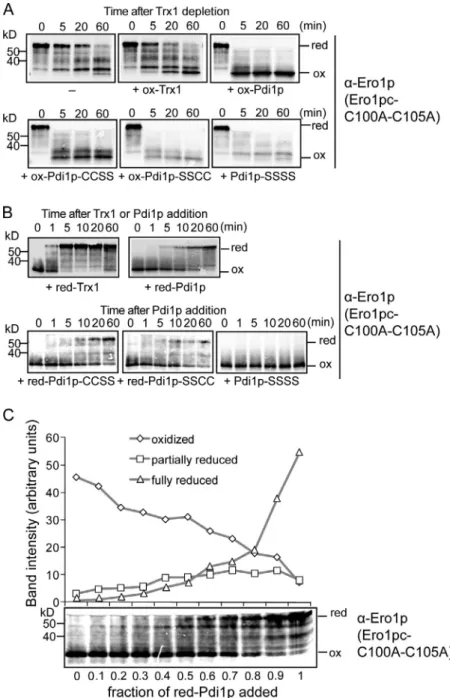
oxidized (Fig. S2) and Trx1 that was almost fully in the oxidized form (not depicted). We found that addition of a 20fold molar excess of oxidized Trx1 had no effect on the rate of reoxida tion of Ero1pcC100AC105A (Fig. 3 A, top), showing that oxidized Trx1 has little or no capability to oxidize the regula tory bonds of Ero1p. In contrast, oxidized Pdi1p dramatically increased the rate of Ero1pcC100AC105A reoxidation (Fig. 3 A, top right). The ability of oxidized Pdi1p to efficiently oxidize the regulatory bonds of Ero1p was not a peculiarity of the Ero1pcC100AC105A mutant, as we could also show that oxidized Pdi1p accelerated the oxidation of wildtype Ero1pc as well as Ero1pcC355S, which is unable to oxidize its regula tory bonds (Fig. S3). These results show that Ero1p can be in activated by direct feedback from Pdi1p.
Next, we tested the converse process, the ability of re duced Pdi1p to reduce and thus activate Ero1p. To assess the activation process selectively, we again used Ero1pcC100A C105A because of this mutant’s greatly diminished capacity for autonomous oxidation of regulatory cysteines once reduced with an exogenous agent (Fig. 2 B, middle). Reduced Pdi1p re quires 60 min to reduce the Ero1p regulatory bonds completely, whereas reduced Trx1 requires <5 min, indicating that Pdi1p is a relatively poor activator of Ero1p (Fig. 3 B, top). In summary, Trx1 is a potent activator but poor inactivator of Ero1p, whereas Pdi1p is a weak activator but potent inhibitor of Ero1p. These contrasting abilities to reduce or oxidize the regulatory bonds of Ero1p can be accounted for by the much greater reducing po tential of Trx1 as compared with Pdi1p. By assaying the rate of activation or inactivation of Ero1p in separate reactions, we could show that reduced Pdi1p activates Ero1p more slowly than oxidized Pdi1p can inactivate Ero1p. This explains why in an in vitro Ero1p oxidation reaction that contains reduced Pdi1p as a substrate, Ero1p rarely, if ever, becomes fully activated, and the reaction proceeds relatively slowly.
The a and a domains of Pdi1p differ in their ability to regulate Ero1p
Yeast Pdi1p has two redoxactive thioredoxinlike domains with different reduction potentials: the a domain (195 mV) and a domain (164 mV; Fig. 1 A; Tian et al., 2006; Vitu et al., 2010). We recently showed that Ero1p preferentially oxidizes the a domain both in vivo and in vitro, suggesting Ero1p provides oxi dizing equivalents to the ER mainly via the a domain (Vitu et al., 2010). Thus, the a domain may play a lesser role in the oxida tion of ER substrates despite its higher reduction potential than the a domain. To fully understand the interactions between Pdi1p and Ero1p, we wished to evaluate the contribution of each of the active Pdi1p domains to reduction and oxidation of Ero1p regu latory bonds.
Recombinant Pdi1p purified from E. coli had a fully oxidized a domain and a partially oxidized a domain (Fig. S2), consistent with the different reduction potentials of the two domains. More over, naturally oxidized purified domain mutants Pdi1pCCSS (a domain cysteines intact) and Pdi1pSSCC (a domain cyste ines intact) had similar oxidation states to the corresponding domains in wildtype Pdi1p (unpublished data). The same assay for Pdi1p oxidation of the Ero1p regulatory bonds was performed bonds would require involvement of additional dithiol/disulfide
exchanges with other cysteines. To determine whether one Ero1p protein can oxidize the regulatory bonds of another Ero1p, we asked whether Ero1pcC355S, which is catalytically inactive and is not capable of autonomous oxidation, could nevertheless be oxidized in trans by active Ero1pc. A maltose binding pro tein (MBP) fusion to Ero1pc was used in this experiment so that MBPEro1pc and Ero1pcC355S could be distinguished by their distinct mobilities on SDSPAGE. An autonomous oxida tion assay was established by combining MBPEro1pc and Ero1pcC355S with reduced Trx1 to reduce the regulatory bonds on both forms of Ero1p. After Trx1 was removed by Ni2+ affinity,
both Ero1pcC355S and MBPEro1pc were reoxidized at nearly equal rates, showing that efficient trans oxidation can occur (Fig. 2 C). Consistent with the oxidation of Ero1pcC355S being a product of MBPEro1pc catalytic function, disruption of the MBPEro1p catalytic cycle by mutating the C100C105 pair (MBPero1C100AC105A) prevented Ero1pcC355S oxi dation (Fig. 2 C). We also tested the effect of dilution of Ero1pc in our autonomous oxidation assay and found that a fourfold dilution of Ero1pc slowed autonomous oxidation of Ero1pc (unpublished data). Together, the trans oxidation experiment and the dilution experiment show that autonomous oxidation of the Ero1p regulatory disulfides occurs predominantly, if not exclu sively, by one Ero1p protein oxidizing the regulatory bonds of another Ero1p. The simplest mechanism for such dithiol/disul fide exchange would be for successive direct transfer of the C100C105 shuttle cysteine disulfide bond to each of the three regulatory cysteine pairs. However, we cannot rule out the pos sibility that more complicated dithiol/disulfide rearrangements occur. Slow oxidation of Ero1pc facilitated by the Ero1pc C100AC105A (but not the Ero1pcC355S) mutant could be observed also at late time points of the autonomous oxidation assay (>20 min), suggesting that the C352C355 active site may contribute directly to oxidation of Ero1pc.
Ero1p is activated slowly but inactivated rapidly by Pdi1p
As we have shown, reduced Trx1 is a potent activator of Ero1p, and the use of Trx1 as an activator and substrate for Ero1p in in vitro assays made it possible to document the dramatic effect that reduction and oxidation of regulatory cysteines have on Ero1p activity (Fig. 1 C; Sevier et al., 2007). However, Pdi1p is the major physiological substrate of Ero1p (Frand and Kaiser, 1999), and we were interested to use the in vitro assay for Ero1p regulation to make a comparable assessment of the ability of Pdi1p to either activate or inactivate Ero1p activity through reduction or oxidation of regulatory bonds.
First, we tested the ability of oxidized Pdi1p to inactivate Ero1p. We prepared activated Ero1pcC100AC105A by treat ment with reduced Trx1 followed by removal of Trx1. The use of the Ero1pcC100AC105A mutant prevented efficient auton omous reoxidation of Ero1p, allowing us to test the ability of exogenous agents to reoxidize Ero1p. Oxidized Pdi1p and Trx1 were prepared by air oxidation during purification from
Esche-richia coli. The endpoint of air oxidation yielded Pdi1p that had the a domain almost fully oxidized and the a domain 50%
on April 18, 2012
jcb.rupress.org
both reduced Pdi1pCCSS and reduced Pdi1pSSCC reduced Ero1p regulatory bonds at similar rates (Fig. 3 B, bottom). As expected, the singledomain mutants reduced the Ero1p regula tory bonds somewhat less efficiently than did wildtype Pdi1p, which has both active sites intact, and the double domain mutant Pdi1pSSSS did not reduce the Ero1p regulatory bonds (Fig. 3 B, bottom right).
The redox state of Ero1p can be set by a mix of reduced and oxidized Pdi1p
Reduced and oxidized Pdi1p promote activation and inactivation of Ero1p, respectively, suggesting that the activity of Ero1p in steady state may be set by the ratio of reduced to oxidized Pdi1p. By taking advantage of Ero1pcC100AC105A, which lacks the abil ity to oxidize Pdi1p efficiently, we could test admixtures of oxidized and reduced Pdi1p for their effect on the steadystate redox balance of the regulatory bonds of Ero1pcC100AC105A. with Pdi1pCCSS or Pdi1pSSCC mutants. Both Pdi1p mutants
accelerated oxidation of the Ero1p regulatory bonds. Despite the lessactive disulfides, Pdi1pSSCC reproducibly accelerated Ero1p oxidation more rapidly than Pdi1pCCSS (Fig. 3 A), con sistent with the higher reduction potential of the a CxxC. A Pdi1pSSSS that lacked both redoxactive sites did not acceler ate oxidation of Ero1p regulatory bonds to the extent of the other Pdi1p variants, but a modest acceleration of Ero1p oxida tion was observed (Fig. 3 A, bottom right). It is possible that the structural (CX6C) disulfide in Pdi1p (Fig. 1 A) has some effect or that Pdi1p may stabilize the oxidized form of Ero1p through a mechanism not involving disulfide exchange with the redox active motifs.
Next, we compared the ability of reduced Pdi1pCCSS or Pdi1pSSCC to reduce the regulatory bonds of Ero1pcC100A C105A. Although reduction potentials measured against a glu tathione standard suggest that the a domain is a better reductant,
Figure 3. Regulation of Ero1p by Pdi1p. (A) Reoxida-tion (ox) of Ero1pc-C100A-C105A, which is impaired for autonomous oxidation, can be stimulated by oxidized Pdi1p but not by oxidized Trx1. Ero1pc-C100A-C105A was reduced (red) by incubation with reduced Trx1 fol-lowed by Trx1 depletion. Reoxidation of Ero1pc-C100A-C105A was followed after addition of a 20-fold molar excess of either oxidized Trx1 (ox-Trx1), oxidized Pdi1p (ox-PDI), or oxidized Pdi1p variants (a mutant–ox-CCSS, a mutant–ox-SSCC, and both the a and a mutant–SSSS). (B) Reduced Pdi1p is capable of reducing Ero1pc-C100A-C105A albeit at a slower rate than reduction by reduced Trx1. Ero1pc-C100A-C105A was incubated with a 20-fold excess of reduced Trx1 (red-Trx1), reduced Pdi1p (red-Pdi1p), or reduced Pdi1p variants (the red-Pdi1p panel is presented again in Fig. 4 C). (C) Oxidized Ero1pc-C100A-C105A was mixed with different admixtures of reduced and oxidized Pdi1p for 1 h (the time required for 100% reduced Pdi1p to reduce Ero1pc-C100A-C105A). The relative amounts of reduced, partially reduced, and oxidized Ero1pc-C100A-C105A were quantified by band intensity.
on April 18, 2012
jcb.rupress.org
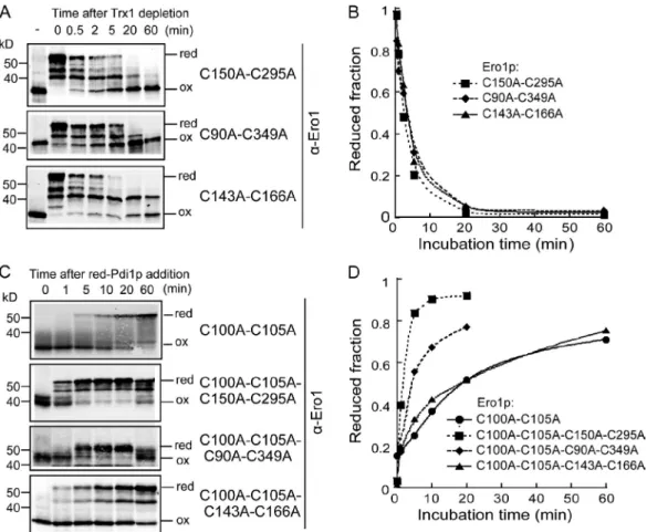
First, the autonomous oxidation of Ero1p regulatory bond mutants Ero1pcC150AC295A, Ero1pcC90A349A, and Ero1pcC143A166A was evaluated for their ability to undergo autonomous reoxidation after being reduced by Trx1. This com parison is complicated by differing band mobility for the oxidized and partially oxidized states of each of the mutants (Fig. 4 A). As a measure of the oxidation rate that could be consistently applied to each of the mutants, we represented the progression of the oxidation process as the decline in the fraction of Ero1p that was in a fully reduced state. All regulatory mutants showed similar overall autonomous oxidation rates (Fig. 4 B), indicat ing that formation of the C150C295 bond does not significantly affect oxidation of other regulatory bonds. Because the pattern of partially oxidized states was more complex for wild type (Fig. 2 B) than for the mutants, it was not possible to make a precise quantitative comparison of rates.
Next, we tested each of the three regulatory bond mutants for their ability to be activated by reduced Pdi1p. This test re quired addition of the shuttle cysteine mutant to each of the three regulatory bond mutants. Reduction of Ero1pcC100AC105A C150AC295A by reduced Pdi1p was significantly faster than for Ero1pcC100AC105A or for the other regulatory bond mutants (Fig. 4, C and D). Ero1pcC100AC105AC90AC349A also Although accurate quantitative amounts cannot be obtained as a
result of the complications that oxidized Pdi1p contains a partially reduced domain and also nonlinear detection by immunoblot analy sis, we found that when the fraction of reduced Pdi1p was >0.8, most Ero1pcC100AC105A was fully reduced, whereas when the fraction of reduced Pdi1p was <0.3, most Ero1pcC100AC105A was oxidized (Fig. 3 C). In a steady state, in vivo Pdi1p is partially reduced (Vitu et al., 2010) like airoxidized recombinant Pdi1p, and almost all Ero1p is in a fully oxidized form (see 0 time point of Figs. 1 B and 5 E). Therefore, we could conclude that partially reduced Ero1pc is at least partially active and will continue to oxidize Pdi1p until a ratio of oxidizedtoreduced Pdi1p is achieved that no longer facilitates reduction and activation of Ero1p.
An Ero1p regulatory mutant is rapidly activated by Pdi1p
Previously, we showed that an Ero1p mutant that lacks only one of the three regulatory bonds, Ero1p(c)C150AC295A, has high enzymatic activity both in vivo and in vitro (Sevier et al., 2007; Vitu et al., 2010). This suggests that in the absence of the C150 C295 regulatory bond, Ero1p is either more readily activated by reduced Pdi1p or less easily inactivated by reaction with oxidized Pdi1p or by autonomous oxidation.
Figure 4. Mutational analysis of the roles of individual regulatory bonds in autonomous reoxidation of Ero1p. (A) Ero1pc-C150A-C295A, Ero1pc-C90A-C349A, or Ero1pc-C143-C166A was reduced (red) by incubation with reduced Trx1 followed by Trx1 depletion and the oxidation (ox) state analysis. (B) The fraction of reduced Ero1p was calculated by the ratio of the band intensity of reduced Ero1p species to the sum of fully reduced and fully oxidized band intensities. (C) Mutational analysis of the roles of individual regulatory bonds in reduction by reduced Pdi1p of Ero1pc-C100A-C105A. The respec-tive mutants were incubated with a 20-fold excess of reduced Pdi1p followed by the oxidation state analysis (the result of Ero1pc-C100A-C105A was reproduced from Fig. 3 B). (D) The fraction of reduced Ero1p determined as in B reveals that the greatest rate enhancement occurs as a consequence of elimination by mutation of the C150-C295 disulfide bond.
on April 18, 2012
jcb.rupress.org
the crude immunoprecipitate by mass spectroscopy identified Pdi1p peptides but failed to identify additional PDI family members, including Eps1p, Eug1p, Mpd1p, or Mpd2p (unpub lished data). This result suggests that Pdi1p is the primary oxi doreductase engaged in oxidation or reduction of the Ero1p regulatory bonds. Loss of any single regulatory cysteine pair in Ero1pC100AC105A did not prevent trapping of a mixed di sulfide bond between Ero1pC100AC105A and Pdi1p, indicat ing that Pdi1p interaction with Ero1pC100AC105A is not limited to a single regulatory cysteine pair (Fig. 5 A). The mixed disulfides between Pdi1p and the various Ero1pC100AC105A regulatory bond mutants showed distinct mobilities on SDS PAGE (Fig. 5 A), which likely corresponded to different vari able combinations of cysteines in Ero1p and Pdi1p that engaged in oxidation and reduction of Ero1p regulatory bonds.
If reduced Pdi1p were essential for activating Ero1p, de pletion of Pdi1p from the ER would be expected to decrease activation of Ero1p by reduction of regulatory bonds even under prevailing reducing conditions in the ER. We have established conditions to deplete a cell of Pdi1p, which is essential for via bility, by glucose repression for 15 h of PGAL1-PDI1 in a pdi1
genetic background (Fig. 5 B; Tachibana and Stevens, 1992; Frand and Kaiser, 1999). As expected, Pdi1p depletion disrupts folding in the ER, as shown by the accumulation of the ER form of carboxypeptidase Y (CPY), similar to that in cells that had been treated with DTT (Fig. 5 C). Radiolabeling of cells to spe cifically follow CPY synthesized after Pdi1p depletion confirmed a complete block of biosynthetic disulfide bond formation in newly synthesized CPY (Fig. S4). However, under the same condi tions of Pdi1p depletion, most Ero1p remains oxidized (Fig. 5 D), indicating that the signal for an insufficiently oxidized ER exhibited slightly faster reduction than Ero1pcC100AC105A or
Ero1pcC100AC105AC143AC166A (Fig. 4, C and D). These results suggest that removal of the C150C295 regulatory bond lowers the threshold for Ero1p activation. Interestingly, both the Ero1pcC100AC105AC150AC295A and Ero1pcC100A C105AC90AC349A mutants exhibited some reoxidation of Ero1p (Fig. 4 C) and oxidation of Pdi1p (not depicted) at the 60min time point, suggesting that Ero1p may have alternative pathways to relay disulfide bonds to Pdi1p when the shuttle disul fide is unavailable (see Discussion).
Pdi1p is the primary in vivo regulator of Ero1p
If Pdi1p regulates Ero1p through reduction and oxidation of its regulatory bonds, it should be possible to trap mixed disulfide complexes between Pdi1p and the regulatory cysteines of Ero1p in vivo. To specifically isolate mixed disulfides with regulatory cysteines, we used FLAG epitope–tagged Ero1pC100AC105A (expressed ectopically in cells that carry wildtype ERO1). Ero1p C100AC105A is unable to support yeast viability as a result of the disruption of its ability to oxidize Pdi1p in what has been shown to be the primary pathway for electron exchange be tween Pdi1p and Ero1p in living cells (Frand and Kaiser, 2000). Therefore, any observable mixed disulfides trapped between Ero1pC100AC105A and Pdi1p in living cells would likely represent thiol/disulfide exchange between the Ero1p regula tory cysteines and Pdi1p.
Ero1pC100AC105AFLAG, isolated from cysteine alkylated cell extracts by immunoprecipitation with FLAG antibody, formed a predominant mixed disulfide complex that also reacted with Pdi1p antiserum (Fig. 5 A, lane 2). Analysis of
Figure 5. Pdi1p-mediated Ero1 regulation in vivo. (A) Pdi1p forms mixed disulfide with the Ero1p regulatory cysteines. Ero1p-C100A-C105A-Flag and its variants were immunopre-cipitated (IP) by anti-flag antibody and analyzed by immunoblots against anti-flag antibody and anti-Pdi1p serum. Lane 1, Ero1p-C100A-C105A-Myc; lane 2, Ero1p-C100A-C105A-flag; lane 3, Ero1p-C100A-C105A-C90A-C349A-flag; lane 4, Ero1p-C100A-C105A-C143A-C166A-flag; lane 5, Ero1p-C100A-C105A-C150A-C295A-flag. The single asterisk indicates a monomer of Ero1p mu-tants, and the double asterisks indicate mixed di-sulfides between Ero1p mutants and Pdi1p. WB, Western blot. (B) Pdi1p was depleted by growing
pdi1 covered with PGAL1-PDI1 in SMM (glucose
medium) for 15 h. (C) Immature CPY accumulates as Pdi1p is depleted. A control for fully reduced environment was prepared by treating cells with 1 mM DTT for 1 h. m, mature CPY; p1/p2, nascent and glycosylated CPY, respectively. (D) Oxidation (ox) states of Ero1p after Pdi1p depletion (glucose medium) in a genetic background with pdi1 or with pdi1 with deletions of all PDI homologs (PDI homolog-). red, reduced. (E) The roles of Pdi1p or glutathione on in vivo activation of Ero1p. Pdi1p-depleted cells (middle) and glutathione-Pdi1p-depleted cells (right) were prepared by growing CKY1081 in glucose medium and in galactose medium with 5 mM BSO, respectively. Cells were treated with 2 mM DTT for the indicated time.
on April 18, 2012
jcb.rupress.org
a pool of reduced glutathione (GSH) in the ER, which in turn could lead to reduction of Pdi1p. To test the possibility that DTT acts indirectly through glutathione, we tested the effect of depleting cells of glutathione by treating cells with 5 mM buthi onine sulfoximine (BSO) for 15 h, which blocks glutathione synthesis by inhibiting glutamylcysteine synthetase. Reduction of Ero1p regulatory bonds by DTT treatment occurred more slowly in glutathionedepleted cells than in untreated cells (Fig. 5 E). This result suggests that the presence of a reducible pool of glu tathione may accelerate the overall process of reduction of Pdi1p by DTT. Alternatively, depletion of glutathione by BSO treat ment may subtly affect Pdi1p expression or have some other indirect effect on the redox state of the ER.
To address the role of glutathione in Ero1p activation more directly, we reconstituted Ero1p activation in vitro using both Pdi1p and GSH as substrates. By itself, Pdi1p is a poor activator of Ero1p because once Pdi1p has become oxidized through Ero1p oxidase activity, there is no longer sufficient reduced Pdi1p avail able to keep Ero1p in an active state, and oxidized Pdi1p can accelerate inactivation of Ero1p. Therefore, fully activated Ero1p was rarely observed during reaction with reduced Pdi1p (Fig. 6 A). Glutathione alone was also a poor activator of Ero1p. 10 mM GSH did not reduce regulatory bonds in either wildtype Ero1pc (Fig. 6 B, left) or Ero1pcC100AC105A (not depicted). However, a mixture of 10 mM GSH and Pdi1p maintained a pool of at least partially activated Ero1p (Fig. 6 B, right). The basis of Ero1p activation in the presence of Pdi1p and GSH appeared to be that cannot be transduced to activate Ero1p in the absence of Pdi1p.
Pdi1pdepleted cells showed very slow activation of Ero1p even when they were treated with the reducing agent DTT, a condi tion that leads to rapid activation of Ero1p in Pdi1pexpressing cells (Fig. 5 E).
In the Pdi1pdepleted cells, a small amount of activated (re duced) Ero1p was detected (Fig. 5 D), and catalytically inactive Ero1 mutants Ero1pC355S and Ero1pC100AC105A exhibited significantly more of the reduced forms (Fig. S5), suggesting that under conditions of Pdi1p depletion, although Ero1p can be re duced to some extent, most of the Ero1p remains in an oxidized state if autonomous reoxidation is possible. We tested the possi bility that the residual reduction of Ero1p in cells that had been depleted for Pdi1p was catalyzed by another PDI homolog. By conducting the depletion experiment in a strain depleted for the other homologs (mpd1, mpd2, eug1, eps1, pdi1, and
CEN-PGAL1-PDI1), we found that reduced Ero1p formed to at
least the same extent as in a pdi1 PGAL1-PDI1 strain (Figs. 5 D
and S5), indicating that no other PDI family members contribute significantly to reduction of Ero1p regulatory bonds.
Glutathione acts synergistically with Pdi1p to activate Ero1p
The rapid Pdi1pdependent reduction of Ero1p regulatory bonds that occurs in cells treated with DTT could be the result of a pool of reduced Pdi1p formed by direct reduction with DTT that had entered the ER. Alternatively, DTT treatment may produce
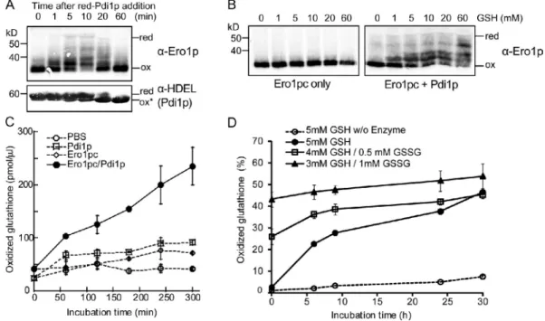
Figure 6. A coupled Pdi1p/GSH system recapitulates Ero1p homeostasis in vitro. (A) Ero1p is only partially activated when Pdi1p is used as a substrate. 1 µM Ero1pc was incubated with 25 µM reduced (red) Pdi1p. Ero1pc and Pdi1p were detected by immunoblotting with anti-Ero1p serum or anti-HDEL antibody. At the endpoint of in vitro oxidation (ox), Pdi1p is in a partially oxidized state, indicated by ox*. (B) Pdi1p is necessary for efficient reduction of Ero1pc by GSH. Ero1pc or a mixture of Ero1pc and oxidized Pdi1p was mixed with different concentrations of GSH for 1 h followed by TCA precipita-tion, NEM modificaprecipita-tion, and Western analysis. (C) Pdi1p is necessary for efficient oxidation of GSH by Ero1pc. 10 mM GSH was incubated with 1 µM Ero1pc in the presence or absence of 10 µM oxidized Pdi1p. The amount of GSSG produced was measured after alkylating GSH followed by reduction with glutathione reductase and assay of free thiols with Ellman’s reagent. (D) The balance of GSH and GSSG can be established by the mixture of Ero1pc and Pdi1p. 10 µM Pdi1p and 5 µM Ero1pc were incubated with three different glutathione mixtures, 5 mM GSH, 4 mM GSH/0.5 mM GSSG, and 3 mM GSH/1 mM GSSG. The natural oxidation of 5 mM GSH without enzymes was also measured. The percentage of oxidized glutathione was calculated for the indicated time points. (C and D) Error bars represent the SEM generated by three independent experiments.
on April 18, 2012
jcb.rupress.org
However, oxidized Trx1 has no capacity to reoxidize the regu latory cysteines of Ero1p. Instead, oxidation of the regulatory cysteines is accomplished by Ero1p itself in a reaction that probably involves the transfer of a disulfide bond from the shut tle cysteines of one Ero1p protein molecule to the regulatory cysteines of another. Thus, the inactivation phase occurs by autonomous oxidation of Ero1p once reduced Trx1 has been depleted by being converted to the oxidized form.
A similar analysis for a reaction that contains Ero1p and its natural substrate Pdi1p revealed that Ero1p is never fully ac tivated in the presence of Pdi1p (Fig. 6 A). Whereas we initially thought that the inability of Pdi1p to activate Ero1p robustly was a consequence of some deficiency in the in vitro reaction, we now have evidence that the balance of the reduced and oxidized forms of Pdi1p is crucial in regulating Ero1p enzyme activity and faithfully reproduces the redox homeostasis set point of the ER.
Pdi1p has two redoxactive domains containing the CxxC motifs. The a domain is not as efficiently oxidized by Ero1p as the a domain (Vitu et al., 2010). However, the a domain does appear to play a greater role than the a domain in oxidizing the Ero1p regulatory bonds; the partially oxidized a domain (Pdi1p SSCC) accelerates oxidation of Ero1p regulatory bonds more rap idly than the fully oxidized a domain (Pdi1pCCSS). Given the relative redox potentials of the two domains, their propensity to be oxidized by Ero1p and their relative ability to oxidize the regu latory bonds of Ero1p provide a consistent explanation for why the a domain is only partly oxidized, whereas the a domain is fully oxidized in cells under standard conditions (Vitu et al., 2010). In contrast to the yeast Pdi1p, the two active domains of the human PDI have similar reduction potentials, and the a domain is the one preferentially oxidized by Ero1 (Wang et al., 2009; Chambers et al., 2010). Therefore, it seems possible that the mechanism of Ero1 regulation via the two domains of the human PDI is distinct from the one in yeast. Specificity in inter action between human and yeast Ero1 and PDI proteins has been demonstrated (Vitu et al., 2010; Masui et al., 2011). Com parison of the activity of human and yeast proteins in vitro shows that the highest enzyme activity is achieved using protein combinations of Ero1 and PDI from the same species (Araki and Nagata, 2011). Low catalytic activity was traced in part to a weaker binding affinity between human Ero1 and yeast Pdi1p, relative to human PDI (Araki and Nagata, 2011). Whether the different redox potentials of the PDI domains in human and yeast PDI also contribute to the differences in Ero1 regulation between species remains to be assessed.
Several lines of evidence point to the existence of multiple disulfide relay pathways from Ero1p to Pdi1p. Best character ized is the flow of disulfides from the C352C355 pair to Pdi1p via the C100C105 shuttle cysteines. This pathway is necessary for full activity of Ero1pc in vitro and is necessary for cell via bility. Nevertheless, Ero1pcC100AC105A retains some activ ity in vitro and shows slow autonomous reoxidation, showing that a disulfide bond can be transferred, albeit slowly, from the active site to the regulatory disulfides without the action of the C100C105 shuttle (Fig. 2 B). Moreover, in vivo experiments have shown that yeast cells with an ero1-C100A mutation are provided by a pool of GSH in great stoichiometric excess to Pdi1p,
which allowed for a significant pool of reduced Pdi1p to be main tained. This pool of reduced Pdi1p in turn could reduce the regu latory bonds of Ero1p, thus overcoming the kinetic barrier for GSH to reduce the Ero1p regulatory bonds. In this coupled reac tion, oxidized glutathione (GSSG) was generated by the com bined action of Pdi1p and Ero1p (Fig. 6 C), and production of GSSG depended on Ero1p enzymatic activity because even in the presence of Pdi1p, catalytically inactive Ero1pcC100AC105A did not produce GSSG actively, whereas hyperactive Ero1pc C150AC295A produced GSSG more rapidly than wild type (not depicted).
To investigate the effects of the balance of GSH to GSSG on Ero1p activity, we reconstituted an in vitro reaction in which re combinant Ero1p and Pdi1p were incubated in the presence of three different ratios of GSH/GSSG. When the reaction was be gun in the presence of GSH only, the rate of glutathione oxidation was initially rapid but then slowed. Evidently, the concomitant generation of GSSG by the action of Ero1p and Pdi1p eventually leads to inactivation of Ero1p (Fig. 6 D, 5 mM GSH curve). Intro duction of GSSG into the coupled reaction slowed the initial rate of GSH oxidation (compare the curves of 5 mM GSH with 8:1 and 3:1 GSH/GSSG; Fig. 6 D). Taking the background of slow nonen zymatic generation of GSSG into account, the Ero1p and Pdi1p catalyzed oxidation of GSH stops at when the molar ratio of GSH/ GSSG is 3:1. Thus, this ratio of GSH/GSSG appears to be the approximate steadystate set point for the coupled reaction.
Discussion
A conserved protein disulfide relay system of the ER, consist ing of Ero1 and PDI, supports correct disulfide bond formation of secretory proteins (Sevier et al., 2001; Sevier and Kaiser, 2006a; AppenzellerHerzog et al., 2008; Braakman, 2009). In
S. cerevisiae, the Ero1PDI relay is essential for biosynthetic disulfide bond formation and is thus essential for cell viability (Frand and Kaiser, 1998; Pollard et al., 1998; Vitu et al., 2010). We previously showed that the activity of S. cerevisiae Ero1p is controlled by an autoregulatory feedback loop that involves the reduction and the reoxidation of three regulatory disulfide bonds (Sevier et al., 2007). Here, we show that not only is Pdi1p the major substrate of Ero1p in the disulfide relay system but that Pdi1p is also the primary regulator of Ero1p activity by its abil ity carry out dithiol/disulfide exchange with the regulatory bonds. We also show that the redox state of glutathione in the ER is important for setting the regulatory state of Pdi1p, and, in this way, the disulfide bond–generating activity of Ero1p is con trolled by the redox status of both Pdi1p and of glutathione.
In a reaction initiated by adding to Ero1p a stoichiometric excess of reduced Trx1, a model substrate, the regulatory bonds in Ero1p are rapidly reduced, and Trx1 is oxidized by active Ero1p. Once Trx1 has been oxidized, the regulatory bonds in Ero1p are reformed, and Ero1p returns to an inactive state. In this paper, we separated the activation and inactivation phases of this cycle to determine the biochemical requirements for each phase. As expected, the reduced form of Trx1 readily reduces regula tory bonds in Ero1p and is thus a potent activator of Ero1p.
on April 18, 2012
jcb.rupress.org
(AppenzellerHerzog et al., 2010). It seems likely that ER redox homeostasis is maintained by the same basic mechanism in yeast and mammalian cells.
Materials and methods
Protein expression and purification
The sequence coding Ero1pc (56–424) or Ero1pc mutants (Ero1pc-C100A-C105A, Ero1pc-C355S, Ero1pc-C150A-C295A, Ero1pc-C90A-C349A, Ero1pc-C143A-C166A, and Ero1pc-C100A-C105A-C143A-C166A) was
fused at the C terminus to His6-tagged MBP linked by the Tobacco etch virus
(TEV) protease cleavage sequence (ENLYFQGS) inserted into the pET17b vector (EMD) between NdeI and XhoI restriction sites. We found that C355S was the only point mutation of the active-site cysteine pair (C352-C355) that could be produced in a soluble FAD-bound state. Ero1pc and all vari-ants were overexpressed in Origami B(DE3) competent cells (EMD) by in-duction with 0.4 mM IPTG supplemented with 10 µM FAD overnight at
room temperature. Ero1pc fusion proteins with His6-tagged MBP were
puri-fied from cell lysates by affinity to amylose (New England Biolabs, Inc.) and then dialyzed into TEV cleavage buffer (50 mM Tris, pH 7.8, 0.5 mM EDTA, and 0.5 mM tris(2-carboxyethyl)phosphine). MBP-fused Ero1pc (MBP-Ero1pc) and Ero1pc-C100A-C105A were also expressed and
puri-fied as described for Ero1pc purification. Ero1pc was cleaved from His6
-tagged MBP by incubating with recombinant His6-tagged TEV protease
(1:100 wt/wt protease to substrate; the TEV protease–encoding plasmid was provided by C. Hill, University of Utah, Salt Lake City, UT) for 15 h at
25°C. Ero1pc and variants cleaved from His6-MBP were collected by
flow-ing through the HiTrap Ni2+ column (GE Healthcare) to remove uncleaved
fusion protein, cleaved His6-tagged MBP, and TEV protease. Some Ero1pc
variants (C105A-C150A-C295A and Ero1pc-C100A-C105A-C90A-C349A) were subcloned in pGEX-4T vector purified by flow-ing though GSTrap (GE Heathcare) followed by thrombin cleavage for
elution and secondary purification with the HiTrap Ni2+ column for removal
of thrombin (Gross et al., 2004). All purified Ero1pc proteins were dia-lyzed against PBS.
The sequence coding Pdi1p (31–522) or Pdi1p variants (C406S-C409S for CCSS, C61S-C64S for SSCC, and C61S-C64S-C406S-(C406S-C409S
viable if they have the capacity to upregulate Ero1p expression through unfolded protein response induction (Frand and Kaiser, 2000), and overexpression of Ero1pC100AC105AC150A C295A was able to partially suppress the temperature sensitiv ity of the ero1-1 strain (unpublished data).
The pathway of dithiol/disulfide exchange reactions used by Ero1pC100AC105A to oxidize Pdi1p remains to be deter mined. Considering that autonomous oxidation of the regulatory cysteines by Ero1pC100AC105A is possible, we hypothesize that the activesite disulfide bond C352355 may first be trans ferred to regulatory cysteine pairs, and then Pdi1p could be oxi dized by reducing these regulatory bonds during the activation process of Ero1p. These pathways were revealed by experi ments with Ero1pcC100AC105A but are not limited to mutant Ero1p and are likely also used by wildtype Ero1p. By transfer ring one or two of the regulatory bonds to Pdi1p, Ero1p would have some capacity to oxidize a stoichiometric quantity of Pdi1p without any Ero1p becoming fully active. The C143C166 bond may be the most labile regulatory bond, as its reduction is the first step of Ero1 activation by Pdi1p in vitro (Heldman et al., 2010) and in turn could readily contribute to Pdi1p oxidation. This pathway may allow the disulfide relay system to respond to modest increases in load with a minimum of excess hydrogen peroxide generation.
When Ero1p is incubated with reduced Pdi1p and GSH, Ero1p can be shown to be catalytically active by its ability to oxi dize a stoichiometric excess of GSH, although little or no Ero1p can be detected in a state in which all regulatory cysteines are in a reduced state (Fig. 6 B). As previously noted (Heldman et al., 2010), this observation indicates that the partially reduced forms of Ero1p have at least some oxidase activity. Thus, the regula tory bonds in Ero1p may act less like an on/off switch than a proportional controller with different degrees of activity accord ing to the redox states of the three regulatory bonds. We have not been able to devise a quantitative test of this attractive hy pothesis because it is not possible to isolate in pure form Ero1p in a partially reduced state.
Toward a biochemical understanding of the mechanism of Ero1p regulation in a highly simplified reaction, we success fully reconstituted the disulfide relay system and the basic prop erties of redox homeostasis in vitro with just their components, Ero1pc, Pdi1p, and glutathione (Fig. 6). The capacity of Pdi1p to sustain Ero1p in an activated state can be increased by addi tion of an excess of GSH in the in vitro reaction. This coupled reaction appears to approximate conditions in the ER (Hwang et al., 1992), in which transfer of disulfide bonds to nascent se cretory proteins and to GSH keeps enough Pdi1p in a reduced state to allow for some capacity to activate Ero1p. In the cou pled reaction, Ero1p enzyme activity is controlled by the ratio of GSH/GSSG, and a 3:1 ratio of GSH/GSSG appears to be close to the steadystate set point at which Ero1p can be activated. Pdi1p, which has both reductase and isomerase activities, can serve as the key link between thiol substrates, including gluta thione, and Ero1p to activate the disulfide relay system only when substrates are present (Fig. 7). In the mammalian cell, it has also been suggested that oxidation of PDI and glutathione is well regulated by an equilibrium between Ero1 and PDI
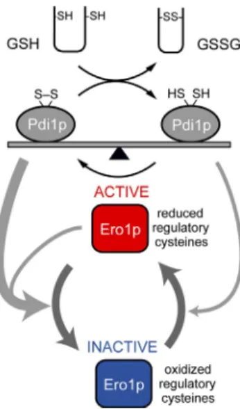
Figure 7. A balance between reduced and oxidized Pdi1p determines ER redox homeostasis. The thiol substrates (reduced protein thiols or re-duced glutathione) present in the ER can be oxidized by Pdi1p, leading to formation of reduced Pdi1p. If sufficient reduced Pdi1p has formed, Ero1p is activated by direct reduction of the Ero1p regulatory bonds by reduced Pdi1p. Once activated, Ero1p oxidizes Pdi1p until a steady-state balance of oxidized and reduced Pdi1p is reestablished. The balance of reduced and oxidized Pdi1p determines the degree of Ero1p activation; once reduced Pdi1p has declined sufficiently, autonomous oxidation of the regulatory bonds of Ero1p will lead to Ero1p inactivation, preventing overoxidation of the ER. Under hyperoxidizing conditions, oxidized Pdi1p would rapidly inactivate Ero1p.
on April 18, 2012
jcb.rupress.org
was calculated by dividing reduced band intensity with the sum of reduced and oxidized band intensity.
To determine the effect of hydrogen peroxide on the oxidation state of Ero1p regulatory bonds (Fig. S1), 1 µM Ero1pc was incubated with 100 µM
reduced His6-tagged Trx1 in PBS containing 0.5 mM EDTA for 2 min, and
Trx1 was selectively removed by affinity for Ni2+ beads. To remove
hydro-gen peroxide hydro-generated during action of Ero1p oxidase, 80 µg/ml cata-lase was added to the reaction. Removal of hydrogen peroxide by catacata-lase was confirmed by measuring the amount of hydrogen peroxide in the reac-tion with Amplex UltraRed reagent (Invitrogen). 1 µM Ero1pc was incu-bated with 100 µM reduced thioredoxin, 50 µM Amplex red, and 0.1 U/ml HRP in the presence or absence of 80 µg/ml catalase for 30 min at room temperature. Fluorescence was measured in a microplate reader with wavelength settings of 530/590 nm. Inactivation of Ero1p under anaero-bic conditions was analyzed as described above except being performed
in the anaerobic chamber flowing N2 and CO2 gases. To test whether
exogenous hydrogen peroxide or oxidized Pdi1p can promote inactivation of Ero1p, 100 µM hydrogen peroxide or 20 µM oxidized Pdi1p was added to Ero1pc, Ero1pc-C100A-C105A, or Ero1pc-C355S after Trx1 de-pletion. Oxidation states of Ero1pc and its mutants were analyzed after cysteine modification with 1 mM AMS by nonreducing SDS-PAGE and immunoblotting with anti-Ero1p serum.
The oxidation states of each catalytic domain of recombinant Pdi1p were determined by introducing a thrombin cleavage site that allows sepa-ration of the two catalytic domains (Fig. S2). The sequence coding N-terminal
His6-tagged Pdi1p (31–522) with a thrombin recognition sequence in the
X-linker was subcloned from pSK42 (Vitu et al., 2010) for E. coli
expres-sion. His6-Pdi1p-thrombin was overexpressed in BL21(DE3)pLysS
compe-tent cells by IPTG and purified from cell lysates with the HiTrap Ni2+ column.
Purified His6-Pdi1p-thrombin was dialyzed into PBS. 10 µg His6-Pdi1p-thrombin
was incubated with 1 mM AMS for 30 min at room temperature followed
by treatment with 0.1 µg thrombin supplemented with 1 mM CaCl2 for 1 h
at 37°C. Fully reduced Pdi1p control was prepared by treatment with 10 mM DTT for 30 min at room temperature. DTT was removed by using Bio-Spin 6. AMS-modified and thrombin-digested samples were analyzed by nonreducing SDS-PAGE.
The oxidation state of newly synthesized CPY in the Pdi1p-depleted ER was analyzed in pdi1 cells with galactose-inducible PDI1 grown to the exponential phase for 15 h in glucose medium to deplete Pdi1p. Cells were suspended in SMM-met, pulse labeled for 10 min at 30°C, and lysed in the presence of 10% chilled TCA to block disulfide exchange. After AMS modification, CPY was immunoprecipitated and resolved by reducing SDS-PAGE. A fully reduced CPY control was prepared by treating cells with 10 mM DTT for 15 min before cell lysis.
Reoxidation of Ero1pc after thioredoxin depletion
1 µM Ero1pc was reduced by incubation with 100 µM reduced His6-tagged
Trx1 for 2 min and then passed through Ni2+ beads to remove Trx1. At
times after Trx1 depletion, the reaction was quenched by mixing with SDS sample buffer containing 1 mM AMS. Depletion of Trx1 was confirmed by analysis with nonreducing SDS-PAGE. When necessary, 40 µM oxidized Trx1, 20 µM oxidized Pdi1p, or 20 µM oxidized Pdi1p mutant was added into Ero1pc or Ero1pc mutants immediately after Trx1 depletion.
Purification of mixed disulfides from yeast
CKY1090 expressing His6-Pdi1p was transformed with pSK64
[ero1-C100A-C105A-flag], pDPS51 [ero1-C100A-C105A-C150A-C295A-flag],
pDPS49 C105A-C90A-C340A], and pDPS50
[ero1-C100A-C105A-C143A-C166A]. Exponentially grown cells were lysed by
agita-tion with glass beads in the presence of chilled 10% TCA. TCA-precipitated protein pellets were washed with acetone and redissolved with 2% SDS and 50 mM Tris, pH 8.0, containing 50 mM NEM to block free cysteines. For effi-cient affinity purification, SDS was removed by SDS-Out reagent (Thermo Fisher Scientific). After clarification by centrifugation, Ero1p-C100A-C105A-flag was released from anti-Ero1p-C100A-C105A-flag affinity beads using 100 µg/ml Ero1p-C100A-C105A-flag pep-tide (Sigma-Aldrich) followed by deglycosylation with PNGaseF for 3 h at 37°C. Samples purified by affinity to anti-flag beads were passed through
an Ni2+ column to capture mixed disulfides with His
6-Pdi1p. Both Ni2+
unbound fraction and bound fraction were collected and analyzed by non-reducing SDS-PAGE and immunoblotting with anti-flag (Sigma-Aldrich) or anti-Pdi1p.
Depletion of Pdi1p and glutathione in yeast
To determine the oxidation states of Ero1p and mutants in Pdi1p- or glutathione-depleted cells, pSK63, pSK64, and pSK66 were transformed into CKY1088 and/or CKY1089. Cells exponentially grown in SMM containing
for SSSS) tagged with His6 at the N terminus was inserted into pET20b
be-tween NdeI and SacI restriction sites. Recombinant His6-tagged Pdi1p,
Pdi1p variants, and His6-tagged E. coli thioredoxin (Trx1; Sevier and Kaiser,
2006b) were overexpressed in BL21(DE3)pLysS competent cells (EMD) by induction with 0.4 mM IPTG for 5 h at 37°C and purified from cell lysates
by affinity to the HiTrap Ni2+ column. Purified, naturally oxidized Pdi1p
and Trx1 were dialyzed into PBS. Reduced forms of Pdi1p and Trx1 were prepared by incubating with 10 mM DTT for 30 min at room temperature and removing DTT with Micro Bio-Spin 6 (Bio-Rad Laboratories).
Yeast strains and plasmids
CKY1044 and CKY1045 and MATa and MAT pdi1::KanMX strains covered by URA3 CEN PDI1 plasmid pCS213 have been previously de-scribed (Vitu et al., 2010). To facilitate scoring of crosses with the pdi1 strain, a NatMX-marked pdi1 strain was constructed by swapping the KanMX marker in CKY1045 with a NatMX4 cassette by homologous re-combination (Goldstein and McCusker, 1999). Individual disruptions of the genes encoding the yeast PDI homolog proteins were made by one-step gene replacement of the entire ORF with KanMX by homologous recombina-tion. The compound deletion strain CKY1086 with the genotype MATa GAL2
ura3-52 leu2-3,112 pdi1::NatMX mpd1::KanMX mpd2::KanMX eug1::KanMX eps1::KanMX [pCS213] was constructed by sequential
rounds of mating, sporulation, and scoring of the initial individual disruption mutant strains. Disruption of all five genes encoding the PDI1 homologs in CKY1086 was confirmed by PCR analysis. Some pdi1 strains also con-tained a genomic-tagged copy of BGL2. Tagging of BGL2 with a triple HA epitope at its C terminus was performed by homologous recombination with a 3HA-KanMX6 module (Longtine et al., 1998). To construct the MATa GAL2
ura3-52 leu2-3,112 pdi1::NatMX BLG2:HA-KanMX [pCS213] double
mutant (CKY1087), individual pdi1 and BLG2:HA strains were mated,
and an MATa NatMX+ KanMX+ segregant was selected after sporulation.
Viability of CKY1086 and CKY1087 depends on a plasmid-borne PDI1 gene provided by plasmid pCS213. CKY1088 and CKY1089 were generated by transformation of CKY1086 or CKY1087, respectively, with the pSK65
[CEN LEU2 PGAL1-PDI1] followed by selection against pCS213 by plating on
supplemented minimal medium (SMM) with 5-FOA. CKY1090 was created
by transformation of CKY1044 with pSK2 [CEN LEU2 His6-PDI1] followed
by selection against pCS213 with 5-FOA.
To create pSK2 [CEN LEU2 His6-PDI1], the sequence coding
HHHH-HGG was inserted right after the signal sequence (1–22) by introducing the NheI restriction site in pCS463, which contains the PDI1 coding region and 875 and 150 bp of the 5 and 3 untranslated regions. Plasmid
pSK65 [CEN LEU2 PGAL1-PDI1] contains the PDI1 coding region and 60
and 150 bp of the 5 and 3 untranslated regions under the GAL1 pro-moter. ERO1-coding URA3 2µ plasmids pSK63 [ERO1-flag], pSK64
[ero1-C100A-C105A-flag], pDPS51 [ero1-C100A-C105A-C150A-C295A-flag],
pDPS49 C105A-C90A-C340A], pDPS50
[ero1-C100A-C105A-C143A-C166A], and pSK66 [ero1-C355S-flag] were generated by
insertion of sequence-encoding flag tag right after the last codon using the NotI restriction site and subcloning or site-directed mutagenesis based on the previously reported plasmid pAF84 (Frand and Kaiser, 1998). Analysis of protein oxidation states
For reduction of Ero1p regulatory bonds in vitro, 1 µM Ero1pc or its variants were incubated with 50 or 20 µM reduced Trx1, reduced Pdi1p, or reduced Pdi1p mutants in PBS containing 0.5 mM EDTA. After incubation at 25°C, the reactions were quenched by mixing with 1× SDS-AMS (4-acetamido-4-maleimidylstilbene-2,2-disulfonic acid) buffer (1× SDS sample buffer without reducing agent and 1 mM AMS [Invitrogen]), which alkylates free cysteine thiols and adds one 500-D moiety to each free thiol. For reac-tions that contained glutathione or DTT, proteins were first precipitated by 10% chilled TCA. Protein precipitants were washed with 0°C acetone and were redissolved with 1× SDS-AMS buffer. Ero1p and Pdi1p were ana-lyzed by nonreducing SDS-PAGE followed by immunoblotting with anti-Ero1p serum or anti-HDEL (Santa Cruz Biotechnology, Inc.). The oxidation state of Trx1 was analyzed by nonreducing SDS-PAGE followed by stain-ing with SimplyBlue SafeStain (Invitrogen).
The in vivo oxidation state of Ero1p was assessed in cells growing in SMM medium incubated with 0, 0.2, or 1 mM DTT for 30 min and then lysed in the presence of chilled 10% TCA by agitating with glass beads. Protein pellets from cell lysates were washed with chilled acetone and re-dissolved with 1× SDS-AMS buffer. Oxidation states of Ero1p and variants were analyzed by nonreducing SDS-PAGE and immunoblotting with anti-Ero1p. Immunoblots were visualized with an Odyssey imaging system (LI-COR Biosciences). The fully reduced or the oxidized band of Ero1pc was quantified by the Odyssey program (v2.0; LI-COR Biosciences), and reduced fraction
on April 18, 2012
jcb.rupress.org
Frand, A.R., and C.A. Kaiser. 1998. The ERO1 gene of yeast is required for oxidation of protein dithiols in the endoplasmic reticulum. Mol. Cell. 1:161–170. http://dx.doi.org/10.1016/S10972765(00)800179 Frand, A.R., and C.A. Kaiser. 1999. Ero1p oxidizes protein disulfide isom
erase in a pathway for disulfide bond formation in the endoplasmic reticulum. Mol. Cell. 4:469–477. http://dx.doi.org/10.1016/S1097 2765(00)801987
Frand, A.R., and C.A. Kaiser. 2000. Two pairs of conserved cysteines are re quired for the oxidative activity of Ero1p in protein disulfide bond forma tion in the endoplasmic reticulum. Mol. Biol. Cell. 11:2833–2843. Goldstein, A.L., and J.H. McCusker. 1999. Three new dominant drug resistance cas
settes for gene disruption in Saccharomyces cerevisiae. Yeast. 15:1541– 1553. http://dx.doi.org/10.1002/(SICI)10970061(199910)15:14<1541:: AIDYEA476>3.0.CO;2K
Gross, E., D.B. Kastner, C.A. Kaiser, and D. Fass. 2004. Structure of Ero1p, source of disulfide bonds for oxidative protein folding in the cell. Cell. 117:601–610. http://dx.doi.org/10.1016/S00928674(04)004180 Gross, E., C.S. Sevier, N. Heldman, E. Vitu, M. Bentzur, C.A. Kaiser, C. Thorpe,
and D. Fass. 2006. Generating disulfides enzymatically: Reaction prod ucts and electron acceptors of the endoplasmic reticulum thiol oxidase Ero1p. Proc. Natl. Acad. Sci. USA. 103:299–304. http://dx.doi.org/10 .1073/pnas.0506448103
Heldman, N., O. Vonshak, C.S. Sevier, E. Vitu, T. Mehlman, and D. Fass. 2010. Steps in reductive activation of the disulfidegenerating enzyme Ero1p.
Protein Sci. 19:1863–1876. http://dx.doi.org/10.1002/pro.473 Hwang, C., A.J. Sinskey, and H.F. Lodish. 1992. Oxidized redox state of glu
tathione in the endoplasmic reticulum. Science. 257:1496–1502. http:// dx.doi.org/10.1126/science.1523409
Inaba, K., S. Masui, H. Iida, S. Vavassori, R. Sitia, and M. Suzuki. 2010. Crystal structures of human Ero1 reveal the mechanisms of regulated and targeted oxidation of PDI. EMBO J. 29:3330–3343. http://dx.doi .org/10.1038/emboj.2010.222
Longtine, M.S., A. McKenzie III, D.J. Demarini, N.G. Shah, A. Wach, A. Brachat, P. Philippsen, and J.R. Pringle. 1998. Additional modules for versatile and economical PCRbased gene deletion and modifi cation in Saccharomyces cerevisiae. Yeast. 14:953–961. http://dx .doi.org/10.1002/(SICI)10970061(199807)14:10<953::AIDYEA293> 3.0.CO;2U
Masui, S., S. Vavassori, Cl. Fagioli, R. Sitia, and K. Inaba. 2011. Molecular bases of cyclic and specific disulfide interchange between human ERO1 protein and proteindisulfide isomerase (PDI). J. Biol. Chem. 286:16261– 16271. http://dx.doi.org/10.1074/jbc.M111.231357
Mezghrani, A., A. Fassio, A. Benham, T. Simmen, I. Braakman, and R. Sitia. 2001. Manipulation of oxidative protein folding and PDI redox state in mammalian cells. EMBO J. 20:6288–6296. http://dx.doi.org/10.1093/ emboj/20.22.6288
Nørgaard, P., V. Westphal, C. Tachibana, L. Alsøe, B. Holst, and J.R. Winther. 2001. Functional differences in yeast protein disulfide isomerases. J. Cell
Biol. 152:553–562. http://dx.doi.org/10.1083/jcb.152.3.553
Pollard, M.G., K.J. Travers, and J.S. Weissman. 1998. Ero1p: A novel and ubiq uitous protein with an essential role in oxidative protein folding in the endoplasmic reticulum. Mol. Cell. 1:171–182. http://dx.doi.org/10.1016/ S10972765(00)800180
Sevier, C.S., and C.A. Kaiser. 2002. Formation and transfer of disulphide bonds in living cells. Nat. Rev. Mol. Cell Biol. 3:836–847. http://dx.doi.org/ 10.1038/nrm954
Sevier, C.S., and C.A. Kaiser. 2006a. Conservation and diversity of the cellular disulfide bond formation pathways. Antioxid. Redox Signal. 8:797–811. http://dx.doi.org/10.1089/ars.2006.8.797
Sevier, C.S., and C.A. Kaiser. 2006b. Disulfide transfer between two con served cysteine pairs imparts selectivity to protein oxidation by Ero1.
Mol. Biol. Cell. 17:2256–2266. http://dx.doi.org/10.1091/mbc.E0505 0417
Sevier, C.S., J.W. Cuozzo, A. Vala, F. Aslund, and C.A. Kaiser. 2001. A flavo protein oxidase defines a new endoplasmic reticulum pathway for biosyn thetic disulphide bond formation. Nat. Cell Biol. 3:874–882. http://dx.doi .org/10.1038/ncb1001874
Sevier, C.S., H. Qu, N. Heldman, E. Gross, D. Fass, and C.A. Kaiser. 2007. Modulation of cellular disulfidebond formation and the ER redox envi ronment by feedback regulation of Ero1. Cell. 129:333–344. http://dx.doi .org/10.1016/j.cell.2007.02.039
Tachibana, C., and T.H. Stevens. 1992. The yeast EUG1 gene encodes an endo plasmic reticulum protein that is functionally related to protein disulfide isomerase. Mol. Cell. Biol. 12:4601–4611.
Tavender, T.J., A.M. Sheppard, and N.J. Bulleid. 2008. Peroxiredoxin IV is an endoplasmic reticulumlocalized enzyme forming oligomeric com plexes in human cells. Biochem. J. 411:191–199. http://dx.doi.org/10 .1042/BJ20071428
1.5% galactose and 1% raffinose were incubated in SMM containing 2% glucose for 15 h to shut off expression of Pdi1p. Glutathione was depleted by growing cells in the presence of 5 mM BSO, an inhibitor of glutathione biogenesis, for 15 h. Pdi1p depletion was confirmed by immunoblotting with anti-Pdi1p (Fig. 5 E), and glutathione depletion was confirmed using Ellman’s reagent.
The maturation of CPY was evaluated in pdi1 cells with galactose-inducible PDI1 grown to exponential phase for 15 h in glucose medium to deplete Pdi1p. For fully reduced cell environment, cells were incubated with 1 mM DTT for 1 h after a 14-h incubation in glucose medium. Cell lysates was prepared in the presence of chilled 10% TCA, and mature and immature CPY were analyzed by immunoblotting with anti-CPY serum.
Measurement of oxidized glutathione
GSSG produced by Ero1pc and Pdi1p was measured by using Ellman’s re-agent (5,5-dithiobis-(2-nitrobenzoic acid) [DTNB]). 1 µM Ero1pc, Ero1pc-C150A-C295A, or Ero1pc-C355S was incubated with 10 mM GSH with or without 10 µM oxidized Pdi1p. For extended reaction, 5 mM GSH, 4 mM GSH/0.5 mM GSSG, or 3 mM GSH/1 mM GSSG was mixed with Ero1p and Pdi1p. At various time points after mixing, 10 µl of reactants was quenched by chilled 5-sulfosalicylic acid (1% final concentration), and precipitated protein pellet was removed. GSH was alkylated by 2% 2-vinylpyridine after neutralizing sulfosalicylic acid with a drop of trietha-nolamine (Sigma-Aldrich) for 1 h at 25°C followed by a 20-fold dilution in DTNB reaction buffer containing 100 mM sodium phosphate, pH 7.0, 1 mM EDTA, 0.2 mM DTNB, and 0.2 mM NADPH. The change in absorbance at 405 nm was recorded after adding 1 U of glutathione reductase. Online supplemental material
Fig. S1 shows that autonomous oxidation and inactivation of Ero1p is not a side effect of hydrogen peroxide produced by aerobic catalysis by Ero1p. Fig. S2 shows that the endpoint of air oxidation yields a form of recombinant Pdi1p with the a domain almost fully oxidized, and the a domain is partially oxidized. Fig. S3 shows that air-oxidized Pdi1p can contribute to the oxidation of the Ero1p regulatory bonds. Fig. S4 shows that when cells are depleted of Pdi1p by growing pdi1 cells with glucose-repressible PDI1 in glucose medium, newly synthesized CPY remains largely in a reduced state. Fig. S5 shows that when cells are depleted of Pdi1p, autonomous oxidation of the regulatory bonds of Ero1p can occur. This oxidation requires active-site cysteine C355 but does not require the shuttle cysteines C100 and C105. Online supplemental material is available at http://www.jcb.org/cgi/content/full/jcb.201110090/DC1.
The authors thank Debbie Fass for thoughtful suggestions.
This work was supported by a grant from the National Institutes of Health (GM46941) to C.A. Kaiser.
Submitted: 20 October 2011 Accepted: 10 February 2012
References
AppenzellerHerzog, C., J. Riemer, B. Christensen, E.S. Sørensen, and L. Ellgaard. 2008. A novel disulphide switch mechanism in Ero1alpha balances ER oxidation in human cells. EMBO J. 27:2977–2987. http://dx.doi.org/ 10.1038/emboj.2008.202
AppenzellerHerzog, C., J. Riemer, E. Zito, K.T. Chin, D. Ron, M. Spiess, and L. Ellgaard. 2010. Disulphide production by Ero1PDI relay is rapid and effectively regulated. EMBO J. 29:3318–3329. http://dx.doi.org/10 .1038/emboj.2010.203
Araki, K., and K. Nagata. 2011. Functional in vitro analysis of the ERO1 protein and proteindisulfide isomerase pathway. J. Biol. Chem. 286:32705–32712. http://dx.doi.org/10.1074/jbc.M111.227181
Baker, K.M., S. Chakravarthi, K.P. Langton, A.M. Sheppard, H. Lu, and N.J. Bulleid. 2008. Low reduction potential of Ero1alpha regulatory disulphides ensures tight control of substrate oxidation. EMBO J. 27:2988–2997. http://dx.doi .org/10.1038/emboj.2008.230
Braakman, I. 2009. Entering a new era with Ero. Nat. Rev. Mol. Cell Biol. 10:503. http://dx.doi.org/10.1038/nrm2741
Chambers, J.E., T.J. Tavender, O.B. Oka, S. Warwood, D. Knight, and N.J. Bulleid. 2010. The reduction potential of the active site disulfides of human pro tein disulfide isomerase limits oxidation of the enzyme by Ero1. J. Biol.
Chem. 285:29200–29207. http://dx.doi.org/10.1074/jbc.M110.156596 Cuozzo, J.W., and C.A. Kaiser. 1999. Competition between glutathione and
protein thiols for disulphidebond formation. Nat. Cell Biol. 1:130–135. http://dx.doi.org/10.1038/11047
on April 18, 2012
jcb.rupress.org