Advanced training model for beating heart coronary artery surgery:
the Zurich heart-trainer
O. Reuthebuch
a,*, A. Lang
b, P. Groscurth
b, M. Lachat
a, M. Turina
a, G. Zu¨nd
aa
Clinic for Cardio-vascular Surgery, University Hospital Zu¨rich, Ra¨mistrasse 100, CH-8091 Zurich, Switzerland
b
Institute of Anatomy, University Zu¨rich, Winterthurerstrasse 190, CH-8057 Zurich, Switzerland Received 29 January 2002; received in revised form 16 April 2002; accepted 1 May 2002
Abstract
Objective: Coronary artery surgery with beating heart technique is gaining increasing popularity. However, it is a challenging technique even for well-trained cardiac surgeons. Thus, a training model for beating heart surgery was developed to increase safety and accuracy of this procedure. Methods: The model consists of differentially hardened polyurethane resembling mechanical properties of the human heart. The covering used in this model is a 1:1 replica of the human thoracic wall with optionally embedded skeletal structures. Sternotomy, lateral thoracotomy or trocar placement is possible to access the lungs, the pericardium and the heart with adjacent vessels. Disposable artificial coronaries variable in size, wall quality or wall thickness are embedded in the synthetic myocardium. Two-layer vessels, which can simulate dissection, are available. Bypass conduits utilize the same material. Coronaries/bypasses as well as part of the ascending aorta are water-tight and can be rinsed with saline. Lungs can be inflated. A purpose-built pump induces heart movement with adjustable or randomized stroke volume, heart rate and arrhythmia induction. Results: The model was tested in a recent ‘Wet-Lab’ course attended by 30 surgeons. All conventional instruments and stabilizers with standard techniques can be used. Training with beating or non-beating heart was possible. Time needed for an anastomosis was similar to clinical experience. Each artificial tissue showed its individual nature-like qualities. Various degrees of difficulty can be selected, according to stroke volume, heart rate, arrhythmia, vessel size and vessel quality. The model can be quickly and easily set up and is fully reusable. Conclusions: The similarity to human tissue and the easy set-up make this completely artificial model an ideal teaching tool to increase the confidence of cardiac surgeons dealing with beating heart and minimally invasive surgery. q2002 Elsevier Science B.V. All rights reserved.
Keywords: Coronary disease; Surgery; Off-pump; Training model
1. Introduction
With every new up-coming surgical technique, surgeons have to be well trained to achieve sufficient skills prior to operating on humans. Animal laboratories are convenient for skilled training [1,2]. However, based on animal protec-tion motives, they are not widely accepted among the popu-lation and thus now-a-days only limited and very restrictive use of these laboratories are made. Therefore the need for realistic artificial training models is gaining increasing importance to keep surgical skills at a high level. This became evident, when minimal-invasive laparoscopic surgery began its flush of victory all over the world, only little more than a decade ago. Several training centers were established and all kinds of training models built [3–5],
including teaching devices for thoracic procedures [1,6] as well as for training microvessel anastomoses [7,8].
However, at present, only few, but otherwise imperfect, models exist to train beating heart coronary artery revascu-larization. Therefore we developed a totally artificial train-ing model for coronary artery bypass grafttrain-ing (CABG) encompassing an easy set-up, beating and non-beating heart features, cardiac accessibility via conventional ster-notomy or lateral thoracotomy as well as reusability with only grafts to be disposable.
Implication of human characteristics such as size, anat-omy, loss of sight due to severe bleeding, tissue quality and fragility of vessels should facilitate transfer from model to human and help to increase the confidence of the trainee for his clinical work.
2. Materials and methods
The prosthetic model is a 1:1 thoracic replica of the
1010-7940/02/$ - see front matter q 2002 Elsevier Science B.V. All rights reserved. PII: S 1 0 1 0 - 7 9 4 0 ( 0 2 ) 0 0 2 6 9 - 5
www.elsevier.com/locate/ejcts
* Corresponding author. Tel.: 141-1-2553679; fax: 141-1-2554446. E-mail address: [email protected] (O. Reuthebuch).
human chest reversibly mounted on a water-tight box, which was already described by Segesser et al. [9]. Main component of the shell is differentially hardened
polyur-ethane resembling numerous mechanical properties.
Various covers are available either with preformed median sternotomy or left lateral thoracotomy. Skeletal structures can be optionally embedded. The rims of the incisions are reinforced to avoid tear down while spreading or while fixing retraction sutures (Fig. 1).
Underneath the hollow cover, separately inflatable lungs consisting of polyurethane are placed in the cavity of the box that also contains the heart. They can be removed indi-vidually. A synthetic pericardium loosely encloses the heart and is fixed at the ascending aorta/pulmonary arteries. It shows a Y-shaped incision with the Y up-side-down to easily access the coronaries. Due to its multi-layered struc-ture, it has extreme tensile strength to endure retraction sutures.
The external aspect of the heart model is a polyurethane
reproduction of a human heart with adjacent vessels such as ascending aorta, pulmonary artery, pulmonary veins and superior and inferior vena cava. It is univentricular inside. The wall thickness and stiffness are adapted to the required amount of movement with the ventricle softened and the adjacent vessels more rigid. Air can be inflated and deflated to simulate the beating heart through a special connector in the inferior vena cava. According to the anatomic course of the three major coronaries (left anterior descending (LAD), circumflex (CX) and right coronary artery (RCA)), small cavities are subepicardially embedded ending up at the distal ascending aorta. A special polyurethane-inlay can be inserted in the ascending aorta to train proximal
anasto-Fig. 1. Replica (1:1) of the human wall used as cover of the training model.
Fig. 2. Partially disposable aorta to train proximal anastomoses.
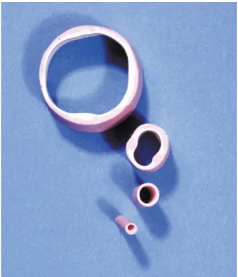
Fig. 3. Cross-section of artificial vessels with various diameters and wall quality.
moses either with partial clamping or with connecting devices. This disposable inlay can be infused with saline under high pressure to control tightness of anastomoses (Fig. 2).
Due to its soft consistence, the heart can be enucleated to reach all coronaries. With a sucking or compressing stabi-lizer supported by deep pericardial retraction sutures, coron-aries can be displayed and stabilized to perform the anastomoses.
The artificial coronaries are inserted in circular cavities with a specially designed flexible hook. At the end, they can be sealed or connected to an infusion system. Effluxing saline will be collected in a water-tight box. Coronaries are made of polyurethane and are available in various sizes (beginning with 1 mm in diameter), lengths, colors, wall thicknesses and wall quality (fragile or durable) (Fig. 3). Even dissection in vessels with non-glued two-layer wall can be simulated (Fig. 4). Bypass conduits utilize the same material.
A purpose-built high-permanence pump induces the heart movement. It is attached to the connector in the vena cava
via a tube. The pump is placed beside or underneath the model. Heart rate can be adjusted to be between 40 and 100 beats/min and the amount of wall motion can be adjusted by regulating the quantity of air. To increase complexity of training stroke-volume, heart rate as well as
Fig. 5. Purpose-built pump to regulate heart rate, stroke volume and arrhythmia induction.
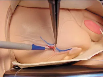
Fig. 6. Approach to LAD, RCX or RCA using stabilizers.
Fig. 7. Nature-like handling of artificial tissue.
Fig. 8. Vessels are water-tight and can be rinsed with saline to control tightness of anastomoses.
arrhythmia induction can be randomized including single or multiple ventricular extrasystolies, ventricular tachycardia or atrial fibrillation (Fig. 5).
3. Results
The model was tested by surgeons in training as well as by experienced surgeons. Recently it was used in a ‘Wet-Lab’ attended by 30 cardiac surgeons (The Heart Lab, Geneva, Switzerland, www.heartlab.org), where it was used to train beating or non-beating heart coronary artery bypass surgery.
Since no biological tissue is required, shipping and storage was extremely easy. The model was easily and quickly set-up either on a conventional table or on an oper-ating table. In general, the model was highly accepted due to its realistic and nature-like features. The preformed thoracic incisions were placed anatomically correct, so that the underlying structures could be easily accessed. All conven-tional retractors could be used.
The pericardium showed to be sufficiently reinforced to be sutured to the skin or to retractors and thus enabling an additional exposure of the antero-lateral and posterior aspect of the heart. The model was used to train surgeons for beating or non-beating heart coronary artery bypass surgeries. Depending on wall thickness and stroke volume, the cardiac movements could be adjusted to the skills of the surgeons. To increase complexity of the procedure, lungs could be additionally ventilated. Due to the flexibility of polyurethane, even the beating heart could be simply shifted and stabilized either with sucking or compressing stabilizers (Fig. 6). Course of coronaries was marked with colors on the surface of the heart for easy detection. All three major coronaries (LAD, RCX, RCA) could be visualized. Using conventional instruments, the epicardial layer could be dissected to visualize the coronaries (Fig. 7). With stay sutures or micro-spreaders, the incision was kept open. If vessels were insufflated, temporary occlusion with bull-dog clamps or sutures were feasible. Insertion of various intra-coronary shunts was simple and terminated bleeding from the anastomotic site. Anastomoses were performed in a standard manner using 7-0 or 8-0 prolene (Fig. 8). Tightness of anastomoses was tested by injection of saline. Setting and placement of different stabilizers could be meticulously done.
After distal anastomoses, proximal anastomoses were taught. The polyurethane inlet in the ascending aorta could be punched with standard instruments and anasto-moses done with conventional sutures or automated connecting devices (U-Clipe, Coalescent Surgical Inc., Sunnyvale,CA; Symmetrye Bypass System, St. Jude Medi-cal, USA). The training model was prepared for the next trainee by simply removing and replacing the artificial coronaries. Removed coronaries could be incised to assess quality of anastomoses.
Degrees of difficulty of beating heart revascularization depended on stroke volume, heart rate, arrhythmia induc-tion, vessel size and vessel quality. Especially coronaries and bypass-grafts of small diameter, thin walls and possible wall dissection proved to be a great challenge not only for the untrained surgeons but also for experienced ones.
The purpose-built pump could be easily set-up and connected with the heart. With several switches and regu-lators, complexity of the procedure was adjusted. Especially randomized arrhythmia induction increased intricacy. Despite pump being in continuous use, it never failed.
4. Discussion
In recent years an increasing shift from on-pump CABG towards off-pump CABG can be observed. This is clearly reflected in the huge variety and diversity of instruments presently available to immobilize the target coronaries [10]. However, beside stabilization, there are other topics to be dealt within off-pump CABG, such as exposure of vessels, temporary occlusion of coronaries, use of intra-coronary shunts or handling of connector devices for prox-imal anastomoses.
Due to the complexity of beating-heart coronary artery surgery, it appears to be mandatory to conduct an appropri-ate training before operating patients. Though in the past this was done in adapted steps in humans, complexity of present procedures may require further efforts. However, we could not find a suitable ex vivo model, although several tools are mentioned in the literature. Izzat et al. [10] have built a box to perform anastomoses with a moving operating field [11]. However, there is no similarity to the human anatomy. Stanbridge et al. report results using the beating heart model created by Limbs and Things, Ltd. (Bristol, UK). Though it was simulated to be life-like, various features were lacking such as variability of vessels, contin-uous infusion of grafts and manipulation of the heart [12].
With this in mind we have developed a totally artificial training model for beating heart coronary artery revascular-ization based on polyurethane. It combines a high similarity in shape and haptics to human tissue with an easy set-up and cost-effective reusability. It can be used in graduated steps beginning with mere end-to-side or end-to-end anastomoses of vessels, followed by revascularization of the arrested heart and finally revascularization of the beating heart.
Due to its nature-like characteristics not only the surgeons can be trained on procedures but also the use and feasibility of newly developed instruments and devices can be tested. Furthermore dexterity of young surgeons can be assessed prior to be accepted for definite training.
The modular concept allows different covers with numer-ous incisions. The surgeons could be trained for beating heart coronary artery surgery by use of sternotomy, lateral thoracotomy or even trocar placement for totally endoscopic coronary artery bypass grafting.
With this model we think to have invented a realistic and helpful educational tool. Training upon this phantom is practicable during an annual workshop (www.heartlab.org). On the other hand it is also commercially available (Med Connect, Pfa¨ffikon, Switzerland) to provide a continuous educational platform in the clinic.
The acceptance of the model in our hospital and during a recent Wet-Lab has encouraged us to keep on developing further training models including intracardiac procedures.
References
[1] Eaton BD, Messent DO, Haywood IR. Animal cadaveric models for advanced trauma life support training. Ann R Coll Surg Engl 1990;72:135–139.
[2] Kirwan WO, Kaar TK, Waldron R. Starting laparoscopic cholecys-tectomy – the pig as a training model. Irish J Med Sci 1991;160(8):243–246.
[3] Hill J, Kiff ES. An abdominal wall jig for surgical craft workshop. Ann R Coll Surg Engl 1990;72:386–387.
[4] Sackier JM, Berci G, Paz-Partlow M. A new training device for laparoscopic cholecystectomy. Surg Endosc 1991;5(3):158–159. [5] Windsor JA. Laparoscopic exploration of the common bile duct: a
training model. J R Coll Surg Edinburgh 1993;38(1):48–49. [6] Franchella A, Shweiki F, Riccipetitoni G, Mandrioloi G, Chendi D,
Pelizzo G, Georgacopulo P. Left lung transplantation in piglets: a training model. Transplant Proc 1994;26(1):214.
[7] Kim DC, Hayward PG, Morrison WA. Training model for microves-sel anastomoses. Microsurgery 1994;15:820–821.
[8] Satur CMR, Gupta NK. Angioscopy-guided training model of coron-ary artery anastomoses. Ann Thorac Surg 1994;57:1343–1345. [9] von Segesser LK, Westaby S, Pomar J, Loisance D, Groscurth P,
Turina M. Less invasive aortic valve surgery: rationale and technique. Eur J Cardiovasc Surg 1999;15:781–785.
[10] Izzat MB, Yim ACP. Cardiac stabilizer for minimally invasive direct coronary artery by pass. Ann Thorac Surg 1997;64:570–571. [11] Izzat MB, El-Zufari MH, Yim APC. Training model for
‘beating-heart’ coronary artery anastomoses. Ann Thorac Surg 1998;66:580– 581.
[12] Stanbridge R, O’Regan D, Cherian A, Ramanan R. Use of a pulsatile beating heart model for training surgeons in beating heart surgery. HSF 1999:1999–8488.