Biological Applications of Weak Polyelectrolyte
Multilayers
by Michael C. Berg B.S., Chemical Engineering
Virginia Polytechnic Institute and State University, 2000
SUBMITTED TO THE CHEMICAL ENGINEERING DEPARTMENT IN PARTIAL FULFILLMENT OF THE REQUIREMENTS FOR THE DEGREE OF
t,,,-+ ,o Dhi: .h.. ;- t"h-;~.l 'l~.,.H ... .. -UU;LU UI UI 11UU11YJ II l II ; IiLa I 1~CCIiIIll-Ir
at the
MASSACHUSETTS INSTITUTE OF TECHNOLOGY MARCH 2005
rr
.
_
_.'#
.
© Massachusetts Institute of Technology, 2005. All rights reserved.
Signature of Author:
Chemical Enginyg Department / March 8, 2005
Certified by: ,, ,
Paula T. Hammond Professor of Chemical Engineq .
A- Thesis Suesrvir Certified by:
Michael F. Rubner Professor of Materials Science and Engineering Thesis Supervisor
Accepted by: . .
Daniel Blankschtein Professor of Chemical Engineering Chairman, Committee for Graduate Students
MASSACUSTTS INSTTE OF TECHNOLOGY
JUN 0 1 2005
Biological Applications of Weak Polyelectrolyte
Multilayers
by Michael C. Berg
Submitted to the Chemical Engineering Department On February 1, 2005 in Partial Fulfillment of the Requirements for the Degree of Doctor of Philosophy
in Chemical Engineering
ABSTRACT
This thesis research focused on biological applications of ultra-thin weak polyelectrolyte multilayers with specific emphasis on cell patterning, drug delivery, and antibacterial coatings. All of these very different applications were studied using three different polymers - polyacrylic acid (PAA), poly(allylamine hydrochloride) (PAH), polyacrylamide (PAAm).
The first part of this thesis focuses on patterning polyelectrolyte multilayers found to resist mammalian cell adhesion, with ligands that promote specific interactions for adhesion. It was found that by patterning PAH on polyelectrolyte multilayers, the patterned functional group density and thickness could be tuned through ink pH adjustment. By changing the surface density of amine groups in the PAH patterns, the ligand density could also be altered using specific chemistry to attach peptides containing the tri-peptide sequence, RGD, which is known to promote cell adhesion in a number of cell types. The RGD density in the patterned regions determined the number of cells attached and the amount of cytoskeletal protein organization.
The second part is an evaluation of porous polyelectrolyte multilayers as a delivery system for controlled release of small molecule drugs. The loading and releasing properties of porous PAH/PAA multilayers were investigated using the two drugs, ketoprofen and
cytochalasin D. It was determined that the amount of drug released was proportional to the number of porous layers. Nanoporous films showed zero-order release, whereas microporous films displayed Fickian diffusion. The efficacy of the released drugs was checked by monitoring the effect of released cytochalasin D on fibroblasts' division.
In the final part of this thesis, the antibacterial properties of both silver-loaded
polyelectrolyte multilayers and superhydrophobic multilayers are examined. It was found that silver loaded multilayers killed bacteria to an extent greater than 99.99% for both airborne and waterborne models. Superhydrophobic films showed excellent anti-fouling properties for proteins, mammalian cells, and bacteria.
Thesis Supervisors: Paula T. Hammond, Associate Professor of Chemical Engineering Michael F. Rubner, TDK Professor of Polymer Materials Science and Engineering
TABLE OF CONTENTS
ABSTRACT ...2
TABLE OF CONTENTS ... ... ... ... 3
LIST OF FIGURES AND TABLES ... ...5
ACKNOWLEDGEMENTS ... 8
Chapter 1 Introduction ... 10
1.1. Introductory Remarks ... ... 10
1.2. General Introduction ... ... 10
1.2.1. Layer-by-Layer Polyelectrolyte Multilayers . . ... 10
1.2.2. pH Tunable Weak Polyelectrolyte Multilayers . . ... 13
1.2.3. Polyelectrolyte Multilayers as Biomaterials ... 15
1.2.4. Patternability of Polyelectrolyte Multilayers ... 18
1.3. Thesis Objectives and Outline ... ... 19
References for Chapter 1 ... ... 21
Chapter 2 Polymer on Polymer Stamping of Weak Polyelectrolytes .. ... 26
2.1. Introduction ... 26
2.2. Experimental Methods ... 29
2.3. Results and Discussion ... ... 32
2.3.1. Characterization of Weak Polyelectrolyte Multilayer Films ... 32
2.3.2. Influence of Ink pH of PAH stamped onto Multilayers as Studied with Atomic Force Microscopy ... 34
2.3.3. Dansyl Chloride Attachment on Stamped PAH as a Function of Ink pH... 40
2.4. Conclusions ... 44
References for Chapter 2 ... ... 46
Chapter 3 Controlling Mammalian Cell Adhesion on Patterned Polyelectrolyte Multilayer Surfaces ... 48
3.1. Introduction... 48
3.2. Experimental Methods ... 52
3.3. Results and Discussion ... ... 57
3.3.1. Functionalization and Characterization of Patterned PAH with RGD Peptides... 57
3.3.2. Effect of Patterned RGD Density on WT NR6 Fibroblasts ... 62
3.3.3. Effect of Patterned RGD Density on Cytoskeletal Protein Organization ... 64
3.4. Conclusions... 68
References for Chapter 3 ... 70
Chapter 4 Controlled Release from Porous Polyelectrolyte Multilayers ... 73
4.1. Introduction ... 73
4.2. Experimental Methods ... 75
4.3. Results and Discussion ... ... 79
4.3.1. Porous Film Characterization ... ... 79
4.3.2. Loading and Releasing Drugs ... 83
4.3.4. Release from nanoporous versus microporous films ... 91
4.3.5. Cytochalasin D Release to Mammalian Cells ... 94
4.4. C onclusions... 96
References for Chapter 4 ... 98
Chapter 5 Bacteria Killing Properties of Silver-Loaded Polyelectrolyte Multilayers ... 101
5.1. Introduction... 101
5.2. Experimental Methods ... 103
5.3. Results and Discussion ... 108
5.3.1. Antibacterial Efficacy ... 108
5.3.2. Cytotoxicity of Silver Loaded Multilayers ... 114
5.3.3. Rate of Silver Release ... 116
5.4. Conclusions... 118
References for Chapter 5 ... 119
Chapter 6 Anti-Fouling Properties of Superhydrophobic Polyelectrolyte Multilayers ... 122
6.1. Introduction ... 122
6.2. Experimental Methods ... 125
6.3. Results and Discussion ... 129
6.3.1. Protein Adsorption Assays ... 129
6.3.2. Mammalian Cell Resistance ... 133
6.3.3. Bacteria Adhesion Prevention ... 135
6.3.4. Patterning by Selective Wetting ... 139
6.4. Conclusions ... 144
References for Chapter 6 ... 146
Chapter 7 Summary and Future Work ... 151
7.1. Thesis Summary ... 151
LIST OF FIGURES AND TABLES
Figure 1-1. Assembly process for layer-by-layer polyelectrolyte multilayer films formed by alternately dipping a substrate in a polycation and a polyanion solution ... 12 Figure 1-2. Chemical structures of the weak polyelectrolytes, (a) PAH and (b) PAA ... 14 Figure 1-3. NR6WT fibroblasts after three days seeded at 10 000 cells/cm2 onto (a) a 6.5/6.5 PAH/PAA multilayer, (b) a 4.0/4.0 PAH/PAA multilayer, (c) a 2.0/2.0 PAH/PAA multilayer, (d) a TCPS control and (e) a 3.0/3.0 PAA/PAAm multilayer (scale bar = 200 pm) ... 17 Figure 1-4. Graphs comparing (a) elastic modulus and (b) cell counts for human microvascular endothelial cells. The modulus was obtained using a nanoindentation technique, and cell counts were preformed seven days post-seeding. The black bars represent data from films with PAA as the outermost layer, and gray bars represent data from films with PAH as the outermost layer. Adapted from work of Thompson et a166... 18 Figure 2-1. Diagram illustrating the polymer-on-polymer stamping process for PAH on a
PAA/PAH multilayer platform ... 33 Table 2-1. Average incremental bilayer thickness (measured by profilometry) and RMS
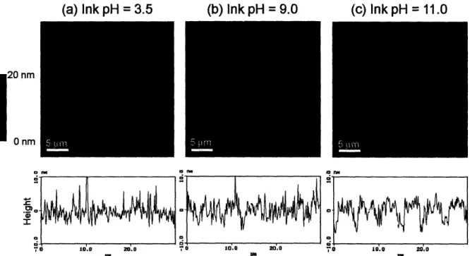
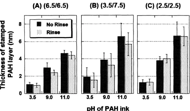
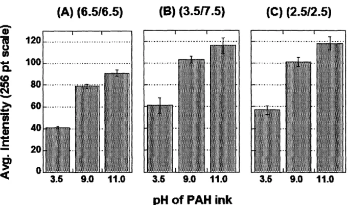
roughness (obtained from AFM) measurements for the three representative 10 bilayer PAA/PAH multilayer films used in stamping study ... 34 Figure 2-2. AFM height images and sectional analyses of 6.5/6.5 PAA/PAH multilayer platforms stamped with PAH at various ink pH's ... 35 Figure 2-3. AFM height images and sectional analyses of 3.5/7.5 PAA/PAH multilayer platforms stamped with PAH at various ink pH's ... 37 Figure 2-4. AFM height images and sectional analyses of 2.5/2.5 PAA/PAH multilayer platforms stamped with PAH at various ink pH's ... 38 Figure 2-5. Thickness measurements from AFM of the stamped PAH layer inked at pH = 3.5, 9.0, and 11.0 before and after rinsing in water for 2 minutes on 6.5/6.5 PAA/PAH multilayers (a), 3.5/7.5 PAA/PAH multilayers (b), and 2.5/2.5 PAA/PAH multilayers (c) ... 40 Figure 2-6. Fluorescent intensities of dansyl chloride labeled PAH stamped onto 6.5/6.5
PAA/PAH multilayers (a), 3.5/7.5 PAA/PAH multilayers (b), and 2.5/2.5 PAA/PAH multilayers (c) ... 41 Figure 2-7. Fluorescent image of a 3.5/7.5 PAA/PAH multilayer platform stamped with PAH at pH 11 and subsequently reacted with dansyl chloride ... 43 Table 2-2. Fluorescence intensity values of multilayer films dipped in the buffer rinse solution or standard PAH solutions ... 44 Figure 3-1. Fluorescence microscopy images of dansyl chloride labeled GRGDSPC sequences attached to PAH (a) line and (b) rectangle patterns stamped at pH = 11.0 (Scale bar, 100 pnm).. 59 Figure 3-2. Fluorescence microscopy image of fluorescein labeled GRGDSPC peptides attached to PAH stamped at pH 3.5 (vertical lines) and pH 11.0 (horizontal lines) using two separate stamps (Scale bar, 50 pm) ... 61 Figure 3-3. Ligand surface density as a function of PAH ink pH calculated from radiolabeling data ... 62
Figure 3-4. Phase contrast images of fibroblast adhesion on 50 gam patterned lines. Adhesion and spreading were compared for PAH stamped from ink solutions of pH (a) 3.5 (b) 7.0 (c) 9.0 (d)
11.0. Dotted lines were added to indicate a few of the patterned lines (Scale bar, 50 im) ... 63 Figure 3-5. Phase contrast micrographs of WT NR6 fibroblasts adhering to patterns of high density GRGDSPC attached to PAH stamped at pH 11.0 on (a) lines with 25 m to 50 inm
widths and (b) 100 m x 200 jim rectangles two days after seeding (Scale bar, 200 inm) ... 64 Figure 3-6. Immunostaining results for cytoskeletal proteins in WT NR6 fibroblasts. Actin stress fibers and vinculin stains were compared for PAH ink pH's of (la and 2a) 3.5, (lb and 2b) 7.0,
(lc and 2c) 9.0, and (id and 2d) 11.0. The upper panel is composed of actin stains, and the lower panel is vinculin stains, and stamped areas are inside dotted lines (Scale bar, 20 nm) ... 66 Figure 3-7. Geometric effects on cytoskeletal organization. Actin stress fiber and vinculin stains
of WT NR6 fibroblasts attached to high density GRGDSPC, 152,000 molecules/imn2, patterns of (la and 2a) 10 m, (lb and 2b) 25 m, and (Ic and 2c) 50 gm width lines. The upper panel is composed of actin stains, and the lower panel is vinculin stains (Scale bar, 10 im) ... 68 Figure 4-1. Chemical structures of (a) cytochalasin D and (b) ketoprofen ... 81
Table 4-1. Thickness of multilayer films before and after porosity transition and thickness increase due to porosity induction. NP designates nanoporous films and MP designates
m icroporous films ... 82 Figure 4-2. AFM images of (a) non-porous (image size = lxl m), (b) nanoporous (image size = lxlm), and (c) microporous (image size = 5x5/im) 20 bilayer PAH/PAA multilayer films... 83 Figure 4-3. A DMSO solution containing 0.2 mg/mL cytochalasin D in the process of wicking into a 50-3(5NP-50) polyelectrolyte multilayer film and a schematic of the process. The change in reflectivity halfway up the film is due to the pores being filled with solution. The schematic next to the photograph is an edge on view of the wicking process. ... 85 Figure 4-4. Plot of the cumulative amount of cytochalasin D released from 50-3(5NP-50) and 15MP films as a function of time (drug loaded from a 0.2 mg/mL DMSO solution in both cases).
... ... ... 86
Figure 4-5. Graphs comparing the (a) total time to release and (b) release flux for ketoprofen and cytochalasin D at different loading concentrations. The solid bars are data from a film loaded with a 0.2 mg/mL DMSO loading solution, and the hatched bars are data from a film loaded with a 1.0 mg/mL solution for cytochalasin D and 10.0 mg/mL solution for ketoprofen ... 87 Table 4-2. Release data for each film and drug tested at a drug loading concentration of
0.2mg/mL . ... 88 Figure 4-6. Total time of drug release normalized by the number of bilayers in the film. Drugs were loaded by absorption from a 0.2 mg/mL DMSO solution. Black bars represent total release time per bilayer for ketoprofen, and hatched bars represent total release time per bilayer for cytochalasin D ... 90 Figure 4-7. Comparison of ketoprofen and cytochalasin D release from (a) 20MP multilayers and (b) 20NP multilayers along with fitted curves from the model discussed in the text ... 92 Figure 4-8. Schematic of porous multilayer films and implications on drug release for (a)
microporous and (b) nanoporous films. The top drawing in each subsection represents an edge-on view of the multilayer, and the bottom drawing in each subsectiedge-on compares cedge-onceptually the overall pore structure of a multilayer film to a single drug-containing vesicle ... 94 Figure 4-9. Microscope images of fibroblasts three days after seeding on 50-3(5NP-50) films with (a) no cytochalasin D and (b) cytochalasin D loaded from a 0.2 mg/mL DMSO solution. Nuclei were stained with DAPI ... 96
Figure 5-1. Surface plasmon resonance peaks of 3.0/3.0 PAA/PAAm polyelectrolyte multilayers with silver nanoparticles ... 109 Figure 5-2. Micrograph of (a) glass control and (b) (PAA/PAAm)3-1 multilayer after being
exposed to airborne bacteria and incubated overnight. Scale bar is 400 pm ... 111 Figure 5-3. Graphs displaying airborne bacteria results for polyelectrolyte multilayers and glass
controls of both (a) E. coli and (b) S. epidermidis. Samples were incubated overnight after spraying with bacteria. The CFU densities are reported relative to the glass controls ... 112 Figure 5-4. Results for waterborne antibacterial testing of (a) E. coli and (b) S. epidermidis on polyelectrolyte multilayers and glass controls. Samples were incubated overnight after 2 hours
of exposure to waterborne bacteria. Results are shown relative to the glass controls ... 114 Figure 5-5. Cytotoxicity results comparing polyelectrolyte multilayers with and without silver
nanoparticles. The data is normalized to the multilayers without silver for both the (a) floating cell assay and (b) RGD functionalization assay ... 116 Figure 5-6. Change in silver content in the polyelectrolyte multilayer films as a function of time
submerged in PBS ... 118 Figure 6-1. Fluorescence images of FITC labeled BSA adsorbed onto (a) a microporous
multilayer and (b) a superhydrophobic multilayer. The films were exposed to a 1.0% FITC-BSA solution in PBS for 30 minutes. Scale bar is 100 m. ... 132 Figure 6-2. Fluorescent images of WT NR6 fibroblasts seeded on (a) microporous and (b)
superhydrophobic multilayers at a density of 10,000 cells/cm2. The cells were stained with LIVEIDEAD stain three days after seeding to visualize the cells through the porous network of
the multilayers films. Scale bar is 100 gm ... 134 Figure 6-3. Results of bacterial growth microporous and superhydrophobic multilayers from an
airborne model (relative to glass samples). The samples were sprayed with solutions of the bacteria strains (a) E. coli and (b) S. epidermidis and left overnight under a slab of agar. Hatched bars represent data from samples with no rinse, and black bars represent data from samples that were rinsed in PBS ... 137 Figure 6-4. Waterborne test results for (a) E. coli and (b) S. epidermidis on microporous and
superhydrophobic multilayers (results relative to glass controls). Samples were submerged in the bacteria solution for 2 hours and then placed under a slab of agar overnight ... 138 Figure 6-5. Photograph of droplets of fluorescent dyes in aqueous solutions on a patterned
superhydrophobic multilayer surface. A UV lamp was held above the sample to excite the fluorophores. Scale bar is 10 mm ... 140 Figure 6-6. FITC labeled lysozyme selectively adsorbed to hydrophilic spots on a
superhydrophobic polyelectrolyte multilayer background. Scale bar is 500 pm. ... 141 Figure 6-7. (a) Fluorescence intensity as a function of number of layers of 8.5/3.5
(F1TC-PAH/PAA) along with a linear fitted trend line. (b) Fluorescence micrograph of 8.5/3.5 (FITC-PAH/PAA) multilayers assembled onto a patterned superhydrophobic multilayer film (10
layers). Scale bar is 200 lm ... 142 Figure 6-8. A fluorescence micrograph of WT NR6 fibroblasts with their nuclei DAPI stained on a pattern with created by adsorbing PAH at pH = 9.0 and attaching RGD. Scale bar is 100 gm.
ACKNOWLEDGEMENTS
First, I would like to thank my thesis advisors, Prof. Paula T. Hammond and Prof. Michael F. Rubner. I have learned a great deal from both, and have become a much better scientist and writer by being a part of their research groups. Both gave me room to grow and come up with my own ideas, but were always there to help at the right time. I enjoyed working with both over my time at MIT, and have always felt that their personalities made my research not only interesting and rewarding, but a great deal of fun. Paula's warmth and friendliness always gave me confidence even when research was not going so well, and Michael's enthusiasm and energy made me feel like I could do anything.
I also would like to thank the rest of my thesis committee, Prof. Robert E. Cohen and Prof. James L. Sherley. Bob's helpfulness and good humor made me feel always welcome in his office. James's expertise made him a valuable resource especially with any "biology" questions, and he made me feel like I was a part of his group.
I would not have been able to complete this thesis without the help and support of my past and current fellow lab members. I would like to thank the Hammond group: Dr. Mitch Anthamatten, Dr. Dean Delongchamp, Dr. Dave Dewitt, Dr. Tarek Farhat, Shoshana Gourdin, LaShanda James-Korley, Dr. Xuiping Jiang, Mark Johnson, Heejae Kim, Ilsoon Lee, Dr. Geoff Lowman, Jodie Lutkenhaus, LaRuth McAfee, Andy Miller, Phuong Nguyen, Juhyun Park, Greg Pollock, Dr. Cathy Santini, Kris Stokes, Marriane Terrot, Dr. Lu Tian, Dr. Hiro Tokuhisa, Dr. Kris van Hege, Eric Verploegen, Ryan Waletko, Kris Wood, Dr. J.-S. Wu, Dr. Pil Yoo, Dr. Bruce Yu, Nicole Zacharia, and Dr. Haipeng Zheng. From the Rubner group, I would like to thank Dr. Hyunku Ahn, Fevzi Cebeci, Dr. Jeeyoung Choi, Anita Chung, Dr. Jeri'ann Hiller, Koji Itano, Daeyeon Lee, Dr. Zifeng Li, Dr. Jonas Mendelsohn, Dr. Takehiro Nishikawa, Adam Nolte, Prof. Masahiro Rikukawa, Dr. Hartmut Ruddman, Dr. Saturo Shimada, Dr. Nobuaki Takane, Dr. Tom Wang, Aleks White, Dr. Zhizoung Wu, Dr. Sung Yun Yang, Dr. Lei Zhai, Toshi,, Xiaxio In addition, I would like to thank the undergraduates that I had the pleasure to work with: Danielle Jensen, Yushan Kim, and Jack Milwid. In addition to my fellow group members in the Hammond and Rubner labs, I would also like to thank the Griffith lab, especially Ada Au, Lily Koo, Ley Richardson, and Kirsty Smith for their help and advice with cell culture. In addition, thanks to Dr. Shuguang Zhang and Dr. Carlos Semino for space to perform cell culture. The research in this thesis would also have not been possible without funding from the DuPont-MIT Alliance (DMA).
I would also like to thank my friends and fellow graduate students at MIT that shared a lot of good times over the years and quite a few pints of Guinness: Brian Baynes, Steve Fox, Kyle Jensen, Jeremy Johnson, Kathyrn Miller, Joe Moritz, Prem Pavoor, Mike Raab, Greg Randall, and Greg Zugates. Also, I would like to thank all of my friends from Delaware and Virginia Tech including Pri Chakkavarthi, Joe Fulcher, Adam Gayzik, Erik Johnson, Monte Marcum, Todd Smith, John Reynolds, Jason Soules, and Brad Turner. In addition, I would like to thank the Philadelphia Eagles, Flyers, Phillies, and Sixers for inspiring me to strive for excellence.
Finally and most importantly, this thesis would not have been possible without the love and support of my family. My parents, Donna and Cliff Berg, and my sister, Jennifer Berg, have always been there for me and believed in me. I would especially like to thank Janice Lansita for her love and support over the last few years; I could not have made it through without her.
Chapter 1
Introduction
1.1. Introductory Remarks
Polyelectrolyte multilayers have become a very highly studied class of materials over the past decade. This chapter serves to introduce important concepts about these thin polymer films before delving into the applications explored in this thesis. First, an overview of layer-by-layer polyelectrolyte multilayers will be presented, which will focus on the development of the research area and important concepts in the field. Second, the importance of a subset of these materials, weak polyelectrolyte multilayers, will be examined. Particular interest will be paid to the effect of assembly pH along with applications of these films, especially the use of polyelectrolyte multilayers as biomaterials. The emphasis of this discussion will be on relevant background information for the applications discussed in later chapters of this thesis. Finally, the objectives and outline of this thesis will be presented. In addition to this background section, each chapter has its own introduction that will give a more specific background to the specific subject matter of the chapter.
1.2. General Introduction
1.2.1. Layer-by-Layer Polyelectrolyte Multilayers
Since polyelectrolyte multilayers were first introduced by Decher et al. over a decade agol' 2, the amount of interest paid to these ultra-thin polymer films has grown exponentially.
The major reasons for this interest are their ease of processing, variety of materials which can be incorporated into their assembly, and the versatility of the technique. Polyelectrolyte multilayers are assembled using a layer-by-layer assembly process that is shown graphically in Figure 1-1. In a typical process, a substrate is dipped into the first polyelectrolyte solution for a long enough time to allow the polyelectrolyte to adsorb to the surface. After rinsing off loosely bound polymer, the substrate is then dipped into a polyelectrolyte solution of opposite charge. This second polyelectrolyte adsorbs to the surface due to electrostatic attraction and actually overcompensates for the surface charge3 resulting in a reversal of the surface charge. The
process is repeated until the desired number of layers is deposited. Each step results in a reversal of surface charge allowing the next layer to be deposited. The multilayers can be applied using any method that allows the polyelectrolyte solution to come into contact with the substrate, with the most common methods including dip-coating', deposition on colloids4, or spin-coating5' 6.
/* K
Polycation Rinse H20 Polyanion Rinse H20
/) C'-CD Polycation Rinse Polyanion Rinse
Figure 1-1. Assembly process for layer-by-layer polyelectrolyte multilayer films formed by alternately dipping a substrate in a polycation and a polyanion solution.
In addition to model polyelectrolytes, many different materials can be incorporated into polyelectrolyte multilayers including proteins7, DNA8, dyes9' lo, light emitting polymersl ' 12, and inorganics 3' 14. Also, the buildup of these thin films is not limited to electrostatic interactions.
Hydrogen bonding'5'8, van der Waals forces'9, and biomolecular interactions2 0 have been explored as well. Hydrogen-bonded multilayers will be discussed in detail in a later section due to their relevance to this thesis.
Polyelectrolyte multilayer assembly compared to other self-assembly techniques such as self-assembled monolayers (SAMs) or Langmuir-Blodgett films is much more versatile. Besides the variety of materials that can be incorporated into the films, a wide range of substrates can be employed including polymers, metals, and glass. With other techniques, the substrate is often limited to a specific surface that interacts with the deposited molecules. An example of this is
I
_ _
f
thiol-containing SAMs that require a gold surface for deposition. In addition, polyelectrolyte multilayers are not limited to simple changes in the surface properties of the material. The interior of the films can be used for many different applications as well including their use as nanoreactors21, 22 and light emitting devices 1' 12.
1.2.2. pH Tunable Weak Polyelectrolyte Multilayers
Polyelectrolyte multilayers made from weak polyelectrolytes have the advantage that their properties can be tuned by simple pH adjustments. For this reason, the pH of weak polyelectrolyte solutions is an extremely important parameter when assembling the films. Unlike strong polyelectrolytes, which remain charged over the entire pH range, the degree of ionization of weak polyelectrolytes depends greatly on solution pH. The Henderson-Hasselbach equation modified to account for polyelectrolytes2 3 explains the relationship between these two
variables:
pH = pKo+ log + 0.434[AFe(a, Cs)IRT] (1.1)
1-a
where pKo is the intrinsic ionization constant, a is the degree of ionization, AFe is the extra work required to add or remove a proton due to neighboring charge groups, Cs is the ionic strength of the solution, R is the gas constant, and T is temperature. The Rubner group has shown that the incremental thickness of adsorbed weak polyelectrolyte layers changes greatly with the degree of ionization24, 25. These thickness differences affect the bulk and surface characteristics of the film. Shiratori et al. produced a matrix of polyelectrolyte multilayer films using two model weak polyelectrolytes, poly(allylamine hydrochloride) (PAH) and polyacrylic acid (PAA). Figure 1-2 shows their chemical structures. PAH is a weak polycation in which the charge density along the polymer chain decreases as the solution pH increases. PAA, a weak polyanion, increases in
charge density as the solution pH increases. The solution pKa (pH where 50% of
polyelectrolyte's repeat units are charged) of PAH is approximately 8.826-28, and the solution pKa for PAA is approximately 6.527'28. The pKa's of both PAA and PAH can change when
incorporated into polyelectrolyte multilayers2 7.
(a) (b)
NH,+ I1.
Figure 1-2. Chemical structures of the weak polyelectrolytes, (a) PAH and (b) PAA.
Films studied by Shiratori et al in the PAHIPAA pH matrix displayed a variety of morphologies. Three multilayer systems with very different properties in this matrix are the 6.5/6.5 PAH/PAA, 7.5/3.5 PAH/PAA, and 2.5/2.5 PAHIPAA films. This notation refers to the pH of the polyanion and polycation solutions during multilayer assembly. In the 6.5/6.5
PAH/PAA system, both polymers are considered fully charged and form thin layers (-3 A~2 that are tightly electrostatically stitched together with very few free acid or amine groups left in the film. In contrast, the 7.5/3.5 PAH/PAA films are very thick with a great deal of polymer loops and tails. This can be explained by the fact that at a pH of 3.5, the PAA is not fully charged and therefore, does not lay flat on the underlying PAH surface. However, due the high pH of the PAH solution, the PAA charges up in the next layering step and a large amount of PAH is attracted to the surface to compensate. The resulting films not only have a high thickness per layer, but have a surface rich in the polyelectrolyte last deposited. This is different from the 2-5/2.5 PAH/PAA multilayers where both solutions are kept at a low pH. This film still contains
a thick layer of PAA for the same reason as the 7.5/3.5 PAH/PAA system; however, a smaller amount PAH is attracted to the film surface during assembly since most of the acid groups in PAA are protonated at pH 2.5. The 2.5/2.5 PAH/PAA multilayers contain an abundance of free acid groups throughout the film for this reason. It should be noted that although we talk about layers of polyelectrolytes, the resulting films are very interpenetrated, and distinct layers are not present. Our group has used various PAH/PAA multilayers in this matrix to selectively adsorb block copolymers2 9, create porous films30'
31, plate nickel3 2, deliver drugs3 3, resist cell adhesion3 4,
pattern polymers3 5, and make superhydrophobic surfaces3 6
1.2.3. Polyelectrolyte Multilayers as Biomaterials
Due to the types of materials that can be incorporated into polyelectrolyte multilayer films, the environmentally friendly assembly conditions, and their biocompatibility, these films make excellent candidates as biomaterials. Biologically relevant materials such as proteins7,
DNA8, polysaccharides3 7, hormones3 8, and enzymes39 have been incorporated into multilayers.
In addition, polyelectrolyte multilayers have been investigated in the areas of biosensors40, drug delivery39 41-46, and surfaces for controlled cell interactions3 4 38, 47-49.
Many research groups have studied polyelectrolyte multilayers as biomaterials. The Schaaf and Voegel research groups have studied protein interactions with multilayers50, the
mechanism of polypeptide multilayer assembly51, cell interactions with multilayer surfaces38' 52,
and bioactive coatings from protein functionalization53. Work from the groups of Mohwald and
Caruso has moved away from flat substrates and focused on using polyelectrolyte multilayer capsules templated on colloids for drug delivery4l' 54 and encapsulation of enzymes39.
nanoparticles55 or are capped with poly(ethylene glycol)56. Others have looked at multilayers as coatings for specific biomedical applications such as stents or other cardiovascular devices that prevent thrombosis57 58, materials to repair blood vessels59, and long-term implantable devices in
the central nervous system6 ' 61. For cardiovascular devices, polyelectrolyte multilayers can interact with blood cells to either reduce or increase coagulation48 62, or can actually be templated onto platelets6 3. Besides the use of only organic species, polyelectrolyte multilayer
research has also utilized inorganic species that can be incorporated and used as biomaterials64' 65
This thesis research is different from previous multilayer biomaterial work, because it takes advantage of tunable weak polyelectrolytes. In the past few years, the Rubner group has explored the use of weak polyelectrolyte multilayers as cell resistant and cell adherent surfaces3 4 49. Mendelsohn et al. found that assembling weak polyelectrolytes under certain conditions led
to films that completely resisted cell adhesion34. Furthermore, these ultra-thin films could be engineered to either support or resist cell adhesion by simply changing the pH of assembly solutions. This phenomenon was found to relate to the swelling of the films in buffered media. The cell adhesion assay results did not correlate well with hydrophilicity, surface charge, or chemical functionality since all of the films were hydrophilic and contained the same polymers. In addition, the results were independent of which polyelectrolyte was left as the top layer. The 2.0/2.0 PAH/PAA multilayers, which resisted the attachment of fibroblasts, swelled nearly 300%. In contrast, the 6.5/6.5 PAH/PAA multilayers swelled approximately 25%, and fibroblasts grew on them in numbers similar to control samples of tissue culture polystyrene (TCPS). In addition to PAH/PAA films, Yang et al. found that polyelectrolyte multilayers composed of PAA and polyacrylamide (PAAm) that also swelled to a high degree in buffer were
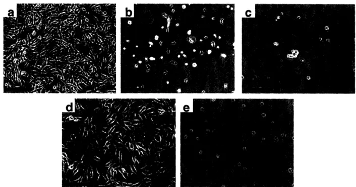
inert to cell adhesion49. Typical results of cell adhesion experiments on the PAH/PAA multilayers, 3.0/3.0 PAA/PAAm multilayers, and a TCPS control are shown in Figure 1-3. As can be seen in the figure, cell growth was dramatically affected by multilayer assembly conditions.
Figure 1-3. NR6WT fibroblasts after three days seeded at 10 000 cells/cm2 onto (a) a 6.5/6.5 PAH/PAA multilayer, (b) a 4.0/4.0 PAH/PAA multilayer, (c) a 2.0/2.0 PAHIPAA multilayer, (d)
a TCPS control and (e) a 3.0/3.0 PAA/PAAm multilayer (scale bar = 200 gm).
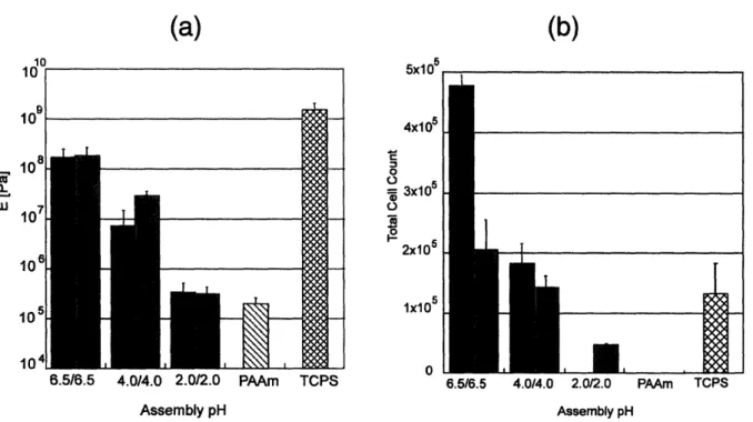
The Rubner and van Vliet groups recently studied various PAH/PAA films using a nanoindentation technique to better quantify the mechanical properties of PAH/PAA multilayers in buffer solution and understand their interactions with mammalian cells66. It was found that the
modulus of the films increased by orders of magnitude as the assembly pH increased, and this correlated with growth of human microvascular endothelial cells as well as Mendelsohn's previous fibroblast results. Figure 1-4 shows that the stiffer films supported cell adhesion, whereas, the more compliant films did not. The fact that nanoscale polyelectrolyte multilayers
can change the chemical and mechanical nature of the surface enough to drastically influence cell behavior is the basis for later chapters of this thesis.
(a)
-6.5/6.5 4.0/4.0 2.0/2.0 PAAm Assembly pH TCPS(b)
5x10⁵ -4x10⁵ o = 3x10⁵ 2x10⁵ 1x10 0 6.5/6.5 4.0/4.0 2.0/2.0 PAAm TPS 2.0/2.0 PAAm TCPS Assembly pHFigure 1-4. Graphs comparing (a) elastic modulus and (b) cell counts for human microvascular endothelial cells. The modulus was obtained using a nanoindentation technique, and cell counts were preformed seven days post-seeding. The black bars represent data from films with PAA as the outermost layer, and gray bars represent data from films with PAH as the outermost layer.
Adapted from work of Thompson et a166.
1.2.4. Patternability of Polyelectrolyte Multilayers
Many of the biological applications that will be discussed in later chapters utilize patterned multilayers. These thin films can be patterned using a variety of methods including polymer-on-polymer stamping (POPS) 67, 68, ink-jet printing18, and photolithography'8. For the
3.0/3.0 PAA/PAAm multilayers, many of these techniques are possible since upon assembly, the hydrogen-bonded films are soluble in neutral water. However, the films can be selectively crosslinked using heat or copolymers with a photo-initiator8. A different approach is presented
in this thesis. In chapter 3, the patterning of multilayers with areas to promote cell adhesion using POPS is studied. The POPS technique, developed by the Hammond group, is a more versatile approach to patterning than its predecessor, microcontact printing6 9 70 since it does not require a specific chemistry or substrate. Jiang et al. showed that POPS can act as a template for multilayer growth6 7 or create a surface containing two different functional groups capable of
selective functionality6 8. The work in this thesis builds on these concepts to study applications that would not be possible with homogenous multilayers.
1.3. Thesis Objectives and Outline
The major objective of this research is to explore applications for weak polyelectrolyte multilayers as biomaterials. The ability to tune these ultra-thin polymer films on the molecular level gives them interesting properties that could help address many important biomedical problems. The second chapter presents a method to pattern polyelectrolyte multilayers on the micron-scale utilizing polymer-on-polymer stamping (POPS). This versatile technique for surface patterning produces patterns with controlled thickness and functional group density. Chapter three provides an application for this surface patterning method; cell patterns and arrays when using a cell-inert polyelectrolyte multilayer film as the background. Using POPS, it was possible to control the ligand density and subsequent cell behavior on the patterned regions due to the tunability of the technique. A much different application, drug delivery, is discussed in the fourth chapter. We found that porous weak polyelectrolyte multilayers provided sustained drug release for days to weeks of both hydrophobic and hydrophilic model drugs. Finally, the fifth and sixth chapters present two different multilayer platforms for killing bacteria and preventing bacterial adhesion, respectively. In these chapters, a silver-loaded multilayer system for killing
bacteria and a superhydrophobic multilayer film that prevents fouling from proteins, cells, and bacteria are presented. In addition, the superhydrophobic multilayers can be patterned for high-throughput screening. Although at first glance these applications seem very different, they are tied together by the types of thin films used. The films were all composed of weak polyelectrolyte multilayers with varying degrees and types of post-treatment processing.
References for Chapter 1
(1) Decher, G.;Hong, J. D.;Schmitt, J. Buildup of Ultrathin Multilayer Films by a Self-Assembly Process: Iii. Consecutively Alternating Adsorption of Anionic and Cationic Polyelectrolytes on Charged Surfaces Thin Solid Films 1992, 210, 831-835.
(2) Decher, G. Fuzzy Nanoassemblies: Toward Layered Polymeric Multicomposites Science 1997, 277, 1232-1237.
(3) Schlenoff, J. B.;Dubas, S. T. Mechanism of Polyelectrolyte Multilayer Growth: Charge Overcompensation and Distribution Macromolecules 2001, 34, 592-598.
(4) Donath, E.;Sukhorukov, G. B.;Caruso, F.;Davis, S. A.;Mohwald, H. Novel Hollow Polymer Shells by Colloid-Templated Assembly of Polyelectrolytes Angew. Chem. Int.
Ed. 1998, 37, 2202-2205.
(5) Chiarelli, P. A.;Johal, M. S.;Casson, J. L.;Roberts, J. B.;Robinson, J. M.;Wang, H.-L. Controlled Fabrication of Polyelectrolyte Multilayer Thin Films Using Spin-Assembly
Adv. Mater. 2001, 13, 1167-1171.
(6) Chiarelli, P. A.;Johal, M. S.;Holmes, D. J.;Casson, J. L.;Robinson, J. M.;Wang, H.-L. Polyelectrolyte Spin-Assembly Langmuir 2002, 18, 168-173.
(7) Lvov, Y.;Ariga, K.;Kunitake, T. Assembly of Alternate Protein Polyion Ultrathin Films
Chem. Lett. 1994, 2323-2326.
(8) Lvov, Y.;Decher, G.;Sukhorukov, G. Assembly of Thin Films by Means of Successive Deposition of Alternate Layers of DNA and Poly(Allylamine) Macromolecules 1993, 26, 5396-5399.
(9) Yoo, D.;Wu, A.;Lee, J.;Rubner, M. F. New Electro-Active Self-Assembled Multilayer Thin Films Based on Alternately Adsorbed Layers of Polyelectrolytes and Functional Dye Molecules Synth. Met. 1997, 85, 1425-1426.
(10) Ariga, K.;Lvov, Y.;Kunitake, T. Assembling Alternate Dye-Polyion Molecular Films by Electrostatic Layer-by-Layer Adsorption J. Am. Chem. Soc. 1997, 119, 2224-2231. (11) Wu, A.;Lee, J.;Rubner, M. F. Light Emitting Electrochemical Devices from Sequentially
Adsorbed Multilayers of a Polymeric Ruthenium (II) Complex and Various Polyanions
Thin Solid Films 1998, 329, 663-667.
(12) Clark, S. L.;Handy, E. S.;Rubner, M. F.;Hammond, P. T. Creating Microstructures of Luminescent Organic Thin Films Using Layer-by-Layer Assembly Adv. Mater. 1999, 11, 1031-1035.
(13) Ariga, K.;Lvov, Y.;Ichinose, I.;Kunitake, T. Ultrathin Films of Inorganic Materials (SiO2 Nanoparticle, Montmorillonite Microplate, and Molybdenum Oxide) Prepared by
Alternate Layer-by-Layer Assembly with Organic Polyions. Appl. Clay Sci. 1999, 15, 137-152.
(14) Schaak, R. E.;Mallouk, T. E. Self-Assembly of Tiled Perovskite Monolayer and Multilayer Thin Films Chem. Mater. 2000, 12, 2513-2516.
(15) Stockton, W. B.;Rubner, M. F. Molecular-Level Processing of Conjugated Polymers. 4.
Layer-by-Layer Manipulation of Polyaniline Via Hydrogen-Bonding Interactions
(16) Wang, L. Y.;Fu, Y.;Wang, Z.;Wang, Y.;Sun, C.;Fan, Y.;Zhang, X. Multilayer
Assemblies of Poly(4-Vinylpyridine) Bearing an Osmium Complex and Poly(Acrylic Acid) Via Hydrogen Bonding Macromol. Chem. Phys. 1999, 200, 1523-1527.
(17) Sukhishvili, S. A.;Granick, S. Layered, Erasable, Ultrathin Polymer Films J. Am. Chem.
Soc. 2000, 122, 9550-9551.
(18) Yang, S. Y.;Rubner, M. F. Micropatterning of Polymer Thin Films with pH-Sensitive and Cross-Linkable Hydrogen-Bonded Polyelectrolyte Multilayers J. Am. Chem. Soc. 2002,
124, 2100-2101.
(19) Kotov, N. A. Layer-by-Layer Self Assembly: The Contribution of Hydrophobic Interactions Nanostruct. Mater. 1999, 12, 789-796.
(20) Cui, X.;Pei, R.;Wang, Z.;Yang, F.;Ma, Y.;Dong, S.;Yang, X. Layer-by-Layer Assembly of Multilayer Films Composed of Avidin and Biotin-Labeled Antibody for
Immunosensing Biosens. Bioelectron. 2003, 18, 59-67.
(21) Joly, S.;Kane, R.;Radzilowski, L.;Wang, T.;Wu, A.;Cohen, R. E.;Thomas, E. L.;Rubner, M. F. Multilayer Nanoreactors for Metallic and Semiconducting Particles Langmuir 2000, 16, 1354-1359.
(22) Wang, T. C.;Rubner, M. F.;Cohen, R. E. Polyelectrolyte Multilayer Nanoreactors for Preparing Silver Nanoparticle Composites: Controlling Metal Concentration and Nanoparticle Size Langmuir 2002, 18, 3370-3375.
(23) Mandel, M. Polyelectrolytes,in Encyclopedia of Polymer Science and Engineering, Kroschwitz, J. I., Editor. 1989, John Wiley & Sons: New York. 739-829.
(24) Yoo, D.;Shiratori, S. S.;Rubner, M. F. Controlling Bilayer Composition and Surface Wettability of Sequentially Adsorbed Multilayers of Weak Polyelectrolytes
Macromolecules 1998, 31, 4309-4318.
(25) Shiratori, S. S.;Rubner, M. F. Ph-Dependent Thickness Behavior of Sequentially Adsorbed Layers of Weak Polyelectrolytes Macromolecules 2000, 33, 4213-4219. (26) Yoshikawa, Y.;Matsuoka, H.;Ise, N. Ordered Structure of Polyallylamine Hydrochloride
in Dilute Solutions as Studied by Small-Angle X-Ray-Scattering Br. Polym. J. 1986, 18, 242-246.
(27) Choi, J.;Rubner, M. F. Influence of the Degree of Ionization on Weak Polyelectrolyte Multilayer Assembly Macromolecules 2005, 38, 116-124.
(28) Petrov, A. I.;Antipov, A. A.;Sukhorukov, G. B. Base-Acid Equilibria in Polyelectrolyte Systems: From Weak Polyelectrolytes to Interpolyelectrolyte Complexes and
Multilayered Polyelectrolyte Shells Macromolecules 2003, 36, 10079-10086.
(29) Choi, J.;Rubner, M. F. Selective Adsorption of Amphiphilic Block Copolymers on Weak Polyelectrolyte Multilayers J. Macromol. Sci. Pure Appl. Chem. 2001, 38, 1191-1206. (30) Mendelsohn, J. D.;Barrett, C. J.;Chan, V. V.;Pal, A. J.;Mayes, A. M.;Rubner, M. F.
Fabrication of Microporous Thin Films from Polyelectrolyte Multilayers Langmuir 2000,
16, 5017-5023.
(31) Hiller, J.;Mendelsohn, J. D.;Rubner, M. F. Reversibly Erasable Nanoporous Anti-Reflection Coatings from Polyelectrolyte Multilayers Nat. Mater. 2002, 1, 59-63.
(32) Wang, T. C.;Chen, B.;Rubner, M. F.;Cohen, R. E. Selective Electroless Nickel Plating on Polyelectrolyte Multilayer Platforms Langmuir 2001, 17, 6610-6615.
(33) Berg, M. C.;Zhai, L.;Cohen, R. E.;Rubner, M. F. Controlled Release from Porous Polyelectrolyte Multilayers In Preparation.
(34) Mendelsohn, J. D.;Yang, S. Y.;Hiller, J.;Hochbaum, A. I.;Rubner, M. F. Rational Design of Cytophilic and Cytophobic Polyelectrolyte Multilayer Thin Films Biomacromolecules 2003, 4, 96-106.
(35) Berg, M. C.;Choi, J.;Hammond, P. T.;Rubner, M. F. Tailored Micropatterns through Weak Polyelectrolyte Stamping Langmuir 2003, 19, 2231-2237.
(36) Zhai, L.;Cebeci, F. C.;Cohen, R. E.;Rubner, M. F. Stable Superhydrophobic Coatings from Polyelectrolyte Multilayers Nano Lett. 2004, 4, 1349-1353.
(37) Richert, L.;Lavalle, P.;Payan, E.;Shu, X. Z.;Prestwich, G. D.;Stoltz, J. F.;Schaaf, P.;Voegel, J.-C.;Picart, C. Layer by Layer Buildup of Polysaccharide Films: Physical Chemistry and Cellular Adhesion Aspects Langmuir 2004, 20, 448-458.
(38) Chluba, J.;Voegel, J.-C.;Decher, G.;Erbacher, P.;Schaaf, P.;Ogier, J. Peptide Hormone Covalently Bound to Polyelectrolytes and Embedded into Multilayer Architectures Conserving Full Biological Activity Biomacromolecules 2001, 2, 800-805.
(39) Caruso, F.;Trau, D.;Mohwald, H.;Renneberg, R. Enzyme Encapsulation in Layer-by-Layer Engineered Polymer Multilayer Capsules Langmuir 2000, 16, 1485-1488. (40) Decher, G.;Lehr, B.;Lowack, K.;Lvov, Y.;Schmitt, J. New Nanocomposite Films for
Biosensors: Layer-by-Layer Adsorbed Films of Polyelectrolytes, Proteins or DNA
Biosens. Bioelectron. 1994, 9, 677-684.
(41) Qiu, X.;Donath, E.;Mohwald, H. Permeability of Ibuprofen in Various Polyelectrolyte Multilayers Macromol. Mater. Eng. 2001, 286, 591-597.
(42) Vazquez, E.;Dewitt, D. M.;Hammond, P. T.;Lynn, D. M. Construction of Hydrolytically-Degradable Thin Films Via Layer-by-Layer Deposition of Hydrolytically-Degradable Polyelectrolytes J.
Am. Chem. Soc. 2002, 124, 13992-13993.
(43) Quinn, J. F.;Caruso, F. Facile Tailoring of Film Morphology and Release Properties Using Layer-by-Layer Assembly of Thermoresponsive Materials Langmuir 2004, 20, 20-22.
(44) Burke, S. E.;Barrett, C. J. Ph-Dependent Loading and Release Behavior of Small Hydrophilic Molecules in Weak Polyelectrolyte Multilayer Macromolecules 2004, 37, 5375-5384.
(45) Nolan, C. M.;Serpe, M. J.;Lyon, L. A. Thermally Modulated Insulin Release from Microgel Thin Films Biomacromolecules 2004, 5, 1940-1946.
(46) Serpe, M. J.;Yarmey, K. A.;Nolan, C. M.;Lyon, L. A. Doxorubicin Uptake and Release from Microgel Thin Films Biomacromolecules 2005, 6, 408-413.
(47) Elbert, D. L.;Herbert, C. B.;Hubbell, J. A. Thin Polymer Layers Formed by
Polyelectrolyte Multilayer Techniques on Biological Surfaces Langmuir 1999, 15, 5355-5362.
(48) Serizawa, T.;Yamaguchi, M.;Matsuyama, T.;Akashi, M. Alternating Bioactivity of Polymeric Layer-by-Layer Assemblies: Anti- Vs Procoagulation of Human Blood on
Chitosan and Dextran Sulfate Layers Biomacromolecules 2000, 1, 306-309. (49) Yang, S. Y.;Mendelsohn, J. D.;Rubner, M. F. New Class of Ultrathin, Higly
Cell-Adhesion-Resistant Polyelectrolyte Multilayers with Micropatterning Capabilities
Biomacromolecules 2003, 4, 987-994.
(50) Ladam, G.;Schaaf, P.;Cuisinier, F. J. G.;Decher, G.;Voegel, J.-C. Protein Adsorption onto Auto-Assembled Polyelectrolyte Films Langmuir 2001, 17, 878-882.
(51) Picart, C.;Lavalle, P.;Hubert, P.;Cuisinier, F. J. G.;Decher, G.;Schaaf, P.;Voegel, J. C. Buildup Mechanism for Poly(L-Lysine)/Hyaluronic Acid Films onto a Solid Surface
Langmuir 2001, 17, 7414-7424.
(52) Richert, L.;Lavalle, P.;Vautier, D.;Senger, B.;Stoltz, J. F.;Schaaf, P.;Voegel, J.-C.;Picart, C. Cell Interactions with Polyelectrolyte Multilayer Films Biomacromolecules 2002, 3, 1170-1178.
(53) Jessel, N.;Atalar, F.;Lavalle, P.;Mutterer, J.;Decher, G.;Schaaf, P.;Voegel, J. C.;Ogier, J. Bioactive Coatings Based on a Polyelectrolyte Multilayer Architecture Functionalized by Embedded Proteins Adv. Mater. 2003, 15, 692-695.
(54) Qiu, X.;Leporatti, S.;Donath, E.;Mohwald, H. Studies on the Drug Release Properties of Polysaccharide Multilayers Encapsulated Ibuprofen Microparticles Langmuir 2001, 17, 5375-5380.
(55) Dai, J.;Bruening, M. L. Catalytic Nanoparticles Formed by Reduction of Metal Ions in Multilayered Polyelectrolyte Films Nano Lett. 2002, 2, 497-501.
(56) Boulmedais, F.;Frisch, B.;Etienne, O.;Lavalle, P.;Picart, C.;Ogier, J.;Voegel,
J.-C.;Schaaf, P.;Egles, C. Polyelectrolyte Multilayer Films with Pegylated Polypeptides as a New Type of Anti-Microbial Protection for Biomaterials Biomaterials 2004, 25, 2003-2011.
(57) Thierry, B.;Winnik, F. M.;Merhi, Y.;Silver, J.;Tabrizian, M. Bioactive Coatings of Endovascular Stents Based on Polyelectrolyte Multilayers Biomacromolecules 2003, 4,
1564-1571.
(58) Tan, Q.;Ji, J.;Barbosa, M. A.;Fonseca, C.;Shen, J. Constructing Thromboresistant Surface on Biomedical Stainless Steel Via Alyer-by-Layer Deposition Anticoagulant
Biomaterials 2003, 24, 4699-4705.
(59) Thierry, B.;Winnik, F. M.;Merhi, Y.;Tabrizian, M. Nanocoatings onto Arteries Via Layer-by-Layer Deposition: Toward the in Vivo Repair of Damaged Blood Vessels J.
Am. Chem. Soc. 2003, 125, 7494-7495.
(60) Ai, H.;Meng, H.;Ichinose, I.;Jones, A. J.;Mills, D. K.;Lvov, Y. M.;Qiao, X.
Biocompatibility of Layer-by-Layer Self-Assembled Nanofilm on Silicone Rubber for Neurons J. Neurosci. Meth. 2003, 128, 1-8.
(61) Ai, H.;Lvov, Y. M.;Mills, D. K.;Jennings, M.;Alexander, J. S.;Jones, S. A. Coating and Selective Deposition of Nanofilm on Silicone Rubber for Cell Adhesion and Growth Cell
Biochemistry and Biophysics 2003, 38, 103-114.
(62) Serizawa, T.;Yamaguchi, M.;Akashi, M. Alternating Bioactivity of Polymeric Layer-by-Layer Assemblies: Anticoagulation Vs Procoagulation of Human Blood
Biomacromolecules 2002, 3, 724-731.
(63) Ai, H.;Fang, M.;Jones, S. A.;Lvov, Y. M. Electrostatic Layer-by-Layer Nanoassembly on Biological Microtemplates: Platelets Biomacromolecules 2002, 3, 560-564.
(64) Koktysh, D. S.;Liang, X.;Yun, B. G.;PastorizaSantos, I.;Matts, R. L.;Giersig,
M.;SerraRodriguez, C.;LizMarzan, L. M.;Kotov, N. A. Biomaterials by Design: Layer-by-Layer Assembled Ion-Selective and Biocompatible Films of Tio2 Nanoshells for Neurochemical Monitoring Adv. Funct. Mater. 2002, 12, 255-265.
(65) Sinani, V. A.;Koktysh, D. S.;Yun, B. G.;Matts, R. L.;Pappas, T. C.;Motamedi, M.;Thomas, S. N.;Kotov, N. A. Collagen Coating Promotes Biocompatibility of Semiconductor Nanoparticles in Stratified Lbl Films Nano Lett. 2003, 3, 1177-1182.
(66) Thompson, M. T.;Berg, M. C.;Tobias, I. S.;Rubner, M. F.;van Vliet, K. J. Quantifying the Role of Compliance in Cell Adhesion and Proliferation: Polyelectrolyte Multilayers as Mechanically Tunable Cell Substrata Submitted to Biomaterials.
(67) Jiang, X.;Hammond, P. T. Selective Deposition in Layer-by-Layer Assembly: Functional Graft Copolymers as Molecular Templates Langmuir 2000, 16, 8501-8509.
(68) Jiang, X.;Zheng, H.;Gourdin, S.;Hammond, P. T. Polymer-on-Polymer Stamping: Universal Approaches to Chemically Patterned Surfaces Langmuir 2002, 18, 2607-2615. (69) Kumar, A.;Biebuyck, H. A.;Whitesides, G. M. Patterning Self-Assembled Monolayers:
Applications in Materials Science Langmuir 1994, 10, 1498-1511.
(70) Kumar, A.;Whitesides, G. M. Patterned Condensation Figures as Optical Diffraction Gratings Science 1994, 263, 60-62.
Chapter 2
Polymer on Polymer Stamping of Weak
Polyelectrolytes
Reproduced in part with permission from Berg, M. C.;Choi, J.;Hammond, P. T.;Rubner, M. F. Tailored Micropatterns through Weak Polyelectrolyte Stamping Langmuir 2003, 19, 2231-2237. Copyright 2003 American Chemical Society
2.1.
Introduction
In recent years, a great deal of effort has been put forth to research thin films made from polyelectrolyte multilayers using the layer-by-layer assembly technique. Since this method for building layers of oppositely charged polyelectrolytes was first developed, many different materials have been employed to produce thin films using this approach 5. Multilayer films
produced in this layer-by-layer process offer many other desirable traits beyond the versatility in building materials. The film thickness and surface properties can be tailored with unequaled control since each layer is added sequentially. In addition, the process is relatively simple and typically utilizes aqueous solutions. Of specific interest to this paper is the behavior of multilayer films assembled with weak polyelectrolytes with pH-tunable charge densities. It has been shown that the thickness of adsorbed weak polyelectrolyte layers changes greatly according to the ionization degree of each polymer involved in the assembly process4' 6. Weak polyelectrolytes, such as poly(allylamine hydrochloride) (PAH) and poly(acrylic acid) (PAA), can deposit on a substrate to create very thick (up to 80 A) layers when the films are fabricated under pH conditions where the polymers are not fully charged. Conversely, the two polymers can be used in their fully charged state to create thin layers similar to strong polyelectrolytes.
This means that films can be created with considerably different bulk and surface properties by using the same two polymers.
Patterning weak polyelectrolyte multilayer films on a micron-scale is highly desirable for various applications such as biosensors, tissue engineering implants, electronic devices, and photonic structures. Biological applications present some of the greatest opportunities particularly considering recent findings that specific PAA/PAH films are completely inert to mammalian cell attachment (specifically mouse fibroblasts), while others promote cell adhesion7. In addition, these multilayers are useful in directing the selective adsorption of block copolymers8 or catalysts for further chemistry9, which can play an important role in biological studies. Soft lithographic patterning techniques in which a chemically patterned surface acts as a template for the deposition of strong or weak polyelectrolyte multilayers have been developed
-12 using microcontact printing methods to create chemical surface templates on gold and oxide
surfaces 3 415. This approach and other methods allow the formation of three-dimensional micron
scale patterned multilayer films. The ability to pattern the topmost surface of a polyelectrolyte multilayer is particularly critical for a number of sensor and array applications; however, micro-contact printing techniques based upon thiol and silane chemistry on metal and metal oxide surfaces are not applicable to the surfaces of polymer multilayers. Recently, a technique, polymer-on-polymer stamping (POPS), has been developed, which involves the direct application of a polymer monolayer onto a surface 6' 7. POPS, which has greatly widened the scope of suitable materials that can be transferred using a stamp, takes advantage of the multivalent nature of polymer chains to form a stable monolayer on the desired substrate. This method was originally developed to pattern a polyethylene oxide graft copolymer onto a polyelectrolyte multilayer surface'6; more recent investigations have reported the use of block
copolymers containing a functional and an anchoring block segment to modify multilayer surfaces via electrostatic, secondary, and covalent interactions, and the ability to pattern strong polyelectrolytes onto oppositely charged polyelectrolyte multilayer surfaces'7 utilizing solely
electrostatic interactions. This approach makes it possible to create micron scale features of differing chemical functionality over large areas on the underlying multilayer. Additional steps can be used to take advantage of the functionality on the patterned polymer or the unstamped region of the substrate, as demonstrated in the use of POPS on multilayer surfaces to template the deposition of additional multilayers 6, colloid deposition , and nickel plating9. Besides
POPS, other soft lithographic methods have been developed to pattern polymer surfaces that take advantage of specific chemistry with the surface2023. Soft lithographic techniques have many
advantages over other patterning methods such as photolithography, including low cost, the ability to create large area patterns, and a wider variation in available surface chemistries. The stamping process does not require a clean room environment or the harsh chemicals necessary for most photolithography techniques, and can be used to obtain features down to one micron or
less in size.
This chapter examines the extension of the POPS process to the transfer of weak polyelectrolytes onto weak polyelectrolyte multilayer platforms. The main advantage of working with weak polyelectrolytes is the ability to influence the charge density of the polymer by simple pH adjustments. In the stamping process, the pH of the polyelectrolyte ink can be adjusted to provide a polymer that is either fully charged in solution or completely uncharged. The pKa of PAH is approximately 9.0. Hence, at this pH, 50% of the functional groups are charged24 .
Therefore, adjustment of the ink solution pH to values well above and below pH 9.0 covers the range of ionization possibilities. The question still remains as to whether or not changing the
charge density of the polymer in the ink solution affects the thickness of the stamped features and the number of free functional groups in the transferred polymers. Because the stamping process is a dry one, it is unclear whether the thickness of the stamped layers can be tailored in manner similar to the adsorption of weak polyelectrolytes6. It has been shown that adsorption of weak polyelectrolytes at pH values that yield low charge can lead to a thicker adsorbed polymer layer. These experiments were designed to check the hypothesis that altering the ionization of the polyelectrolyte would change the thickness of the transferred polymer layer upon stamping as well as available functional groups.
This chapter also addresses issues such as the stability of the stamped pattern in a buffered solution as would be commonly used in biological experiments. We believe that these films are suitable for biological applications due to recent findings that weak polyelectrolyte multilayers can be created to resist cell adhesion. Patterning these particular films with different functional groups could allow directed chemistry to these regions on a micron scale. Such chemistry could present specific biological ligands for cell attachment, creating a template for controlling cell growth on virtually any surface. Also, multilayers offer the possibility of utilizing the functional groups within the film itself in addition to those on the surface.
2.2. Experimental Methods
1. Materials. Poly(acrylic acid) (PAA) (MW = 90,000) was purchased from Polysciences as a 25% aqueous solution. Poly(allylamine hydrochloride) (PAH) (MW = 70,000) and dansyl chloride were purchased from Aldrich. Poly(dimethylsiloxane) (PDMS) stamps were created by curing Sylguard 184 (a two ingredient elastomer kit purchased from Dow Chemical) on top of a silicon master with a striped photoresist pattern. All materials were used without any
further purification. Dulbecco's Phosphate Buffered Saline (PBS) solution was purchased from Gibco.
2. Substrate Preparation. As detailed in previous work4, multilayer thin films of weak
polyelectrolytes were made by a layer-by-layer dipping assembly technique, which utilized an HMS programmable slide stainer from Zeiss, Inc. to automate the process. For making the multilayer films, 10-2 M (based on repeat unit of the polymer) polyelectrolyte solutions were prepared with 18 MQ Millipore water, and were pH-adjusted with either HC1 or NaOH. Clean glass substrates were first submerged into the polyelectrolyte solution (PAH first) for 15 minutes, and then taken through a series of three rinse baths of water (pH unadjusted -5.5) for 2, 1, and 1 minute, respectively. Afterwards, the slides were immersed in the oppositely charged polyelectrolyte solution for 15 minutes followed by the same rinse procedure. The process was then repeated until 20 layers were assembled to leave PAA as the outermost layer.
3. Stamping of weak polyelectrolytes. PDMS stamps were inked by immersion in 0.05
M (based on polymer repeat unit) polyelectrolyte solution (PAH) for 1 hour, similar to a previously described method2 5. The pH of the PAH ink was adjusted using HC1 or NaOH. The stamps were then briefly rinsed with water and blown dry with air. The stamp was still wet when rinsed, and the pH of the rinse water was not adjusted. To ensure the degree of ionization of the PAH was not changed, the rinse step was kept less than 2 seconds. Finally, the stamp was brought into contact with a polyelectrolyte multilayer platform for 30 seconds. Some samples were then rinsed with water for 2 minutes to compare the stamped features before and after rinsing.
4. Dansyl Chloride Attachment. Samples stamped with PAH were reacted with dansyl
chemistry. When dansyl chloride is bound to the primary amines of PAH in the stamped region of the films, the compound can be seen using fluorescence microscopy. For the treatment, a 10-2 M stock solution of dansyl chloride was prepared in N,N - dimethyl formamide (DMF). From this stock solution, 5 mL were added to 50 mL of 0.1 M sodium bicarbonate buffer solution at pH = 9.0 and stirred vigorously. Samples were then placed in this mixed solution and allowed to incubate for 2 minutes. The time was kept short to avoid having the dansyl chloride diffuse into the bulk of the film. Following the process, samples were rinsed for 2 minutes in water. The relative intensity of the fluorescent images was checked using the National Institute of Health's (NIH) ImageJ image analysis software. The reported values are not meant to be quantitative for the number of reacted amine groups, but are meant to compare the relative number of reacted groups for each stamping condition. The reported values were obtained by analyzing 6 spots from images taken of 3 different samples under the same lens and exposure time.
5. PBS Stability. The stability of the stamped patterns in a pH 7.4 buffered solution was checked using PBS. The samples were immersed in PBS for 3 days to check the stability of the patterns in biological applications.
6. Characterization. The thickness of the films was checked using a Tencor P-10
Surface Profiler (Tencor, Santa Clara, CA) to perform profilometry4. Reported values represent
an average of at least 6 measurements for each sample. The Digital Instruments Dimension 3000 atomic force microscope (AFM) (Digital Instruments, Santa Barbara, CA) was used in tapping mode to obtain topographical information about the stamped area including heights of stamped features. Stamp height values were obtained by examining the sections of AFM data. Reported values represent an average of at least 10 data points taken from different areas of the samples. Four different samples were used for each stamping condition and multilayer platform. For
visualization of the PAH stamped regions labeled with dansyl chloride, a Zeiss Axioplan 2 fluorescence microscope (Carl Zeiss Inc., Thornwood, NY) was utilized. When using a DAPI filter, the stamped regions appear blue-green. The intensity of the fluorescence images was checked using the NIH ImageJ software.
7. Statistical Analysis. Analysis of variance (ANOVA) was employed to test the
statistical significance of differences in stamped thickness values. ANOVA tests whether the variance between thickness values from different stamping conditions is greater than the variance within a reported thickness value for a given stamping condition. If this is the case, then the difference between two stamping conditions is statistically significant. The final result of the ANOVA is the p-value (or probability value). If the p-value is less than a significance level of 0.05 (based on a 5% level), than the groups are statistically different.
2.3.
Results and Discussion
2.3.1. Characterization of Weak Polyelectrolyte Multilayer Films
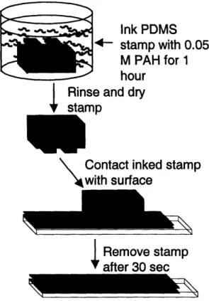
In this study, we stamped PAH onto PAA/PAH multilayer films where PAA was the top layer. Figure 2-1 contains a schematic diagram of the POPS process; adhesion of the polymer to the multilayer surface can be based on electrostatic, hydrogen bonding, acid-base or covalent interactions. The process of submerging the entire PDMS stamp in the ink solution has been utilized in previous research2 5. As the diagram indicates, we believe that the polyelectrolyte
Ink PDMS stamp with 0.05
M PAH for 1
hour Rinse and dry stamp
\
Contact inked stamp with surfaceI Remove stamp
Figure 2-1. Diagram illustrating the polymer-on-polymer stamping process for PAH on a PAA/PAH multilayer platform.
We chose three different weak polyelectrolyte multilayer platforms to check the versatility of the POPS process when the surface roughness and chemical characteristics of the multilayer film are changed. Each of these three 10 bilayer PAA/PAH multilayer films was built using a different pH combination to create platforms that differ dramatically in architecture (a bilayer being one layer of PAH and PAA). Multilayer films of PAA/PAH assembled at pH
6.5/6.5, 3.5/7.5, and 2.5/2.5 (notation refers to the assembly pH of PAA and PAH) were
prepared. The molecular architecture of the various PAA/PAH multilayer films has previously been described in detail6. Briefly, both polyelectrolytes are fully charged when deposited at
6.5/6.5, resulting in extremely thin layers. In this case, most of the polyion charged groups are paired with oppositely charged groups from the complimentary polyelectrolyte. The 6.5/6.5 films are relatively smooth (4 A RMS roughness), with highly interpenetrated layers. There are
few remaining free functional groups from either PAA or PAH left in the interior of the film or at the surface due to the high degree of ionic crosslinking. The 3.5/7.5 PAA/PAH layers are very thick, loopy structures with a higher RMS roughness of 18 A. The 2.5/2.5 films exhibit a bilayer thickness and RMS roughness that falls between those of the 6.5/6.5 and 3.5/7.5 PAA/PAH multilayers. The resulting films are rich in free acid groups both in the bulk and on the surface regardless of the top layer of the film. The 2.5/2.5 films are also of particular interest due to their ability to resist mammalian cell adhesion7. Table 2-1 compares the thickness and roughness
values measured on dried films for each of the PAA/PAH multilayer films prepared for this study.
Table 2-1. Average incremental bilayer thickness (measured by profilometry) and RMS roughness (obtained from AFM) measurements for the three representative 10 bilayer PAA/PAH
multilayer films used in stamping study. PAA/PAH Avg. Bilayer Avg. RMS
Film Thickness (A) Roughness (A)
6.5/6.5 6 4
2.5/2.5 42 10
3.5/7.5 106 18
2.3.2. Influence of Ink pH of PAH stamped onto Multilayers as Studied with
Atomic Force Microscopy
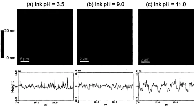
PAH was stamped onto the three different PAA/PAH multilayer systems with PAA as the top layer. For all cases, the PDMS stamp was immersed in a 0.05 M PAH solution at room temperature for 1 hour. Three different ink solutions were used to present PAH with varying degrees of ionization, which were approximately 100% (pH = 3.5), 50% (pH = 9.0), and 0% (pH = 11.0). The pH values were chosen based on previous work that estimated the charge density of PAH in solution as a function of pH24, 26. After inking the stamp with PAH, the stamp was rinsed
brought into contact with the multilayer surface, which consisted of 10 bilayers of alternating PAH and PAA, for 30 seconds. This process was repeated for each ink pH and multilayer combination to yield a total of 9 different conditions. Following stamping, the patterned samples were characterized with AFM, rinsed for 2 minutes in water, and then re-characterized to explore the effects of the rinse step on the stamped regions. The pattern that was used for all of the stamping conditions contained lines of varying widths from 5 m to 10 pm. In the resulting images, the thicker lines always indicate the stamped regions. The width of stamped PAH lines matched the PDMS stamp features, so no spreading was observed on the multilayer surface.
(a) Ink pH = 3.5 (b) Ink pH = 9.0 (c) Ink pH = 11.0
20 nm
Onm
, - . 20.0
Io 10.0 20.0
Figure 2-2. AFM height images and sectional analyses of 6.5/6.5 PAA/PAH multilayer platforms stamped with PAH at various ink pH's.
The 6.5/6.5 multilayer films were by far the smoothest platforms in this study, and consequently, the patterns were the easiest to visualize using AFM. Figure 2-2 shows AFM data for the 6.5/6.5 PAA/PAH multilayers at the three different stamping pH conditions after rinsing
nm
o
o