doi:10.1017/S1751731112001802
Intrauterine crowding impairs formation and growth of
secondary myofibers in pigs
C. E. Pardo
1,2, J. Be´rard
1,2a, M. Kreuzer
2and G. Bee
1-1Agroscope Liebefeld-Posieux, Research Station ALP, 1725 Posieux, Switzerland;2ETH Zurich, Institute of Agricultural Science, Universita¨tstrasse 2, 8092 Zurich, Switzerland
(Received 10 May 2012; Accepted 13 July 2012; First published online 1 October 2012)
There are indications that intrauterine crowding may cause intrauterine growth retardation with the possibility of an impaired myofiber hyperplasia. The aim of the study was to confirm this by generating large differences in uterine space using sows that were unilaterally hysterectomized-ovariectomized (HO; crowded) or unilaterally oviduct ligated (OL; non-crowded). In the study, seven HO and seven OL Swiss Large White third parity sows were used. At farrowing, litter size and litter birth weight were determined. Subsequently, within each litter two male and two female progenies each with the respectively lowest (L) and highest (H) birth weight were sacrificed. Internal organs and brain were weighed, andlongissimus(LM) andsemitendinosusmuscle (SM) samples were collected. Histological analyses were performed in both muscles using mATPase staining after preincubation at pH 4.3 and 10.2. Myosin heavy chain (MyHC) polymorphism was determined in the LM by means of SDS-PAGE. The number of piglets born alive was similar in both sow groups, but litter size expressed per uterine horn was lower (P,0.05) in OL than HO sows. Consequently, OL progeny were markedly heavier (P,0.01). Regardless of gender, the organs, the brain and the SM were heavier (P,0.001) in OL and H compared with HO and L offspring, respectively. Compared with HO pigs, the SM of OL offspring tended (P,0.1) to have more myofibers, which were of larger (P,0.05) size. However, myofiber density appeared to be lower (P,0.1) in the SM of OL than HO pigs. The impact of birth weight on myofiber characteristics was limited to the lower (P,0.05) myofiber density in the SM and the larger (P,0.01) myofiber size in the light portion of the SM of H than L offspring, whereas myofiber hyperplasia did not differ between birth weight categories. The SM, but not the LM, of male offspring had a greater (P,0.05) myofiber density. This did not affect total SM myofiber number. The relative abundance of fetal and type I MyHC in the LM was lower (P,0.05) and that of type II MyHC was greater (P,0.001) in OL than HO pigs. The current data suggest that regardless of birth weight and gender, in the LM and SM of individuals born from a crowded environment, not only hyperplasia but also hypertrophy of myofibers is impaired and their maturity seems delayed.
Keywords: birth weight, intrauterine crowding, myogenesis, pig
Implications
In sows, selection for increasing litter size has led to a decrease in litter birth weight as a likely result of elevated intrauterine crowding. It is important to better understand how uterine space availability affects fetal development. This study validated the connection between the level of intra-uterine crowding and the degree of fetal growth retardation. This includes reduced myofiber size and number and ulti-mately impaired pre- and postnatal muscle development. These results will help to convey the litter size where negative effects of intrauterine crowding are not counterbalancing the economic advantage given by extra offspring.
Introduction
Intrauterine growth retardation in newborn pigs has been hypothesized to be linked to intrauterine crowding. This gets apparent in lower overall fetal growth, markedly lower birth weight and impaired myofiber hyperplasia especially of that of the secondary myofibers in thesemitendinosus(SM) and
psoas majormuscle (Be´rardet al., 2010). Primiparous sows of the common Large White breed kept in Switzerland were shown to have a very high ovulation rate of on average 31.3 (Be´rard and Bee, 2010). Thus, naturally occurring intrauterine crowding cannot be excluded. Individual birth weight may be of importance for the actual effects of intrauterine crowding because of its influence on postnatal growth. There seems to be a close relationship between birth weight and low meat quality, resulting in part from low myofiber numbers and an
aPresent address: Institut Agricole Re´gionale La Roche`re, Aosta, Italy.
accelerated fiber hypertrophy in piglets with low birth weights (Bee, 2004; Gondret et al., 2006). Some studies have even proposed that a birth weight 3 gender interaction exists regarding postnatal development, suggesting that the negative effects of low birth weight exhibit an influence in adult life more in female than in male pigs (Poore and Fowden, 2004; Rehfeldtet al., 2008).
The effects of differences in available uterine space on fetal development and maternal metabolism have been previously studied with different experimental models such as unilateral ovary-hysterectomy (HO) (Huanget al., 1987; Be´rardet al., 2010) or unilateral oviduct ligation (OL; Town
et al., 2004) or both (Pe`reet al., 1997). However, only Town
et al. (2004) and Be´rard et al. (2010) studied the effects of intrauterine crowding on muscle development using these experimental approaches. Compared with OL sows, HO sows have only half the uterine space, whereas the overall ovulation rate is similar for both experimental models (Christensonet al., 1987). Therefore, combining the HO and OL experimental model makes uterine conditions regarding space and nutrient availability per fetus even more divergent and allows to better determine the effects of intrauterine crowding on fetal development and myogenesis. In addition, the use of OL also prevents the possibility of intrauterine crowding, which might happen in intact sows with a high ovulation rate as observed by Be´rard and Bee (2010).
The aim of the present study was to confirm or disprove effects of intrauterine crowding on organ and muscle development by generating large and unbiased differences in uterine space with the combined use of both, HO and OL, models. For that purpose, progeny were selected at birth in a way that birth weight extremes and both genders were included. This approach would help identifying which type of piglet will be most adversely affected.
Material and methods
Animals and treatments
All procedures imposed on the animals were approved by the Committee for Animal Care and Use of the Canton Fribourg, Switzerland.
In all, 14 third parity Swiss Large White gilts were used in the present study (seven pairs of siblings). The HO sows originated from a previous experiment (Be´rard
et al., 2010). In their intact sisters, unilateral OL surgery of the left oviduct was performed two days after weaning of the second parity. Before the surgery, sows were sedated with a mix of 25 mg romidifin (Sedivet, Boehringer Ingelheim, Basel, Switzerland) and 30 mg butorphanol (Morphasol-10, Dr. E. Gra¨ub AG, Bern, Switzerland). Subsequently, ligation of the oviduct was carried out under general anesthesia induced with 15 mg/kg BW of ketamine (Ketasol-100, Dr. E. Gra¨ub AG) and maintained with isoflurane at a saturation of 2% to 3.5% (Attane, Provet AG, Lyssach, Switzerland), via a face mask, with 2 l/min of oxygen. Post-operative analgesia was provided by administration of 400 mg carprofen (Rimadyl Rind, Pfizer GmbH Tiergesundheit,
Karlruhe, Germany). No postsurgical problems occurred and the wounds healed completely.
For estrus stimulation, 5 ml/day of Regumate Porcine (Intervet International B.V., Boxmeer, the Netherlands) was injected subcutaneously during 18 days. At day 18, 5 ml of Folligon (Intervet International B.V.) and, 4 days later, 1.5 ml of Chorulon 1500 (Intervet International B.V.) were injected subcutaneously. During estrus, the 14 gilts were artificially inseminated three times with unfrozen semen from Swiss Large White boars (Suisag, Sempach, Switzerland).
From mating to farrowing, gestating sows were reared with other multiparous gilts in group pens equipped with an automatic feeder (Compident, Model 2000, Schauer, Prambachkirchen, Austria). Sows were offered 2.5 kg/day of a gestation diet, which was formulated according to the Swiss nutrient recommendations for pigs (Agroscope Liebefeld Posieux Research Station (ALP), 2012). The ingredient com-position (g/kg as fed) was: barley, 242; oat, 200; dried whole maize plant, 200; wheat bran, 100; soybean meal, 100; rapeseed meal, 50; molasses, 50; 1 : 1 lard–tallow mixture, 30.2; potato protein, 10; NaCl, 4.8; calcium carbonate, 11.7; dicalcium phosphate, 0.8; lysine HCl, 0.9; L-threonine, 0.6;
vitamin–mineral premix, 4.0; binder (Pellan, Mikro-Technik GmbH & Co. KG, Bu¨rgstadt, Germany), 4.0. The premix sup-plied per kg of diet: all-trans retinol, 1.2 mg; cholecalciferol, 0.006 mg; vitamin E, 9.9 mg; riboflavin, 2.8 mg; vitamin B-6, 1.3 mg; vitamin B-12, 0.015 mg; vitamin K3, 0.2 mg; pan-tothenic acid, 102 mg; niacin, 10 mg; folic acid, 0.48 mg; Fe as FeSO4, 84 mg; I as Ca(IO3)2, 0.56 mg; Se as Na2Se, 0.2 mg; Cu
as CuSO4, 9.2 mg; Zn as ZnO2, 81 mg; Mn as MnO2, 2.5 mg;
choline, 196 g; biotin, 0.99 mg. The estimated composition per kg dry matter was: digestible energy, 12.1 MJ; total ash, 62.2 g; ether extract, 68.4 g; CP, 133 g; crude fiber, 94.0 g. The animals had free access to water.
Data and tissue sample collection at farrowing
At farrowing, one HO sow gave birth to only two piglets and was excluded from the experiment for that reason. At the end of farrowing, the number of piglets born alive and stillborn and their individual BWs were recorded. Then, where possible, the male and female piglets with the highest (H) and the male and female piglets with the lowest (L) birth weight of each litter were sacrificed. Pigs with a birth weight of ,800 g were considered runts and were not used for sample collection. One OL sow had only male progeny, one OL sow had no low birth weight male (L) and two OL sows did not give birth to either a H or a L female, respectively. Consequently, only 47 instead of the 56 piglets intended to be collected from the 14 sows were selected. The selected newborn pigs were anesthetized using isoflurane at a 4% saturation in oxygen with a flow rate of 2 l/min, for at least 5 min and euthanized by exsanguination. Subsequently, kidney, spleen, liver, lung, heart and brain were dissected and weighed. From the left side of the carcass caudal to the 10th rib, a 3 cm long section of thelongissimus dorsimuscle (LM) was collected. In addition, the entire SM consisting of a light (SMlight) and dark (SMdark) portion was excised and weighed.
These muscles and sections were selected because in slaughter pigs they differ in their metabolic and contractile properties where the LM and SMlightwill be predominately
composed of fast-twitch glycolytic myofibers and the SMdark
of slow-twitch oxido-glycolytic myofibers (Bee, 2004). The two muscles were then snap frozen by submersion in 2-methylbutane cooled in liquid nitrogen and stored at 2808C for subsequent histochemical analysis.
Histochemical analysis
The number of primary and secondary myofibers, the second-ary : primsecond-ary myofiber ratio and the cross-sectional muscle area were determined histochemically as described previously by Be´rardet al. (2010). Briefly, 10 mm thick cross-sections of the muscles were prepared and stained for the determination of myofibrillar ATPase activity after alkaline (pH 10.3) or acid (pH 4.5) preincubation. In the SMdarkand the LM, the primary
myofibers stain light and the secondary myofibers dark using the basic preincubation condition, whereas the opposite occurs after acid preincubation. In addition, primary myofibers can be differentiated by size from the secondary myofibers. This was possible in the LM and the SMdark, whereas, in accordance
to observations of Lefaucheur et al. (1995), primary and secondary myofibers were not distinguishable in the SMlightof
newborn pigs using this mATPase histochemistry assay. To determine the cross-sectional area of the total SM, SMdark
and SMlight, cross-sections of the muscles were stained using
anti-slow myosin heavy chain (MyHC) monoclonal antibodies (Novocastra lyophilized mouse monoclonal antibody myosin heavy chain (NCL-MHCs) diluted 1 : 20 in ultrapure water; Novocastra, Newcastle, UK). The number of primary myofibers was determined in the mATPase sections after acid pre-incubation, where ,750 primary myofibers were counted in an area of 0.89 mm2. The number of primary myofibers, counted in the selected area, and the cross-sectional area of the SMdark was used to estimate the total number of
primary myofibers. The number of secondary myofibers was determined in mATPase sections after alkaline preincuba-tion. Four images containing at least 33 primary myofibers were selected, and all the surrounding secondary myofibers were counted ( ,1000 secondary myofibers). This permitted estimating the total secondary myofiber number as well as calculating the secondary to primary myofiber ratio. Total number of myofibers in the SMdarkwas calculated from the
respective estimated total number of primary and secondary myofibers. In the SMlight, the number of myofibers was
determined in mATPase sections after acid preincubation, where ,3000 myofibers were counted in an area of 1.47 mm2. The number of counted myofibers, the according measurement area (1.47 mm2) and the cross-sectional area of the SMlight were used to estimate the total number of
myofibers. Different from the SM, myofiber alignment in the LM is not perpendicular to the muscle axis, which makes it difficult to estimate the total myofiber number. As an indicator of myofiber density, the number of primary and secondary myofibers was expressed per mm2 in both muscles. The diameter of at least 60 primary and 180 secondary myofibers
was determined in each LM and SMdark. In the SMlightthe
diameter of at least 300 myofibers was measured.
MyHC isoform distribution in the LM
The MyHC isoform distribution was determined in the LM of 47 newborn pigs using the SDS-PAGE. The samples (500 mg) were dissolved in a solution of 5 ml of homogenization buffer (g/l: NaH2PO40.54, Na2HPO41.08, SDS 20, pH 7.0), 5 ml of
phenylmethylsulfonyl fluoride 10 mM and 50 ml of cysteine protease inhibitor E64 1 mM (Merck KGaA, Calbiochem, Darmstadt, Germany), and centrifuged at 1500 3 g for 15 min. From the supernatant, the protein concentration was determined using the bicinchoninic acid assay (Micro BCA, Thermo Scientific, Rockford, IL, USA). To obtain the final protein concentration of 1 mg/ml, samples were diluted using the homogenization buffer and 400 ml of La¨mmli buffer (126 mM Tris-HCl, pH 6.8, 126 g/l glycerol, 50 g/l SDS, 0.02 g/l bromophenol blue and 50 ml/l 2-mercaptoethanol). Denaturation of the protein was carried out at 958C for 5 min. From each sample, 1 mg of protein was used for loading onto a 7% polyacrylamide separating gel. Gel preparation, running conditions and staining were per-formed as previously described by Be´rardet al. (2010). The gels were scanned using a densitometer (GS-710 BioRad Laboratories, Reinach, Switzerland) and the proportion of the fetal, type I and type II MyHC isoforms, identified according to Lefaucheuret al. (1995), were quantified using Quantity One software (BioRad Laboratories). The relative proportion of fetal, type I and type II MyHC was expressed as a percentage of the sum of all MyHC isoforms detected.
Statistical analyses
The data were analyzed as an incomplete block design, considering the sow sisters as block, using the MIXED procedure of SAS (version 9.1; SAS Institute Inc., Cary, NC, USA). The statistical model used for analyzing the reproductive performance data from the sows included the surgical treat-ment (OL and HO) as fixed effects and the sire as random effect. Data from the piglets were analyzed by considering surgical treatment, birth weight category, gender and the two-and three-way interactions as fixed two-and sire as a rtwo-andom effect. However, none of the interactions were statistically significant. Therefore, they were removed from the final model. The piglet was the experimental unit. Least squares means were compared using the PDIFF option with probability levels of P,0.05 being considered significant, and levels of
P,0.1 were referred to as tendencies. The results are pre-sented in the tables as least squares means together with the pooled standard error. Pearson correlation coefficients were calculated between MyHC isoforms and myofiber density using the CORR procedure of SAS.
Results
Sow performance
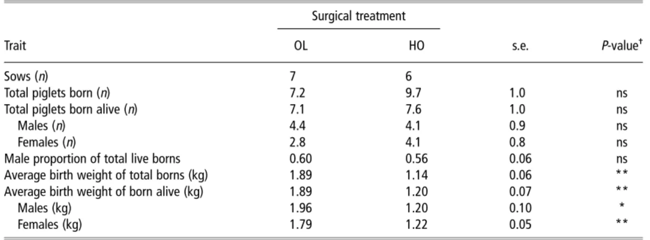
The total number of piglets born per sow was non-significantly lower in OL than HO sows (Table 1). However, as
the number of stillborn in OL sows also tended to be lower (0.1v. 2.1;P,0.1), the total number of piglets born alive were almost the same in the two sow groups. When litter size was expressed per uterine horn, OL sows gave birth to less than half the number of piglets than HO sows (3.6v. 9.7;
P,0.05). Per litter, the average OL progeny were 750 g heavier (P,0.01) at birth compared with the HO progeny. Compared with average male and female HO offspring born alive, OL progeny were heavier by 760 and 570 g (P,0.05 for each), respectively. Furthermore, the number of female but not of male offspring tended to be lower (P,0.1) in OL compared with HO sows.
Birth weight and morphometric measurements of the piglets selected at birth
As expected, birth weight was greater (P,0.001) in OL and H than HO and L offspring, respectively (Table 2). Regardless of surgical treatment or birth weight category, the selected females tended to be lighter (P,0.1) than their male littermates. Absolute weights of heart, liver, kidney, lung,
spleen, brain and SM were greater (P,0.01) in piglets from OL sows and the H birth weight category compared with piglets from HO sows and the L birth weight category, respectively. Gender had no effect on absolute organ and muscle weights. However, except for the brain and SM, relative organ weights, expressed as percentage of the birth weight, were similar in offspring from OL and HO sows and the H and L birth weight category (data not shown), indicating that the observed treatment differences found in absolute organ weights paralleled those in birth weights. By contrast, the relative muscle weights were slightly but sig-nificantly greater (P,0.05) in OL compared with HO off-spring (0.22% v. 0.21%) and greater in female than male piglets (0.22%v. 0.20%). Relative weights of the brain were greater (P,0.001) in piglets from HO sows and the L birth weight category compared with piglets from OL sows (2.80%v. 1.89%) and the H birth weight category (2.55%v. 2.13%). Brain : liver ratio was lower in OL and H progeny, whereas no difference was observed between male and female piglets.
Table 1Reproductive characteristics of unilaterally OL and unilaterally HO sows at farrowing Surgical treatment
Trait OL HO s.e. P-value
-Sows (n) 7 6
Total piglets born (n) 7.2 9.7 1.0 ns
Total piglets born alive (n) 7.1 7.6 1.0 ns
Males (n) 4.4 4.1 0.9 ns
Females (n) 2.8 4.1 0.8 ns
Male proportion of total live borns 0.60 0.56 0.06 ns
Average birth weight of total borns (kg) 1.89 1.14 0.06 **
Average birth weight of born alive (kg) 1.89 1.20 0.07 **
Males (kg) 1.96 1.20 0.10 *
Females (kg) 1.79 1.22 0.05 **
OL 5 oviduct ligated; HO 5 hysterectomized-ovariectomized.
-*P,0.05; **P,0.01; ns, not significant.
Table 2Birth weight, body organs, semitendinosus muscle weight and brain to liver weight ratio of selected male and female littermates with the respectively highest and lowest birth weight born to unilaterally OL and unilaterally HO sows
Surgical treatment Birth weight category Gender P-value
-Trait OL HO High Low Male Female s.e. Trt BtW Gender
Observations (n) 23 24 24 23 25 22 Birth weight (kg) 1.86 1.23 1.71 1.38 1.59 1.50 0.03 *** *** ** Heart (g) 12.6 8.1 11.5 9.3 10.6 10.1 0.3 *** *** ns Liver (g) 49.2 32.5 45.6 36.1 41.2 40.5 2.5 *** *** ns Kidney (g) 14.9 10.3 13.7 11.5 12.6 12.5 0.4 *** *** ns Lung (g) 25.8 16.8 23.9 18.7 21.9 20.7 0.7 *** *** ns Spleen (g) 1.6 1.2 1.6 1.3 1.5 1.4 0.1 *** *** ns Brain (g) 34.2 31.8 34.0 32.1 33.1 33.0 0.4 *** *** ns Semitendinosus muscle (g) 4.1 2.5 3.6 3.0 3.3 3.3 0.3 *** *** ns
Brain to liver weight ratio 0.75 1.16 0.86 1.06 0.94 0.97 0.05 *** *** ns
OL 5 oviduct ligated; HO 5 hysterectomized-ovariectomized; Trt 5 surgical treatment; BtW 5 birth weight category.
Muscle characteristics of the piglets selected at birth
Because of the lower (P,0.01) density of primary but not of secondary myofibers, the secondary : primary myofiber ratio of the LM was greater (P,0.05) in OL than HO progeny (Table 3). Furthermore, the secondary myofibers of OL off-spring were larger (P,0.1) and the primary tended also to be larger in size (P,0.1) than those of HO offspring. Regardless of the sow group, in H progeny primary but not secondary myofibers tended (P,0.1) to be smaller than in L progeny, whereas density of primary and secondary myo-fibers as well as secondary : primary ratio were not different between the two birth weight categories.
Overall myofiber density tended (P,0.1) to be lower in the SM of OL compared with HO progeny (Table 4). This difference resulted mainly from a lower (P,0.01) primary and secondary myofiber density in the SMdark of OL
off-spring. Conversely to the LM, the secondary : primary ratio did not differ. In the SMdarkthe secondary myofibers were
larger (P,0.001), but not the primary, and in the SMlight
overall the myofibers were larger (P,0.05) in the OL than the HO offspring. Compared with the HO piglets, the SM cross-sectional area of OL piglets was greater (P,0.001) because of the greater (P,0.05) cross-sectional area of both, the SMlightand SMdark. In addition, OL progeny tended
Table 3Muscle-fiber related traits of the longissimus muscle of selected male and female littermates with the respectively highest and lowest birth weight born to unilaterally OL and unilaterally HO sows
Surgical treatment Birth weight category Gender P-value
-Trait OL HO High Low Male Female s.e. Trt BtW Gender
Observations (n) 23 24 24 23 25 22
Prim density 212 307 243 276 262 257 50 ** ns ns
Sec density 6087 7382 6574 6896 6679 6790 1135 ns ns ns
Sec : Prim ratio 28.8 24.9 27.7 26.0 26.3 27.4 1.4 * ns ns
Prim diameter (mm) 15.7 13.8 13.9 15.6 14.7 14.8 0.8 ns ns ns
Sec diameter (mm) 9.2 7.4 8.2 8.4 8.2 8.4 0.9 *** ns ns
OL 5 oviduct ligated; HO 5 hysterectomized-ovariectomized; Trt 5 surgical treatment; BtW 5 birth weight category; Prim 5 primary myofiber; Sec 5 secondary myofiber; Sec : Prim ratio 5 ratio of primary and secondary myofibers; Density 5 density of myofibers was expressed as number of myofibers per mm2of the
longissimusmuscle cross-sectional area.
-Probability values for Trt, BtW and gender. *P,0.05; **P,0.01; ***P,0.001; ns, not significant.
Table 4Muscle-fibre related traits of the semitendinosus muscle of selected male and female littermates with the respectively highest and lowest birth weight born to unilaterally OL and unilaterally HO sows
Surgical treatment Birth weight category Gender P-value
-Trait OL HO High Low Male Female s.e. Trt BtW Gender
Observations (n) 23 24 24 23 25 22
Dark portion
CSA (mm2) 39.8 32.3 36.2 35.9 36.2 35.8 1.9 * ns ns
Prim density 193 236 201 228 227 202 10 ** * *
Sec density 7042 8337 7519 7858 8127 7252 375 ** ns *
Sec : Prim ratio 37.2 36.4 38.3 35.3 37.2 36.4 1.6 ns ns ns
Prim diameter (mm) 19.4 18.9 19.6 18.7 19.2 19.1 0.5 ns ns ns Sec diameter (mm) 10.8 9.1 10.2 9.8 9.9 10.1 0.6 *** ns ns Light portion CSA (mm2) 55.1 45.9 52.8 48.2 49.4 51.6 2.7 * ns ns Myofiber diameter (mm) 9.1 8.4 9.1 8.3 8.7 8.8 0.2 * ** ns Myofiber density 9410 10 007 9120 10 298 10 096 9321 630 ns ** ns Total muscle CSA (mm2) 95.1 77.7 89.1 83.7 85.4 87.4 3.0 *** ns ns Myofiber density 8452 9286 8457 9282 9261 8478 564 ns * * Myofiber number (3 105) 7.913 7.125 7.467 7.571 7.701 7.336 0.302 ns ns ns
OL 5 oviduct ligated; HO 5 hysterectomized-ovariectomized; Trt 5 surgical treatment; BtW 5 birth weight category; CSA 5 cross-sectional area; Prim 5 primary myofiber; Sec 5 secondary myofiber; Sec : Prim ratio 5 ratio of primary and secondary myofibers; Density 5 density of myofibers was expressed as number of myofibers per mm2of the dark, light and totalsemitendinosusmuscle cross-sectional area; Myofiber number 5 total myofiber number in thesemitendinosusmuscle
was calculated from the estimated total number of Prim and Sec myofibers in the dark portion and in the light portion by estimating the total number of myofibers in mATPase sections after acid preincubation.
(P,0.1) to have a greater total myofiber number than HO progeny. Regardless of the surgical treatment, H offspring had a lower (P,0.05) primary but similar secondary myofiber density in the SMdark. However, the secondary myofibers
tended (P,0.1) to be larger. As a result of the greater (P,0.01) myofiber diameter in the SMlightof the H pigs, the
myofiber density was lower (P,0.01) than in the L offspring. Compared with males, females had fewer (P,0.05) primary and secondary myofibers per mm2in the SMdarkand myofiber
number per mm2tended (P,0.1) to be lower in the SMlight.
MyHC polymorphism in the LM of the piglets selected at birth
The MyHC isoform distribution in the LM was affected by surgical treatment and birth weight but not gender (Table 5). The LM of OL offspring had a lower (P,0.05) relative abundance of the fetal and type I MyHC isoforms and a greater (P,0.001) abundance of the type II MyHC isoform compared with the OL progeny. In H piglets, relative abun-dance of the type I MyHC was lower (P,0.05) than in L piglets. The number of primary myofibers was positively correlated (r50.51;P,0.001) with the abundance of the type I and negatively correlated (r5 20.43;P,0.001) with the abundance of the type II MyHC isoforms.
Discussion
Combining the OL and HO sow model, the effects of different degrees of intrauterine crowding on fetal development were studied. The present results demonstrated the suitability of this approach to generate the expected effects on litter size and litter birth weight. When expressed per uterine horn, HO sows gave birth to 1.7 times more piglets and, although average birth weight was lower by 750 g, total litter birth weight was 0.6 times greater compared with OL sows. Regardless of gender and birth weight category, the greater intrauterine crowding markedly hampered the development of internal organs and myogenesis.
Effects of intrauterine crowding on litter characteristics
It is well established that uterine capacity is a limiting factor for fetal survival and development, thereby affecting litter
size, number of born alive and of stillborn as well as litter birth weight (Dziuk, 1968; Fenton et al., 1972, Fordet al., 2002). By determining the estrone sulfate concentration, which is a good indicator of the numbers of viable embryos (Gaustad-Aaset al., 2002), in the plasma of HO and intact sows at day 24 of gestation, Be´rardet al. (2010) suggested that implantation rate was not impaired by different extents of intrauterine crowding in primiparous Swiss Large White sows. However, at birth they observed a greater number of mummies in the HO sows compared with the intact sows suggesting that uterine capacity limited prenatal survival rate in late gestation in these gilts. In the current study, neither smaller nor larger mummies were found. Smaller mummies (length: ,1 cm) would represent fetal death at about days 35 to 40 of gestation and larger mummified fetuses (length: .10 cm) represent fetal losses from day 55 of gestation to parturition (van der Lende and van Rens, 2003). However, it cannot be excluded that smaller and very small mummified fetuses may have gone unnoticed at far-rowing in the present study as experienced by van der Lende and van Rens (2003). In addition, the uterus is able to reabsorb non-calcified bone tissues, thus additionally ham-pering the detection at birth of fetal death occurring from implantation to around day 40 of gestation. However, in the current study a greater number of stillborns occurred in HO compared with OL sows. The relatively high average birth weight of the stillborn (0.86 kg) compared with the litter-mates born alive suggests that they perished just before or during farrowing. Under these conditions where the uterine horn was occupied by 9.7 piglets during the whole gesta-tional period, not only uterine space but also competition for maternal nutrient resources might have restricted the normal development of the entire litter. When the number of embryos/fetuses exceeds 14 (seven per uterine horn), normal offspring development is considered to be limited by the uterine space and placental development and efficiency (Town et al., 2004; Foxcroft et al., 2007). Therefore, with each additional fetus a linear decrease in the average litter birth weight can be expected (Foxcroft et al., 2007). In HO sows the aforementioned threshold in fetus number was exceeded by on average 2.6 piglets, which clearly explains the observed low average birth weight of the HO offspring.
Table 5Myosin heavy-chain isoform distribution in the longissimus muscle of selected male and female littermates with the respectively highest and lowest birth weight born to unilaterally OL and unilaterally HO sows
Surgical treatment Birth weight category Gender P-value
-Trait-
-OL HO High Low Male Female s.e. Trt BtW Gender
Observations (n) 23 24 24 23 25 22
Myosin heavy-chain isoforms (%)
Fetal 37.8 43.0 41.4 39.4 40.5 40.2 2.8 * ns ns
Type I 12.1 15.4 12.4 15.1 13.9 13.6 2.6 * * ns
Type II 50.0 40.8 45.8 45.1 45.1 45.7 5.2 *** ns ns
OL 5 oviduct ligated; HO 5 hysterectomized-ovariectomized; Trt 5 surgical treatment; BtW 5 birth weight category.
-Probability values for Trt, BtW and gender. *P,0.05; ***P,0.001; ns, not significant.
-
Furthermore, Leenhouwers et al. (1999) found a positive relationship between the number of stillbirths and pre-weaning mortality of live borns, indicating an overall lower viability of litters in which stillbirths occur. Altogether, these findings allow concluding that increased intrauterine crowding will negatively affect birth weight as well as pre-and perinatal survival rate. In the case of the OL offspring, intrauterine competition was minimal, which permitted an adequate placental development and, consequently, an improved nutrient exchange between fetuses and dam. These intrauterine conditions allowed the fetuses to reach maximal prenatal growth, which is reflected in the markedly higher birth weight compared with OH offspring.
Effects of intrauterine crowding on organ and muscle development
Regardless of the birth weight and gender, the selected HO progeny were lighter by 34% than the OL offspring. Hearts, livers, kidneys and lungs of HO offspring were lighter to a similar extent (range between 30% and 36%), indicating that at around birth allometric growth paralleled that of the whole body. However, it has to be pointed out that these results do not allow to exclude that allometric growth of these organs was constant during gestation (Vallet and Freking, 2006). Surprisingly, at farrowing, the reduction of the SM weight relative to birth weight was, with 39%, slightly larger than for the other organs in HO compared with OL offspring. Similarly, Powell and Aberle (1981) as well as Wigmore and Stickland (1983) found that growth retarded piglets with a low birth weight had relatively smaller muscles than their larger littermates. One possible explanation for this greater growth restriction is that secondary myofiber hyperplasia, which is predominantly responsible for prenatal muscle growth (Handel and Stickland, 1987), occurs at a time in the intrauterine development when nutrient require-ments for overall fetal growth dramatically increase. Because of the aforementioned great intrauterine crowding in HO sows, muscle development might therefore have been more than proportionately impaired. By contrast, and in agree-ment with other studies (Bauer et al., 2003; Town et al., 2004; Be´rard et al., 2010), brain growth was partly main-tained through compensatory mechanisms as the difference in absolute brain weight between HO and OL offspring amounted only to 7%. This phenomenon, also known as brain sparing effect, illustrates that the maintenance of brain weight is of primary importance for growth restricted piglets (McMillenet al., 2001). Similarly, brain weight was only 6% lower in L than H offspring, whereas weights of the SM and those of the other organs were lower to a similar extent (19%) as birth weight.
Effects of intrauterine crowding on myofiber characteristics and MyHC polymorphism
Increased intrauterine crowding, as induced by HO, resulted in impaired myofiber hyperplasia and, as a consequence, a smaller muscle cross-sectional area of the SM. These findings confirm results reported by Town et al. (2004)
showing lower secondary myofiber number and smaller cross-sectional area of the SM in 90 day old fetuses sub-jected to modest intrauterine crowding. However, in that study, like as in the present study, a confounding effect between intrauterine crowding and birth weight exists, because offspring born from a crowded compared with an un-crowded intrauterine environment were also of lower birth weight. However, Be´rard et al. (2010) demonstrated that independent of birth weight, intrauterine crowding negatively affects myofiber hyperplasia (shown in thepsoas major muscle and SMdark); this probably through
malnutri-tion of the fetuses (Pe`re and Etienne, 2000). Unexpectedly, in the present study, myofiber hyperplasia in the SM of HO offspring was impaired even though there was a greater myofiber density. This discrepancy might be explained by the differences observed in myofiber diameter. Thus, intrauterine crowding not only seems to reduce hyperplasia but also impair hypertrophy of myofibers.
In the LM, the muscle cross-sectional area was not determined but, as reported earlier (Bee, 2004), a similar positive relationship between the cross-sectional area of the LM and birth weight can be expected as that found with the SM in the present study. Because the secondary myofiber density was similar, the secondary : primary ratio was lower and primary and secondary myofiber sizes were smaller in the HO offspring; it is likely that, as in the SM, the LM had fewer myofibers than that of the OL offspring as well. Furthermore, in the LM, the relative abundance of fetal and type I MyHC was greater in HO offspring compared with OL offspring. Lefaucheur et al. (1997) showed that embryonic and fetal MyHC isoforms are transitional MyHC isoforms, which within 2 weeks after birth mature to become the adult type II isoforms. Therefore, this result indicates a delayed muscle development in HO offspring.
Effects of intrauterine crowding on piglets with different birth weight and gender
The complete lack of surgical treatment 3 birth weight interactions suggests that contrasting levels of intrauterine crowding similarly affected muscle development of H and L offspring. However, the effect of birth weight on myofiber characteristics was very limited in the present study. Only in the SM, myofiber density was greater and myofiber size was smaller, whereas total myofiber number did not differ between L and H offspring. On a first glance, this was unexpected considering numerous previous evidence that birth weight has a distinct effect on myogenesis (Wigmore and Stickland, 1983; Rehfeldt and Kuhn, 2006; Tristanet al., 2009). However, in the present study the average birth weight even of the L pigs was rather high with 1.38 kg and the weight difference to the H pigs amounted to only 320 g. On the basis of earlier results where similar birth weight ranges were compared, no differences in myofiber hyper-plasia were observed as well (Rehfeldt and Kuhn, 2006; Be´rardet al., 2010).
In a similar way, the average birth weight of both genders was high with an average of 1.54 kg and the weight
difference between them amounted only to 80 g. As there was also no significant interaction between surgical treat-ment and gender, it can be assumed that intrauterine crowding has a similar effect on these high average weight piglets. Although myofiber densities in the SM were greater in male compared with female piglets, these differences were not sufficiently large to affect total myofiber number of the muscle. Because myofiber diameter and cross-sectional area were similar in both genders, myofibers were more densely packed within the muscle of the males. The lack of a general gender effect on myofiber number is in agreement with previous results obtained in the SM of slaughter pigs (Bee, 2004). The author observed a heavier muscle in gilts than barrows, related to a greater myofiber hypertrophy but not hyperplasia. By contrast, Be´rard et al. (2010) reported greater myofiber hyperplasia in the SMdarkandrhomboideus
muscle, the latter being also larger in size, of male compared with female newborns. This discrepancy of results might be related to differences in birth weight of the newborn pigs between studies (on average 130 g lighter in the study by Be´rardet al., 2010).
Conclusion
The present results revealed that HO offspring carried all negative phenotypic characteristics that are indicative of intrauterine growth retardation such as lower birth weight, reduced organ and muscle weights as well as brain sparing effects. Furthermore, elevated intrauterine crowding impaired hyperplasia and growth of myofibers along with a delayed myofiber maturation. On the basis of the current knowledge about the relationship between prenatal muscle develop-ment and postnatal growth, carcass characteristics and meat quality, an impaired myogenesis induced by intrauterine crowding can be expected in common genotypes with important adverse effect on economically relevant traits of pig production.
Acknowledgments
This study was supported by the Fondation Sur-la-Croix (Basel, Switzerland). We are grateful to G. Maı¨koff and his team for taking care of the animals, Dr A. Gutzwiller for the surgical procedures on the sows and Dr P. Silacci and his team for the help during sample collection and muscle analysis.
References
Agroscope Liebefeld Posieux Research Station (ALP) 2012 . Fu¨tterungsempfeh-lungen und Na¨hrwerttabellen fu¨r Schweine (Feeding Recommendations and Nutrient Tables for Pigs). Retrieved April 13, 2012, from http://www.agroscope. admin.ch/futtermitteldatenbank/04835/index.html?lang5de
Bauer R, Walter B, Brust P, Fuchtner F and Zwiener U 2003. Impact of asymmetric intrauterine growth restriction on organ function in newborn piglets. European Journal of Obstetrics Gynecology and Reproductive Biology 110, S40–S49.
Bee G 2004. Effect of early gestation feeding, birth weight, and gender of progeny on muscle fiber characteristics of pigs at slaughter. Journal of Animal Science 82, 826–836.
Be´rard J and Bee G 2010. Effects of dietaryL-arginine supplementation to gilts
during early gestation on foetal survival, growth and myofiber formation. Animal 4, 1680–1687.
Be´rard J, Pardo CE, Bethaz S, Kreuzer M and Bee G 2010. Intra-uterine crowding decreases average birth weight and affects muscle fiber hyperplasia in piglets. Journal of Animal Science 88, 3242–3250.
Christenson RK, Leymaster KA and Young LD 1987. Justification of unilateral hysterectomy–ovariectomy as a model to evaluate uterine capacity in swine. Journal of Animal Science 65, 738–744.
Dziuk PJ 1968. Effect of number of embryos and uterine space on embryo survival in the pig. Journal of Animal Science 27, 673–676.
Fenton FR, Bazer FW, Ulberg LC, Schwartz FL and Robison OW 1972. Stage of gestation when uterine capacity limits embryo survival in gilts. Journal of Animal Science 35, 383–388.
Ford SP, Vonnahme KA and Wilson ME 2002. Uterine capacity in the pig reflects a combination of uterine environment and conceptus genotype effects. Journal of Animal Science 80 (E. suppl. 1), E66–E73.
Foxcroft GR, Bee G, Dixon W, Hahn M, Harding J, Patterson J, Putman T, Sarmento S, Smit M, Tse W-Y and Town SC 2007. Consequences of selection for litter size on piglet development. In Paradigms in pig science (ed. J Wiseman, MA Varley, S McOrist and B Kemp), pp. 207–229. Nottingham University Press, Nottingham, UK.
Gaustad-Aas AH, Ropstad E, Karlberg K, Hofmo PO and Dahl E 2002. Oestrone sulphate measurements for the prediction of small or large litters in pigs. Acta Veterinaria Scandinavica 43, 157–164.
Gondret F, Lefaucheur L, Juin H, Louveau I and Lebret B 2006. Low birth weight is associated with enlarged muscle fiber area and impaired meat tenderness of the longissimus muscle in pigs. Journal of Animal Science 84, 93–103.
Handel SE and Stickland NC 1987. Muscle cellularity and birth weight. Animal Production 44, 311–317.
Huang YT, Johnson RK and Eckardt GR 1987. Effect of unilateral hysterectomy and ovariectomy on puberty, uterine size and embryo development in swine. Journal of Animal Science 65, 1298–1305.
Leenhouwers JI, van der Lende T and Knol EF 1999. Analysis of stillbirth in different lines of pig. Livestock Production Science 57, 243–253.
Lefaucheur L, Edom F, Ecolan P and Butler-Browne GS 1995. Pattern of muscle fiber type formation in the pig. Developmental Dynamics 203, 27–41. Lefaucheur L, Hoffman R, Okamura C, Gerrard D, Leger JJ, Rubinstein N and Kelly A 1997. Transitory expression of alpha cardiac myosin heavy chain in a subpopulation of secondary generation muscle fibers in the pig. Developmental Dynamics 210, 106–116.
McMillen IC, Adams MB, Ross JT, Coulter CL, Simonetta G, Owens JA, Robinson JS and Edwards LJ 2001. Fetal growth restriction: adaptations and consequences. Reproduction 122, 195–204.
Pe`re MC and Etienne M 2000. Uterine blood flow in sows: effects of pregnancy stage and litter size. Reproduction Nutrition Development 40, 369–382.
Pe`re MC, Dourmad JY and Etienne M 1997. Effect of number of pig embryos in the uterus on their survival and development and on maternal metabolism. Journal of Animal Science 75, 1337–1342.
Poore KR and Fowden AL 2004. The effects of birth weight and postnatal growth patterns on fat depth and plasma leptin concentrations in juvenile and adult pigs. Journal of Physiology 558, 295–304.
Powell SE and Aberle ED 1981. Skeletal muscle and adipose tissue cellularity in runt and normal birth weight swine. Journal of Animal Science 52, 748–756.
Rehfeldt C and Kuhn G 2006. Consequences of birth weight for postnatal growth performance and carcass quality in pigs as related to myogenesis. Journal of Animal Science 84, E113–E123.
Rehfeldt C, Tuchscherer A, Hartung M and Kuhn G 2008. A second look at the influence of birth weight on carcass and meat quality in pigs. Meat Science 78, 170–175.
Town SC, Putman CT, Turchinsky NJ, Dixon WT and Foxcroft GR 2004. Number of conceptuses in utero affects porcine fetal muscle development. Reproduction 128, 443–454.
Tristan F, Rivero MA, Albors OL, Ramis G, Vazquez JM, Martinez M, Martinez JS and Gil F 2009. Relationship of birth weight with the size, number and
proportion of fibres in the pig semitendinosus muscle. Anatomia, Histologia, Embryologia 38, 275–278.
Vallet JL and Freking BA 2006. Changes in fetal organ weights during gestation after selection for ovulation rate and uterine capacity in swine. Journal of Animal Science 84, 2338–2345.
van der Lende T and van Rens BTTM 2003. Critical periods for foetal mortality in gilts identified by analysing the length distribution of mummified foetuses and frequency of non-fresh stillborn piglets. Animal Reproduction Science 75, 141–150. Wigmore PM and Stickland NC 1983. Muscle development in large and small pig fetuses. Journal of Anatomy 137, 235–245.