Carduogenesb vol.15 no.5 pp.837-843, 1994
High promutagen activating capacity of yeast microsomes
containing human cytochrome P-450 1A and human
NADPH-cytochrome P-450 reductase
Christian Sengstag, Hans-Pietro Eugster and
Friedrich E.Wiirgler
Institute of Toxicology, Swiss Federal Institute of Technology and University of Zflrich, Schorenstrasse 16, CH-8603 Schwerzenbach, Switzerland
Yeast Saccharomyces cerevisiae strains have been constructed
that co-express cDNAs coding for the human cytochrome
P-450 enzymes CYP1A1 or CYP1A2 in combination with
human NADPH-cytochrome P-450 reductase (oxidoreductase).
Microsomal fractions prepared from the strains were able to
efficiently activate various drugs to Salmonella mutagens.
These experiments demonstrated that a functional interaction
occurred between the respective human enzymes in the yeast
microsomes. For every drug tested, the microsomes containing
CYP enzymes and oxidoreductase were 2- to 4-fold better in
activation than the corresponding microsomes that contained
CYP alone. Interestingly, co-expression of CYP1A2 with
oxidoreductase resulted in a decrease of
7-ethoxyresorufin-0-deethylase activity, a problem which is related to this
specific substrate. Using the microsomes, it was demonstrated
that aflatoxin B, was activated to a mutagen not only by
CYP1A2 but also by CYP1A1. In contrast, benzo[a]pyrene
was exclusively activated by CYP1A1 whereas CYP1A2 was
inactive. The drug 3-amino-l-methyl-5//-pyrido[4,3-ft]indole
(Trp-P-2) was activated by CYP1A2 and to a lesser extent
by CYP1A1. A strong substrate specificity was observed with
the two structurally related heterocyclic arylamines
3,4-dimethyUmidazo[4,5-/]quinoUne (MelQ) and
2-amino-3,8-dimethyUmidazo[4,5-/]quinoxaline (MelQ*). MeIQx was
activated efficiently by both CYP enzymes, whereas MelQ
was only activated by CYP1A2 and not by CYP1A1. The fact
that microsomes from vector transformed control strains were
unable to activate any of the drugs studied underlines the
suitability of these microsomes for metabolic studies.
Moreover, the presence of suitable marker genes in the yeast
strains will enable us to study mitotic recombination and gene
conversion events induced by drugs that require metabolic
activation.
Introduction
Carcinogenesis is often accompanied by specific alterations in
two important classes of genes, the proto-oncogenes and the
tumor suppressor genes. The products of these genes make up
a complicated molecular network involved in cell-cycle regulation
and differentiation. It is not surprising that subtle disturbances
in this delicate network can have profound detrimental effects
•Abbreviations: oxidoreductase, NADPH cytochrome P-450 reductase; PAHs, polycyclic aromatic hydrocarbons; BaP, benzo(a]pyrene; CYP, cytochrome P-45O, PMSF, phenylmethylsulfonylfiuoride; TPCK, A'-tosyl-L-phenylalanine chloro-methyiketone; Trp-P-2, 3-amino-l-methyl-5tf-pyrido[4,3-6]indole; DMSO, dimethyl suffoxide; AFB1, aflatoxin B(; MelQ, 2-amino-3,4-dimethyl-imidazo(4,5/lquinoline; MelQ,, 2-arniro-3,8-dimethvlimidazo(4,5-/l<piinoxaline; EROD, 7-anoxyresorufin-Ckleethylase.like neoplasia. Mutations that arise spontaneously or are induced
by environmental agents in the two classes of genes have been
recognized as an important factor of carcinogenesis. Therefore,
several test systems have been developed where the mutagenic
effect of a given compound on various organisms from bacteria
(1,2) to mouse (3,4) can be recognized.
Apart from mutations, the genetic event described as
homo-logous mitotic recombination also plays an important role in
carcinogenesis (5). Mitotic recombination provides an attractive
mechanism for the loss of heterozygous tumor suppressor genes.
It has indeed been recognized that heterozygous wild-type tumor
suppressor gene copies are not only inactivated by further
mutations, but recombination-mediated loss has clearly been
demonstrated in a few cases (6,7). Such a loss of heterozygosity
can simply be explained by a single crossover occurring during
the G
2phase in the homologous DNA between the gene locus
and the corresponding chromosomal centromere. Chromatid
exchange will lead to a segregation of the two alleles in half of
the following mitotic divisions. Thereby clones are created that
have become homozygous for the gene locus of interest. For this
reason it is not only important to learn the mutagenic activity
of a chemical compound to which humans are exposed, but a
potential recombinagenic activity of the compound should also
be considered in cancer risk assessments.
In contrast to a large battery of test systems designed for
the identifiation of mutagens, only few systems exist where
recombinagens can be detected (8-10). One of these is based
on the lower eukaryotic yeast Saccharomyces cerevisiae and uses
strain D7 which has been constructed by Zimmermann and
colleagues (11). Strain D7 allows for the simultaneous phenotypic
detection of mitotic recombination, gene conversion and reversion.
To extend the metabolic capacity of this strain we have genetically
engineered it to confer on it specifically defined human enzyme
activities. For this purpose we have introduced plasmids that
direct the expression of human cytochrome P-4501A cDNAs
in combination with a NADPH-cytochrome P-450 reductase
(oxidoreductase*) cDNA. In this study we have asked whether
the newly introduced cDNAs were functionally expressed in me
yeast strains. To this end we have prepared microsomal fractions
from the strains and used them as alternative activation systems
in the Ames test. Here we show that two important classes of
carcinogens, polycyclic aromatic hydrocarbons (PAHs) and
heterocyclic arylamines are efficiently activated to Salmonella
mutagens. As a member of the first class of compounds we chose
benzo[a]pyrene (BaP) which is activated by a combination of
cytochrome P-450 (CYP) mediated epoxidation and hydrolysis
by epoxide hydrolase to the ultimate carcinogen (for review, see
ref 12). BaP and other PAHs are produced in combustion
processes and are present as urban environmental pollutants. The
other class of compounds, heterocyclic arylamines, are natural
constitutents of our diet and they have been identified particularly
in broiled and fried fish and meat (13). Heterocyclic arylamines
are activated by hydroxylation of me amino group, which is
predominantly catalyzed by cytochrome CYP1A2 to an
inter-mediate which is subsequently acetylated by a N-acetyltransferase
C.Sengstag, H.-P.Eugster and F.E.Wurgler
to yield the ultimate mutagen (for review, see ref 14). Our results
demonstrate that both classes of compounds are efficiently
activated by the yeast microsomes. Therefore, the constructed
strains represent a promising tool for the study of drug induced
mitotic recombination.
Materials and methods
Chemicals
3-Amino-l-methyl-5//-pyrido[4,3-fc]indole (Trp-P-2) was purchased from Pharmacare (BrOggen, Switzerland) and was dissolved m methanol; all other pro-mutagens were dissolved in dimethyl sulfoxkle (DMSO). Aflatoxin B, (AFB1) and BaP were purchased from Ftuka (Buchs, Switzerland). 2-Amino-3,4-dimethyl-imidazo[4,5-/]quinoline (MelQ) and 2-amirK>3,8-dimeuiylimidazo[4,5-/]quin-oxaline (MelQJ were gifts from Dr P.Morgenthaler (Nestec Ltd, Vers-Chez-Les Blanc, Switzerland).
Strains and media
S.cerevisiae strain YHE2 (MATa/MATa, ade2-4Olade2-\\9, trp5-\2ltrp5-21, ilvl-92/itvl-92, ura5A5/ura3A5) has been described (15). It was transformed according to standard methods (16) and transformants were cultured in minimal medium (17) containing 0.67% yeast nitrogen base w/o amino acids, 2% glucose and supplemented with 20 /ig/ml adenine and tryptophane, 30 /ig/ml isoleucine and leucine and 150 /ig/ml valine. Untransformed strains were propagated in YPD medium (17). For mkrosomal preparations cultures were grown in synthetic complete medium lacking uracil (SD-ura) (17). Strain YNW64 (AM7b, ade2-\ 19, lrp5, cprJ ::um3A5, ilvl-92, ura3AS) was provided by N.Wittekindt and derived from strain YES9 (18) by disrupting the URA3 gene present in the cpr l:\URA3 allele. Details of the disruption will be described elsewhere.
Escherichia coli strain DH5aF' [ F \ endAl, hsdR17, supE44, lhi-1, recAl, gyrA96, relAl, 4>&M/lacZAM15, A(lacZYA argF) U169] was used for plasmid constructions, transformed according to standard protocols (19) and propagated in LB medium (19) (containing 150 jjg/ml ampicillin when appropriate).
Salmonella typhimurium strain TA98 (hisD3052, rfa, AuvrB, pKMlOl) was used for mutagenicity tests and was grown in NB medium (20).
Plasmid constructions
The constructions of plasmids pHElO (15), pHE36 (21) and pSB229 (18) have been described. To construct plasmid pCS316, a 3.1 kb Sail fragment containing the oxidoreductase expression cassette including promoter and terminator was isolated from pSB229 and inserted into the Sal I site of vector pUC7 (22). From there it was recovered as a BamHl fragment and inserted into the BamHl site of pBLSK-Not-Not. This latter plasmid was constructed from pBLSK (Strategene, La Jolla) by ligating the two phosphorylated and annealed oligonucleotides 5TCGACAGCGGCCGCTG3' and 5'GTACCAGCGGCCGCTG3' into the plasmid's Ajp718 and Sail sites, thereby providing a second Notl site. The oxidoreductase expression cassette was subsequently recovered as a Notl fragment and inserted into the Ate I sites in plasmid pCS289 and pCS290 in both orientations, to result in the plasmids pCS311, 312, 316 and 339. For the construction of the intermediates pCS289 and pCS290, a 2.2 kb Sad fragment containing die CYP1A2 expression cassette was isolated from plasmid pHE36 (21) and inserted into the Sad she of plasmid pNW144. This vector was a gift from N.Wittekindt and was derived from vector pDP34 (23) by converting the BamWl site to a Notl site by linker addition. All restriction enzymes were used according to the manufacturer's recommendations.
Preparation of yeast microsomes
MicTosomes were prepared essentially as described (15) with the following modifications: The procedure was scaled up to 6 1 yeast cultures. Sonication of the spheroblasts was performed for 2 x 30 s in cell washing buffer containing 0.8 mM phenylmethylsulfonylfluoride (PMSF) as well as leupeptin, pepstatin and A'-tosyl-L-phenylalanine chloromethylketone (TPCK) each at 4 pg/ml. Cell debris was removed by two centrifugations for 15 min at 11 000 g and 25 000 g. After the subsequent ultracentriftigatjon for 1 h at 102 000 g, die tube walls were cleaned with a paper tissue and the pellet was resuspended in 100 mM sodium pyrophosphale, 1 mM EDTA, pH 7.5, by gently grinding with a cold reagent tube. Ultraceatrifugation was repeated, die pellet dissolved in 5 ml 100 mM sodium phosphate buffer, pH 7.4, and homogenized with a Dounce potter. Then aliquots were frozen in liquid nitrogen and kept at - 8 0 ° C .
Enzyme activities
The determination of 7-ethoxyresorufin-O-deemylase (EROD) activities of intact yeast cells and of cytochrome c reductase activities of microsomes were done as described (18). EROD activities of microsomes were determined as described (21) using 5 /d microsomes and a substrate concentration of 3 fiM. Ames tests with yeast microsomes as activating system
An NADPH regenerating system containing 150 mM potassium phosphate buffer, pH 7.4, 0.74 mM NADP+, 15 mM glucose-6-phosphate, 10 mM MgCl2 and
838
14 units glucose-6-phosphate dehydrogenase per ml was freshly prepared. To 700 )A of the regenerating system, 100 /d of an overnight culture of strain TA98 was added, followed by the addition of 15 - 3 0 ^1 yeast microsomes. After gently mixing, promutagens were added in a volume of 30 yX. Preincubation was done at 37 "C for 20 min, then 2 ml molten top agar (20) were added and poured on minimal plates (20) containing 150 /jg/ml ampicillin. Plates were incubated for 48 h and revertam colonies were counted.
Western blots
Detection of antigenic proteins was done as described (21).
Results
Co-expression of cytochrome P-450 and oxidoreductase cDNAs
in S.cerevisiae
The expression of human CYP1A1 (15), CYP1A2 (21) and the
co-expression of CYP1A1 with human oxidoreductase (18) in
S.cerevisiae strain YHE2 has previously been described. The
expression plasmids used in this study are shown in Figure 1.
Previous experiments have shown that co-expression with
oxidoreductase resulted in a 16-fold increase in the CYP1A1
specific EROD activity compared to expression of CYP1A1 alone
(Figure 2). This result prompted us to co-express human CYP1A2
with oxidoreductase. For this purpose a plasmid was constructed
where the two human cDNAs coding for CYP1A2 and
oxido-reductase are transcribed in yeast cells under control of the same
constitutive GAPDH promoter. This plasmid (pCS311) and a
similar plasmid lacking oxidoreductase (pHE36) were introduced
into yeast by transformation and transformants were cultured in
uracil-deficient medium to select for the plasmids.
To test whether a functional interaction occurred between the
heterologous enzymes, EROD activities were determined in
^micron
1**
Jmtaor
Fig. 1. Expression plasmids used in this study. The E.coli/S.cerevisiae shuttle plasmids replicate at high copy number in yeast due to die presence of 2 micron DNA. They are stable in boui organisms under selection for ampicillin resistance and uracil prototrophy respectively. Individual human cDNAs (open arrows) are expressed by the S.cerevisiae GAPDH promoter (filled arrows denoted by p). The stippled boxes (denoted t) represent the terminator region of die S.cerevisiae PH05 gene. The origin of replication in E.coli and die oxidoreductase cDNA are denoted by on and hOR respectively.
Promutagen activating capacity of yeast mlcrosomes
exponentially growing cells (Figure 2). Unlike our previous
observation regarding co-expression of oxidoreductase with
CYP1 Al, the analogous co-expression with CYP1A2 surprisingly
did not result in an increased EROD activity. In contrast,
transformants containing plasmid pCS311 even exhibited less
EROD activity than pHE36 transformants. Since the relative
orientation of the two expression cassettes in plasmid pCS311
was different from that in pSB229, we tested whether the
expression was influenced by the particular orientations of the
cassettes. For this purpose the CYP1A2 and hOR expression
cassettes were inserted into the vector in all four possible
orientations. Nevertheless, none of these plasmids conferred
CYP1A1 PHEIO — m PSB229-CYP1A1 EHOD 8 136 CYP1A1
m
pHE38 CYP1A2 hOfl p c s 3 i i — m m PCS312- pCS316-CYP1A2 him—>«-CYP1A2 KOR 1.5 PCS339 > CYP1A2
Fig. 2. CYP1A specific EROD activities of exponentially growing yeast strains. EROD activities of corresponding YHE2 transformants are given as pmol/min/3 x 107 cells, where 107 cells contain - 1 0 0 #ig protein. The
expression cassettes for CYP1A1, CYP1A2 and oxidoreductase (hOR) are represented by bold arrows and the orientations of the arrows give the direction of transcription. Only the relevant parts of the plasmids are shown. No activity was detectable in vector transformed controls.
a higher EROD activity than was obtained with pHE36
transformants (Figure 2).
In order to discover why co-expression of oxidoreductase with
CYP1A2 was unable to increase the cells' EROD activity, the
presence of the heterologous enzymes in the yeast cells was tested.
Crude extracts or microsomal fractions were prepared from the
respective transformants and were subjected to Western blot
analysis using an antisenim that recognized rat cytcchrome P-450
enzymes from family 1. Figure 3 demonstrates that the strains
transformed with plasmids pHElO, pSB229, pHE36 and pCS316
contained antigenic CYP proteins to a similar extent, whereas
control strains lacked heterologous CYP. Evidence for the
presence of human oxidoreductase in the yeast transformants
was obtained indirectly. For this purpose the plasmids were
transformed into the cprlA strain YNW64 which lacked
endogenous yeast oxidoreductase due to a disruption of the CPR1
gene. Such crplA strains are viable on normal media but they
react supersensitively towards the antifungal drug ketoconazole
(24). The supersensitive phenotype is rescued by expression of
human oxidoreductase cDNA in such strains (18). YNW64
transformants containing individual plasmids were tested for their
sensitivity towards low doses of ketoconazole. These experiments
demonstrated that plasmids pSB229 and pCS316 were both able
to rescue the cprlA mutation and to confer their host strain
ketoconazole resistant to a similar degree (data not shown).
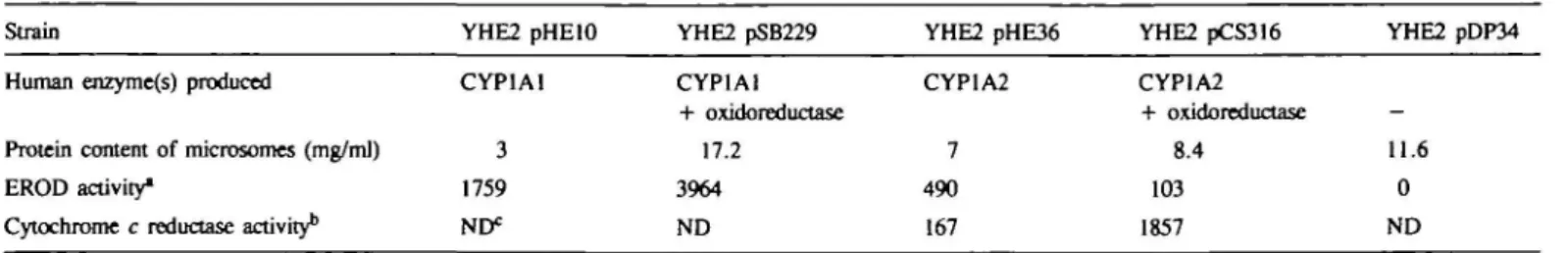
In addition, the > 10-fold increase in cytochrome-c-reductase
activity in pCS316 microsomes compared to pHE36 microsomes
(Table I) suggested that oxidoreductase was functional. Therefore,
the lack of increase in EROD activity of pCS316 transformants
did not reflect a problem in the production of the two human
enzymes but was presumably related to the specific substrate
7-ethoxy resorufin.
OJ * - c\j • * r-- OJ co LU m _ i X CO Q a. a. ¥ CD CD W •» !D i -C\l CO CO CO m Q- LU coco a x o
a. a. CL a.Fig. 3. Western blots demonstrating the presence of human CYP enzymes in yeast. 300 ng whole cell proteins (left) or 50 /tg microsomal proteins (right) were electrophoresed on 10% polyacrylamide—SDS gels. Proteins were transferred to nitrocellulose and detected with a polyclonal antisenim directed against rat CYP enzymes from family 1 (P-450cd). CYP1A1 and CYP1A2 enzymes were present in all yeast transformants except in strains transformed with vector (pDP34) or with plasmid pHE71 (18) containing the oxidoreductase expression cassette alone. An antigenic band of the same mobility was seen in microsomes from human kidney donor liver (KDL34).
Microsomal fractions from yeast transformants activate
promutagens in vitro
Batch cultures of the yeast transformants were grown and
microsomal fractions were prepared. The protein content and
EROD activities of the microsomes are presented in Table I. To
test whether the human enzymes present in the microsomes were
able to activate promutagens, various drugs were incubated with
the microsomes in vitro and the formation of mutagenic
metabolites was monitored by performing an Ames test. Table
II shows that the mycotoxin and liver carcinogen AFB1 was
activated to a mutagenic metabolite by microsomes containing
CYP1A1 and oxidoreductase. No significant activation was
observed when the microsomes or the NADPH-regenerating
system were omitted from the reaction or when microsomes were
used from a control strain that produced no heterologous enzymes.
From this result it was concluded that human CYP1A1 which
is present in the microsomes in combination with oxidoreductase
Table I. Enzymic activities of yeast microsomesStrain
Human enzyme(s) produced
Protein content of microsomes (mg/ml) EROD activity*
Cytochrome c reductase activity1'
YHE2 pHElO CYP1A1 3 1759 ND* YHE2 pSB229 CYP1A1 + oxidoreductase 17.2 3964 ND YHE2 pHE36 CYP1A2 7 490 167 YHE2 pCS316 CYP1A2 + oxidoreductase 8.4 103 1857 YHE2 pDP34 -11.6 0 ND "EROD activities are given as pmol resorufin formed per mg protein per min.
bCytochrome c reductase activities are given as nmol cytochrome c reduced per mg protein per min.
C.Sengstag, H.-P.Eugster aDd F.E.WQrgier
Table n . Activation of AFBl to a mutagenic product by yeast microsomes nmol AFBl 15 3 15 3 15 0 15 3 0
Yeast microsomes Yeast microsomes (516 Mg) CYPlAl containing (348 ^tg) containing and oxidoreductase + + -+ +
-5
os
3 1 5 5M
• E 5 c no human protein _ -+ + + 1000 ' 900 ' 800 ' 7 0 0 • 6 0 0 • 5 0 0 • 400 • 300 • 200 1 0 0 •y
htr w—^—o— 0 5 NADPH regenerating system + + + + -+ + + + >y
1A1 „ vectofi _ _ ^ T 10 15 nmol AFB1 His revertants of strain TA98 per plate 1896 327 133 50 87 58 95 40 305
* •
? g
j I1
—
1800 1600 1400 1200 1000 800 600 400 200 -o : 1A2 + hORFig. 4. Activation of AFBl to a mutagen by yeast microsomes. ABF1 was incubated with 300—500 ng mkrosomal protein and His+ revertants of
strain TA98 were determined in an Ames test. The results are shown as mean values and standard deviations from two independent experiments. 1A1 and 1A2 denote microsomes from strains YHE2 pHElO and YHE2 pHE36 that contain CYPlAl and CYP1A2 respectively, hOR denotes the additional presence of oxidoreductase in the microsomes prepared from strains YHE2 pSB229 and YHE2 pCS316, and vector denotes microsomes from strain YHE2 pDP34 that contain no human protein.
is able to activate AFBl to a mutagenic metabolite. A weak
mutagenicity of AFBl at the highest dose in the absence of yeast
microsomes has consistently been observed and presumably
reflects the contamination of our batch by a mutagenic metabolite.
In order to test whether cytochrome CYPlAl was able to
activate AFBl in the absence of human oxidoreductase,
microsomes from strain YHE2 pHElO were used as activating
system. Figure 4 shows that human CYPlAl activated AFBl
to a mutagen which is reflected by a dose dependent increase
in the observed number of revertants. The AFBl activating
potential of CYPlAl was however markedly increased when
human oxidoreductase was present in the microsomes. This result
reflected the observation of increased EROD activities of the
microsomes containing oxidoreductase and it argued that the
3 0 0 2 SO -700 ' ISO • too so -O 1 / /
h
/
1
y
1A1 + hOfl • "f1
1A2 1/U.hOfl n c * » i . AFig. 5. Activation of BaP to a mutagen by yeast microsomes. The data points are represented as mean values and standard deviations calculated from 2 - 4 independent experiments. The amounts of microsomal protein in the test were 350 ,ig (pSB229), 60 Mg (pHElO), 170 ^g (pCS316), 140 pg
(pHE36) and 230 /ig (pDP34). The number of induced revertants were standardized to 200 fig microsomal protein. For clarify error bars were omitted in the curves for 1A2, 1A2 + hOR and vector. The standard errors for these data points were all below 22 revertants. The symbols are the same as in Figure 4.
Fig. 6. Activation of Trp-P-2 to a mutagen by yeast microsomes. The amounts of microsomal proteins in the test varied between 50 and 350 /ig. The number of induced revertants were standardized to 100 pg microsomal protein. Data points were calculated from double determinations. The plot represents the data from one out of 2—5 experiments performed with similar outcome. No activation was observed at the highest dose when microsomes were omitted. The number of revertants in the solvent control was 34 =t 9.
amount of endogenous yeast oxidoreductase might be limiting
during heterologous cytochrome P-450 expression.
The lower part of Figure 4 shows the activation of AFBl by
human CYP1A2 enzyme. As was observed with CYPlAl,
co-expression of oxidoreductase and CYP1A2 clearly increased
the activating potency of CYP1A2. From this and additional
results (see below) it was concluded that human oxidoreductase
and human CYP1A2 present in YHE2 pCS316 microsomes
indeed functionally interacted, although no increase in the EROD
activity of the microsomes was detectable.
The same microsomes were used to study the activation of the
PAH BaP. Figure 5 shows that YHE2 pHElO and YHE2 pSB229
but not the other microsomes activated BaP to a mutagen. Thus,
the activation of this compound could clearly be assigned to
CYPlAl, whereas CYP1A2 seems to possess no activity for this
substrate. Again, the presence of human oxidoreductase increased
the activating potency of the heterologous CYP enzyme.
The activation of a heterocyclic aromatic amine to a mutagen
by human CYP enzymes was studied with the drug Trp-P-2.
Promutagen activating capacity of yeast mkrosomes
8
I"
Fig. 7. Activation of MelQ and MelQj to mutagens by yeast microsomes. The symbols for the data points are the same as in Figure 4. The amounts of microsomal protein in the tests were 126 pg (pCS316), 105 fig (pHE36), 516 Mg (pSB229) and 348 /»g (pDP34). The upper plot shows the activation of MelQ,. The data for 1A1 + hOR microsomes represent mean values of two independent experiments. The other dam are from single experiments. The lower plot shows the activation of MelQ. The data for 1A1 + hOR and 1A2 + hOR microsomes are mean values of two independent experiments. The other data are from individual experiments.
Figure 6 shows that both human enzymes CYP1 Al and CYP1A2
activated this drug to a potent mutagen. As was the case with
BaP, the mutagenicity of Trp-P-2 was absolutely dependent on
metabolic activation since no revertants were induced in the
absence of microsomes. Cytochrome CYP1A2 was more potent
than CYP1A1 in activating Trp-P-2 and the presence of
oxido-reductase again increased the activating potency of both enzymes.
Two other structurally related heterocyclic aromatic amines,
MelQ and MelQj, were tested with the same system. Figure 7
shows that CYP1A2 alone or in combination with human
oxidoreductase activated both drugs to a potent mutagen.
Considering the amount of drug present in the test, metabolic
activation of MelQ resulted in a slightly stronger mutagen than
activation of MelQ,. A different behaviour was observed with
CYP1A1. In contrast to CYP1A2 which activated both drugs,
the CYP1A1 enzyme in combination with oxidoreductase
exclusively activated MelQ, to a mutagen (upper plot) whereas
the same enzyme combination was unable to activate MelQ (lower
plot). This observed selective behaviour of the heterologously
expressed enzymes underlines the usefulness of the yeast
microsomes for studies concerning the enzymes' substrate
specificities. The technical simplicity in the preparation of the
microsomes circumvents the need for complex enzyme
purifications.
Our experiments provided direct evidence for the functionality
of the human enzymes in the yeast microsomal fractions.
Moreover, the presence of the human oxidoreductase considerably
increased the promutagen activating potency of the microsomes.
It will therefore be interesting to see whether the drugs used are
also metabolized in the intact yeast strains and whether the
activated drugs will be able to induce genetic alterations other
than point mutations, i.e. mitotic recombination and gene
conversion. Such experiments are currently in progress and will
be described elsewhere.
Discussion
This paper describes a set of yeast strains which contain human
CYP1A1 and CYP1A2 enzymes that are involved in the
activation of PAHs and heterocyclic aromatic amines to genotoxic
compounds. In humans, CYP1A2 is exclusively expressed in the
liver (25,26). In contrast, CYP1A1 is expressed upon induction
in most other tissues, e.g. in lung and placenta of cigarette
smoking individuals (27-29). Cytochrome P-450 enzymes are
part of the monooxygenase complex which also contains
oxidoreductase. Oxidoreductase is necessary for the electron
transfer and thus for the functionality of cytochrome P-450. The
lower eukaryote yeast S.cerevisiae contains an endogenous
oxidoreductase (30) which can couple with heterologous CYP
enzymes from other organisms. However, from our previous
studies of heterologous CYP cDNA expression, it was concluded
that the level of yeast oxidoreductase was limiting or that
interaction between yeast oxidoreductase and human CYP was
suboptimal (18). Here we show for both CYPs from family 1
that co-expression of human oxidoreductase significantly increases
their promutagen potency. Therefore, for metabolic studies,
microsomes from co-expressing strains will be superior over the
microsomes from our previously constructed strains (15,21).
A similar limitation in the amount of endogenous oxidoreductase
has also been observed by other groups. Kedzie and colleagues
(31) reported that the activity of heterologously expressed
CYP2B1 was markedly increased by the addition of purified rat
oxidoreductase to the yeast microsomes. Moreover, expression
of human CYP1A1 in a strain that overexpressed yeast
oxidoreductase resulted in a 2-fold higher EROD activity and a
5-fold higher turnover number than without overexpression (32).
To our surprise, co-expression of CYP1A2 and oxidoreductase
did not result in an increased EROD activity of the yeast culture
nor of microsomal fractions thereof. However, individual tests
for the presence of the two human enzymes as well as an
increased promutagen activating potency clearly demonstrated
that both enzymes were present and functionally interacted in
the expected way. Therefore, other explanations for the lack of
increased EROD activity had to be considered and a similar case
described in the literature provided some evidence that the
problem might be linked to the specific substrate
7-edioxy-resorufin. Resorufin ethers have been shown to be substrates not
only of CYP enzymes, but also of oxidoreductase (33). The planar
molecule resorufin converts to a non-planar derivative upon
reduction by oxidoreductase (34). It has further been shown that
the reduced form of pentoxyresorufin is not metabolized by rat
CYP2B1 whereas the oxidized form is (33). In our case it might
therefore be possible that the non-planar reduced form of
7-ethoxyresorufin is metabolized by CYP1A1 but not by
CYP1A2. Increased levels of oxidoreductase would consequently
reduce the pool of oxidized 7-ethoxyresorufin, available as
substrate for CYP1A2.
By using the described yeast microsomes evidence was obtained
that AFB1 was activated to a mutagen by CYP1A1 enzyme. This
was particularly clear when the microsomes also contained
oxidoreductase. Studies done in the past few years have shown
the involvement of several CYP enzymes in the activation of
AFB1 to a mutagenic and/or cytotoxic product(s). The major
contribution was assigned to CYP1A2 (35-37), CYP3A4 (38,
39,40-42), CYP2A3 (35,43,44) and the activating potency
was suggested to decrease in this order (45). Apart from these
enzymes, minor contributions of CYP2B1 and CYP3A3 enzymes
to AFB1 activation were also shown (35,46). A possible function
of CYP1A1 enzyme in AFB1 activation was however less clear.
C.Stngstag, H.-P.Eugster and F.E.Wurgler
No correlation was detected between the CYP1A1 content of
human liver microsomes and the mutagenicity of AFB1 incubated
with the microsomes (40). Similarly, no correlation was found
between the level of CYP1A1 specific mRNA and AFB1
metabolism in mouse liver (47). Furthermore, a human
lympho-blastoid cell line (AHH-1) that naturally expressed CYP1A1
exhibited only a weak activation of AFB1 to a mutagen (48).
However, engineering the AHH-1 cell line towards the expression
of a CYP1A1 cDNA increased the cell line's sensitivity towards
AFB1 by a factor of two (49). Also, a 25-fold increase in the
cytotoxicity of AFB1 was observed when a monkey CYP1A1
cDNA was expressed in CHO cells which contain CYP1A1 at
very low levels (50) and purified rat CYP1A1 converted AFB1
to a genotoxic product (51). Moreover, Sawada and colleagues
(52) recently reported on die co-expression of mouse
oxido-reductase arid monkey CYP1A1 cDNAs in CHO cells observing
a 9-fold higher sensitivity towards AFB1 than the parental
cell line that expressed the CYP cDNA clone alone. In full
agreement with their data, our data also argue that human
CYP1A1 is indeed involved in AFB1 activation, and when
supplemented with oxidoreductase, CYP1A1 is only twice less
effective than CYP1A2.
The activation of BaP to a Salmonella mutagen could almost
exclusively be attributed to CYP1A1 since no significant
mutagenicity was obtained upon incubation with CYP1A2
containing microsomes. This result is in agreement with other
reports (32,53,54). A similar weak activation of BaP by human
CYP1A2 was also demonstrated by vaccinia virus expression in
hepatoma cells (55).
Heterocyclic aromatic amines are known to be metabolized via
a hydroxylation of the amino group (13,14) and the reaction is
predominantly catalyzed by CYP1A2 enzyme (39,56). From
induction studies, where rat pulmonary CYP1A1 or CYP1A2
enzymes were specifically induced, it was concluded that Trp-P-2
was also activated by CYP1A1 (57). In contrast, cDNA
expression in HeLa cells of mouse CYP1A2 but not of mouse
CYP1A1 provided cell lysates that activated Trp-P-2 to a
mutagen. Our results clearly show that human CYP1A1 also
activates Trp-P-2. It is therefore possible that the difference
between mouse and human CYP1A1 with respect to Trp-P-2
activation reflects a species difference between the two enzymes.
Minor discrepancies between our results and other reports
concerning the activation of MelQ and MelQ, were also noted.
Although it seemed clear mat bodi drugs were activated by
CYP1A2 (39,56,58), the involvement of CYP1A1 was more
doubtful. Expression studies of human CYP cDNAs in COS cells
revealed that CYP1A1 did barely activate these two drugs (58).
However, from enzyme inhibition studies using rat and chick
hepatocytes it was indirectly concluded that MelQ was also
activated by CYP1A1 (59). In contrast, the CYP1A1 inducer
5,6-benzoflavone did not increase the activating potency of rat
liver microsomes, arguing against a role of this enzyme in MelQ
activation (57). Our results demonstrate that both heterocyclic
aromatic amines MelQ and MelQ, are activated by human
CYP1A2, but that only one of the drugs, MelQ,, is metabolized
to a mutagen by CYP1A1, whereas MelQ is clearly not. Thus,
minor variations in the molecular structure of the two drugs may
determine whether they are accepted as substrates by the two
human enzymes or not.
In the present paper we have described a set of genetically
engineered yeast strains that produce human xenobiotics
metabolizing enzymes. Microsomes from the strains have been
shown to activate various promutagens to mutagens. For every
drug tested, the presence of human oxidoreductase has resulted
in a higher activating potency compared to microsomes containing
CYP alone. Therefore, co-expression of oxidoreductase might
also be considered in future expression studies that aim at high
activity of heterologous CYP enzymes in yeast. Moreover, the
described strains might also prove useful to detect drug induced
genetic alterations other than point mutations, e.g. mitotic
recombination and gene conversion, which represent important
mechanisms for the loss of heterozygous tumor suppressor genes,
and the various marker genes present in strain YHE2 should
enable us to perform such studies. Preliminary experiments have
indeed demonstrated that AFB1 is metabolized by our strains to
a product which significantly induces gene conversions. These
results will be described elsewhere.
Acknowledgements
We are grateful to B.Weibel for excellent technical assistance and we thank P.Morgenthaler for promutagens and N.Wittekindt for plasmid pNW144 and strain YNW64. We further thank G.Acufia and E.Ehrenhofer for fruitful discussions. This study was supported in part by grant 31-30219.90 from the Swiss National Science Foundation.
References
1. Ames.B.N., Durston,W.E., Yamasaki.E. and Lee.F.D. (1973) Carcinogens are mutagens: a simple test system combining liver homogenates for activation and bacteria for detection. Proc. Nail. Acad Sd. USA, 70, 2281-2285. 2. Ames.B.N., Lee.F.D. and Durston.W.E. (1973) An improved bacterial test system for the detection and classification of mutagens and carcinogens. Proc. Nail. Acad. Sd. USA, 70, 782-786.
3. Kohler.S.W., Provost.G.S., Kretz.P.L. ,Fieck,A., SorgeJ.A. andShorUM. (1990) The use of transgenic mice for short-term, in vivo mutagenicity testing. Genet. Anal. Tech. AppL, 7, 212-218.
4. Kohler.S.W., Provost.G.S., Fieck.A., Kretz.P.L, Bullock.W.O., Putman.D.L., Sorge.J.A. and ShortJ.M. (1991) Analysis of spontaneous and induced mutations in transgenic mice using a lambda ZAP/lacI shuttle vector. Environ. Mol. Mutagen., 18, 316-321.
5. Morley^A.A. (1991) Mitotic recombination in mammalian cells in vivo. Mutat. Res., 250, 345-349.
6. Cavenee.W.K., Dryja.T.P., Phillips.R.A., Benedkt.W.F., Godbout.R., Gallie.B.L., Murphree.A.L., Strong.L.C. and White.R.L. (1983) Expression of recessive alleles by chromosomal mechanisms in retinoblastoma. Nature, 305, 779-784.
7.Baker,S.J., Fearon.E.R., Nigro.J.M., Hamilton.S.R., Preisinger.A.C, JessupJ.M., vanTuinen.P., Ledbetter.D.H., Barker.D.F., Nakamura.Y., White,R. and Vogelstein.B. (1989) Chromosome 17 deletions and p53 gene mutations in colorectal carcinomas. Science, 244, 217-221.
8. Fahrig.R. and NeuhSuser-Klaus.A. (1985) Similar pigmentation characteristics in the specific-locus and the mammalian spot test. A way to distinguish between induced mutation and recombination. J. Hered., 16, 421-426.
9. Hoffmarm.G.R. (1992) Bacterial assays for recombinagens. Mutat. Res., 284, 125-146.
10. Vogel.E.W. (1992) Tests for recombinagens in somatic cells of Drosophila. Mutat. Res., 284, 159-175.
11. Zimmermann,F.K., Kern.R. and Rasenberger.H. (1975) A yeast strain for simultaneous detection of induced mitotic crossing over, mitotic gene conversion and reverse mutation. Mutat. Res., 28, 381-388.
12. Conney.A.H. (1982) Induction of microsomal enzymes by foreign chemicals and carcinogenesis by polycyclic aromatic hydrocarbons: G.H.A.Clowes Memorial Lecture. Cancer Res., 42, 4875-4917.
13.Overvik,E. and Gustafsson.J.A. (1990) Cooked food mutagens: current knowledge of formation and biological significance. Mutagenesis, 5, 437—446. 14. Aeschbacher,H.U. and Turesky.R.J. (1991) Mammalian cell mutagenicity
and metabolism of heterocyclic aromatic amines. Mutat. Res., 259, 235-250. 15. Eugster,H.P., Sengstag.C, Meyer.U.A., Hirmen,A. and WQrgler.F.E. (1990) Constitutive and inducible expression of human cytochrome P450IA1 in yeast Saccharomyces cerevisiae: an alternative enzyme source for in vitro studies. Biochem. Biophys. Res. Commun., 172, 737-744.
16. Klebe,RJ., HarrissJ.V., Sharp.Z.D. and Douglas.M.G. (1983) A general method for polyethylene-glycol-induced genetic transformation of bacteria and yeast. Gene, 25, 333-341.
17. Sherman.F. (1991) Getting started with yeast. Methods EnzymoL, 194,3-21. 18. Eugster.H.P., Bartsch,S., Wurgler.F.E. and Sengstag,C. (1992) Functional co-expression of human oxidoreductase and cytochrome P450 1A1 in Saccharomyces cerevisiae results in increased EROD activity. Biochem. Biophys. Res. Commun., 185, 641-647.
Promntagen activating capacity of yeast mkrosomes 19. Sambrook.J., Fritsch.E.F. and Maniatis.T. (1989) Molecular Cloning: a
Laboratory Manual. Cold Spring Harbor Laboratory Press, Cold Spring Harbor, NY.
20. Maron.D.M. and Ames.B.N. (1983) Revised methods for the Salmonella mutagenesis test. Mutat. Res., 113, 173-215.
21. Eugster.H.P., Probst,M., WOrgler.F.E. and Sengstag.C. (1993) Caffeine, estradiol, and progesterone interact with human CYP1A1 and CYP1A2. Evidence from cDNA-directed expression in Saccharomyces cerevisiae. Drug Metab. Dispos. BioL Fate Chem., 21, 4 3 - 4 9 .
22. Vieira.J. and MessingJ. (1982) The pUC plasmids, an M13mp7-derived system for insertion mutagenesis and sequencing with synthetic universal primers. Gene, 19, 259-268.
23. Meyhack.B., Hinnen.A. and Heim.J. (1989) Heterologous gene expression in Saccharomyces cerevisiae. In Hershberger.C.L., Queener,S.W. and Hegeman.G. (eds), Genetics and Molecular Biology of Industrial Microorganisms. American Society for Microbiology, Washington, pp. 311-321.
24. Sutter.T.R. and LoperJ.C. (1989) Disruption of the Saccharomyces cerevisiae gene for NADPH-cytochrome P45O reductase causes increased sensitivity to ketoconazole. Biochem. Biophys. Res. Commun., 160, 1257—1266. 25. Wrighton.S.A., Campanile,C, Thomas.P.E., Maines.S.L., Watkins.P.B.,
Parker.G., Mendez-Picon.G., Haniu.M., ShivelyJ.E., Levin.W. and Guzelian,P.S. (1986) Identification of a human liver cytochrome P-450 homologous to the major isosafrole-inducible cytochrome P-450 in the rat. Mol. Pharmacol., 29, 405-410.
26.de Waziers.I., Cugnenc.P.H., Yang.C.S., LerouxJ.P. and Beaune.P.H. (1990) Cytochrome P 450 isoenzymes, epoxide hydrolyase and glutathione transferases in rat and human hepatic and extrahepatk tissues. J. Pharmacol Exp. Ther., 253, 387-394.
27. Song.B.J., Gelboin.H.V., Park.S.S., Tsokos.G.C. and Friedman.F.K. (1985) Monoclonal antibody-directed radioimmunoassay detects cytochrome P-450 in human placenta and lymphocytes. Science, 228, 490-492.
28. McLemore.T.L., Adelberg.S., Czerwinski.M., Hubbard.W.C, Yu.S.J., Storeng,R., Wood.T.G., Hines.R.N. and Boyd.M.R. (1989) Altered regulation of the cytochrome P4501A1 gene: novel inducer-independent gene expression in pulmonary carcinoma cell lines. /. Natl. Cancer Inst., 81,
1787-1794.
29. Quattrochi.L.C. and Tukey,R.H. (1989) The human cytochrome CyplA2 gene contains regulatory elements responsive to 3-methylcholanthrene. Mol. Pharmacol, 36, 6 6 - 7 1 .
30. Yabusaki.Y., Murakami,H. and Ohkawa.H. (1988) Primary structure of Saccharomyces cerevisiae NADPH-cytochrome P450 reductase deduced from nucleotide sequence of its cloned gene. /. Biochem., 103, 1004-1010. 31. Kedzie,K.M., Philpot.R.M. and Halpert.J.R. (1991) Functional expression
of mammalian cytochromes P450HB in the yeast Saccharomyces cerevisiae. Arch. Biochem. Biophys., 291, 176-186.
32. Gautier.J.C, Urban,P., Beaune.P. and Pompon.D. (1993) Engineered yeast cells as model to study coupling between human xenobiotic metabolizing enzymes. Simulation of the two first steps of benzo(a]pyrene activation. Eur. J. Biochem., 211, 6 3 - 7 2 .
33. Dutton.D.R. and Parltinson.A. (1989) Reduction of 7-alkoxyresorufins by NADPH-cytochrome P450 reductase and its differential effects on their 0-dealkylation by rat liver microsomal cytochrome P450. Arch. Biochem. Biophys., 268, 617-629.
34. Dutton.D.R., Reed.G.A. and Parkinson.A. (1989) Redox cycling of resorufin catalyzed by rat liver microsomal NADPH<ytochrome P450 reductase. Arch. Biochem. Biophys., 268, 605-616.
35. Aoyama.T., Yamano.S., Guzelian.P.S., Gelboin.H.V. and Gonzalez.F.J. (1990) Five of 12 forms of vaccinia vims-expressed human hepatic cytochrome P450 metabolically activate aflatoxin B,. Proc. Natl Acad. Sci. USA, 87, 4790-4793.
36. Crespi.C.L., Steimel.D.T., Aoyama.T., Gelboin.H.V. and Gonzalez.F.J. (1990) Stable expression of human cytcchrome P450IA2 cDNA in a human lymphoblastoid cell line: role of the enzyme in the metabolic activation of aflatoxin Bl. Mol. Carcinogenesis, 3, 5 - 8 .
37. Trottier.Y.. Waithe.W.I. and Anderson^. (1992) Kinds of mutations induced by aflatoxin B, in a shuttle vector replicating in human cells transiently expressing cytochrome P4501A2 cDNA. Mol. Carcinogenesis, 6, 140-147. 38. Shimada.T. and Guengerich.F.P. (1989) Evidence for cytochrome P-450NF,
a nifedrpine oxidase, being the principal enzyme involved in the bioactivation of aflatoxins in human liver. Proc. NatL Acad. Sci. USA, 86, 462-465. 39. Shimada.T., Iwasaki.M., Martin.M.V. and Guengerich.F.P. (1989) Human liver microsomal cytochrome P-450 enzymes involved in the bioactivation of procarcinogens detected by umu gene response in Salmonella typhimuriwn TA 1535/pSK1002. Cancer Res., 49, 3218-3228.
40. Forrester.L.M., Neal.G.E., Judah.D.J., Glancey.M.J. and Wolf.C.R. (1990) Evidence for involvement of multiple forms of cytcchrome P-450 in aflatoxin B, metabolism in human liver. Proc. NatL Acad. Sci. USA, 87, 8306-8310.
41. Kitada,M., Taneda.M., Ohta.K., Nagashima.K., Itahashi.K. and Kamataki.T. (1990) Metabolic activation of aflatoxin B! and 2-amino-3-methylimid-azo{4,5-/]quinoline by human adult and fetal livers. Cancer Res., SO, 2641-2645.
42. Ramsdell.H.S., Parkinson^., Eddy.A.C. and Eaton.D.L. (1991) Bioactivation of aflatoxin Bl by human liver microsomes: role of cytochrome P450 JJIA enzymes. ToxicoL Appl. PharmacoL, 108, 436—447.
43. Brian,W.R., Sari.M.A., Iwasaki.M., Shimada.T., Kaminsky.L.S. and Guengerich.F.P. (1990) Catalytic activities of human liver cytochrome P-450 IIIA4 expressed in Saccharomyces cerevisiae. Biochemistry, 29, 11280-11292.
44. Crespi.C.L., Penman,B.W., Leaky.J.A., Arlotto.M.P., Stark,A., Paritinson.A., Turner.T., Steimel.D.T., Rudo.K., Davies.R.L. and Langenbach.R. (1990) Human cytochrome P450HA3: cDNA sequence, role of the enzyme in the metabolic activation of promutagens, comparison to nitrosamine activation by human cytochrome P450DE1. Carcinogenesis, 11, 1293-1300.
45. Crespi,C.L., Penman.B.W., Steimel.D.T., Gelboin.H.V. and Gonzalez.F.J. (1991) The development of a human cell line stably expressing human CYP3A4: role in the metabolic activation of aflatoxin B[ and comparison to CYP1A2 and CYP2A3. Carcinogenesis, 12, 355-359.
46. Doehmer.J., Dogra.S., Friedberg.T., Monier.S., Adesnik.M., Glatt.H. and Oesch.F. (1988) Stable expression of rat cytochrome P-45OHB1 cDNA in Chinese hamster cells (V79) and metabolic activation of aflatoxin B1. Proc. NatL Acad. Sci. USA, 85, 5769-5773.
47. Koser.P.L., Faletto.M.B., Maccubbin.A.E. and Gurtoo.H.L. (1988) The genetics of aflatoxin B, metabolism. Association of the induction of aflatoxin B!-4-hydroxylase with the transcnptional activation of cytochrome P3-450 gene. J. Biol. Chem., 263, 12584-12595.
48. Crespi.C.L. and Thilly.'W.G. (1984) Assay for gene mutation in a human lymphoblast line, AHH-1, competent for xenobiotic metabolism. Mutat. Res., 128, 221-230.
49. Crespi.C.L., Langenbach.R., Rudo.K., Chen.Y.T. and Davies.R.L. (1989) Transfection of a human cytochrome P-450 gene into the human lymphoblastoid cell line, AHH-1, and use of the recombinant cell line in gene mutation assays. Carcinogenesis, 10, 295-301.
50. Sawada.M., Khamura,R. and Kamataki.T. (1992) Stable expression of monkey cytochrome P-450IA1 cDNA in Chinese hamster CHL cells and its applica-tion for detecapplica-tion of mutagenicity of aflatoxin Bj. Mutat. Res., 265, 23—29. 51. Shimada.T., Nakamura.S., Imaoka.S. and Funae.Y. (1987) Genotoxic and mutagenic activation of aflatoxin Bi by constitutive forms of cytochrome P-450 in rat liver microsomes. Taxicol. Appl. Pharmacol., 91, 13—21. 52. Sawada.M., Khamura.R., Ohgiya.S. and Kamatalti.T. (1993) Stable
expression of mouse NADPH-cytochrome P450 reductase and monkey P4501A1 cDNAs in Chinese hamster cells: establishment of cell lines highly sensitive to aflatoxin B. Arch. Biochem. Biophys., 300, 164-168. 53. Shimada.T., Martin,M.V., Pruess.S.D., Marnett.L.J. and Guengerich.F.P.
(1989) Roles of individual human cytcchrome P-450 enzymes in the bioactivation of benzo(a)pyrene, 7,8-dihydroxy-7,8-dihydrobenzo(a)pyrene, and other dihydrodiol derivatives of polycyclic aromatic hydrocarbons. Cancer Res., 49,6304-6312.
54. Snyderwine.E.G. and Battula.N. (1989) Selective mutagenic activation by cytochrome P3-45O of carcinogenic arylamines found in foods. J. NatL Cancer Inst., 81, 223-227.
55. Aoyama.T., Gonzalez.F.J. and Gelboin.H.V. (1989) Mutagen activation by cDNA-expressed P( 1)450, P(3)450, and P450a. MoL Carcinogenesis, 1, 253-259.
56. Trotrier.Y., Waithe,W.I. and Andereson.A. (1992) The detection of promutagen activation by extracts of cells expressing cytochrome P450IA2 cDNA: preincubation dramatically increases revertant yield in the Ames test. Mutat. Res., 281, 3 9 - 4 5 .
57. Shimada.T., Yamazaki.H., Mimura.M. and Guengerich.F.P. (1992) Rat pulmonary microsomal cytcchrome P-450 enzymes involved in the activation of procarcinogens. Mutat. Res., 284, 233-241.
58. McManus.M.E., Burgess.W.M., Veronese.M.E., Huggett.A., Quattrochi, L.C. and Tukey.R.H. (1990) Metabolism of 2-acetylaminofluorene and benzo(a)pyrene and activation of food-derived heterocyclic amine mutagens by human cytochromes P^»50. Cancer Res., 50, 3367-3376.
59. SinclairJ.F., Schaeffer.B.K., Wood.S.G., Lambrecht.L.K., Gorman.N., Bement,W.J., Smith.E.L., Sindair.P.R. and Waldren.C.A. (1992) 2-Amino-3,4-dimethylimidazo[4,5-/]quinoline induces and inhibits cytcchrome P450 from the IA subfamily in chick and rat hepatocytes. Cancer Res., 52, 3615-3621.
Received on November 30, 1993; revised on February 2, 1994; accepted on February 10, 1994