HAL Id: pasteur-01402751
https://hal-pasteur.archives-ouvertes.fr/pasteur-01402751
Submitted on 25 Nov 2016
HAL is a multi-disciplinary open access
archive for the deposit and dissemination of
sci-entific research documents, whether they are
pub-lished or not. The documents may come from
teaching and research institutions in France or
abroad, or from public or private research centers.
L’archive ouverte pluridisciplinaire HAL, est
destinée au dépôt et à la diffusion de documents
scientifiques de niveau recherche, publiés ou non,
émanant des établissements d’enseignement et de
recherche français ou étrangers, des laboratoires
publics ou privés.
cells
Matthias Lochner, Caspar Ohnmacht, Laura Presley, Pierre Bruhns,
Mustapha Si-Tahar, Shinichiro Sawa, Gérard Eberl
To cite this version:
Matthias Lochner, Caspar Ohnmacht, Laura Presley, Pierre Bruhns, Mustapha Si-Tahar, et al..
Microbiota-induced tertiary lymphoid tissues aggravate inflammatory disease in the absence of RORγt
and LTi cells. Journal of Experimental Medicine, Rockefeller University Press, 2011, 208 (1), pp.125
- 134. �10.1084/jem.20100052�. �pasteur-01402751�
The Rockefeller University Press $30.00 J. Exp. Med. Vol. 208 No. 1 125-134 www.jem.org/cgi/doi/10.1084/jem.20100052
125
In mammals, the development of LNs and
Peyer’s patches (PPs) is programmed during
ontogeny in the sterile environment of the
fetus (Mebius, 2003). In contrast, isolated
lym-phoid follicles (ILFs) are induced to develop
after birth in the intestinal lamina propria by
the colonizing bacterial microbiota (Hamada
et al., 2002; Pabst et al., 2006; Bouskra et al.,
2008). The development of both types of
lym-phoid tissues is initiated by lymlym-phoid tissue
in-ducer (LTi) cells, which express and require the
nuclear hormone receptor RORt for their
generation (Eberl and Littman, 2004; Eberl
et al., 2004). In the fetus, LTi cells aggregate in
LN and PP anlagen where they activate stromal
cells through membrane-bound lymphotoxin
(LT)
1
2and LTR interaction, which results
in the expression of adhesion molecules and
chemokines involved in the recruitment and
organization of lymphocytes (Mebius, 2003).
After birth, LTi cells cluster into cryptopatches
(CPs) located between intestinal crypts.
Bacte-ria activate CPs through the shedding of
pepti-doglycans recognized by NOD-1 in epithelial
cells and the release of -defensin-3 and CCL20
which activate CCR6
+LTi cells and B cells
(Bouskra et al., 2008). As a result, CPs collect
B cells through an LTR-dependent
mecha-nism and form ILFs (Lorenz et al., 2003).
Tertiary lymphoid tissues (tLTs), which
re-semble ILFs (Eberl and Lochner, 2009), develop
in a variety of inflammatory lesions both in
mouse and man (Aloisi and Pujol-Borrell,
2006). Upon infection with influenza A virus,
mouse lungs develop large numbers of inducible
CORRESPONDENCE Gérard Eberl: gerard.eberl@pasteur.fr Abbreviations used: DSS, dex-tran sulfate sodium; iBALT, inducible bronchus-associated lymphoid tissue; ILF, isolated lymphoid follicle; IVIG, i.v. IgG; LT, lymphotoxin; LTi, lymphoid tissue inducer; NOD, nonobese diabetic; PP, Peyer’s patch; tLT, tertiary lymphoid tissue.
Microbiota-induced tertiary lymphoid tissues
aggravate inflammatory disease
in the absence of RORt and LTi cells
Matthias Lochner,
1,4,5Caspar Ohnmacht,
1,4Laura Presley,
1,4Pierre
Bruhns,
2,6Mustapha Si-Tahar,
3,7Shinichiro Sawa,
1,4and Gérard Eberl
1,41Lymphoid Tissue Development Unit, 2Unité d’Allergologie Moléculaire et Cellulaire, and 3Unité de Défense Innée
et Inflammation, Institut Pasteur, 75724 Paris, France
4URA1961, Centre National de la Recherche Scientifique, 75724 Paris, France
5Institute of Infection Immunology, Twincore, Centre for Experimental and Clinical Infection Research,
Medical University Hannover and Helmholtz Centre for Infection Research, 30625 Hannover, Germany
6INSERM U760, and 7INSERM U874, 75724 Paris, France
The programmed development of lymph nodes and Peyer’s patches during ontogeny requires
lymphoid tissue inducer (LTi) cells that express the nuclear hormone receptor RORt. After
birth, LTi cells in the intestine cluster into cryptopatches, the precursors of isolated lymphoid
follicles (ILFs), which are induced to form by symbiotic bacteria and maintain intestinal
homeostasis. We show that in RORt-deficient mice, which lack LTi cells, programmed
lymphoid tissues, ILFs, and Th17 cells, bacterial containment requires the generation of large
numbers of tertiary lymphoid tissues (tLTs) through the activity of B cells. However, upon
epithelial damage, these mice develop severe intestinal inflammation characterized by
extensive recruitment of neutrophils and IgG
+B cells, high expression of activation-induced
deaminase in tLTs, and wasting disease. The pathology was prevented by antibiotic treatment
or inhibition of lymphoid tissue formation and was significantly decreased by treatment with
intravenous immunoglobulin G (IVIG). Our data show that intestinal immunodeficiency, such
as an absence in RORt-mediated proinflammatory immunity, can be compensated by
increased lymphoid tissue genesis. However, this comes at a high cost for the host and can
lead to a deregulated B cell response and aggravated inflammatory pathology.
© 2011 Lochner et al. This article is distributed under the terms of an Attribution– Noncommercial–Share Alike–No Mirror Sites license for the first six months after the publication date (see http://www.rupress.org/terms). After six months it is available under a Creative Commons License (Attribution–Noncommercial–Share Alike 3.0 Unported license, as described at http://creativecommons.org/licenses/ by-nc-sa/3.0/).
The Journal of Experimental Medicine
on December 3, 2016
Downloaded from
/content/suppl/2010/12/17/jem.20100052.DC1.html Supplemental Material can be found at:
IL-10 (McGeachy et al., 2009). Most
persuasively, a gain-of-function
mu-tation in the IL-23R predisposes
patients to the development of
inflam-matory bowel disease (Duerr et al.,
2006). Th17 cells, which depend on
RORt for their generation (Ivanov
et al., 2006), have been shown to
be required for disease development
in an adoptive transfer model of colitis (Leppkes et al.,
2009). Furthermore, IL17R-deficient mice are resistant to
trinitrobenzenesulfonic-induced colitis, even in the presence
of increased levels of IL-12 and IFN- (Zhang et al., 2006).
It is therefore suggested that antagonists of IL-17R and
RORt could prevent colitis. However, other models of
coli-tis are associated with a Th1 type or a Th2 type of immune
responses, which may limit the effectiveness of a therapeutic
targeting of the IL-17/23 pathway (Uhlig and Powrie, 2009).
In this paper, we demonstrate that tLTs can be induced
both in influenza A–infected lungs and during colitis in the
absence of RORt and LTi cells. Instead, in the DSS model
of mouse colitis, we show that the formation of tLTs is
dependent on LT expressed by B cells. The lack of Th17
cells, as well as the lack of other populations of IL-17– and
IL-22–producing T cells and innate lymphoid cells in
RORt-deficient mice (Ivanov et al., 2006; Satoh-Takayama
et al., 2008; Sanos et al., 2009), does not protect mice
from inflammatory disease. On the contrary, the absence of
RORt
+cells to contain the intestinal microbiota is
com-pensated by the formation of a large number of tLTs that
leads to severe inflammatory pathology upon epithelial
damage, which is characterized by increased B cell
recruit-ment and differentiation. Disease progression is prevented by
concomitant treatment of the mice with a broad spectrum
antibiotic cocktail and with an antagonist to the LTR
that blocks the formation of tLTs and is mitigated by
treat-ment with i.v. IgG (IVIG), which inhibits B cell–induced
bronchus-associated lymphoid tissues (iBALTs) that promote
local immunity and memory to the virus (Moyron-Quiroz
et al., 2004, 2006). The formation of iBALT is independent of
RORt
+LTi cells. In that context, LTi function may be
per-formed by abundant effector lymphocytes, such as B cells,
that are recruited to the infected lung and, similar to LTi cells,
express LT
1
2(Ansel et al., 2000). In the pancreas of aged
nonobese diabetic (NOD) mice, tLTs develop that provide a
positive-feedback loop to local inflammation and exacerbate
the pathology (Lee et al., 2006). The requirement for LTi cells
in the formation of pancreatic tLTs has not been formally
as-sessed, but central to this process is the recruitment of islet
antigen-specific T cells. In that case, the ligand activating
LTR on stromal cells is not LT
1
2but LIGHT (TNFSF14).
During intestinal inflammation induced by dextran sulfate
sodium (DSS), a high number of tLTs are induced in mice
that lack LNs and PPs and the disease is aggravated (Spahn
et al., 2002). It was suggested that the pathological
inflamma-tion resulted from a failure to engage regulatory pathways in
the absence of LNs. The role of LTi cells has not been
investi-gated in that model.
Recent studies show that the IL-17–IL-23 signaling
path-way is involved in several chronic inflammatory pathologies,
including colitis. IL-23, a cytokine produced by DCs,
mono-cytes and macrophages (Kastelein et al., 2007) and shown to
be essential in several experimental colitis models in mice
(Uhlig and Powrie, 2009), promotes maturation of
proinflam-matory Th17 cells and blocks the production of regulatory
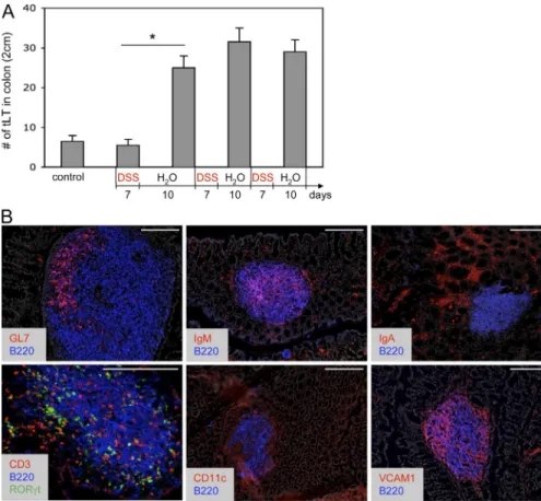
Figure 1. Supernumerary and mature tLTs induced by DSS-mediated colitis.
(A) Quantification of tLTs in the colon of wild-type mice before and after multiple cycles of DSS treatment. During each cycle, mice were treated with DSS for a period of 7 d, followed by a 10-d recovery period without DSS. Data are shown for one representative experiment out of two with three mice per group. Statis-tical significance was assessed by the paired Student’s t test. *, P < 0.05. Error bars are SD. (B) Structure of colonic tLTs in sections of colons from wild-type mice after two cycles of DSS treatment. Sections were stained with the indicated antibodies and with DAPI for nuclear staining (shown in gray). RORt is visualized through GFP expression in Rorc(gt)-GfpTG reporter mice. Bars, 200 µm.
on December 3, 2016
JEM VOL. 208, January 17, 2011 127
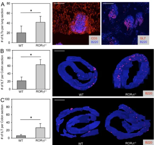
The LTi-independent formation of tLTs
Even though the generation of LTi cells and the
subse-quent development of LNs, PPs, and ILFs require
expres-sion of RORt (Eberl and Littman, 2004; Eberl et al.,
2004), tLTs induced by influenza A virus infection of the
lungs, termed iBALT, develop normally in RORt-deficient
mice (Moyron-Quiroz et al., 2004). In NOD mice, the
formation of tLTs in the inflamed pancreatic islets depends
on LIGHT expression by reactive T cells rather than LT
(Lee et al., 2006), thus presumably in the absence of LTi
cells. We observed that RORt-deficient mice generated
an increased number of mature germinal center–containing
iBALTs in response to influenza A virus infection as
com-pared with infected wild-type controls (Fig. 2 A). Similarly,
in the colon, exposure to DSS induced a threefold higher
number of mature tLTs, as well as extensive neutrophil
in-filtration, in RORt-deficient mice as compared with
control mice (Fig. 2 B and
Fig. S1
). Interestingly, an
in-creased number of tLTs is present already in unexposed
RORt-deficient mice (Fig. 2 C). These data show, first,
that tLTs can be efficiently generated in the absence of LTi
cells during infection and inflammation and, second, that
the intestinal environment is prone to spontaneous tLT
formation in RORt-deficient mice that lack LNs, PPs,
ILFs, and several IL-17– and/or IL-22–producing
lym-phoid cells (Ivanov et al., 2006; Satoh-Takayama et al., 2008;
Sanos et al., 2009).
inflammatory responses (Nimmerjahn and Ravetch, 2008).
Our data indicate that inhibition of the Th17 pathway and
of the normal function of lymphoid tissues may exacerbate
inflammatory bowel disease through the generation of tLTs
and a deregulated B cell response.
RESULTS
The formation of mature tLTs during intestinal inflammation
It was previously reported that colitis induced by 7 d of
DSS oral treatment did not induce significant numbers of
tLTs when compared with mice receiving water (Spahn et al.,
2002). We confirm this observation but observed a marked
increase in the number of tLTs after a 10-d period of rest
that followed the initial 7 d of DSS (Fig. 1 A). The number
of tLTs increased only marginally upon a second cycle of
DSS treatment, followed by another rest period, and
re-mained stable during subsequent cycles of treatment. These
tLTs were significantly larger than ILFs found in untreated
mice. Yet, like conventional ILFs (Hamada et al., 2002; Eberl
and Littman, 2004), these structures were composed of a well
defined B cell follicle surrounded by a shell of DCs and
contained mostly IgM
+B cells, a germinal center,
dis-persed LTi cells, and T cells, but no defined T cell zone
(Fig. 1 B). Stromal cells expressed VCAM-1, which is typical
of stromal cells found in lymphoid tissues, collectively
termed lymphoid stromal cells (Honda et al., 2001; Peduto
et al., 2009).
Figure 2. tLTs form in the absence of LTi cells. (A) Quantification of influenza virus A–
induced iBALT. RORt/ or wild-type control mice were infected intranasally with 50 pfu influenza virus A and sacrificed 3 wk later. Histograms show mean numbers of lymphoid follicles in sections of whole lungs. Data shown are the mean for seven mice per group from two independent experiments. *, P < 0.05. Sections were stained with the indicated anti-bodies. Bars, 200 µm. (B and C) Quantification of tLTs in the colon of RORt/ and wild-type control mice. The number of colonic tLTs was assessed as described in Materials and methods and is indicated as the mean number of tLTs per whole colon section. Data shown for adult mice that were either treated with two cycles of 2.5% DSS (B) or left untreated (C). Figures show data compiled from 10–15 mice per group from three independent ex-periments. *, P < 0.001. Histology shows rep-resentative colon samples from RORt/ or wild-type control mice either treated with two cycles of 2.5% DSS or left untreated. Sections were stained with anti-CD45R/B220 antibodies to visualize tLTs and DAPI for nu-clear staining. Single pictures of 100× magni-fied sections were assembled to generate an integral view of a colon. Bars, 0.5 cm. Statisti-cal significance was assessed by the paired Student’s t test. Error bars are SD.
on December 3, 2016
tLTs in the pancreas of aged NOD mice (Lee et al., 2006).
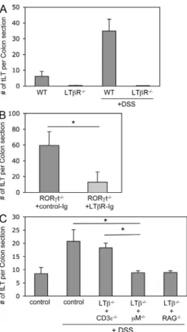
As tLTs generated in the colon of DSS-treated mice consist
mostly of mature B cell follicles, we hypothesized that a subset
of B cells expressing LT
1
2(Ansel et al., 2000) may function
as LTi cells in that context. In that regard, it has been reported
that LT
1
2-expressing B cells, in addition to LTi cells, are
required for the full development of ILFs (Lorenz et al., 2003).
Bone marrow chimeras were thus generated that lacked
ex-pression of LT on T cells, on B cells, or both. In the absence
of LT expressed by T cells, the number of tLTs induced
during colitis was similar to the number of tLTs induced in
unmanipulated wild-type mice (Fig. 3 C). In contrast, in the
absence of LT expressed by B cells, or both B and T cells, the
number of tLTs dropped to the number of tLTs generated in
the absence of colitis. Thus, DSS-mediated colitis induces the
formation of tLTs through the LTi function of LT
1
2-
expressing B cells. Nevertheless, it remains formally possible
that LT
1
2-expressing cells types, in addition to
lympho-cytes, are involved in the formation of tLTs during DSS-
mediated colitis in the absence of LTi cells, although no such
cells have been identified yet.
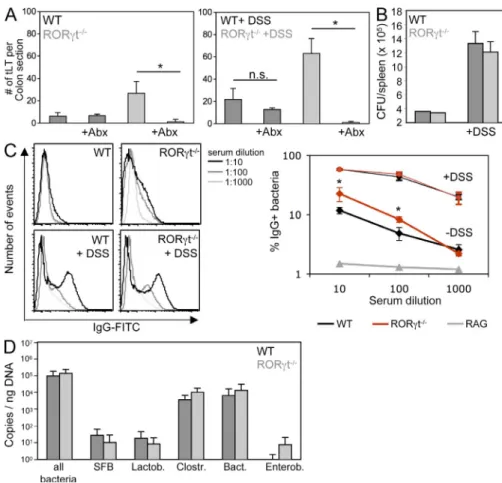
Containment of microbiota in the absence of RORt
+cells
Despite the absence of LTi cells, RORt-deficient mice
de-velop significantly higher numbers of mature intestinal tLTs,
both during steady state and exposure to DSS (Fig. 2, B
and C). As DSS-mediated colitis is dependent on microbiota
(Hans et al., 2000) and RORt-deficient mice lack lymphoid
tissues and cells involved in intestinal homeostasis and defense
(Eberl and Littman, 2004; Eberl et al., 2004; Ivanov et al.,
2006; Satoh-Takayama et al., 2008; Eberl and Lochner, 2009;
Sanos et al., 2009), we assessed whether microbiota was
responsible for the increased lymphoid tissue genesis. In
RORt-deficient mice treated with a large-spectrum cocktail
of antibiotics during exposure to DSS, induction of tLTs was
ab-rogated (Fig. 4 A). The induction of tLTs was also abab-rogated
in antibiotic-treated RORt-deficient mice during steady
state. In contrast, antibiotics had no visible effect on the
gen-eration of lymphoid tissues in the colon of wild-type mice
during steady state, indicating that several of these colonic
lymphoid tissues are LTi cell dependent and programmed
lymphoid tissues, such as colonic patches (Eberl and Lochner,
2009) and, thus, not tLTs.
We next assessed whether the microbiota-induced
forma-tion of tLTs in RORt-deficient mice was a consequence of
decreased containment of the intestinal microbiota. It was
re-cently reported that mice deficient in both Myd88 and TRIF,
which are involved in the signaling of toll-like receptors,
show deficient containment of the microbiota (Slack et al.,
2009). As a consequence, an increased number of live bacteria
was recovered from the spleen, and microbiota-specific IgG
was detected in the serum. Although exposure to DSS increased
the number of live bacteria found in the spleen, no significant
difference was observed between RORt-deficient mice and
wild-type controls during steady state and colitis (Fig. 4 B).
In contrast, markedly higher titers of serum IgG directed
An LTi function for B cells
We first assessed whether tLTs generated during DSS-mediated
colitis were induced through the canonical LTR pathway.
In both LTR-deficient mice and RORt-deficient mice
treated with LTR-Ig fusion protein, the induction of tLTs
during DSS-mediated colitis was markedly inhibited (Fig. 3,
A and B). LTR has two known ligands, LT
1
2and LIGHT
(Gommerman and Browning, 2003). Whereas LT
1
2is
re-quired for the LTi-mediated generation of LNs, PPs, and ILFs
(Hamada et al., 2002; Mebius, 2003; Eberl and Littman, 2004),
LIGHT is involved in the T cell–mediated generation of
Figure 3. The LTi function of B cells. (A) Quantification of tLTs inthe colons of LTR-deficient and wild-type control mice that were either untreated or treated with two cycles of 2.5% DSS. Data are shown for one representative experiment out of two with three mice per group. (B) Quantification of colonic tLTs in RORt/ mice that were treated with two cycles of 2.5% DSS and by weekly i.p. injections of LTR-Ig fusion protein or control Ig. Data are shown for one representative experiment out of two with six mice per group. *, P < 0.005. (C) Quantification of tLTs in the colon of wild-type mice and mixed bone marrow chimeras after two cycles with 2% DSS. For mixed bone marrow chimeras, irradiated adult C57BL/6 wild-type mice received bone marrow from LT/ mice that was mixed 1:1 with bone marrow from M/, CD3/, or RAG/ mice to create mice that lack LT expression in B cells, T cells, or both B and T cells. DSS treatment was initiated 6 wk after transfer. Data are shown for one representative experiment out of three with five mice per group. *, P < 0.001. Statistical significance was assessed by the paired Student’s t test. Error bars are SD.
on December 3, 2016
JEM VOL. 208, January 17, 2011 129
mice was prevented
by the
administra-tion of antibiotics,
as expected, as well
as by blocking the
formation of tLTs with LTR-Ig fusion protein (Fig. 5, C and
D; and
Fig. S2
; Rennert et al., 1998). These data demonstrate
that the supernumerary intestinal tLTs exacerbate the
inflam-matory pathology caused by the intestinal microbiota in
DSS-treated RORt-deficient mice.
Furthermore, in the absence of RORt required for the
generation of several IL-17– and/or IL-22–producing
lym-phoid cells (Ivanov et al., 2006; Satoh-Takayama et al., 2008;
Sanos et al., 2009), the intestinal immune response to DSS
shifted from a Th17 type of response to a IFN-–dominated
Th1 type of response (Fig. 5 E,
Fig. S3 A
, and
Fig. S4
).
How-ever, neutralization of IFN- in DSS-treated RORt-deficient
mice did not protect from severe colitis (Fig. 5 F) and had
no impact on the number of tLTs (Fig. S3B). In contrast,
complementation of deficient mice with
RORt-sufficient spleen cells, but not RORt-deficient cells, partially
protected from colitis (
Fig. S5
), indicating that RORt
+cells
contribute to protection from pathology and thus are involved
in intestinal homeostasis. Together, these data show that in the
absence of RORt
+cells, including Th17 cells and lymphoid
tissues induced by RORt
+LTi cells, mice develop aggravated
colitis induced by microbiota and supernumerary tLTs.
against bacterial microbiota were found in RORt-deficient
mice at steady state, a difference which nevertheless vanished
during colitis (Fig. 4 C). Thus, although wild-type mice can
contain microbiota during steady state without the formation
of large numbers of intestinal tLTs and increased serum IgG,
RORt-deficient mice need to increase the number of
intes-tinal tLTs and the production of systemic microbiota-specific
IgG to reach a similar levels of containment. Furthermore, upon
epithelial damage, RORt-deficient mice required even larger
numbers of intestinal tLTs to be able to develop a level of
con-tainment comparable to that of wild-type mice. In that context,
the bacterial microbiota was not significantly different between
wild-type and RORt-deficient mice (Fig. 4 D), indicating
that RORt-deficient mice were still able to develop a level of
selective pressure on the microbiota that was comparable to the
selective pressure developed by wild-type mice.
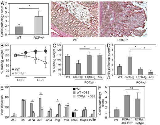
Supernumerary tLTs exacerbate colonic pathology
Even though an increased number of intestinal tLTs may
compensate for the immunodeficiencies of RORt-deficient
mice, at least for the containment of the intestinal microbiota,
this comes at a high cost for the organism. When exposed
to DSS, and in our experimental conditions (Fig. 1 A),
RORt-deficient mice developed severe colitis (Fig. 5 A)
and wasting disease (Fig. 5 B), whereas wild-type mice
developed a mild colitis and no wasting disease. The severe
colitis and wasting disease developing in RORt-deficient
Figure 4. Containment of microbiota in the absence of RORt+ cells. (A) RORt/ and wild-type control mice were treated from birth with a cocktail of antibiotics. 8-wk-old mice were exposed or not to two cycles of DSS before quantification of tLTs. Data are shown for one representative experiment out of two with five mice per group. *, P < 0.0001. Statistical significance was assessed by the paired Student’s t test. n.s., not significant; Abx, antibiotic treated. (B) The bacterial con-tent in the spleen of RORt/ and wild-type control mice was determined by plating spleen extracts from three individual mice exposed or not to DSS on blood agar plates. CFU, colony forming unit. Data are shown for one representative experiment out of two. (C) The IgG response specific for intestinal bacteria was determined in the sera from the peripheral blood of RORt/ and wild-type control mice exposed or not to DSS. Data from one representative experiment out of three and three mice per group show the percentage of bacteria positive for IgG bind-ing. *, P < 0.05. As a baseline control, bacteria were directly stained with anti–mouse IgG without the addition of serum (not depicted). (D) Colon biofilms were collected from 8-wk-old RORt/ and wild-type control mice and the bacterial content was determined by quantitative PCR. Data are shown for one representative experiment out of two with five mice per group. SFB, segmented filamen-tous bacteria; Lactob, Lactobacillaceae; Clostr, Clostridiales; Bact, Bacteroides; Enterob, Enterobacteriaceae. Error bars are SD.
on December 3, 2016
DISCUSSION
tLTs form within numerous types of chronic inflammatory
lesions (Aloisi and Pujol-Borrell, 2006) and have been shown
to function like secondary lymphoid tissues in the induction
of effector B and T cells (Lee et al., 2006; Moyron-Quiroz
et al., 2006; Nasr et al., 2007). These inducible tissues can
provide protection to virus infection in the absence of LNs
or aggravate inflammatory disease. Therefore, in the latter
context, it has been suggested that the formation of tLTs may
be blocked as a strategy to prevent or mitigate inflammatory
disease. The development of LNs and PPs in the fetus (Eberl
et al., 2004), and of ILFs in the intestinal lamina propria
(Eberl and Littman, 2004), requires RORt
+LTi cells, and in
the absence of RORt, these lymphoid tissues do not
de-velop. RORt is also required to generate the
proinflamma-tory Th17 cells (Ivanov et al., 2006) and IL-22–producing
NKp46
+innate lymphoid cells (Satoh-Takayama et al., 2008;
Luci et al., 2009). Thus, it has been suggested that RORt
antagonists may be developed to block excessive immunity
and chronic inflammation in several pathological settings.
However, the role of RORt
+LTi cells in the formation of
tLTs during inflammation remained to be clearly assessed,
and the effect of an absence of functional RORt, which is
involved in the generation of both lymphoid tissues and
Th17 cells, must be carefully measured during
inflamma-tory disease.
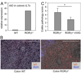
Hyperactive tLTs and B cells as a cause of aggravated
colonic pathology
Intestinal tLTs, as ILFs, are primarily B cell follicles harboring
a germinal center but no distinct T cell zone (Fig. 1 B; Eberl
and Lochner, 2009). We therefore tested whether tLTs
aggra-vate colonic pathology in DSS-treated RORt-deficient
mice by inducing a hyperactive B cell compartment. In
ac-cordance with this view, tLTs in such mice express a 30-fold
increase in transcripts for the activation-induced deaminase
AID (Fig. 6 A), which is required for gene switch
recombina-tion and somatic hypermutarecombina-tion of Ig genes (Muramatsu
et al., 2000). Possibly as a consequence, even though it is
dif-ficult to assess, IgG
+B cells were present in the lamina propria
of DSS-treated RORt-deficient mice but not in wild-type
mice (Fig. 6 B). To demonstrate that Igs were involved in the
pathology induced by tLTs, we treated RORt-deficient
mice concomitantly with DSS and IVIG, which have a
thera-peutic effect against a broad range of hematological and
im-munological disorders, essentially through the saturation and
activation of Fc receptors (Nimmerjahn and Ravetch, 2008).
IVIG treatment significantly decreased the colonic
pathol-ogy induced by tLTs in RORt-deficient mice (Fig. 6 C).
Together, our data indicate that in the absence of RORt, the
supernumerary tLTs induced by the microbiota are nurturing
a hyperactive B cell compartment that contributes to the
ag-gravated colonic pathology.
Figure 5. tLTs aggravate DSS-mediated colitis. (A) Histological disease score in
RORt/ and wild-type control mice exposed to two cycles of DSS. Scores are shown for eight mice per group from three independent experiments. *, P < 0.01. H&E staining of rep-resentative sections from distal colon of RORt/ and wild-type control mice after exposure to two cycles of DSS is shown. Bars, 200 µm. (B) Wasting disease. RORt/ and wild-type control mice were exposed to DSS cycles as indicated, and mouse body weight was assessed at the beginning of every cycle. Shown are weights relative to the starting weight. Data derive from eight mice per group from three independent experiments. *, P < 0.01. (C and D) LTR blockade ameliorates DSS colitis. Wild-type and RORt-deficient mice were exposed to two cycles of DSS. RORt/ mice were either treated by weekly i.p. injections of LTR-Ig protein or with a control Ig. One group of RORt/ mice was treated from birth with a cocktail of antibiot-ics. Data are shown for six mice per group from two to three independent experiments. *, P < 0.01. Shown is mouse weight at the end of the second DSS cycle in percentage of the starting weight (C) and histological disease score (D). Abx, antibiotic treated. (E) Quantitative real-time PCR on whole colon tissue from untreated wild-type controls and RORt/, as well as wild-type control mice after exposure to DSS. Ct values were normalized to Gapdh expression. Data shown are for one representative experiment out of three with two mice per group. *, P < 0.05. (F) RORt/ mice were treated with two i.p. injections of 250 µg of neutralizing anti–IFN- antibody before the first and the second DSS cycle. Data are shown for one representative experiment out of two with three mice per group. ns, not specific. P > 0.05. Statistical significance was assessed by the paired Student’s t test. Error bars are SD.
on December 3, 2016
JEM VOL. 208, January 17, 2011 131
We show that the formation of tLTs in RORt-deficient
mice is induced by microbiota through the LTi function of
B cells, even though we do not formally exclude an LTi
func-tion for other cell types in that context. So how does
micro-biota induce the recruitment of LT
+B cells? We had shown
previously that CCL20 was required for the recruitment of
B cells and the formation of ILFs (Bouskra et al., 2008) but
expression of CCL20 was undetectable in RORt-deficient
mice. The only cytokine found to be increased in
RORt-deficient mice treated with DSS was IFN-; however,
block-ing IFN- with neutralizblock-ing antibody had no effect on the
number of tLTs and the severity of disease. The intestine of
DSS-treated RORt-deficient mice nevertheless showed an
important infiltration of IgG
+B cells. We therefore suggest
that the microbiota-induced inflammation unfolding in
DSS-treated RORt-deficient mice eventually leads to the sustained
recruitment of B cells, which induce the formation of tLTs
through their expression of LT
1
2. Given that tLTs are
pri-marily B cell follicles, this pathway can generate a
positive-feedback loop in B cell activation and differentiation and in
the formation of tLTs.
In RORt-deficient mice during steady state or that
have been exposed to DSS, a vast network of tLTs develops
that contains approximately three times the number of tLTs
found in wild-type mice subjected to the same treatments.
RORt is required for the development of LNs and PPs, as
well as for the generation of a collection of lymphoid cells
producing IL-17 and/or IL-22, such as Th17 cells and IL-22
+NKp46
+cells. The latter cell type was recently shown to be
involved in protection against infection by Citrobacter
roden-tium and DSS-induced colitis (Satoh-Takayama et al., 2008),
and IL-17 and IL-22 synergize in the activation of epithelial
cells to produce antibacterial peptides (Liang et al., 2006).
Thus, it might be expected that the absence of lymphoid
tis-sues and of IL-17/22–producing lymphoid cells in
RORt-deficient will be matched by the increased activity in other
immune compartments, such as tLT formation, to maintain
a similar level of containment of the intestinal microbiota.
Such a compensatory mechanism has been reported by Lorenz
et al. (2003); the inhibition of development of secondary
lymphoid tissues through the administration of LTR-Ig
pro-tein to pregnant mothers induced the formation of
numer-ous tLTs or ILFs. However, when exposed to DSS, which
injures the epithelial cell layer, RORt-deficient mice
ap-pear only to be able to contain microbiota at the price of
an additional increase in intestinal tLTs and B cell activity.
This increase in the number of tLTs and in B cell activity is,
however, not tolerated by the intestine, which develops
severe inflammation and leads the host to wasting disease.
This pathology is possibly a consequence of the formation
of immune complexes consisting of bacteria and specific
IgG, which activate IgG receptor-bearing inflammatory
cells, such as neutrophils. This hypothesis is supported by
the antiinflammatory effect of IVIG treatment, which
is shown to depend on IgG receptors (Nimmerjahn and
Ravetch, 2008).
We find that during colitis induced by DSS, mature colonic
tLTs develop that consist of a well structured B cell follicle
con-taining predominantly IgM
+B cells and a germinal center.
Similar tLTs termed iBALTs were reported in influenza A–
infected lungs (Moyron-Quiroz et al., 2004). In the absence of
LTi cells in RORt-deficient mice, both iBALT and colonic
tLTs develop, indicating that other cells can take over the
func-tion of LTi cells for the inducfunc-tion of lymphoid tissues. In the
case of tLTs induced in the pancreas of aged NOD mice, the
LTi function is taken over by autoreactive T cells (Lee et al.,
2006). Central to the development of LNs, PPs, and ILFs
is LTR-mediated activation of stromal cells by LT
1
2-
expressing LTi cells (Mebius, 2003). Activated stromal cells
produce the structural chemokines CC19, CCL21, and CXCL13,
which are involved in the recruitment and organization of
lym-phocytes and DCs (Dejardin et al., 2002). In the inflamed
pan-creas, T cells induce the formation of tLTs through the alternative
LTR ligand LIGHT (Lee et al., 2006). The formation of iBALTs
is independent of LT (Moyron-Quiroz et al., 2004), and the
involvement of LIGHT remains to be assessed. In the case of
DSS-induced colonic tLTs, B cells perform the LTi function
through LT, and thus, presumably, through its
membrane-bound LT
1
2heterotrimer. Together, these data show that
tLTs can develop during chronic inflammation through similar
mechanisms but distinct lymphocyte or lymphoid cell subsets.
Figure 6. Hyperactive tLTs and B cells as a cause of aggravated colonic pathology. RORt/ and wild-type control mice were treated with two cycles of 2.5% DSS. (A) The expression of AID was assessed by quantitative RT-PCR in laser-captured tLTs from DSS-treated RORt/ and wild-type control mice. Data are shown for one representative experi-ment out of two with two mice per group. Ct values were normalized to Gapdh expression. (B) Colon sections were stained for IgG, B220, and DAPI for nuclear staining (shown in gray). IgG+ B plasma cells express low levels of B220. Bars, 200 µm. Data are shown for one representative ex-periment out of two with three mice per group. (C) RORt/ mice were treated with two i.p. injections of IVIG. The histological disease score was assessed as described in Materials and methods. Data are shown for one representative experiment out of two with two to three mice per group. Statistical significance was assessed by the paired Student’s t test. *, P < 0.05. Error bars are SD.on December 3, 2016
40 sections of a whole colon were taken at regular intervals and fixed for 5 min in acetone at 20°C. For staining, slides were first hydrated in PBS-BS (PBS containing 1% normal bovine serum; Sigma-Aldrich) for 5 min and blocked with 10% bovine serum in PBS for 1 h at room temperature. Slides were then incubated with anti-phycoerythrin–conjugated anti-CD45R/B220 mAb (clone RA3-6B2) in PBS-BS at room temperature for 1 h, washed three times for 5 min with PBS-BS, incubated with DAPI (Sigma-Aldrich) for 5 min at room temperature, washed three times for 5 min, and mounted with Fluoromount-G (SouthernBiotech). Numbers of tLTs per colon section were calculated as the mean number of tLTs per section in 40 sections.
Influenza virus A infection. Mice were infected intranasally with 50 PFU
influenza A virus (H3N2 strain Scotland/20/74). Mice were sacrificed 21 d after infection.
Immunofluorescence histology. For immunofluorescence histology,
tis-sues were fixed and stained as previously described (Peduto et al., 2009). In brief, tissues were washed and fixed overnight at 4°C in a fresh solution of 4% paraformaldehyde (Sigma-Aldrich) in PBS. The samples were then washed for 1 d in PBS, incubated in a solution of 30% sucrose (Sigma- Aldrich) in PBS, and embedded in OCT. Frozen blocks were cut at 8-µm thickness and sections collected onto Superfrost Plus slides (VWR). For staining, slides were first hydrated in PBS-XG (PBS containing 0.1% Triton X-100 and 1% normal goat serum; Sigma-Aldrich) for 5 min and blocked with 10% bovine serum in PBS-XG for 1 h at room temperature. Slides were then incubated with primary polyclonal antibody or conjugated mAb (in general 1/100) in PBS-XG overnight at 4°C, washed three times for 5 min with PBS-XG, incubated with DAPI for 5 min at room temperature, washed three times for 5 min, and mounted with Fluoromount-G. Slides were exam-ined under a fluorescence microscope (AxioImager M1; Carl Zeiss, Inc.) equipped with a charge-coupled device camera and images were processed with AxioVision software (Carl Zeiss, Inc.).
Laser capture microdissection. Colon tissues were embedded in OCT
compound 4583 (Sakura), frozen in a bath of isopentane cooled with liquid nitrogen, and stocked at 80°C. Frozen blocks were cut at 10-µm thickness and serial sections collected onto Superfrost/Plus slides. Sections were im-mediately fixed for 5 min in acetone at 20°C, dried, and stored at 80°C. Serial sections were then thawed and immediately stained for 5 s with histo-gen (MDS Analytical Technologies), washed briefly in RNase-free water supplemented with ProtectRNA (Sigma-Aldrich), dehydrated successively in one bath of 70% ethanol for 30 s, two baths of 95% ethanol for 1 min, two baths of water-free ethanol (VWR) for 2 min, and two baths of xylene for 5 min, and air-dried. Slides were transferred immediately into a Veritas Laser Capture Microdissector (MDS Analytical Technologies), microdissected, and captured with Capsure Macro LCM caps (MDS Analytical Technologies). RNA was isolated using the PicoPure RNA Isolation kit (MDS Analytical Technologies), and its quality was assessed using the 2100 Bioanalyzer system (Agilent Technologies).
Antibodies. The following mAbs were purchased from BD: PE-conjugated
anti–mouse IgM (R6-60.2); from eBioscience: allophycocyanin-conjugated anti-CD45R/B220 (RA3-6B2), PE-conjugated anti-CD106 (VCAM-1; 429), anti-CD11c (N418), anti-IgA (11–44-2), biotinylated anti–Ly-77 (GL7), and purified CD3 (500A2); and from Serotec: biotinylated anti-neutrophil (7/4). Cy3-anti–armenian or –syrian hamster and DyLight 488 donkey anti–mouse IgG were purchased from Jackson Immuno-Research Laboratories. Cy3-conjugated streptavidin was purchased from Sigma-Aldrich.
Colitis disease score. Swiss rolls of whole colons were directly frozen in
OCT and sections of 7-µm thickness were stained with hematoxylin and eosin (H&E). Histological scoring was performed using a modified scoring system described previously (Hartmann et al., 2000). In brief, the presence of rare inflammatory cells in the lamina propria were counted as: 0, increased num-bers of inflammatory cells; 1, confluence of inflammatory cells; 2, extending
RORt controls the proinflammatory IL-17 pathway
(Ivanov et al., 2006), which is shown to be involved in
auto-immune pathology through the recruitment of neutrophils
(Weaver et al., 2006). The IL-17/23 pathway is involved in
several colitis models in mice (Uhlig and Powrie, 2009), and
patients with a defective IL-23R show resistance to the
devel-opment of the disease (Duerr et al., 2006). Therefore, it can be
expected that the absence of RORt, or antagonizing RORt
function during the initial phase of inflammation, protects
from progression to inflammatory disease. We show that
coli-tis induced by exposure to DSS was actually more severe in
RORt-deficient mice as compared with RORt-sufficient
mice. In the absence of RORt, the colon developed
pro-found tissue damage, and mice suffered from marked wasting
disease, whereas in the presence of RORt, mice endured
mild intestinal inflammation under the regimen applied and
grew normally. Furthermore, complementation of
RORt-deficient mice with RORt-sufficient spleen cells
signifi-cantly decreased the severity of the disease, demonstrating a
protective effect of RORt
+cells in intestinal pathology. We
suggest that RORt
+cells, including Th17 cells and IL-22–
producing NKp46
+cells, limit DSS-induced intestinal
in-flammatory disease by strengthening antibacterial immunity,
such as the production of antibacterial peptides by epithelial
cells (Liang et al., 2006). Thus, our data show that a narrow
road has to be followed to prevent the pathological effect of
immunity during colitis while maintaining the essential
func-tions of immunity for intestinal homeostasis and defense.
MATERIALs And METHOds
Mice. RORt-deficient (Rorc(t)Gfp/Gfp) mice (Eberl et al., 2004) and BAC
transgenic Rorc(gt)-GfpTG mice (Lochner et al., 2008) have been described
previously. Mice deficient in LTR (Fütterer et al., 1998), LT (Alimzhanov et al., 1997), mu chain (Kitamura et al., 1991), CD3 epsilon (Malissen et al., 1995), and RAG2 (Shinkai et al., 1992) have been described before. All mice were kept in specific pathogen-free conditions and all animal experiments were approved by the committee on animal experimentation of the Institut Pasteur and by the French Ministry of Agriculture.
In vivo treatments. For antibiotic treatment, pregnant mothers were
treated with a mixture of 1 mg/ml ampicillin, 1 mg/ml colistin, and 5 mg/ml streptomycin together with 5% glucose (all from Sigma-Aldrich) in their drinking water. After birth, treatment was continued until analysis. For LTR-Ig treatment, mice were treated with LTR-Ig fusion protein (gift from J. Browning, Biogen Idec, Cambridge, MA; Browning et al., 1997) by weekly i.p. injections of 10 µg/mg of body weight during the course of the experiment. Control mice were treated with the Ig fusion partner. For IFN- neutralization, mice received two i.p. injections of 250 µg of neutralizing anti–IFN- (clone R4-6A2; eBioscience) or isotype control antibodies before the first and second DSS cycle. Colitis was induced using DSS salt (mol wt = 36,000–50,000; MP Biomedicals) dissolved in the drinking water at a con-centration of 2.5% (m/v). Mice were exposed to DSS for 7 d, followed by a recovery period of 10 d without DSS. This cycle was repeated once or twice and weight was monitored at the end of every cycle. For IVIG treatment, DSS-treated mice received two i.v. injections of Gamunex 10% (Talecris Biotherapeutics) at a concentration of 1 g/kg at the end of a first DSS cycle (day 5) and 1 d before a second DSS cycle (day 11).
Quantification of tLTs. Whole colons were frozen as swiss rolls in OCT
compound 4583 (Sakura) and frozen blocks were cut as 7-µm sections.
on December 3, 2016
JEM VOL. 208, January 17, 2011 133 on Brucella agar plates containing 5% horse blood. Colonies were counted after 2 d of culture at 37°C.
Online supplemental material. Fig. S1 shows the structure of tLTs and
neutrophil recruitment in DSS-treated RORt-deficient mice. Fig. S2 shows the histology of the effect of LTR-Ig and antibiotics on colitis progression. Fig. S3 shows the increased expression of IFN- by CD4+ T cells and the
lack of impact of neutralizing IFN- in disease severity. Fig. S4 shows the expression of transcripts for LIGHT, LT, LT, and LTR in colonic tissue. Fig. S5 shows the effect of RORt-sufficient spleen cells upon transfer into irradiated RORt-deficient mice. Online supplemental material is available at http://www.jem.org/cgi/content/full/jem.20100052/DC1.
We thank the members of the DTL laboratory for discussions and critical reading of the manuscript, and Lucette Polomack and Sophie Dulauroy for technical assistance.
This work was supported by the Institut Pasteur, grants from the Mairie de Paris, the Agence Nationale de la Recherche, and an Excellence Grant from the European Commission. M. Lochner was supported by the Deutsche Forschungsgemeinschaft and the Schlumberger Foundation.
The authors have no competing financial interests.
submitted: 7 January 2010 Accepted: 24 november 2010
REFERENCES
Alimzhanov, M.B., D.V. Kuprash, M.H. Kosco-Vilbois, A. Luz, R.L. Turetskaya, A. Tarakhovsky, K. Rajewsky, S.A. Nedospasov, and K. Pfeffer. 1997. Abnormal development of secondary lymphoid tissues in lymphotoxin -deficient mice. Proc. Natl. Acad. Sci. USA. 94:9302– 9307. doi:10.1073/pnas.94.17.9302
Aloisi, F., and R. Pujol-Borrell. 2006. Lymphoid neogenesis in chronic inflam-matory diseases. Nat. Rev. Immunol. 6:205–217. doi:10.1038/nri1786 Ansel, K.M., V.N. Ngo, P.L. Hyman, S.A. Luther, R. Förster, J.D.
Sedgwick, J.L. Browning, M. Lipp, and J.G. Cyster. 2000. A chemo-kine-driven positive feedback loop organizes lymphoid follicles. Nature. 406:309–314. doi:10.1038/35018581
Bouskra, D., C. Brézillon, M. Bérard, C. Werts, R. Varona, I.G. Boneca, and G. Eberl. 2008. Lymphoid tissue genesis induced by commensals through NOD1 regulates intestinal homeostasis. Nature. 456:507–510. doi:10.1038/nature07450
Browning, J.L., I.D. Sizing, P. Lawton, P.R. Bourdon, P.D. Rennert, G.R. Majeau, C.M. Ambrose, C. Hession, K. Miatkowski, D.A. Griffiths, et al. 1997. Characterization of lymphotoxin- complexes on the surface of mouse lymphocytes. J. Immunol. 159:3288–3298.
Dejardin, E., N.M. Droin, M. Delhase, E. Haas, Y. Cao, C. Makris, Z.W. Li, M. Karin, C.F. Ware, and D.R. Green. 2002. The lymphotoxin- receptor induces different patterns of gene expression via two NF-kappaB pathways. Immunity. 17:525–535. doi:10.1016/S1074- 7613(02)00423-5
Duerr, R.H., K.D. Taylor, S.R. Brant, J.D. Rioux, M.S. Silverberg, M.J. Daly, A.H. Steinhart, C. Abraham, M. Regueiro, A. Griffiths, et al. 2006. A genome-wide association study identifies IL23R as an inflammatory bowel disease gene. Science. 314:1461–1463. doi:10.1126/science.1135245 Eberl, G., and D.R. Littman. 2004. Thymic origin of intestinal T cells
revealed by fate mapping of ROR+ cells. Science. 305:248–251. doi:10
.1126/science.1096472
Eberl, G., and M. Lochner. 2009. The development of intestinal lymphoid tissues at the interface of self and microbiota. Mucosal Immunol. 2:478– 485. doi:10.1038/mi.2009.114
Eberl, G., S. Marmon, M.J. Sunshine, P.D. Rennert, Y. Choi, and D.R. Littman. 2004. An essential function for the nuclear receptor RORgamma(t) in the generation of fetal lymphoid tissue inducer cells. Nat. Immunol. 5:64–73. doi:10.1038/ni1022
Fütterer, A., K. Mink, A. Luz, M.H. Kosco-Vilbois, and K. Pfeffer. 1998. The lymphotoxin receptor controls organogenesis and affinity matura-tion in peripheral lymphoid tissues. Immunity. 9:59–70. doi:10.1016/ S1074-7613(00)80588-9
into the submucosa; and 3, transmural extension of the inflammatory cell infil-trate. For epithelial damage, absence of mucosal damage was counted as 0, discrete focal lymphoepithelial lesions were counted as 1, mucosal erosion/ ulceration was counted as 2, and a score of 3 was given for extensive mucosal damage and extension through deeper structures of the bowel wall. The two subscores were added and the combined histological score ranged from 0 (no changes) to 6 (extensive cell infiltration and tissue damage).
RNA isolation and quantitative PCR. To perform gene expression
analysis, whole tissue from the middle and terminal part of the colon was immediately frozen in liquid nitrogen upon animal sacrifice. Tissue was homogenized using Ultra Turrax T8 (IKA-Werke) in TRIZOL regent, and total RNA was purified according to the manufacturer’s protocol (Invitrogen). RNA was subjected to DNase I digestion and additional purification using the RNeasy Mini kit (QIAGEN). 1 µg of total RNA was transcribed into cDNA using Superscript III reverse transcription (Invitrogen) according to the manufacturer’s protocol. Quantitative real-time PCR was performed using RT2 qPCR Primer sets and RT2 SYBR-Green master mix (QIAGEN) on
a PTC-200 thermocycler equipped with a Chromo4 detector (Bio-Rad Laboratories). Data were analyzed using Opticon Monitor software (Bio-Rad Laboratories).
Bone marrow and spleen transfer. 107 bone marrow cells from CD3/,
M/, or RAG/ mice were mixed with 107 bone marrow cells from
LT/ mice and injected i.v. into 10-Gy irradiated wild-type mice. After 4 wk,
reconstitution of the mice was assessed in peripheral blood. In reconstituted mice, DSS-mediated colitis experiments were performed 6 wk after transfer. For spleen transfer, RORt/ mice were sublethally irradiated (500 rad) and
trans-ferred with 107 splenocytes from either RORt-deficient or RORt-sufficient
littermate mice. 10 d later, mice were treated with two cycles of DSS.
Intestinal biofilm collection and analysis. Large intestine was isolated
immediately after animal sacrifice. Fecal contents were removed using forceps pressed along the whole length of the organ, and the tissue was placed into a Petri dish containing sterile ice-cold PBS. The intestine was cut into 3-cm sections and then cut longitudinally and vigorously rinsed with scraping using a Pasteur pipette. Tissue was then removed and PBS containing the mucosal biofilm was transferred to a 50-ml falcon tube. Biofilm was separated by centrifugation for 15 min at 4,000 rpm and 4°C, and the supernatant was discarded. DNA extraction was performed using FastDNA Spin kit (MP Biomedicals) using Lysis buffer CLS-Y, according to the manufacturer’s in-structions. Quantitative PCR was performed on a DNA Engine thermal cycler (Bio-Rad Laboratories). QuantiTect SYBR green (QIAGEN) master mix was used in 25-µl reactions. Primers and reaction conditions were de-scribed previously (Bouskra et al., 2008). Absolute numbers of bacteria were determined from standard curves established by quantitative PCR with serial dilutions of reference plasmids harboring 16S rDNA.
Bacterial IgG FACS. Mouse serum was diluted 10-fold in PBS and
heat-inactivated at 60°C for 30 min. After centrifugation (10 min, 13,000 rpm in a Microcentrifuge; Eppendorf ), the supernatant was further diluted through 1:10 serial dilutions in PBS. To isolate fecal bacteria, 0.1 g of feces from RAG-2–deficient mice was suspended in 1 ml PBS and spun on lowest setting to remove fecal matter. 20 µl of supernatant was collected and washed with PBS (1 min at 8,000 rpm). Bacteria were stained with DAPI 1 µg/ml for 5 min and washed twice with PBS. Bacteria were then resuspended in 25 µl PBS and 25 µl of serum dilutions was added. After 1 h of incubation on ice, bacteria were washed twice and stained with DyLight488 anti–mouse-IgG Antibody (Jackson) for 30 min at 4°C. After washing twice, bacteria were resuspended in PBS 1% PFA and analyzed on a FACSCanto II cytometer (BD).
Counting bacterial CFU in the spleen. Spleens were pressed through a
70-µm cell strainer into 2 ml PBS and cells were resuspended by pipetting several times up and down. 100 µl of cell suspension was mixed with 900 µl PBS/0.1% Triton X-100, and 10 µl of this dilution was plated as triplicates
on December 3, 2016
Gommerman, J.L., and J.L. Browning. 2003. Lymphotoxin/light, lym-phoid microenvironments and autoimmune disease. Nat. Rev. Immunol. 3:642–655. doi:10.1038/nri1151
Hamada, H., T. Hiroi, Y. Nishiyama, H. Takahashi, Y. Masunaga, S. Hachimura, S. Kaminogawa, H. Takahashi-Iwanaga, T. Iwanaga, H. Kiyono, et al. 2002. Identification of multiple isolated lymphoid follicles on the antimesenteric wall of the mouse small intestine. J. Immunol. 168:57–64.
Hans, W., J. Schölmerich, V. Gross, and W. Falk. 2000. The role of the resident intestinal flora in acute and chronic dextran sulfate sodium- induced colitis in mice. Eur. J. Gastroenterol. Hepatol. 12:267–273. doi:10 .1097/00042737-200012030-00002
Hartmann, G., C. Bidlingmaier, B. Siegmund, S. Albrich, J. Schulze, K. Tschoep, A. Eigler, H.A. Lehr, and S. Endres. 2000. Specific type IV phosphodiesterase inhibitor rolipram mitigates experimental colitis in mice. J. Pharmacol. Exp. Ther. 292:22–30.
Honda, K., H. Nakano, H. Yoshida, S. Nishikawa, P. Rennert, K. Ikuta, M. Tamechika, K. Yamaguchi, T. Fukumoto, T. Chiba, and S.I. Nishikawa. 2001. Molecular basis for hematopoietic/mesenchymal in-teraction during initiation of Peyer’s patch organogenesis. J. Exp. Med. 193:621–630. doi:10.1084/jem.193.5.621
Ivanov, I.I., B.S. McKenzie, L. Zhou, C.E. Tadokoro, A. Lepelley, J.J. Lafaille, D.J. Cua, and D.R. Littman. 2006. The orphan nuclear receptor RORt directs the differentiation program of proinflammatory IL-17+
T helper cells. Cell. 126:1121–1133. doi:10.1016/j.cell.2006.07.035 Kastelein, R.A., C.A. Hunter, and D.J. Cua. 2007. Discovery and
biol-ogy of IL-23 and IL-27: related but functionally distinct regulators of inflammation. Annu. Rev. Immunol. 25:221–242. doi:10.1146/annurev .immunol.22.012703.104758
Kitamura, D., J. Roes, R. Kühn, and K. Rajewsky. 1991. A B cell-deficient mouse by targeted disruption of the membrane exon of the immuno-globulin mu chain gene. Nature. 350:423–426. doi:10.1038/350423a0 Lee, Y., R.K. Chin, P. Christiansen, Y. Sun, A.V. Tumanov, J. Wang, A.V.
Chervonsky, and Y.X. Fu. 2006. Recruitment and activation of naive T cells in the islets by lymphotoxin beta receptor-dependent tertiary lym-phoid structure. Immunity. 25:499–509. doi:10.1016/j.immuni.2006.06.016 Leppkes, M., C. Becker, I.I. Ivanov, S. Hirth, S. Wirtz, C. Neufert, S.
Pouly, A.J. Murphy, D.M. Valenzuela, G.D. Yancopoulos, et al. 2009. RORgamma-expressing Th17 cells induce murine chronic intestinal in-flammation via redundant effects of IL-17A and IL-17F. Gastroenterology. 136:257–267. doi:10.1053/j.gastro.2008.10.018
Liang, S.C., X.Y. Tan, D.P. Luxenberg, R. Karim, K. Dunussi-Joannopoulos, M. Collins, and L.A. Fouser. 2006. Interleukin (IL)-22 and IL-17 are coex-pressed by Th17 cells and cooperatively enhance expression of antimicrobial peptides. J. Exp. Med. 203:2271–2279. doi:10.1084/jem.20061308 Lochner, M., L. Peduto, M. Cherrier, S. Sawa, F. Langa, R. Varona, D.
Riethmacher, M. Si-Tahar, J.P. Di Santo, and G. Eberl. 2008. In vivo equi-librium of proinflammatory IL-17+ and regulatory IL-10+ Foxp3+ RORt+
T cells. J. Exp. Med. 205:1381–1393. doi:10.1084/jem.20080034 Lorenz, R.G., D.D. Chaplin, K.G. McDonald, J.S. McDonough, and R.D.
Newberry. 2003. Isolated lymphoid follicle formation is inducible and dependent upon lymphotoxin-sufficient B lymphocytes, lymphotoxin receptor, and TNF receptor I function. J. Immunol. 170:5475–5482. Luci, C., A. Reynders, I.I. Ivanov, C. Cognet, L. Chiche, L. Chasson, J.
Hardwigsen, E. Anguiano, J. Banchereau, D. Chaussabel, et al. 2009. Influence of the transcription factor RORgammat on the development of NKp46+ cell populations in gut and skin. Nat. Immunol. 10:75–82. doi:10.1038/ni.1681
Malissen, M., A. Gillet, L. Ardouin, G. Bouvier, J. Trucy, P. Ferrier, E. Vivier, and B. Malissen. 1995. Altered T cell development in mice with a targeted mutation of the CD3-epsilon gene. EMBO J. 14:4641–4653.
McGeachy, M.J., Y. Chen, C.M. Tato, A. Laurence, B. Joyce-Shaikh, W.M. Blumenschein, T.K. McClanahan, J.J. O’Shea, and D.J. Cua. 2009. The interleukin 23 receptor is essential for the terminal differen-tiation of interleukin 17-producing effector T helper cells in vivo. Nat. Immunol. 10:314–324. doi:10.1038/ni.1698
Mebius, R.E. 2003. Organogenesis of lymphoid tissues. Nat. Rev. Immunol. 3:292–303. doi:10.1038/nri1054
Moyron-Quiroz, J.E., J. Rangel-Moreno, K. Kusser, L. Hartson, F. Sprague, S. Goodrich, D.L. Woodland, F.E. Lund, and T.D. Randall. 2004. Role of inducible bronchus associated lymphoid tissue (iBALT) in respiratory immunity. Nat. Med. 10:927–934. doi:10.1038/nm1091
Moyron-Quiroz, J.E., J. Rangel-Moreno, L. Hartson, K. Kusser, M.P. Tighe, K.D. Klonowski, L. Lefrançois, L.S. Cauley, A.G. Harmsen, F.E. Lund, and T.D. Randall. 2006. Persistence and responsiveness of immunologic memory in the absence of secondary lymphoid organs. Immunity. 25:643–654. doi:10.1016/j.immuni.2006.08.022
Muramatsu, M., K. Kinoshita, S. Fagarasan, S. Yamada, Y. Shinkai, and T. Honjo. 2000. Class switch recombination and hypermutation require activation-induced cytidine deaminase (AID), a potential RNA editing enzyme. Cell. 102:553–563. doi:10.1016/S0092-8674(00)00078-7 Nasr, I.W., M. Reel, M.H. Oberbarnscheidt, R.H. Mounzer, F.K. Baddoura,
N.H. Ruddle, and F.G. Lakkis. 2007. Tertiary lymphoid tissues gener-ate effector and memory T cells that lead to allograft rejection. Am. J. Transplant. 7:1071–1079. doi:10.1111/j.1600-6143.2007.01756.x Nimmerjahn, F., and J.V. Ravetch. 2008. Anti-inflammatory actions
of intravenous immunoglobulin. Annu. Rev. Immunol. 26:513–533. doi:10.1146/annurev.immunol.26.021607.090232
Pabst, O., H. Herbrand, M. Friedrichsen, S. Velaga, M. Dorsch, G. Berhardt, T. Worbs, A.J. Macpherson, and R. Förster. 2006. Adaptation of soli-tary intestinal lymphoid tissue in response to microbiota and chemokine receptor CCR7 signaling. J. Immunol. 177:6824–6832.
Peduto, L., S. Dulauroy, M. Lochner, G.F. Späth, M.A. Morales, A. Cumano, and G. Eberl. 2009. Inflammation recapitulates the ontogeny of lymphoid stromal cells. J. Immunol. 182:5789–5799. doi:10.4049/ jimmunol.0803974
Rennert, P.D., D. James, F. Mackay, J.L. Browning, and P.S. Hochman. 1998. Lymph node genesis is induced by signaling through the lymphotoxin receptor. Immunity. 9:71–79. doi:10.1016/S1074-7613(00)80589-0 Sanos, S.L., V.L. Bui, A. Mortha, K. Oberle, C. Heners, C. Johner, and
A. Diefenbach. 2009. RORgammat and commensal microflora are required for the differentiation of mucosal interleukin 22-producing NKp46+ cells. Nat. Immunol. 10:83–91. doi:10.1038/ni.1684 Satoh-Takayama, N., C.A. Vosshenrich, S. Lesjean-Pottier, S. Sawa, M.
Lochner, F. Rattis, J.J. Mention, K. Thiam, N. Cerf-Bensussan, O. Mandelboim, et al. 2008. Microbial flora drives interleukin 22 produc-tion in intestinal NKp46+ cells that provide innate mucosal immune defense. Immunity. 29:958–970. doi:10.1016/j.immuni.2008.11.001 Shinkai, Y., G. Rathbun, K.P. Lam, E.M. Oltz, V. Stewart, M.
Mendelsohn, J. Charron, M. Datta, F. Young, A.M. Stall, et al. 1992. RAG-2-deficient mice lack mature lymphocytes owing to inability to initiate V(D)J rearrangement. Cell. 68:855–867. doi:10.1016/ 0092-8674(92)90029-C
Slack, E., S. Hapfelmeier, B. Stecher, Y. Velykoredko, M. Stoel, M.A. Lawson, M.B. Geuking, B. Beutler, T.F. Tedder, W.D. Hardt, et al. 2009. Innate and adaptive immunity cooperate flexibly to maintain host-microbiota mutualism. Science. 325:617–620. doi:10.1126/science.1172747 Spahn, T.W., H. Herbst, P.D. Rennert, N. Lügering, C. Maaser, M. Kraft,
A. Fontana, H.L. Weiner, W. Domschke, and T. Kucharzik. 2002. Induction of colitis in mice deficient of Peyer’s patches and mesenteric lymph nodes is associated with increased disease severity and formation of colonic lymphoid patches. Am. J. Pathol. 161:2273–2282.
Uhlig, H.H., and F. Powrie. 2009. Mouse models of intestinal inflammation as tools to understand the pathogenesis of inflammatory bowel disease. Eur. J. Immunol. 39:2021–2026. doi:10.1002/eji.200939602
Weaver, C.T., L.E. Harrington, P.R. Mangan, M. Gavrieli, and K.M. Murphy. 2006. Th17: an effector CD4 T cell lineage with regulatory T cell ties. Immunity. 24:677–688. doi:10.1016/j.immuni.2006.06.002 Zhang, Z., M. Zheng, J. Bindas, P. Schwarzenberger, and J.K. Kolls.
2006. Critical role of IL-17 receptor signaling in acute TNBS- induced colitis. Inflamm. Bowel Dis. 12:382–388. doi:10.1097/01.MIB .0000218764.06959.91