Endocrine Disorders as a Contributory Factor to Neoplasia in SJLjJ Mice
l,2W.
Pierpaoli,3
N.
Haran-Ghera,4
E.
Bianchi,5
J.
Muller,6
A.
Meshorer,4
andM. Bree
4,7SUMMARY-We studied the endocrine status of SJLjJ mice. Light and electron microscopy revealed that the adenohypophyses of both sexes became progressively infiltrated with an abnormal number of gonadotropin-producing cells that probably secreted large amounts of luteotropic hormone. The ovaries had numerous large corpora lutea even in animals over 1 year of age with reticulum cell neoplasms. The adrenal cortexes of female mice showed no regression of the reticular zone. In ac-cordance with the anomalous condition of the adenohy-pophysis and ovary, females had abnormal estrous cycles, with prolonged diestrus and consequent reduction in fer-tility. These data were discussed in the context of hormone environment versus onset of systemic neoplastic disease and the relationship between hormone dependence and leukemic virus expression.-J Natl Cancer Inst 53: 731-744,1974.
SJLjJ MICE, which develop a high incidence of spontaneous reticulum cell sarcomas or neoplasms at an early age, have been investigated intensively by oncologists. This reticulum cell neoplasm, defined as type B (RCNB) by Dunn (1), kills virtually all SJLjJ mice of both sexes between 8 and 14 months of age (mean survival time: 13.3 months). Since it resembles Hodgkin's disease in humans, it is sometimes called Hodgkin's-like disease of mice. Descriptions of this disease in SJLj J and other strains of mice have been detailed in (1-7). Study of this neoplastic proc-ess in the SJLj J strain is especially compelling, inasmuch as reticulum cell neoplasms, contrary to lymphoma and lymphosarcoma, are generally rare in young mice and found only sporadically in old animals (8).
One of the most prominent aspects of the disease in SJLj J mice is the early diffuse or focal proliferation of modified reticulum cells, histiocytes, plasma cells, and lymphocytes, mostly in the lymph nodes but also in other lymphoid organs such as spleen and thymus. At an early age, these mice show hyperplasia of ,the thymus and peripheral lymphatic tissues (9). Their immune status in relation to age and development of reticulum cell neoplasms has been explored by Haran-Ghera et al. (10).
Arnesen (11) showed that AKR mice, which develop spontaneous leukemia, have morphologic abnormalities in the adrenals. Also, Levine and Treiman (12) found that the plasma corticosterone response to stress in AKR mice is significantly lower than in other strains.
Since proliferation and differentiation of lymphoid and hematopoietic cells largely depend on the hor-mone environment, a reasonable assumption was tha t a primary hormone derangement could be responsible for the early and progressive alterations of lymphatic tissues in SJLjJ mice. Moreover, a specific chronic endocrine stimulus could be
respon-sible for the changes observed concomitantly with viral agents or could possibly alone induce the onset of RCNB. Therefore, studies designed to test the validity of this assumption were based largely on determining the endocrine status of SJLIJ mice of different ages and sex.
MATERIALS AND METHODS
Animals.-Inbred SJLjJ and C57BLj6 mice, cesarean-originated and barrier-sustained, were raised and maintained in the Animal Breeding Center, The Weizmann Institute of Science.
Histology.-Groups of 3-4 females and in some cases, males, 1 day to 12 months of age were killed. The tissues were fixed in Bouin's fluid and embedded in paraffin, and the sections were stained with hematoxylin-eosin-phosphomolybdic acid-light green. Sections of the following tissues were prepared and examined: brain; hypophysis; heart; thoracic aorta; thymus; axillary, mesenteric and inguinal lymph nodes; liver; spleen; thyroid; pancreas; kidneys; adrenals; testes; and ovaries. Histologic examination was repeated in groups in which abnormalities were found. Because of difficulties in identification of dif-ferent cell types in the adenohypophysis by light microscopy, sections of the adenohypophyses of the mice were also analyzed by electron microscopy.
Light micrographs of sagittal sections of the adrenal glands at the vena centralis level of the medulla were taken to evaluate possible structural differences. Our criterion for judging the size of the zona fasciculata was not based on overall magnification of the gland but rather on the ratio between the size of the cortical zones and the size of the medulla.
Electron microscopv.-The adenohypophyses of 2- and 5-month-old male and female SJLjJ and G57BLj6 mice (4 in each group) were fixed in glutaraldchyde-osmium, embedded, sectioned, and examined as pre-viously described by Bianchi et al. (13). Identically oriented and located ultrathin sections of the latero-distal part of the adenohypophysis were selected by light microscopy and used to compare and identify cell types by electron microscopy.
1 Received December 27, 1973; revised I\1arch 29, 1974;
accepted May 30, 1974.
2 Supported in part by the Swiss National Science Fund (grant #3.8750.72) and by the Swiss Cancer League.
3 Schweizerisches Forschungsinstitut, :Medizinische Abteilung,
7270 Davos-Platz, Switzerland.
4 Department of Chemical Immunology, The Wcizmann
Institute of Science, Rchovot, Israel.
S Pediatric Clinic, University ofPavia, Ital).
65teroidlabor, I\,1edizinische Universitatsklinik, Kantons-spital, Zurich, Switzerland.
7 We thank Dr. Martin Haas for stimulating discussions and
Dr. I\,1aurice Landy for revising the lTJ.alluscript. The generous contribution and advice of Dr. :Max Keller, Endocrinology Laboratory, Hoffmann-La Roche and Co., Diagnostica, Schweizerhalle, Switzerland, art. gratefully acknowledged.
732 PIERPAOLI ET AL. Hormone dettrminations.-Pooled sera of groups of
5-10 mice of both strains were obtained by rapid collection of blood from the retro-orbital plexus under light ether anesthesia. The animals were always bled in the late morning (10:00-11 :00 a.m.) and under the same conditions to minimize stress and the daily physiologic variations of hormone levels in blood. Serum or plasma was kept frozen at -200
C until measurements were made. Corticosterone was meas-ured by a double isotope dilution derivative assay (14, 15). Determinations of hormones were repeated 3-4 times in animals of same age and sex.
Yestfor hypophyseal and adrenalfunction.-To evaluate
hypophyseal function for adrenocorticotropic hormone (ACTH) secretion under stress or production of corticosterone under exogenous ACTH treatment, we stressed groups of 2-, 4-, and 6-month-old female
SJLjJ mice (10 mice per group) by keeping them for
30 minutes at +5° C and pinching the tails with pincettes, or by giving them intraperitoneal (ip) injections of 10 J..Lg ACTH (Synacthen; Ciba-Geigy, Basel, Switzerland) in 0.5 ml saline. Controls were either untouched or given injections of 0.5 ml saline. Immediately after the stressing procedure or 1 hour after the ACTH injection, the mice were bled. The stressing procedure increased the release of endog-enous ACTH in the adenohypophysis and indicated whether the cortex of the adrenal gland responded normally with augmented production of corticos-terone. Inoculation of ACTH revealed whether the adrenal cortex responded with increased output of corticosterone to stimulation with excess exogenous ACTH.
Estrous cycle.-Daily vaginal smears were taken
from female SJLjJ mice 2 and 5 months of age, and the pattern of the estrous cycle was evaluated.
Operative procedures.-For histologic evaluation of
the possible direct effect of thymus and ovaries on the structure of the adrenals and of the thymus on the structure of the ovaries (16,17), groups of 10-12 new-born SJLIJ mice were thymectomized by suction within 12 hours after delivery. Controls were sham operated. They were all killed at 30 days of age and the tissues examined histologic ally. Other groups of female SJLjJ mice (10 animals in each group) were thymectomized or ovariectomized at 30 days of age. Another group of the same age was thymectomized
and ovariectomized. The mice were killed 15 or 30
days after the operations and the tissues examined histologic ally .
RESULTS
Light Microscopy Findings
The histopathology of all lymphatic organs of
SJLjJ mice before and during development of the
disease was described in (1-10); we found further alterations and pathologic changes in the adeno-hypophysis, adrenal cortex, ovaries, and small vessels.
Adenohypophysis.-The anterior pituitary glands of SJLj J mice of both sexes had a greater number of
chromophobic or slightly acidophilic cells than did
C57BLj6 mice of the same age and sex. This cell type
was clearly identified by electron microscopy (see
below). The number increased steadily with age, and clusters of cells formed especially in the laterodistal portion of the anterior pituitary gland normally occupied by somatotropin-producing (STH) acid-ophilic cells in C57BL/6 and other strains of mice. Although present, STH cells in SJLjJ mice were fewer than in other strains.
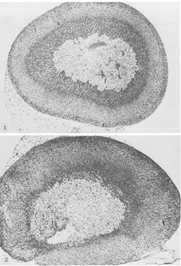
Adrenals.-The adrenal cortexes of SJLjJ mice had
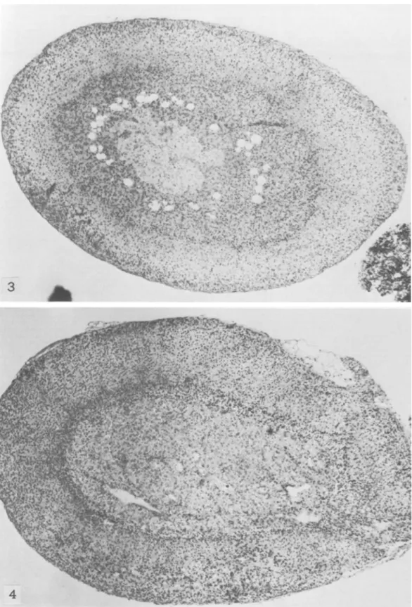
a remarkably smaller zona fasciculata and an ab-normally enlarged zona reticularis occupying most of the cortex (figs. 1, 2). What we call "zona retic-ularis" corresponds to the much discussed transitory or x-zone of the adrenal cortex of mice (18-22). We use this terminology only for the topographic de-scription of the histologic findings. This zona retic-ularis, which normally in other strains of mice can be seen until puberty in males and until the first preg-nancy in females, disappeared also in male SJL/J mice at puberty; it was maintained in virgin SJLjJ females for a long time and could be seen even at the age of 6-8 months (figs. 3-5), whether reticulum cell sarcomas were already present or not.
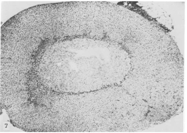
The overall picture indicated a hypofunction of the zona fasciculata, which is under hypophyseal control through ACT H. This zone is responsible for secretion of glucocorticosteroids such as corticosterone. The width of the zona fasciculata decreased even further with age in SJLjJ mice and was reduced to a narrow strip in 8- to 12-month-old animals (figs. 6, 7). Absence of the thymus in athymic nude mice has been recently shown to influence the size and duration of the zona fasciculata (16). However, neither neonatal thymectomy, gonadectomy, nor removal of both thymus and gonads at 30 days of age seemed to affect the formation, size, and duration of the zona reticularis or the size of the zona fasciculata in SJLjJ mice. This agrees with the suggestion that this reticular zone (or x-zone) in mouse adrenal cortex is directly controlled by gonadotropins (22).
Numerous large foci of extramedullary hemato-poiesis were observed in the adrenal cortexes of
SJLjJ mice until they were 10 days of age.
Ovaries. -The fertility of SJLj J females (expressed
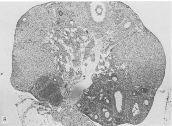
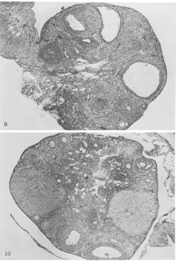
as average number of litters per female) was much more reduced than in other strains (4.5-9 litters; A. Meshorer, personal communication). The ovaries of SJLjJ mice maintained large numerous corpora lutea (figs. 8, 9). These abnormally large corpora lutea were visible until 7-8 months of age, when ovulation apparently ceased (fig. 10); sometimes they were still presen t in animals 1 year of age, with diffuse or localized RCNB (figs. 11, 12). The fallopian tubes and the other components of the ovaries of 7- to 8-month-old SJLjJ mice appeared sclerotic. Neonatal or adult thymectomy did not affect forma-tion, development, number, or size of corpora lutea in the ovaries of these mice.
Vessels.-Diffuse vasculitis was confined to the
small arterioles in almost all organs. The aorta did not seem to be involved. The alteration, thickening, and damage of the small arteries was remarkable. In view of the endocrine alterations in SJLjJ mice, the pathogenesis is being investigated.
ENDOCRINE DISORDERS IN SJL/J MICE 733
Hormone Levels
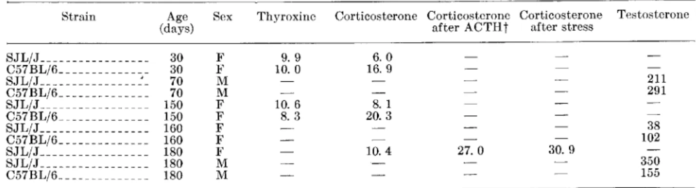
Thyroxine levels in SJLj J mice were normal, as compared to C57BLj6 mice. By contrast, levels of corticosterone in the blood of SJLjJ mice were a half to a third those in C57BLj6 mice of the same age and sex (table 1). The stress test as well as injection of ACTH induced approximately threefold increases in this parameter. The level of testosterone was normal or below normal in young male SJL/J mice, whereas it reached high levels in 6-month-old mice (table 1).
Estrous Cycle
Most female SJLjJ mice had a remarkably pro-longed diestrus, and therefore the whole cycle was lengthened (8-9 days instead of 4-5 days).
Electron M icroscopy
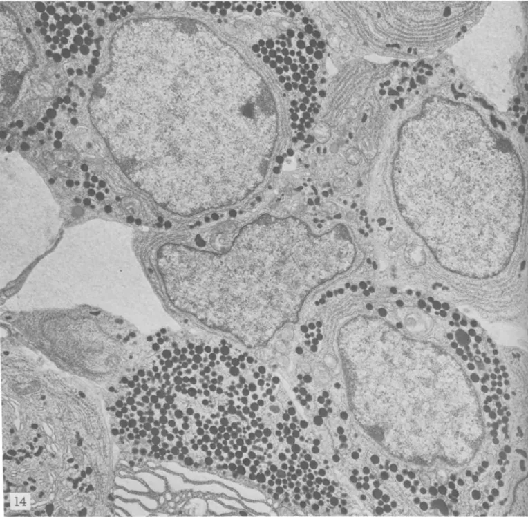
As confirmed by light microscopy, indirectly by the presence of gigantic long-lasting corpora lutea in the ovaries, and by the prolonged diestrus, the adeno-hypophyses of male and female SJLjJ mice showed massive infiltration with luteotropic cells [e-cells, according to the classification of Barnes (23)]. These cells occupied most of the gland, but especially the laterodistal portions, and increased in number with age. They constituted a majority and infiltrated all portions of the gland both in males and females. Other cell types were present, though the number of STH cells was greatly reduced in SJLjJ mice as compared to C57BLj6 and other strains (13, 24) (figs. 13, 14). Therefore, a massive hyperplasia of luteotropic cells was present in the anterior pituitaries of SJLjJ mice. Figures 13 and 14 show selected fields exemplifying the predominance of luteotropic cells in the adenohy-pophyses of SJLjJ mice.
DISCUSSION
This investigation has shown that, by various criteria, SJLjJ mice are endocrinologically abnormal. The cytology of the adenohypophysis deviates from that of other strains of mice in that it contains many gonadotropic cells
Cl
u teotropic cells, e-cells) (13), characterized by polymorphic granuli in the Golgi zone (figs. 13, 14). The abnormality is present at 2months of age and increases progressively with age. At 5 months, most cells of the anterior pituitary gland are luteotropic, but there is a sharp reduction in number of STH cells which, in other strains of mice of the same age and sex, normally constitute the major-ity in the same portions of the adenohypophysis. In parallel with the anticipated high production of gonadotropins, the ovaries of female SJLjJ mice contain numerous large corpora lutea that occupy most of the gland (figs. 8-12). They are maintained even in the atrophic glands of 10-l2-month-old mice, in which no oocytes and follicles are left and no ovu-lation occurs. From these data we infer that produc-tion of gonadotropins is abnormally and chronically high in SJLj J mice. As expected, the estrous cycle in adult female SJLjJ mice is abnormal and diestrus, which is maintained by luteotropic hormone and progesterone, is prolonged.
A collateral observation, which supports the con-cept of a pathologic and progressively increasing high input of gonadotropins in SJLjJ mice and thus exces-sive stimulation of testicular function, is that male SJLjJ mice cannot be housed in the same cage because their extreme aggressiveness causes continued fighting and death. In fact, the level of testosterone in sera of 2-month-old SJLjJ males is comparable to that in C57BLj6 males, but is much higher in 6-month-old mice (table 1). All long-term experiments with SJLjJ mice must therefore be done with females. The adrenal cortexes of SJLjJ mice are abnormal. The reticular zone is large and, in virgin females, does not show the normal involution, whereas the zona fasciculata is thin (figs. 1-7). The decreased serum corticosterone level in SJLj J mice may be due to an abnormally low level of corticosterone-binding globulin in these animals. However, the narrow width of the zona fascicula ta of the adrenal cortex is more suggestive of a decreased rate of corticosterone pro-duction. The normal increase in plasma corticosterone in response to exogenous ACTH excludes a primary adrenocortical lesion, such as a defect in corticosteroid biosynthesis. The normal response to the stress dem-onstrates that the pituitary gland can still secrete ACTH on demand and is evidence against a primary failure of the ACTH-producing pituitary cells. It seems more likely that a lowered baseline secretion of
TABLE I.-Levels of thyroxine, testosterone, and corticosterone in sera of SJL/J and C57BL/6 mice*
Strain Age Sex Thyroxine Corticosterone Corticosterone Corticosterone
(days) after ACTHt
SJL/J _________________ 30 F 9. 9 6. 0 C57BL/6- - - ___________ 30 F 10. 0 16. 9 SJL/J _______________ ~_ 70 M C57BL/6 ______________ 70 M SJL/J _________________ 150 F 10.6 8.1 C57BL/6 ______________ 150 F 8.3 20. 3 SJL/J _________________ 160 F C57BL/6 ______________ 160 F SJL/J _________________ 180 F 10. 4 27.0 SJL/J _________________ 180 M C57BL/6 ______________ 180 M
*Values of thyroxine and corticosterone are expressed as I-Ig/IOO ml serum; values of testosterone, as ng/lOO ml serum. tThe mice Were bled 1 hour after ip injection of 10 I-Ig ACTH.
after stress 30. 9 Testosterone 211 291 38 102 350 155
734 PIERPAOLI ET AL. corticosterone might be due to a functional
disturb-ance of the hypothalamus.
Hormones profoundly affect all steps in the onto-genic maturation and proliferation of thymolymphatic and hematopoietic tissues (24, 25). Thus any change in hormone environment will be reflected in variations in the cell dynamics. Cells of the thymolymphatic, hematopoietic, and reticular tissues are highly sensi-tive targets of hormones during ontogeny. In SJL/J mice, a specific congenital tendency to increased production of gonadotropins probably affects both gonadal and adrenal functions. The consequences of the hormone imbalance in SJL/J mice is most dramat-ically expressed in the cytology, development, and alteration of the thymolymphatic, reticular, and hematopoietic tissues, though direct proof must still be provided. In SJL/J mice, cells of the thymus, lymph nodes, and bone marrow and reticulum cells are all affected, but certainly the cell type that is involved most and responds with massive proliferation in this SJL/J-specific hormone imbalance is the reticulum cell. An indication of the far-reaching implications of this relationship is that oral treatment of SJL/J mice with dimethyl benzanthracene (DMBA), which destroys oocytes in the ovaries (26) and pro-vokes a deep and obvious change in the hormone environment, results in early appearance of lympho-sarcoma in the thymus rather than reticulum cell sarcoma in the lymph nodes (3).
Hellmann and Fowler (27) and Fowler et al. (28) found that estrogens activate the expression of antigens of murine C-type virus (which has been identified as the cause of leukemia in mice) and promote synthesis of RNA-directed DNA polymerase. The obligatory role of hormones for replication of tumor virus has also been recognized recently in mammary cancer of mice (29, 30) . Therefore, the links to consider now are those between hormones, cell types, viruses, and en-zymes. For example, removal of the thymus, a sensi-tive target for hormones (31), prevents appearance of leukemia induced by X-irradiation (32) or carcino-gens (33). Clearly the thymus is, in that specific instance, species, and circumstance, the organ in which the fortuitous combination of this organ, cell, hormone and virus factors are operative. Change in one of these conditions-as thymectomy or, as in
SJLj J mice, the hormone imbalance-will either prevent or delay onset of leukemia. In this strain thymectomy does not prevent onset of DMBA-induced
lymphatic leukemia (3).
The similarity between type-B reticulum cell sarcoma (Hodgkin's-like disease in mice) and Hodg-kin's disease in humans is still doubtful (34); however, the hormone conditions in preleukemic and leukemic states and in Hodgkin's disease have been almost completely neglected and warrant some consideration. Certain· pre-existent congenital, subpathologic, or paraphysiologic hormone imbalances in families with high incidence of certain neoplasms might affect onset of the tumors.
'Whelher or not systemic neoplasms in humans can be shown to have a viral causation, clearly viruses need certain hormones to express themselves and their
carcinogenic properties. For example, the notion that estrogens "promote" leukemia in mice should now be recast as a possible estrogen-dependent expression or promotion and stimula tion of replication of ubiquitous viruses through increased synthesis of RNA-dependent DNA polymerase, which has a high concentration in the thymus. In this situation the quantitative aspect is clear because removal of the thymus, which represents the privileged target for hormones and viruses, effectively prevents the onset of leukemia.
We are now attempting to induce, change, or "modulate" the onset, pattern, and development of leukemias and RCNB in SJLjJ and other strains of mice by manipulation of their hormone environment. REFERENCES
(1) DUNN TB: Normal and pathologic anatomy of the reticular tissue in laboratory mice, with a classification and discussion of neoplasms. J Natl Cancer Inst 14: 1281-1433, 1954
(2) MURPHY ED: SJL/J, a new inbred strain of mouse with a high, early incidence of reticulum-cell neoplasms. Proc Am Assoc Cancer Res 4: 46, 1963
(3) HARAN-GHERA N, KOTLER M, MESHORER A: Studies on leukemia development in the SJL/J strain of mice. J Natl Cancer Inst 39:653-661, 1967
(4) DUNN TB, DERlNGER MK: Reticulum cell neoplasm, type B, or the "Hodgkin's-like lesion" of the mouse. J Natl Cancer Inst 40:771-821, 1968
(5) SIGLER R, RICH MA: Pathogenesis of reticulum cell sarcoma in mice. J Natl Cancer Inst 41: 125-143, 1968 (6) DETKAczEvsKI LZ, W ANEBO HJ: Electron microscopy of murine SJLIJ disease. In t J Cancer 4: 533-547, 1969 (7) EAST J: Immunopathology and neoplasms in New Zealand Black (NZB) and SJL/J mice. Prog Exp Tumor Res 13:84-134, 1970
(8) TUCKER MJ, BAKER SB DE C: Diseases of specific pathogen-free mice. In Pathology of Laboratory Rats and Mice. Oxford, Blackwell, 1967, p 799
(9) MESHORER A, HARAN-GHERA N: Unpublished observa-tions
(10) HARAN-GHERA N, BEN-YAAKov M, PELED A, et al: The immune status of SJL/J mice in relation to age and spontaneous tumor development. J Natl Cancer Inst 50: 1227-1235, 1973
(11) ARNEsEN K: Constitutional difference in lipid content of adrenals in two strains of mice and their hybrids. Acta Endocrinol (Kbh) 18:396-401, 1955
(12) LE VINE S, TRElMAN DM: Differential plasma corti-costerone response to stress in four inbred strains of mice. Endocrinology 75: 142-144, 1964
(13) BlANCHI E, PlERPAOLI W, SORKlN E: Cytological changes in the mouse anterior pituitary after neonatal thymec-tomy: A light and electron microscopical study. J Endocrinol 51: 1-6, 1971
(14) KLIMAN B, PETERSON RE: Double isotope derivative assay of aldosterone in biological extracts. J BioI Chem 235:1639-1648, 1960
(15) :MUELLER J: Aldosterone stimulation in vitro. I. Evalu-ation of assay procedure and determinEvalu-ation of aldo-sterone-stimulating activity in a human urine extract. Acta Endocrinol (Kbh) 48 :283-296, 1965
(16) PlERPAOLI W, SORKlN E: Alterations of adrenal cortex and thyroid in mice with congenital absence of the thymus. Nature [New BioI] 238:282-285, 1972 (17) BEsEDOVSKY HO, SORKIN E: Thymus involvement
in female sexual maturation. Nature (Lond) 249 :356-358, 1974
(18) HOWARD-MlLLER E: A transitory zone in the adrenal cortex which shows age and sex relationships. Am J Anat40:25l-293,1927
(19) W ARING H: The development of the adrenal gland of the mouse. QJ Microsc Sci 78 :329-366, 1935
ENDOCRINE DISORDERS IN SJL/J MICE
735
(20)
(21)
(22)
MOOG F, JACKSON BENNETT C, DEAN CM JR: Growth and cytochemistry of the adrenal gland of the mouse from birth to maturity. Anat Rec 120:873-891, 1954 HOWARD E, l\1IGEON CJ: Sex hormone secretion by the
adrenal cortex. In Handbuch der Experimentellen Pharmakologie, part 1. Berlin, Springer-Verlag, 1962, pp 593-597
DUNN TB: Normal and pathologic anatomy of the adrenal gland of the mouse, including neoplasms. J Natl Cancer Inst44:1323-1389, 1970
(23) BARNES GB: Electron microscope studies on the secretory cytology of the mouse anterior pi tui tary. Endocrinology 71 :618-628, 1962
(24)
(25)
PIERPAOLI W, BIANCHI E, SORKIN E: Hormones and the immunological capacity. V. Modification of growth hormone-producing cells in the adenohypophysis of neonatally thymectomized germ-free mice: An electron microscopical study. Clin Exp Immunol 9:889-901, 1971
PIERPAOLI W, FABRIS N, SORKIN E: Developmental hormones and immunological maturation. In Hor-mones and the Immune Response. Ciba Foundation Study Group No. 36. London, Churchill, 1970, pp 126-143
(26) KRARUP T: Effect of 9,1O-dimethyl-l,2-benzanthracene on the mouse ovary. Acta Endocrinol (Kbh) 64:489-507, 1970
(27) HELLMANN A, FOWLER AK: Hormone-activated expres-sion of the C-type RNA tumour virus genome. Nature [New BioI] 233:142-144,1971
(28) FOWLER AK, REED CD, TODARO GJ: et al: Activation of C-type RN A virus markers in mouse uterine tissue. Proc Natl Acad Sci USA 69:2254-2257, 1972
(29) MCGRATH CM: Replication of mammary tumor virus in tumor cell cultures: Dependence on hormone-induced cellular organization. J Natl Cancer Inst 47: 455-467, 1971
(30) MCGRATH CM, NAND! S, YOUNG L: Relationship between organization of mammary tumors and the ability of tumor cells to replicate mammary tumor virus and to recognize growth-inhibitory contact signals in vitro. J ViroI9:367-376, 1972.
(31) PIERPAOLI W. SORKIN E: Hormones, thymus and lym-phocyte functions. Experientia 28: 1385-1389, 1972 (32) KAPLAN HS: Influence of thymectomy, splenectomy, and gonadectomy on incidence of radiation-induced lymph-oid tumors in strain C57BL mice. J Natl Cancer Inst 11 :83-90, 1950
(33) LAw LW, MILLER JH: The influence of thymectomy on the incidence of carcinogen-induced leukemia in strain DBA mice. J Natl Cancer Inst 11 :425-438, 1950 (34) McINTIRE KR, LAw LW: Abnormal serum
immuno-globulins occurring with reticular neoplasms in an inbred strain of mouse. J Natl Cancer Inst 39: 1197-1211, 1967
Figures 1-7.-Adrenal glands of virgin female mice. Hematoxylin-eosin-phosphomolybdic acid-light green.
FIGURE l.-SJL/J mouse 2 months old. Note broad zona reticularis and reduced width of zona fasciculata. X 36
FIGURE 2.-C.57BL/6 mouse 2 months old. Note broad zona fasciculata and absence of zona reticularis. X 40
3
~AFIGURE 3.-SJL/J mouse 5 months old. Note persistence of zona reticularis. X 40
FIGURE 4.-C57BL/6 mouse 5 months old. Note absence ofzona reticularis. X 36
FIGURE 5,-S]L/] mouse 7 months old. Note persistence of zona reticularis. X 32
FIGURE 6.-S]L/] mouse 8 months old. Note remarkably reduced width of zona fasciculata. X 36
FIGURE 7.-C57BL/6 mouse 8 months old. Note normal width of zona fasciculata. X 40
Figures 8-12.-0varies of virgin female mice. Hematoxylin-eosin-phosphomolybdic acid-light green.
FIGURE 8.-SJL/J mouse 5 months old. Note large corpora lutea and follicles at different stages of maturation. X 28
FIGURE 9.--C57BL/6 mouse 5 months old. X 32
FIGURE lO.-SJL/J mouse 8 months old. Note large corpora lutea and initial atrophy of residual part of gland. X 28
FIGURE 11.-SJL/J mouse 9 months old, bearing large reticulum cell neoplasm. Note large corpo'ra lutea and initial atrophy of residual parts of gland. X 28
FIGURE 12.-SJL/J mouse 12 months old. Note persistence of large corpus luteum and advanced stage of sclerosis of residual parts of gland. X 32
Figures 13, 14.-ldentically oriented and located sections of the anterior pituitary glands of S-month-old virgin
SJL/
J
and CS7BL/6 Females. Gluteraldehyde-osmium. X 10,000FIGURE 13.-SJL/J mouse. The field is completely occupied hy gonadotropin-produciilg cells containing typical polymorphic hormone granuli (luteotropic hormone-producing cells).
FIGURE 14.--C57BL/6 mouse. Note prevalence of growth hormone-secreting cells containing typical round hormone granuli in cyto-plasm.
![FIGURE 5,-S]L/] mouse 7 months old. Note persistence of zona reticularis. X 32](https://thumb-eu.123doks.com/thumbv2/123doknet/14903910.655046/8.833.125.713.68.927/figure-mouse-months-old-note-persistence-zona-reticularis.webp)