TNF inhibits malaria hepatic stages in vitro
via synthesis of IL-6
Andreas Nussler, Sylviane Pied, Joseph Goma, Laurent Renia,
Francois Miltgen
1, Georges E. Grau
2, and Dominique Mazier
INSERM U313, Groupe Pitie-Salpetriere, 911 Bd de l'H6prtal, 75013 Paris, France 'Museum National d'Histoire Naturelle, 61, rue Button, 75006 Paris, France ZWHO-IRTC, Dept of Pathology, University of Geneva, Switzerland
Key words: Cytokines, effector mechanism, hepatocytes, infection, liver, pre-erythrocytic stages,
non-parenchymal cells
Abstract
We examined the capacity of murlne recomblnant tumor necrosis factor (rmTNF) to induce an inhibitory effect at the hepatic stage on malaria induced by Plasmodium yoelii sporozoltes. When injected three times, 1.0 /ig of rmTNF was found to protect 78% of mice against a sporozolte challenge. In contrast, whatever the dose and the schedule of administration, no inhibition was observed when purified hepatocyte cultures were infected with P. yoelii. The addition of non-parenchymal hepatic cells to hepatocyte cultures restored the capacity of TNF to modulate hepatic stage development, leading to up to 44% Inhibition. Antibodies to Interleukin 6 reversed the anti-parasite activity in the co-culture system.
Introduction
Tumor necrosis factor alpha/cachectin (TNF), initially described for its ability to induce hemorrhagic necrosis in vivo (1,2), is a cytokine with various activities, including striking viral (3), anti-bacterial (4) and anti-parasitic effects (5). In malaria, it has been shown that TNF administration in vivo protects mice challenged with parasitized erythrccytes of P. chabaudi (6) and reduces the development of hepatic forms of P. berghei (7). Although a previous report indicates that TNF reduced development of
P. berghei in a Hep. G2 hepatoma cell line (7), preliminary data
obtained in our laboratory using fresh hepatocytes did not confirm this effect. The aim of the present study was to understand the reasons for such discrepancies and to clarify the role of TNF, at the hepatic level, in sporozoite induced malaria.
Methods Animals
Three-month-old BALB/c, C3H/HeJ, and C57B1/6 mice were purchased from Charles River Breeding Laboratories, Saint-Aubin les Elbeuf, France.
Cytokines and anti-cytokine antibodies
Murine recombinant TNF alpha (rmTNF) was a kind gift from Dr B. Allet, Glaxo 1MB, Garouge, Switzerland. Rabbit anti-mouse TNF IgG was prepared as previously described (8). 6B4, a rat
lgG1 anti-mouse interleukin 6 (IL-6) (9) and murine recombinant IL-6 (rmlL-6) were a gift from Dr J. Van Snick, Ludwig Institute, Brussels, Belgium.
Cytokine assays
Supernatants from hepatocyte cultures were assayed for IL-6 in a bioassay using the 7TD1 cell line as described (10). Secretion of IFN-gamma in supernatants after stimulation with rmTNF was measured with an enzyme-linked immunoassay (ELISA) using a murine monoclonal antibody to IFN-gamma (11). Interleukin 1 (IL-1) was assayed by the standard thymocyte co-mitogen assay as previously described (12,13).
Parasites
Sporozoites of the 17X strain of Plasmodium yoelii yoelii were obtained from infected salivary glands of Anopheles stephensi mosquitoes, 16 to 21 days after an infective mouse blood meal. After aseptic dissection, salivary glands were homogenized in a glass grinder and diluted in culture medium or sterile phosphate-buffered saline (PBS) (14).
In vivo experiments
Balb/c mice were divided into four groups. In the first group, each mouse received 1.0 /tg of rmTNF intravenously (i-v)- After 24 h, each animal was injected with 3500 P. yoelii sporozoites i.v. Each mouse in group 2 received 0.5 /ig rmTNF i.v. 24 h before, during,
Correspondence to: D. Mazier
and 24 h after sporozoite inoculation. The third group was treated as the second group but the rmTNF dose was 1.0 /xg. In the control group rmTNF was replaced by 0.2 ml of PBS and the regimen was as for group 2. Blood smears, taken daily from the third to the seventeenth day after sporozoite inoculation, were stained with Giemsa and examined for erythrocytic stages of
P. yoeiii.
Culture of hepatic stages of malaria parasites
Monoculture of hepatocytes. Rodent hepatocyte monolayers
(BALB/c, C3H/HeJ, C57BL/6) were isolated by collagenase perfusion of liver fragments as previously described (15). Briefly, 60 000 cells were cultured in eight-chamber plastic Lab-Teck slides (Miles Lab. Inc., USA) in minimal essential medium supplemented with 10% fetal bovine serum and incubated with 5% CO2 at 37°C for 24 h before use in experiments.
Co-culture of hepatocytes and non-parenchymal cells (NPC).
NPC were obtained as already described (16). Briefly, livers from mice (BALB/c, C57BL/6) were perfused with HEPES buffer followed by perfusion with 0.05% collagenase (Collagenase H, Boehringer Mannheim, Germany). Then the distended and blanched liver was teased and suspended in 30 ml collagenase solution and maintained for 45 min at 37°C under magnetic agitation. The suspension was then centrifuged at 300 g for 10 min, the supernatant discarded, and the pellet resuspended in 5 ml HEPES. The liver cell suspension was overlaid with 7 ml 30% w/v metrizamide (Nicomed, Oslo, Norway), dissolved in HEPES without NaCI. The liver cell metrizamide gradient was centrifuged at 1400 g for 20 min. NPC were collected from the top layer, washed twice in HEPES and resuspended in tissue-culture medium. Sixty thousand viable NPC were added to primary hepatocyte cultures 3 h after the culture was set up (ratio 1:1).
Treatment of monocultures and co-cultures. Both culture types
were incubated with 0.5,1.0 or 2.5 ^g/ml rmTNF. Control cultures were maintained without rmTNF. Forty thousand P. yoeiii sporozoites were added 24 h after rmTNF addition. Three and 24 h after sporozoite inoculation culture supernatants were taken for cytokine assays and replaced either with fresh medium or with medium containing rmTNF. Forty-eight hours after sporozoite inoculation, supernatants were taken again for cytokine assays and cultures were stopped and stained with Giemsa to find schizonts. In addition, other cultures of both types were incubated with 10 ILJ/ml rmlL-6 for 24 h before sporozoites were inoculated; these cultures were stopped 48 h later.
Neutralization of IL-6 by an anti-IL-6 mAb. Using the same
protocol as described in the previous paragraph, anti-murine IL-6 mAB (1 /100) was added to cultures either at the time of cytokine incubation or 30 minutes before sporozoite inoculation. The cultures were then maintained as described above.
Results
In vivo experiments
Injection of 1.0 ng rmTNF the day before, at the time of, and the day after inoculation of 3500 sporozoites prevented occurrence of parasitemia in 78% of the mice. This was a consistent finding,
Table 1. Effect of in vivo TNF administration on P. yoeiii
sporozoite infection in BALB/c mice
Treatment rmTNF (/jg/mouse) - 2 4 h 0.5 1 1 0 + 3 h 0.5 1 0 + 24 h 0.5 1 0 Protection (number of mice) 0/5 1/10 16/20 2/20 Delay8 1 day ±0.8 4 days ±1.5 5 days±1 0
aDe)ay represents the specific mean±SE in the appearance of parasitemia in non-protected mice, compared to controls injected with PBS.
Effect of TNF in the presence of NPC
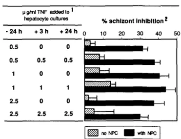
V g/ml TNF added to '
hepatocyte cultures %8Chlzont Inhibition
2 • 2 4 h 0.5 0.5 1 1 2.5 2.5 + 3h 0 0.5 0 1 0 2.5 + 2 4 h0 10 20 30 40 51
0 5 ^ M ^ ^ ^ ^ ^ ^ ^ ^ _ _ ,
0 ^ ^ ^ ^ ^ ^ ^ ^ ^ ^ ^ ~0 tt^^^H^B-H
2.5 ^ ^ ^ ^ ^ ^ ^ ^ M . no NPC wtti NPCFig. 1. Effect of TNF in the presence of NPC. 'rmTNF was added at
different times (indicated in hours) after sporozoite inoculation. Percentage inhibition was estimated by counting numbers of 48-h schizonts in cultures with or without rmTNF. Results are expressed as means±SEM of six individual cultures. The controls were either hepatocytes without NPC (Schizont number: 65 ±3) or hepatocyte with NPC (Schizont number: 70 ±6).
mice being aparasitaemic 17 days after infection. In the remaining 20% of treated but non-protected mice, the rise in parasitemia was delayed by a mean of 5 days compared to PBS treated mice. A single injection of rmTNF (1.0 ^g/mouse) 24 h before sporozoite inoculation was much less protective (Table 1). A lower dose of rmTNF (0.5 ^g/mouse), even when administered three times (the day before, on the day of, and 24 h after sporozoite inoculation) had no protective effect.
In vitro experiments
Effects of TNF on parasite development in vitro. Using BALB/c
hepatocyte monocultures (Fig. 1), no significant parasite inhibi-tion could be observed when cultures were incubated with 0.5 /ig/ml rmTNF 24 h before sporozoite inoculation. This effect was not improved by repeating the TNF administration (24 h before, during, and 24 h after infection), nor by increasing the dose to 2.5 /tg/ml. Similar results were obtained with primary cultures from
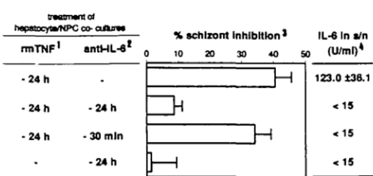
HHiU>*.iune>Cco-aAnm— % schizont Inhibition » IL-6 In «/n nnTNF1 antHL-6l 0 10 20 30 40 SO (U/Ull) - 2 4 h - 2 4 h - 2 4 h -. - 2 4 h -30 min - 2 4 h It r*i
H
H
H
123.0 t38.1effects (36±5.6% inhibition; Fig. 2), despite the fact that IL-6 remained undetectafcde after 3 h. Neither IFN-7 nor IL-1 could be detected in primary or in co-culture supernatants at 3,24 and 48 h after sporozoite inoculation, incubated or not with rmTNF.
Effects of murine recombinant IL-6 and its neutralization by an anti-IL-6 mAb. Incubation of rmlL-6 inhibited parasite
develop-ment up to 42 ±3.3% in primary cultures, while 86 ± 7.0% inhibi-tion was reached in co-cultures. With an anti-murine IL-6 mAb (1/100) nearly 90% of the rmlL-6 (10 Ill/ml) induced inhibition could be neutralized in both primary and co-cultures (Table 2).
Fig. 2. IL-6 levels in culture supernalants. 11.0 /ig/ml, final
concentra-tion 21:100, final dilution. 3Results of six individual cultures, expressed
as means ±SEM, compared to infected hepatocyte cultures without TNF. The schizont number in untreated control cultures was 86 ± 7. 4 Assayed
3 h after sporozoite inoculation.
Table 2. Effects of in vitro IL-6 incubation on the pre-erythrocytic
parasite development of P. yodii in Balb/c hepatocyte cultures, in the presence and absence of non-parenchymal cells (NPC)
Treatment Inhibition^ rmlL-68 - 2 4 h - 2 4 h - 2 4 h -anti-IL-6b _ - 2 4 h - 3 0 min - 2 4 h - 3 0 mm no NPC 4 2 2 * 3 3 5.2±4 5 37.6±6.4 4 . 0 ± 3 8 3.2±4.0 with NPC 86.5 ±7.0 7.3±4.0 79.0±7.5 2.5±6.5 6 . 8 ± 2 0
a10 Ill/ml, final concentration. b1/100, final dilution
cPercentage inhibition of schizont development of six individual
cultures; expressed as means±SEM, compared to infected non-treated hepatocyte cultures. The schizont number in control cultures was without NPC 75±4, and with NPC 69 ±5.
C3H/HeJ and C57/BL6 mice (data not shown). The addition of NPC to hepatocyte cultures (ratio 1:1), in the presence of 0.5
fig TNF resulted in 3 1 % schizont inhibition (Fig. 1). The degree
of inhibition was not improved by repeating the addition of TNF (24 h before, during, and 24 h after inoculation) nor by increasing the dose to 2.5 /xg/ml. Identical results were obtained using co-cultures from C57/BL6 and C3H/HeJ mice (data not shown). However, incubation of lower doses of rmTNF were less effec-tive than those shown in Fig. 1 (data not shown).
Effects of TNF-induced IL-6 secretion and its neutralization in culture supernatants. Purified munne hepatocytes were cultured
alone or in the presence of NPC (co-culture). Both culture types were incubated with 1.0 nglm\ rmTNF 24 h before sporozoite inoculation and IL-6 levels were measured in supernatants (Fig. 2). It was found that IL-6 levels were high in co-cultures 3 h after sporozoite inoculation (mean: 123 U/ml, Fig. 2) and rose to a mean of 490 U/ml after 48 h, whereas IL-6 was not detectable in monocultures. Addition of anti-IL-6 mAb to co-cultures at the time of rmTNF incubation virtually abolished the inhibitory effect induced by TNF. In contrast, the addition of anti-IL-6 mAb 30 min before sporozoite inoculation did not modify the rmTNF
Discussion
The in vitro studies presented here were performed in order to define the site and mode of action of TNF. It is shown that when primary hepatocyte cultures are used instead of tumor cell lines, TNF fails to induce a protective effect, whatever the dose and the schedule of administration and whatever the strain of mice. These results are in contrast to a recent study, where TNF was found to inhibit development of P. berghei in a Hep. G2 cell line (7). We have shown that acute-phase proteins (APPs) secreted after cytokine stimulation (17 -19) inhibit parasite development
in vitro and in vivo (20 - 22). Therefore, the fact that TNF induces
different APPs in tumor cell lines and in normal rat hepatocytes (23,24) might explain the differences observed in these two experimental models. Since a TNF dose of 1 ^g/mouse clearly protects in vivo without inducing necrosis (25) but has no effect on hepatocytes in vitro, we questioned whether an in vitro model containing only hepatocytes was valid. We found in these experiments that the addition of NPC to hepatocytes restored the capacity of TNF to modulate parasite hepatic stage develop-ment (Fig. 1). In our co-culture assay, NPC essentially consist of pit cells (large granular lymphocytes), Kupffer cells, T-cells, natural killer cells, endothelial cells from sinusoids, epithelial cells, fibroblasts and Ito cells. Some of these cells are known to produce one or several of the following cytokines: IFN-7, IL-1, and IL-6 (26-28), all of which are known to interfere with the hepatic stages (29-32). We recently demonstrated that IL-6 induces various effector mechanisms including oxygen radicals (31) and nitrites (32).
In these co-cultures, markedly elevated IL-6 concentrations were found upon addition of TNF, whereas IL-1 and IFN-7 remained undetectable. To confirm that IL-6 was directly involved in the TNF-induced parasite inhibition, anti-IL-6 mAb was added to mono- and co-cultures of hepatocytes. Anti-IL-6 mAb dramatically decreased the TNF-induced inhibition when added 24 h before sporozoite inoculation. The preservation of TNF effects when addition of anti-IL-6 mAb was delayed until 30 min before sporozoite inoculation showed that the release of IL-6 and the subsequent modification inside the hepatocytes takes place in the very early phase of culture after TNF introduction. This is consistent with the observation of high IL-6 concentrations in 24 and 48 h culture supernatants (data not shown), even when initial IL-6 production had been neutralized by the addition of anti-IL-6 mAb. The specific inhibitory effect of IL-6 on pre-erythrocytic stages of parasite development was furthermore demonstrated by incubating hepatocyte cultures directly with rmlL-6 (Table 2). These results strongly suggest that IL-6 is a crucial mediator of the observed TNF effect. The higher inhibitory effect of IL-6 on
the parasite development observed in the presence of NPC suggests a stimulation in NPC of additional factors such as yet to be identified cytokines or substances that act synergistically with IL-6 (33).
TNF has also been demonstrated to be involved in L-arginine-dependent cytotoxic effector mechanisms in macrophages (34) which can have parasiticidal effects (32,35,36).
In malaria the production of various cytokines, and particularly of TNF, is increased during the blood stage (37,38), and these cytokines might modulate infection by liver stages. Furthermore, it has been shown that cytotoxic T cells are involved in the protec-tion induced by immunizaprotec-tion with irradiated sporozoites (39,40). We recently demonstrated that immunization with peptide corresponding to the non-repetitive part of the circumsporozoite of P. yodii, induced CD4+ and CD8+ T cells effective in the destruction of cultured hepatic schizonts (41; Renia et al., in preparation). Moreover, these T cells are known to produce cytokines which include TNF and IL-6 (42). Taken together, these data further illustrate the interdependence of different stages of development in malaria parasites.
Acknowledgements
This research was supported in part by the United Nations Development Program-World Bank-Health Organisation Special Program on training and research in Tropical Diseases, the Commission des Communautes Europeennes, the Fondatons Raoul Foflereau and the Conseil Scientifique Pitie-Salpetriere. We wish to thank Mr Bertho for assistance with IL-1 assays, Dr Draper and Miss N. Uwechue for reviewing the manuscript, and Prof. G Target for critical discussion. Furthermore we would like to thank Dr van Snick for providing the recombinant murine IL-6 and the monoclonal antibody 6B4. Abbreviations APP IFN-7 IL-1 IL-6 mAb NPC rmTNF References acute-phase protein interferon gamma interteukin 1 interteukin 6 monoclonal antibody non-parenchymal cell munne recombinant TNF
1 Carswell, E. A., Old, L. J., Kassel, R. L., Green, S., Fwre, N., and Williamson, B. 1975. An endotoxin-induced serum factor that causes necrosis of tumors. Proc. Natl Acad. Set. USA 720:3666 2 Tracey, K. J , Vlassara, H., and Cerami, A. 1989. Cachectin/tumor
necrosis factor. Lancet 1:1122.
3 Ito, M. and O'Malley, J. A. 1987. Antiviral effects of recombinant human tumor necrosis factor. Lymphokine Res. 6:309.
4 Havell, E. A. 1989. Evidence that tumor necrosis factor has an impor-tant role in antibacterial resistance. J. Immunol. 143.2894. 5 Trtus, R. G., Sherry, B., and Cerami, A. 1989. Tumor necrosis factor
plays a protective role in experimental murine cutaneous letshmariasis. J. Exp. Med. 170:2097.
6 Clark, I. A., Hunt, N. H., Butcher, G. A., and Cowden, W. B. 1987. Inhibiton of munne malaria (Plasmodium chabaudfjin vivo by recom-binant interferon gamma or tumor necrosis factor, and its enhance-ment by butylated hydroxyanisole. J. Immunol. 139:3493. 7 Schofiekj, L, Nussenzweig, R. S., and Nussenzwetg, V. 1988. CD8+
T-cells and gamma-interferon required for immunity to sporozoite challenge. Report of the tenth meeting of the scientific working group on the immunology of Malaria, p. 17.
8 Grau, G. E. and Gretener, D. 1987. Prevention of murine cerebral
malaria by low-dose cyclosponn A. Immunology 61 521 9 Vink, A., Coulie, P., Wauters, P., Nordan, R P , and Van Snick, J.
1988. Bcell growth and differentiation activity of interieukin-HP1 and related murine plasmacytoma growth factor. Synergy with mterieukin 1. Eur. J. Immunol. 18:607.
10 Van Snick, J., Cayphas, S., vink, A., Uyttenhove, C , Coulie, P. G., Rubira, M., and Simpson, R. J. 1986. Purification and NH2-terminal
amino add sequence of a T-cell derived lymphokine with growth factor activity for B-cell hybridomas. Proc. Natl Acad. Sci. USA 83:9679. 11 Slade, S. J. and Langhome, J 1989. Production of interferon gamma during infection of mice with Plasmodium chabaudi chabaudi. Immunobiology 179.353.
12 Mizel, S. B., Oppenheimer, J. J., and Rosenstreich, D. L. 1978. Characterisation of lymphocyte-activating factor (LAF) produced by the macrophage cell line P388D. I. Enhancement of LAF production by activated T lymphocytes J Immunol 1201487.
13 Koopman, W. J., Farrar, J. J., and Fuller-Bonar, J 1978. Evidence for identification of LAF as the adherent cell-derived mediator respon-sible for enhanced antibody synthesis by nude mouse spleen cells. Ceil Immunol. 35.92.
14 Mazier, D., Landau, I., Miltgen.F , Druilhe, P., Lambiotte, M., Baccam, D., and Gentilini, M. 1982. Infestation in vitro d'hepatocytes de Thamnomys adulte par des sporozoites de Plasmodium yoeii'v. schizogonie et liberation de merozoites infestants. C. R. Acad. Sci. Paris 294:963.
15 Mazier, D , Mellouk, S., Beaudoin, R. L , Texier, B., Druilhe, P., Hockmeyer, W., Trosper, P , Paul, C , Charoenvrt, Y., Young, J., Miltgen, F., Chedid, L, Chigot, J. P., Galley, B , Brandicourt, O , and Gentilini, M. 1986. Effect of antibodies to recombinant and synthetic peptides on Plasmodium falaparum sporozoites in vitro Science 231:156.
16 Kirn, A., Steffan, A. M., Bmgen, A., Cinqualbre, J., and Gendrault, J. L. 1980. Isolement et culture de cellules de Kupffer humaines. C. R. Acad. Sci. Paris 291:249.
17 Ramadori, G., Sipe, J. D., Dinarello, G. A., Onzel, S. B. B., andColten, H. R. 1985 Pretranslational modulation of acute phase hepatic protein synthesis by murine recombinant mterieukin 1 (IL-1) and purified IL-1. J. Exp Med. 162.930.
18 Mortensen, R F., Shapiro, J., Lin, B. F., Douches, S , and Neta, R. 1988 Interaction of recombinant IL-1 and recombinant tumor necrosis factor in the induction of mouse acute phase proteins. J. Immunol. 140:2260.
19 Jablons, D. M., Mule, J. J., Mclntosh, J. K , Sehgal, P. B., May, L T , Huang, C. M., Rosenberg, S. A., and Lotze, M. 1989. IL-6/IFN-beta-2 as a circulating hormone. Induction by cytokine administra-tion in humans. J. Immunol 142:1542.
20 Mazier, D , Miltgen, F., Nudelman, S., Nussler, A., Renia, L., Pied, S., Goma, J., and Gentilini, M. 1988. Pre-erythocytic stages of plasmodia Role of specific and non-specific factors. Bid. Cell 64:165. 21 Pied, S., Nussler, A , Pontet, M., Miltgen, F., Matile, H., Lambert, P. H., and Mazier, D. 1989. C-reactive protein protects against pre-erythrocytic stages of malaria Infect Immun. 57.278.
22 Nussler, A., Pied, S., Pontet, M., Miltgen, F., Renia, L., Gentilini, M., and Mazier, D. 0000. Inflammatory status and pre-erythocytic stages of malaria. Role of the C-reactive protein. Exp. Parasitol. 000:000. 23 Andus, T , Heinrich, P. C , Bauer, J., Tran-Thi, T., Decker, K., Mannel, D., and Northoff, H. 1987. Discrimination of hepatocyte-stimulating activity from human recombinant tumor necrosis factor alpha. Eur J. Immunol. 17:1193.
24 Perfmutter, D. H., Dinarello, C A , Punsel, P. I., and Colten, H. R. 1986. Cachectin/tumor necrosis factor regulates hepatic acute-phase gene expression. J. Clin. Invest. 78:1378.
25 Piguet, P. F., Grau, G. E., andVasselli, P. 1990. Subcutaneous perfu-sion of tumor necrosis factor induced local proliferation of fibroblasts, capillaries, and epidermal celts, or massive tissue necrosis. Am. J. Pathol. 136:103.
26 Lotze, M., Jink, F., Kabouridis, P., Tsoukas, C , Hirano, T , Kishimoto, T., and Carson, D. A. 1988. B cell stimulating factor 2/interieukin 6 is a costimulant for human thymocytes and T lymphocytes. J. Exp. Med. 167:1253.
27 Katz, Y and Strunk, R. C. 1989. Simaarities and differences in stimula-tion of expression of alternative pathway of complement and IFN-beta2/lL-6 genes in human fibroblasts. J. Immunol. 142:3862.
28 Konase, M., Hendriksen-DeStefano, D., May, L., Vfcek, J., and Sehgal, P. B. 1986. Induction of beta-2-interferon by tumor necrosis factor: a homeostatic mechanism in the control of cell proliferation. Cell 45659.
29 Mellouk, S., Maheshwan, R. K., Rhodes-Feuillette, A., Beaudom, R. L, Berbiguier, N., Matile, H., MMgen, F., Landau, I., Pied, S., Chigot, J. P., Friedman, R. M , and Mazier, D. 1987. Inhibitory activity of interferons and interieukin 1 on the development of Plasmodkim falciparum in human hepatocyte cultures. J. Immunol. 139:4192. 30 Ferreira, A., Schofield, L, Enea, V., Schellekens, H , Van Der Meide,
P , Collins, W E., Nussenzweig, R. S., and Nussenzweig, V. 1986. Inhibition of development ot exoerythrccytic fofms of malaria parasites by gamma interferon. Science 232:881.
31 Pied, S., Renia, L , Nussler, A., Miltgen, F., and Mazier, D. 1990. Inhibitory activity of IL-6 on malaria hepatic stages. Parasite Immunol. 000:000.
32 Nussler, A., Drapier, J. C , Renia, L, Pied, S., Miltgen, F., Gentilini, M., and Mazier, D 1990. L-Arginine-dependent destruction of intrahepatic malaria parasites in response to tumor necrosis factor and/or interieukin 6 stimulation.
33 Schincfler, R., Manalla, J , Endres, S , Ghorbani, R., ClarK S. C , and Dmarello, C A 1990. Correlation and interactions in the production of mterleukin-6 (IL—6), IL-1, tumor necrosis factor (TNF) in human blood mononuclear cells. IL-6 suppress IL-1 and TNT. Blood 75.40. 34 Drapier, J. C , Wietzerbin, J., and Hibbs, J. B. 1988 Interferon gamma
and tumor necrosis factor induce the L-arginine-dependent cytotoxic effector mechanism in murine macrophages. Eur. J Immunol. 18.1587.
35 Green, S. J., Monte, M. S., Hibbs, J. B., Jr, and Nacy, C. A. 1990.
Activated macrophages destroy intracellular Leishmania major amastigotes by the L-arginine-dependent killing mechanism. J. Immunol. 144:278.
36 Adams, L. B., Hibbs, J. B., Jr., Taintor, R. R., and Krahenbuhl, J. L. 1990. Microbiostatic effect of murine-activated macrophages for Toxopiasma gondii. Role for synthesis of inorganic nitrogen oxides from L-arginine. J. Immunol. 1442725.
37 Bate, C. A. W., Taverne, J., and Playfair, J. H. L. 1989. Soluble malarial antigens are toxic and induce the production of tumor necrosis factor in vivo. Immunology 66:600.
38 Grau, G. E., Fajardo, L. F., Piguet, P. F., Allet, B., Lambert, P. H., and Vassali, P. 1987. Tumor necrosis factor (cachetin) as an essen-tial mediator in murine cerebral malaria. Science 237:1210 39 Schofield, L., Villaquiran, J., Ferreira, A., Schellekens, H.,
Nussenz-weig, R., and NussenzNussenz-weig, V. 1987. Gamma-interferon, CD8+ T cells and antibodies required for immunity to malaria sporozoites. Nature 330.664.
40 Weiss, W. R., Sedegah. M., Beaudoin, R L , Miller, L. H., and Good, M. F. 1988. CD8+ T cells (cytotoxic/suppressors) are required for protection in mice immunized with malaria sporozortes. Proc. Natl Acad. Sci. USA 85:573
41 Mazier, D., Renia, L., Nussler, A., Pied, S., Marussig, M., Goma, J , Gnllot, D , Miltgen, F., Drapier, J. C , Corradin, G., del Giudice, and Grau, G. E. 1990 Hepatic phase of malaria is the target of cellular mechanisms induced by previous and subsequent stages. A crucial role for liver nonparenchymal cells. Immunol Lett. 25:65. 42 Mosmann, T. R. andCoffman, R. L. 1989. Heterogeneity of cytokine
secretion pattern and functions of helper T cells. Adv, Immunol 46:111.