. . . .
. . . .
Prognostic value of cardiac hybrid imaging
integrating single-photon emission computed
tomography with coronary computed
tomography angiography
Aju P. Pazhenkottil
1, Rene´ N. Nkoulou
1, Jelena-Rima Ghadri
1, Bernhard A. Herzog
1,
Ronny R. Buechel
1, Silke M. Ku¨est
1, Mathias Wolfrum
1, Michael Fiechter
1,
Lars Husmann
1, Oliver Gaemperli
1, and Philipp A. Kaufmann
1,2*
1
Cardiac Imaging, University Hospital Zurich, Ramistrasse 100, CH-8091 Zurich, Switzerland; and2
Zurich Center for Integrative Human Physiology (ZIHP), University of Zurich, Zurich, Switzerland
Received 19 November 2010; revised 25 January 2011; accepted 1 February 2011; online publish-ahead-of-print 14 February 2011
This paper was guest edited by Prof. Jeroen Bax, Department of Cardiology, Leiden University Medical Center, Leiden, The Netherlands
Aims Although cardiac hybrid imaging, fusing single-photon emission computed tomography (SPECT) myocardial perfusion imaging with coronary computed tomography angiography (CCTA), provides important complementary diagnostic information for coronary artery disease (CAD) assessment, no prognostic data exist on the predictive value of cardiac hybrid imaging. Hence, the aim of this study was to assess the prognostic value of hybrid SPECT/CCTA images.
Methods and results
Of 335 consecutive patients undergoing a 1-day stress/rest99mTc-tetrofosmin SPECT and a CCTA, acquired on stand-alone scanners and fused to obtain cardiac hybrid images, follow-up was obtained in 324 patients (97%). Sur-vival free of all-cause death or non-fatal myocardial infarction (MI) and free of major adverse cardiac events (MACE: death, MI, unstable angina requiring hospitalization, coronary revascularizations) was determined using the Kaplan – Meier method for the following groups: (i) stenosis by CCTA and matching reversible SPECT defect; (ii) unmatched CCTA and SPECT finding; and (iii) normal finding by CCTA and SPECT. Cox’s proportional hazard regression was used to identify independent predictors for cardiac events. At a median follow-up of 2.8 years (25th – 75th percentile: 1.9 – 3.6), 69 MACE occurred in 47 patients, including 20 death/MI. A corresponding matched hybrid image finding was associated with a significantly higher death/MI incidence (P , 0.005) and proved to be an independent predictor for MACE. The annual death/MI rate was 6.0, 2.8, and 1.3% for patients with matched, unmatched, and normal findings.
Conclusion Cardiac hybrid imaging allows risk stratification in patients with known or suspected CAD. A matched defect on hybrid image is a strong predictor of MACE.
-Keywords Coronary artery disease † SPECT/CCTA hybrid imaging † CT angiography † Myocardial perfusion imaging † Major adverse cardiac events † Outcome
Introduction
The definition of functional relevance of a given coronary ste-nosis by purely morpho-anatomical criteria has remained
controversial, despite many technical advances in invasive angiography over the past decades. A coronary stenosis .50% is generally perceived to confer haemodynamic relevance, although many parameters which cannot be fully
*Corresponding author. Tel:+41 44 255 4196, Fax: +41 44 255 4414, Email:[email protected]
elucidated by documenting coronary luminology alone may determine whether a given lesion may eventually cause stress-induced ischaemia or not. Therefore, according to evidence-based guidelines, the proof of ischaemia is essential for best clinical practice prior to any revascularization pro-cedure.1 Although in the recent prospective randomized FAME trial,2 the superiority of the evidence-based approach has been once more impressively documented; in many instances, a more angiography-based approach has remained standard in daily clinical practice. As revascularization of a non-flow-limiting stenosis is not of benefit to the patient in terms of prognostic or symptomatic improvement,2 single-photon emission computed tomography (SPECT)-myocardial perfusion imaging (MPI) has been suggested as a gatekeeper for invasive coronary examinations.3 The recent introduction of cardiac hybrid imaging integrating morphological information obtained from non-invasive coronary computed tomography angiography (CCTA) with the functional information from the nuclear MPI now allows a comprehensive non-invasive assess-ment of coronary artery disease (CAD). This can be equally obtained from hybrid scanners4 or from software fusion of CCTA and SPECT images separately acquired on stand-alone scanners.5 The initial experience on the added clinical value of hybrid imaging has provided encouraging results4,6 and has been confirmed by several subsequent reports.7 In fact, these studies support that hybrid images offer superior diagnostic information with regard to the identification of the culprit vessel and may potentially allow an improved risk stratification. There are, however, no outcome data available so far. There-fore, the aim of the present study was to assess the prognostic predictive value of cardiac hybrid imaging.
Methods
Patient population and follow-up
We enrolled 335 consecutive patients who were referred for the evaluation of known or suspected CAD by SPECT and CCTA and therefore underwent a 1-day adenosine stress/rest99mTc-tetrofosmin SPECT and a CCTA 2 + 10 days apart from each other. The CCTA and SPECT-MPI images were then fused to obtain cardiac hybrid images. Follow-up was obtained with the following endpoints: all-cause death (as declared in the medical charts) and non-fatal myocardial infarction (MI) as defined by Thygesen et al.8 In addition, following major adverse cardiac events (MACE) were included as combined end-points: death, MI, unstable angina requiring hospitalization, and coron-ary revascularization. The first event in each patient was used for the survival analysis. All patients with revascularizations within the first 30 days were excluded because during this period revascularization could potentially be directly triggered by the MPI or by the CCTA test result, which would introduce a confounder between the diagnostic and the prognostic value. The study protocol was approved by the institutional review board (local Ethics Committee) and written informed consent was obtained from each patient before enrolment. The pre-test likeli-hood of CAD was determined using the Diamond and Forrester9 method, with a risk threshold of ,13.4% for low risk, between 13.4 and 87.2% for intermediate risk, and .87.2% for high risk, as reported previously.
Single-photon emission computed
tomography-myocardial perfusion imaging
All patients underwent a 1-day electrocardiography (ECG)-gated stress/ rest protocol. Pharmacological stress was induced by infusion of adeno-sine at a standard rate of 140 mg/kg/min and a dose of 300 – 350 MBq99m
Tc-tetrofosmin was injected 3 min into the pharmacological stress. After a delay of 45 – 60 min, the ECG-gated stress images were acquired. Then, a three-fold higher dose 99mTc-tetrofosmin was administered followed by a delay of 45 – 60 min before acquisition of the ECG-gated rest data. The SPECT-MPI acquisition was performed on a dual-head camera (Millenium VG and Hawkeye or Ventri, both GE Healthcare, Mil-waukee, WI, USA) with a low-energy, high-resolution collimator, a 20% symmetric window at 140 keV, and data were stored in a 64× 64 matrix. X-ray-based attenuation correction was performed as reported pre-viously.10Image analysis was performed using a commercially available software package (Cedars QGS/QPS; Cedars-Sinai Medical Center, Los Angeles, CA, USA). Reversible perfusion defects were identified as reported previously.11In brief, myocardial tomograms were divided into 20 segments for each patient. Segments were scored by consensus of two experienced readers using following five-point scoring system (0, normal; 1, equivocal; 2, moderate; 3, severe reduction in radioisotope uptake; and 4, absence of detectable tracer in a segment). A scan was categorized as abnormal if two or more segments had stress scores ≥2. A reversible perfusion defect was defined as one in which a stress defect was associated with a rest score≤1 or a stress defect score of 4 with a rest score of 2. Only reversible defects were considered for further analysis as ischaemia-driven patient management is most evidence-based ascertaining best clinical practice.1,2Radiation dose for SPECT-MPI was calculated as 99mTc-tetrofosmin activity times 7.9 mSv/GBq.
Coronary computed tomography
angiography
All scans were performed on a 64-detector CT scanner (LightSpeed VCT, GE Healthcare) with helical scanning (until 2007; n ¼ 241) or prospective (from 2007; n ¼ 61) ECG-triggering as previously described in detail.6,12,13To achieve a target heart rate ,65 b.p.m., intravenous metoprolol (5 – 20 mg) was administered prior to the CCTA examination if necessary. Furthermore, all patients received 2.5 mg sublingual isosorbiddinitrate 2 min prior to the scan.
The CCTA data sets were analysed using axial source images, multi-planar reformations, and thin-slab maximum intensity projections on a remote workstation (Advantage workstation 4.3, GE Healthcare). Cor-onary lesions were visually assessed with regard to luminal stenosis. A diameter stenosis≥50% was considered clinically significant. Effective radiation dose for CCTA was estimated as dose – length product times a conversion coefficient for the chest k ¼ 0.014 mSv/(mGy cm).14
Image fusion
The image fusion of SPECT with CCTA was performed on a dedicated workstation (Advantage Workstation 4.3, GE Healthcare) using the CardIQ Fusion software package (GE Healthcare) as previously described in detail5(Figure1). In brief, the fusion software provides tools for the optimal alignment of axial source SPECT and CCTA images and for SPECT image projection on the left ventricular (LV) epi-cardial surface. The window presets for the colour scale projected on the LV epicardium are adopted from the corresponding separate SPECT images and remain unchanged during the fusion process. The three-dimensional (3D) volume-rendered fusion images were dis-played in different views including anterior, posterior, lateral, and
apical views. The right ventricle could be faded away by a cardiac trans-parency tool for better visualization of the septal wall.
Data interpretation
The fused SPECT and CCTA images were analysed by consensus of two experienced nuclear cardiologists with regard to reversible per-fusion defects and morphologically significant lesions (≥50%). A matched hybrid imaging finding was defined as a reversible SPECT-MPI defect in a territory subtended by a coronary artery with a significant stenosis. All other combinations of pathological findings were classified as unmatched. Thus, in order to assess the prognostic value of hybrid imaging, all patients were assigned to one of the following three cat-egories: (i) matched: CCTA and matched (reversible) SPECT findings as defined above; (ii) unmatched: any unmatched pathological finding from CCTA and/or SPECT; and (iii) normal: i.e. normal CCTA or any luminal narrowing ,50% and no (fixed or reversible) defect by SPECT. Figure1illustrates a patient with matched and a patient with unmatched findings.
Statistical analysis
SPSS software (SPSS 15.0, SPSS Inc.) was used for statistical testing. Quantitative variables were expressed as median and range and categ-orical variables as frequencies or percentages. P-values for continuous variables were calculated by one-way ANOVA. P-values for categorical variables were calculated by thex2test. Differences in event-free sur-vival over time were analysed by the Kaplan – Meier method. The log-rank test was used to compare the survival curves. Univariate and multivariate Cox’s proportional hazard regression models were used to identify independent predictors of cardiac events. Variables were selected in a stepwise forward selection manner; entry and retention sets with P , 0.05 were considered to indicate a significant difference. Variables included in the models were age, male gender, more than two risk factors (i.e. hypertension, hypercholesterolaemia, smoking, diabetes mellitus, and a positive family history for CAD), abnormal perfusion, stenosis≥50%, and a matched hybrid finding. A variable’s risk was expressed as hazard ratio with corresponding 95% confidence interval. P-values from two-sided tests of ,0.05 were con-sidered statistically significant.
Results
Patient characteristics
Single-photon emission computed tomography and CCTA were performed in 335 patients. Follow-up was successful in 324 patients (97%). Of these, 22 patients were excluded due to early revascular-ization (,30 days). Baseline characteristics of the remaining 302 patients included in the final analysis are given in Table1.
Single-photon emission computed
tomography and coronary computed
tomography angiography findings
Single-photon emission computed tomography revealed normal perfusion in 244 patients (81%). An abnormal perfusion was found in 58 patients (19%), of which 40 patients (13%) had a par-tially (n ¼ 6) or fully (n ¼ 34) reversible defect. A normal CCTA examination (i.e. no coronary wall changes or non-stenotic coron-ary plaques) was observed in 198 patients (66%). Coroncoron-ary com-puted tomography angiography identified a significant stenosis in 104 patients (34%). Matched pathological hybrid findings (signifi-cant CCTA stenosis with a reversible MPI defect) were observed in 37 patients (12%). Unmatched findings were present in 69 patients (22%), whereas the remaining patients were normal by both imaging methods (n ¼ 196, 67%). Among the 69 patients with unmatched findings, the abnormal finding was confined to SPECT in 2 (reversible defects) and to CT in 66 patients (including 18 patients with fixed defects in SPECT), whereas a pathological finding was found in both SPECT (reversible defects) and CCTA but in non-corresponding territories in 1 patient.
The estimated radiation dose for the CCTA was 15.0 + 4.9 mSv when helical scanning was used (n ¼ 241). After introducing pro-spective triggering for CCTA, the effective radiation dose was 1.8 + 0.6 mSv (n ¼ 61). The respective value for stress/rest SPECT-MPI was 10.3 + 1.8 mSv.
Figure 1 Visualization of matched and unmatched cardiac hybrid images. Cardiac hybrid single-photon emission computed tomography/cor-onary computed tomography angiography images of a 40-year-old patient show a stenosis [(A), arrow] and a matched anterolateral reversible perfusion defect [arrowhead; stress (B)/rest (C )], whereas the hybrid image of a 59-year-old patient shows an unmatched finding, i.e. stenosis in coronary computed tomography angiography [(D), arrow] but no findings in the hybrid image (E).
Outcome data
During a median follow-up of 2.8 years (25th – 75th percentile: 1.9 – 3.6 years), 69 MACE occurred in 47 patients (16%), including 12 all-cause deaths and 8 non-fatal MIs. In the matched group, 21 MACE occurred; of which, for 5 MACE, no information was avail-able on whether the event occurred in the target vessel with the matched finding or not (4 deaths without autopsy and 1 hospital-ization for angina pectoris without coronary angiography). For the remaining 16 MACE, we could (angiographically) assign 13 to the index vessel identified by hybrid images, whereas in 3 patients with angiographic total occlusion, the neighbouring artery was revascularized, as origin of collaterals serving the target territory. A total of 42 invasive coronary angiograms were performed during follow-up.
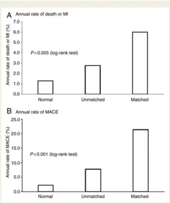
According to the Kaplan – Meier curves, the most favourable event-free survival was found in the normal followed by the unmatched group, whereas the matched group had the most unfavourable outcome with regard to death and non-fatal MI (P , 0.005; Figure 2A) and combined MACE (P , 0.001; Figure2B). The predictive value of matched SPECT/CCTA findings proved to be significant by Cox’s regression analysis (P , 0.001; Table 2). In addition, by multivariate analysis, the presence of a matched finding was confirmed as an independent predictor of combined MACE (P , 0.01), although this fell short of statistical significance for death and non-fatal MI. When the matched findings were expanded to include fixed defects, this turned to significance (multivariate hazard ratio 5.44, P , 0.005), reflecting the fact that infarct tissue contributes to a significant risk of future events, although there is less certainty about the appropriateness of target vessel revascularization of infarcted territories.
Finally, the overall annual rate of death or MI was 2.2%. First-year rates of death or MI were 8.1, 5.8, and 1.0% for patients with matched, unmatched, and normal findings, respectively, whereas the first-year rates of MACE were 27.0, 11.7, and 2.1%
for the respective patient groups. Similarly, in patients with matched hybrid findings, the annual rate of death or MI was highest at 6.0%, whereas for patients with unmatched and normal findings, the respective values were 2.8 and 1.3% (P , 0.005; Figure3A). The respective values for annual rate of MACE were 21.0, 7.8, and 2.2% (,0.001; Figure3B).
Discussion
Our results demonstrate that cardiac hybrid imaging with fused SPECT/CCTA allows improved risk stratification in patients with known or suspected CAD as a matched finding on hybrid image is a strong predictor of MACE. Although in the past years a number of studies have reported on the added diagnostic value of cardiac hybrid imaging,6,7,15 the present study is the first to document the prognostic value of the concurrent fused assess-ment of coronary morphology and myocardial perfusion. In fact, the results of the present study reveal that patients with stenosis in CCTA and a matched reversible perfusion defect in SPECT are at highest risk for future cardiac events.
Recommendations for the interventional coronary catheteriza-tion1suggest for best clinical practice that the addition of coronary physiological measurements should complement traditional angio-graphic information because this is essential for evidence-based accurate clinical decision-making as has been recently underlined by the prospective randomized FAME trial.2Combining MPI with CCTA allows non-invasive comprehensive CAD assessment which may contribute to avoid unnecessary invasive angiographies.3,6 Accordingly, hybrid imaging has shown to provide an added diagnos-tic information for culprit lesion identification and for guiding target vessel revascularization.6 The present data extend these findings, documenting that hybrid images allow accurate risk stratification.
A wealth of data has been published on the diagnostic accuracy and the prognostic value of MPI. In contrast, only limited data are
. . . .
. . . .
. . . . Table 1 Baseline characteristics
All Normal Unmatched Matched P-value
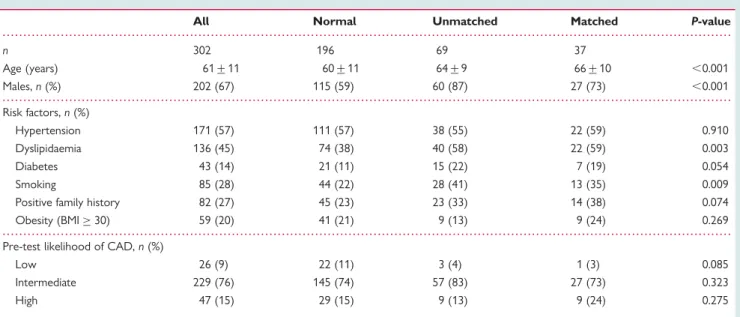
n 302 196 69 37 Age (years) 61+11 60+11 64+9 66+10 ,0.001 Males, n (%) 202 (67) 115 (59) 60 (87) 27 (73) ,0.001 Risk factors, n (%) Hypertension 171 (57) 111 (57) 38 (55) 22 (59) 0.910 Dyslipidaemia 136 (45) 74 (38) 40 (58) 22 (59) 0.003 Diabetes 43 (14) 21 (11) 15 (22) 7 (19) 0.054 Smoking 85 (28) 44 (22) 28 (41) 13 (35) 0.009
Positive family history 82 (27) 45 (23) 23 (33) 14 (38) 0.074 Obesity (BMI≥ 30) 59 (20) 41 (21) 9 (13) 9 (24) 0.269 Pre-test likelihood of CAD, n (%)
Low 26 (9) 22 (11) 3 (4) 1 (3) 0.085
Intermediate 229 (76) 145 (74) 57 (83) 27 (73) 0.323
High 47 (15) 29 (15) 9 (13) 9 (24) 0.275
available on the prognostic value of anatomic imaging with CCTA16–18or on the combination of CT and SPECT.19In patients with overall risk comparable to our study population, annual rates of death or MI between 4.320and 5.1%21have been reported for MPI abnormalities and between 2.718 and 5.3%17 for significant
lesions in CCTA. The annual rate of death or MI for matched find-ings in the present study (6.0%) lies above this range, suggesting the superiority of hybrid imaging for risk stratification. This superiority may at least in part be due to the fact that 3D hybrid imaging pro-vides additional information about haemodynamic lesion relevance and facilitates lesion interpretation by allowing exact allocation of perfusion defects to its subtending coronary artery. This cannot reliably be achieved by mental integration of the side-by-side CCTA and SPECT-MPI scan results as recently reported, because standard myocardial distribution territories correspond in only 50 – 60% with the real anatomic coronary tree.6Although in a low pre-test-probability population, a CCTA alone may be appropriate to rule out CAD, the identification of matched findings by hybrid imaging in an intermediate-risk population such as in the present study allows discriminating patients with substantial risk exceeding considerably the margin set to define ‘high risk’ by the ACC/AHA guidelines for stable angina at 3 – 5%.22 In order to provide clinically meaningful information on haemodynamically rel-evant lesions in the present study, only reversible SPECT-MPI find-ings were considered as a matched finding because ischaemia in territories subtended by stenotic coronaries constitutes an indi-cation for revascularization, whereas scar tissue does not. Our results support the clinical importance of functional lesion charac-terization particularly for prognostically relevant target vessel revascularization and confirm that also fixed defects confer a pre-dictive value for adverse events.
In addition to its prognostic value, hybrid imaging on hybrid devices or in dedicated imaging facilities with stand-alone scanners may confer the advantage of increasing the probability that patients are getting a comprehensive anatomic and functional non-invasive assessment before being sent to invasive catheterization. This could contribute to substantially increase the currently reported low yield of elective diagnostic invasive coronary angiography23 and potentially facilitate evidence-based coronary interventions.2,3
Study limitations
We acknowledge the following limitations: first, all-cause mortality was used although this is not a direct cardiac endpoint. However, an important advantage of all-cause death is the fact that it is easily ascertained, it is not affected by adjudication bias, and therefore constitutes the most valid endpoint.24 Secondly, the patients
Figure 2 The Kaplan – Meier survival curves showing the prog-nostic value of cardiac hybrid imaging. Cardiac hybrid findings predict (A) all-cause death or non-fatal myocardial infarction (MI) and (B) major adverse cardiac events (death, MI, unstable angina requiring hospitalization, and coronary revascularization).
. . . . . . . . Table 2 Predictors of events at univariate and multivariate analyses (n 5 302)
Predictors MACE Death or MI
Univariate HR (95% CI) P-value Multivariate HR (95% CI) P-value Univariate HR (95% CI) P-value Age 1.04 (1.01 – 1.07) 0.005 NA NS 1.09 (1.03 – 1.14) 0.002 Male gender 1.29 (0.68 – 2.45) 0.433 NA NS 2.12 (0.60 – 7.44) 0.241 ≥3 risk factors 2.62 (1.48 – 4.65) ,0.001 NA NS 3.74 (1.39 – 10.05) 0.009 Reversible perfusion defect 6.23 (3.51 – 11.08) ,0.001 NA NS 3.88 (1.41 – 10.68) 0.009 Stenosis≥50% 6.56 (3.46 – 12.45) ,0.001 3.12 (1.56 – 6.23) ,0.001 4.83 (1.68 – 13.91) 0.006 Matched SPECT/CCTA finding 7.48 (4.21 – 13.29) ,0.001 3.80 (1.76 – 8.21) 0.002 4.49 (1.63 – 12.37) 0.005
group with matched hybrid findings was relatively small. This is due to the fact that the probability of early coronary revascularization (defined as exclusion criteria) is much higher in patients with matched findings. Hence, many of these patients with highest risk were excluded from further outcome analysis which leads to an underestimation of the risk assessment. Nevertheless, there was good risk discrimination between the matched and the other groups, further supporting our results. Thirdly, in the univariate analysis, the presence of ischaemia was an independent predictor of adverse outcome, although this fell short of statistical signifi-cance for the multivariate analysis in contrast to the previous studies. This is at least in part due to the fact that the present study included into the multivariate analysis CCTA, hybrid, and MPI imaging results, i.e. findings which are linked by interaction. Fourthly, the additive radiation burden from combined SPECT and CCTA is a limitation and may have hampered its widespread use in daily clinical practice. However, the radiation dose can be decreased significantly when dedicated dose reduction techniques are implemented for SPECT25,26
and CCTA.13,27In fact, the last 61 patients of the present study were scanned using prospective ECG triggering, resulting in an average radiation dose of 1.8 + 0.6 mSv for the CCTA. Combined with new SPECT protocols including low-dose and stress-only MPI scanning,12,25radiation dose optim-ization allows to reach values below 5 mSv for hybrid SPECT/ CCTA scanning.28These dose reductions may improve the clinical value of hybrid imaging, as the balance of harms and benefits is shifted to the favourable end allowing such combined non-invasive
assessment of CAD to gain importance. Finally, data for MPI and CCTA were acquired on separate stand-alone scanners and hybrid images obtained by software fusion whereby misalignment could result in mismatch. The fact that despite potential misalign-ment matched findings allowed good risk stratification further strengthens our results. In addition, the accuracy of the fusion soft-ware and its clinical validity has been previously validated.5,6
Conclusions
This is the first study to show the independent prognostic value of cardiac hybrid imaging findings. The unique advantage of cardiac hybrid imaging is the complete non-invasive assessment of ana-tomic and functional data. By revealing both coronary stenosis and its functional relevance, the hybrid approach can provide com-prehensive information to guide management decisions in CAD patients. This, however, requires further validation in multicentre studies. In addition to being intuitively convincing and to providing incremental diagnostic information on functionally relevant coron-ary stenosis,6 the present study documents that cardiac hybrid imaging integrating SPECT with CCTA allows improved risk stratification.
Acknowledgements
We are grateful to Patrick von Schulthess, Ennio Mueller, Edlira Loga, Mirjam De Bloeme, Raji Kanagasabai, and De´sire´e Beutel for their excellent technical support.
Funding
The study was supported by a grant from the Swiss National Science Foundation.
Conflict of interest: none declared.
References
1. Kern MJ, Lerman A, Bech JW, De Bruyne B, Eeckhout E, Fearon WF, Higano ST, Lim MJ, Meuwissen M, Piek JJ, Pijls NHJ, Siebes M, Spaan JAE. Physiological assess-ment of coronary artery disease in the cardiac catheterization laboratory—a scientific statement from the American Heart Association Committee on Diag-nostic and Interventional Cardiac Catheterization, Council on Clinical Cardiology. Circulation 2006;114:1321 – 1341.
2. Tonino PA, De Bruyne B, Pijls NH, Siebert U, Ikeno F, van’ t Veer M, Klauss V, Manoharan G, Engstrom T, Oldroyd KG, Ver Lee PN, MacCarthy PA, Fearon WF. Fractional flow reserve versus angiography for guiding percutaneous coronary intervention. N Engl J Med 2009;360:213 – 224.
3. Gaemperli O, Husmann L, Schepis T, Koepfli P, Valenta I, Jenni W, Alkadhi H, Luscher TF, Kaufmann PA. Coronary CT angiography and myocardial perfusion imaging to detect flow-limiting stenoses: a potential gatekeeper for coronary revascularization? Eur Heart J 2009;30:2921 – 2929.
4. Namdar M, Hany TF, Koepfli P, Siegrist PT, Burger C, Wyss CA, Luscher TF, von Schulthess GK, Kaufmann PA. Integrated PET/CT for the assessment of coronary artery disease: a feasibility study. J Nucl Med 2005;46:930 – 935.
5. Gaemperli O, Schepis T, Kalff V, Namdar M, Valenta I, Stefani L, Desbiolles L, Leschka S, Husmann L, Alkadhi H, Kaufmann PA. Validation of a new cardiac image fusion software for three-dimensional integration of myocardial perfusion SPECT and stand-alone 64-slice CT angiography. Eur J Nucl Med Mol Imaging 2007;34:1097 – 1106.
6. Gaemperli O, Schepis T, Valenta I, Husmann L, Scheffel H, Duerst V, Eberli FR, Luscher TF, Alkadhi H, Kaufmann PA. Cardiac image fusion from stand-alone SPECT and CT: clinical experience. J Nucl Med 2007;48:696 – 703.
7. Santana CA, Garcia EV, Faber TL, Sirineni GK, Esteves FP, Sanyal R, Halkar R, Ornelas M, Verdes L, Lerakis S, Ramos JJ, Aguade-Bruix S, Cuellar H, Candell-Riera J, Raggi P. Diagnostic performance of fusion of myocardial perfusion imaging (MPI) and computed tomography coronary angiography. J Nucl Cardiol 2009;16:201 – 211.
Figure 3 Annual event rate according to hybrid findings. Patients with matched hybrid findings have a higher annual rate of death or non-fatal myocardial infarction (MI) (A) as well as a higher annual MACE rate (B) than patients with unmatched or normal findings. MACE, major adverse cardiac event.
8. Thygesen K, Alpert JS, White HD. Universal definition of myocardial infarction. J Am Coll Cardiol 2007;50:2173 – 2195.
9. Diamond GA, Forrester JS. Analysis of probability as an aid in the clinical diagnosis of coronary-artery disease. N Engl J Med 1979;300:1350 – 1358.
10. Fricke E, Fricke H, Weise R, Kammeier A, Hagedorn R, Lotz N, Lindner O, Tschoepe D, Burchert W. Attenuation correction of myocardial SPECT perfusion images with low-dose CT: evaluation of the method by comparison with per-fusion PET. J Nucl Med 2005;46:736 – 744.
11. Berman DS, Kiat H, Friedman JD, Wang FP, van Train K, Matzer L, Maddahi J, Germano G. Separate acquisition rest thallium-201/stress technetium-99m sesta-mibi dual-isotope myocardial perfusion single-photon emission computed tom-ography: a clinical validation study. J Am Coll Cardiol 1993;22:1455 – 1464. 12. Husmann L, Herzog BA, Gaemperli O, Tatsugami F, Burkhard N, Valenta I,
Veit-Haibach P, Wyss CA, Landmesser U, Kaufmann PA. Diagnostic accuracy of computed tomography coronary angiography and evaluation of stress-only single-photon emission computed tomography/computed tomography hybrid imaging: comparison of prospective electrocardiogram-triggering vs. retrospective gating. Eur Heart J 2009;30:600 – 607.
13. Husmann L, Valenta I, Gaemperli O, Adda O, Treyer V, Wyss CA, Veit-Haibach P, Tatsugami F, von Schulthess GK, Kaufmann PA. Feasibility of low-dose coronary CT angiography: first experience with prospective ECG-gating. Eur Heart J 2008;29:191 – 197.
14. Einstein AJ, Moser KW, Thompson RC, Cerqueira MD, Henzlova MJ. Radiation dose to patients from cardiac diagnostic imaging. Circulation 2007;116: 1290 – 1305.
15. Sato A, Nozato T, Hikita H, Miyazaki S, Takahashi Y, Kuwahara T, Takahashi A, Hiroe M, Aonuma K. Incremental value of combining 64-slice computed tomogra-phy angiogratomogra-phy with stress nuclear myocardial perfusion imaging to improve noninvasive detection of coronary artery disease. J Nucl Cardiol 2010;17:19 – 26. 16. Gaemperli O, Valenta I, Schepis T, Husmann L, Scheffel H, Desbiolles L,
Leschka S, Alkadhi H, Kaufmann PA. Coronary 64-slice CT angiography predicts outcome in patients with known or suspected coronary artery disease. Eur Radiol 2008;18:1162 – 1173.
17. Min JK, Shaw LJ, Devereux RB, Okin PM, Weinsaft JW, Russo DJ, Lippolis NJ, Berman DS, Callister TQ. Prognostic value of multidetector coronary computed tomographic angiography for prediction of all-cause mortality. J Am Coll Cardiol 2007;50:1161 – 1170.
18. Chow BJ, Wells GA, Chen L, Yam Y, Galiwango P, Abraham A, Sheth T, Dennie C, Beanlands RS, Ruddy TD. Prognostic value of 64-slice cardiac computed tomogra-phy severity of coronary artery disease, coronary atherosclerosis, and left ventri-cular ejection fraction. J Am Coll Cardiol 2010;55:1017 – 1028.
19. van Werkhoven JM, Schuijf JD, Gaemperli O, Jukema JW, Boersma E, Wijns W, Stolzmann P, Alkadhi H, Valenta I, Stokkel MP, Kroft LJ, de Roos A, Pundziute G, Scholte A, van der Wall EE, Kaufmann PA, Bax JJ. Prognostic value of multislice computed tomography and gated single-photon emission com-puted tomography in patients with suspected coronary artery disease. J Am Coll Cardiol 2009;53:623 – 632.
20. Patel AD, Abo-Auda WS, Davis JM, Zoghbi GJ, Deierhoi MH, Heo J, Iskandrian AE. Prognostic value of myocardial perfusion imaging in predicting outcome after renal transplantation. Am J Cardiol 2003;92:146 – 151.
21. Schinkel AF, Elhendy A, van Domburg RT, Bax JJ, Roelandt JR, Poldermans D. Prognostic value of dobutamine-atropine stress (99m)Tc-tetrofosmin myocardial perfusion SPECT in patients with known or suspected coronary artery disease. J Nucl Med 2002;43:767 – 772.
22. Gibbons RJ, Abrams J, Chatterjee K, Daley J, Deedwania PC, Douglas JS, Ferguson TB Jr, Fihn SD, Fraker TD Jr, Gardin JM, O’Rourke RA, Pasternak RC, Williams SV. ACC/AHA 2002 guideline update for the management of patients with chronic stable angina—summary article: a report of the American College of Cardiology/American Heart Association Task Force on practice guidelines (Committee on the Management of Patients With Chronic Stable Angina). J Am Coll Cardiol 2003;41:159 – 168.
23. Patel MR, Peterson ED, Dai D, Brennan JM, Redberg RF, Anderson HV, Brindis RG, Douglas PS. Low diagnostic yield of elective coronary angiography. N Engl J Med 2010;362:886 – 895.
24. Hachamovitch R, Di Carli MF. Methods and limitations of assessing new noninva-sive tests: Part II: outcomes-based validation and reliability assessment of nonin-vasive testing. Circulation 2008;117:2793 – 2801.
25. Herzog BA, Husmann L, Buechel RR, Pazhenkottil AP, Burger IA, Valenta I, Altorfer U, Wolfrum M, Nkoulou RN, Ghadri JR, Wyss CA, Kaufmann PA. Rapid cardiac hybrid imaging with minimized radiation dose for accurate non-invasive assessment of ischemic coronary artery disease. Int J Cardiol 2010; doi:10.1016/j.ijcard.2010.08.023.
26. Pazhenkottil AP, Herzog BA, Husmann L, Buechel RR, Burger IA, Valenta I, Landmesser U, Wyss CA, Kaufmann PA. Non-invasive assessment of coronary artery disease with CT coronary angiography and SPECT: a novel dose-saving fast-track algorithm. Eur J Nucl Med Mol Imaging 2010;37:522 – 527.
27. Herzog BA, Husmann L, Burkhard N, Gaemperli O, Valenta I, Tatsugami F, Wyss CA, Landmesser U, Kaufmann PA. Accuracy of low-dose computed tom-ography coronary angitom-ography using prospective electrocardiogram-triggering: first clinical experience. Eur Heart J 2008;29:3037 – 3042.
28. Herzog BA, Husmann L, Landmesser U, Kaufmann PA. Low-dose computed tom-ography coronary angitom-ography and myocardial perfusion imaging: cardiac hybrid imaging below 3mSv. Eur Heart J 2009;30:644.
![Figure 1 Visualization of matched and unmatched cardiac hybrid images. Cardiac hybrid single-photon emission computed tomography/cor- tomography/cor-onary computed tomography angiography images of a 40-year-old patient show a stenosis [(A), arrow] and a ma](https://thumb-eu.123doks.com/thumbv2/123doknet/14907683.656925/3.892.78.823.87.361/visualization-unmatched-cardiac-emission-tomography-tomography-tomography-angiography.webp)