. . . .
. . . .
Influence of smoking on the prognostic value
of cardiovascular computed tomography
coronary angiography
Jacob M. van Werkhoven
1,2, Joanne D. Schuijf
1, Aju P. Pazhenkottil
3,
Bernard A. Herzog
3, Jelena R. Ghadri
3, J. Wouter Jukema
1,2, Eric Boersma
4,
Lucia J. Kroft
5, Albert de Roos
5, Philipp A. Kaufmann
3,6, and Jeroen J. Bax
1*
1
Department of Cardiology, Leiden University Medical Center, Albinusdreef 2, 2333 ZA Leiden, The Netherlands;2
The Interuniversity Cardiology Institute of the Netherlands, Utrecht, The Netherlands;3
Cardiac Imaging, University Hospital Zurich, Zurich, Switzerland;4
Department of Cardiology, Erasmus Medical Center, Rotterdam, The Netherlands; 5
Department of Radiology, Leiden University Medical Center, Leiden, The Netherlands; and6
Zurich Integrative Human Physiology, University of Zurich, Zurich, Switzerland Received 17 May 2010; revised 20 September 2010; accepted 13 October 2010; online publish-ahead-of-print 7 December 2010
Aims Computed tomography coronary angiography (CTA) is an important non-invasive imaging modality increasingly used
for the diagnosis and prognosis of coronary artery disease (CAD). The purpose of the current study was to deter-mine the influence of smoking status on the prognostic value of CTA in patients with suspected or known CAD.
Methods and results
In 1207 patients (57% male, age 57 + 12 years) referred for CTA, the presence of significant CAD (≥50% stenosis)
was determined. During follow-up (FU) the following events were recorded: all cause mortality, and non-fatal infarc-tion. The prognostic value of CTA in smokers and non-smokers was compared using an interaction term in the Cox proportional hazard regression analysis. Significant CAD was observed in 327 patients (27%), and 273 patients (23%) were smokers. During a median FU time of 2.2 years, an event occurred in 50 patients. After correction for baseline characteristics including smoking in a multivariate model, significant CAD remained an independent predictor of events. Furthermore, a significant interaction (P , 0.05) was observed between significant CAD and smoking. The annualized event rate in smokers with significant CAD was 8.78% compared with 0.99% in smokers without signifi-cant CAD (P , 0.001). In non-smokers with signifisignifi-cant CAD the annualized event rate was 2.07% compared with 1.01% in non-smokers without significant CAD (P ¼ 0.058).
Conclusion The prognostic value of CTA was significantly influenced by smoking status. The event rates in patients with significant CAD were approximately four-fold higher in smokers compared with non-smokers. These findings suggest that smoking cessation needs to be aggressively pursued, especially in smokers with significant CAD.
-Keywords Ateriosclerosis † Smoking † Prognosis
Introduction
The introduction of multi-slice computed tomography coronary angiography (CTA) has changed the field of non-invasive imaging. In contrast to functional imaging techniques assessing myocardial perfusion and wall motion, CTA can provide direct non-invasive anatomic assessment of the coronary arteries. Because of the high negative predictive value for detection of significant coronary
artery disease (CAD) (defined as≥50% stenosis),1the technique
is increasingly used as a gatekeeper for further diagnostic testing. In the last 3 – 4 years, several single and multicenter studies have suggested that CTA may also provide important prognostic infor-mation. These studies have shown that patients with significant CAD detected on CTA are associated with worse outcome
com-pared with patients without significant CAD.2–7
Although the prognostic value of CTA and its incremental value over baseline clinical variables have thus been previously described, no reports have specifically focused on the prognostic value of
*Corresponding author. Tel:+31 71 5262020, Fax: +31 71 5266809, Email:[email protected]
CTA in smokers. This may be of interest, as smoking is an impor-tant but also modifiable risk factor resulting in an approximately two to four times increased risk of coronary heart disease
com-pared with non-smokers.8,9 Furthermore, smoking has recently
been shown to significantly increase the risk of events in asympto-matic individuals with evidence of atherosclerosis according to the coronary calcium score (CS), when compared with non-smokers
with a similar calcium burden.10 It is conceivable that smoking
has a similar effect on risk stratification with CTA. The purpose of the current study was therefore to determine the influence of smoking status on the prognostic value of CTA in patients with suspected or known CAD.
Methods
The study population consisted of patients who were clinically referred for CTA because of chest pain symptoms or a high risk profile for car-diovascular disease. Patients were enrolled at the University Hospital in Zurich, Switzerland, and at the Leiden University Medical Center, The Netherlands. Results from this prospective registry have been pre-viously published.5Exclusion criteria were: cardiac arrhythmias, renal insufficiency (defined as a glomerular filtration rate ,30 mL/min), known hypersensitivity to iodine contrast media, and pregnancy. In addition, patients with an uninterpretable CTA examination or coron-ary artery bypass grafts were excluded. Clinical patient characteristics were collected by the referring physician. Patients provided informed consent and the study was approved by the local ethics committees in both participating centres.
Computed tomography coronary
angiography acquisition and data analysis
Patients were scanned using a 64-row CT scanner (Aquilion64, Toshiba Medical Systems, Otawara, Japan; and General Electrics Light-Speed VCT, Milwaukee, WI, USA) or with a 320-row CT scanner (Toshiba Multi-slice Aquilion ONE system, Toshiba Medical Systems, Otawara, Japan). Before the examination, the patient’s heart rate and blood pressure were monitored. In the absence of contraindications, patients with a heart rate exceeding 65 beats per minute were admi-nistered beta-blocking medication (50 – 100 mg metoprolol, oral or 5 – 10 mg metoprolol, intravenous). All scan parameters have been previously published.11–13
Post-processing of the CTA examinations was performed on dedi-cated workstations (Vitrea2 and VitreaFx, Vital Images, USA; and Advantage GE Healthcare, USA). Computed tomography coronary angiography examinations were read by two experienced readers at both participating centres, blinded to follow-up (FU) results. Coronary anatomy was assessed using a 17 segment model according to a modi-fied American Heart Association classification.14 Normal CTA was defined as completely normal anatomy or minimal wall irregularities ,30%, non-significant CAD was defined as the presence of luminal narrowing with a maximal luminal diameter stenosis ,50%, and signifi-cant CAD was defined as the presence of a lesion exceeding≥50% maximal luminal diameter stenosis.
Follow-up results
Patient FU data were gathered using clinical visits or standardized tele-phone interviews. A composite endpoint was constructed using all cause mortality, and non-fatal myocardial infarction. Non-fatal infarc-tion was defined based on criteria of typical chest pain, elevated cardiac enzyme levels, and typical changes on the electrocardiogram
(ECG).15Patients with stable complaints undergoing an early elective revascularization within 60 days after CTA were excluded from the survival analysis.
Statistical analysis
Normally distributed continuous variables were expressed as mean values (+standard deviation). Non-normally distributed continuous variables were expressed as median values with a 25 – 75th percentile. Categorical baseline data were expressed in numbers and percentages. Differences between smokers and non-smokers were compared using the Student t and x2tests. Cox regression analysis was used to deter-mine the prognostic value of significant (≥50% luminal narrowing) CAD on CTA. First univariate analysis of baseline clinical variables, and CTA was performed using a composite endpoint of all cause mor-tality, and non-fatal infarction. For each variable a hazard ratio with a 95% confidence interval (95% CI) was calculated. A multivariate model was created to assess the independent prognostic value of CTA. To compare the prognostic value of CTA in smokers and non-smokers a final multivariate model was constructed to test for inter-action between smoking and CTA. Multivariate models were created using stepwise backward elimination; first all baseline clinical variables were included in the model, subsequently the least significant variable was excluded one at a time until all variables in the model reached a P-value of ,0.5. Annualized event rates were calculated based on the number of events per 100 patient years FU. Survival curves were estimated with the Kaplan – Meier method, and curves were com-pared using the log-rank test. Statistical analyses were performed using SPSS software (version 16.0, SPSS Inc., Chicago, IL, USA). A P-value of ,0.05 was considered statistically significant.
Results
The study population consisted of 1467 patients presenting at the University Hospital Zurich (n ¼ 468), and at the Leiden University Medical Center (n ¼ 999). In 44 (3%) patients the CTA examin-ation was uninterpretable due to the presence of motion artefacts, increased noise due to a high body mass index, and breathing. In addition, 117 patients (8%) were lost to FU. Finally 99 patients (7%) were excluded due to early revascularization. After exclusion, a total of 1207 remained for analysis. The majority of patients were symptomatic (67%), the remaining 33% of patients were referred because of a high risk profile with or without an abnormal exercise ECG. An overview of the baseline characteristics of the study
population is presented in Table1.
Computed tomography coronary
angiography results
Significant CAD was observed on CTA in 327 patients (27%). In the remaining 880 patients (73%) non-significant CAD was observed in 425 patients (35%) and 455 patients (38%) were
classi-fied as normal. Figure1illustrates the prevalence of significant CAD
on CTA according to smoking status. In non-smokers (n ¼ 934), significant stenosis was observed on CTA in 229 patients (25%), compared with 98 (36%) of the 273 patients who smoked (P , 0.001).
Follow-up results
The median FU time was 2.2 years (25 – 75th percentile: 1.3 – 3.2 years). During the FU period a myocardial infarction occurred in
12 patients and all cause mortality was registered in 40 patients. The composite endpoint of all cause mortality and myocardial infarction occurred in 50 patients. This resulted in an event rate of 1.8 per 100 patient years FU.
Survival analysis
The presence of significant CAD on CTA was a significant
univari-ate predictor of events (Table 2). After correction for baseline
clinical variables including smoking status, significant CAD
remained an independent predictor of events (Table 2). An
event rate of 4.01 events per 100 patient years FU was observed in patients with significant CAD compared with 1.0 event per 100 patient years FU in patients without significant CAD.
To assess the prognostic value of significant CAD on CTA in smokers and non-smokers, a second multivariate model was
con-structed to test for interaction (Table3). The prognostic value of
CTA was significantly higher in smokers compared with the prog-nostic value of CTA in non-smokers (interaction P ¼ 0.031). The event rate in smokers with significant CAD was 8.78 events per 100 patient years FU compared with 0.99 events per 100 patient years FU in smokers without significant CAD (P , 0.001). In
non-smokers with significant CAD the event rate was 2.07 events per 100 patient years FU compared with 1.01 events per 100 patient years FU in non-smokers without significant CAD (P ¼ 0.058). The survival rate following CTA according to
smoking status is illustrated in Figure2.
Discussion
The main finding of the current study comparing the prognostic value of CTA in smokers and non-smokers is that the prognostic value of significant CAD on CTA was significantly influenced by smoking status. The event rate in patients with significant CAD was approximately four-fold higher in smokers compared with non-smokers. On the other hand, in patients without significant CAD, the event rate was similar in smokers and non-smokers.
Although several studies have been published on the prognostic value of CTA, to our knowledge this is the first report to describe the effect of smoking on risk stratification with CTA. The effect of smoking on the prognostic value of atherosclerosis as detected by
CS has been studied.10Calcium score is generally used in
asympto-matic cohorts as a measure of atherosclerotic plaque burden, and elevated CS are associated with an increased risk of events. In the study by Shaw et al. in a large cohort of 10 377 asymptomatic individ-uals, the value of CS for risk stratification has been compared between smokers and non-smokers. The authors observed a signifi-cant interaction between smoking and CS for the prediction of all cause mortality. In each CS category the event rates in smokers were higher than observed in non-smokers. In addition to this imaging study in asymptomatic individuals, elevated event rates in smokers when compared with non-smokers have also been reported in symptomatic patients with established CAD. For instance, several studies have shown that following revascularization,
smokers have a higher event rate than non-smokers.16–18 The
results of the current study are in line with these findings and further strengthen the evidence that smokers with CAD have a higher risk of events than non-smokers with similar levels of CAD.
. . . .
. . . .
. . . .
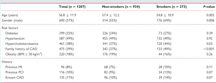
Table 1 Patient characteristics
Total (n 5 1207) Non-smokers (n 5 934) Smokers (n 5 273) P-value
Age (years) 56.8 + 11.9 57.4 + 12.2 54.8 + 10.9 0.002 Gender (male) 690 (57%) 514 (55%) 176 (64%) 0.006 Risk factors Diabetes 299 (25%) 226 (24%) 73 (27%) 0.39 Hypertension 587 (49%) 455 (49%) 132 (49%) 0.92 Hypercholesterolaemia 461 (38%) 341 (37%) 120 (44%) 0.03 Family history of CAD 475 (39%) 342 (37%) 133 (49%) ,0.001 Obesity (BMI≥ 30 kg/m2) 220 (18%) 176 (19%) 44 (16%) 0.26 History
Previous MI 96 (8%) 68 (7%) 28 (10%) 0.11
Previous PCI 116 (10%) 82 (9%) 34 (13%) 0.07
Known CAD 135 (11%) 96 (10%) 39 (14%) 0.07
BMI, body mass index; CAD, coronary artery disease; MI, myocardial infarction; PCI, percutaneous coronary intervention.
Figure 1 Relationship between computed tomography coron-ary angiography findings and smoking.
The observations in the current study may be explained in part by the influence of smoking on the formation and progression of atherosclerosis through its negative effects on vasomotor
dys-function, inflammation and lipid modification.19 Indeed multiple
reports have described the effects of smoking on the formation
of atherosclerosis both at autopsy,20 as well as in clinical
studies using coronary angiography,21,22 CS,23–25 and
intima-media thickness (IMT) measurements.26,27 Coronary angiography
studies have described that smoking is an important and indepen-dent predictor of CAD, which is in line with the increased
preva-lence of significant CAD observed in the current study.21,22 Of
interest, the atherosclerotic process seems to occur earlier in
life in smokers.25,28 Earlier formation of CAD explains the
increased levels of CAD observed in smokers; however, this may also be linked to increased progression of CAD. Smoking has been associated with CAD progression both on coronary angiography, IMT, and CTA. In a substudy of the CCAIT trial,
Waters et al.29 observed that smoking resulted in both plaque
progression and new plaque formation on serial quantitative coronary angiography.
The rapid decrease in the risk of myocardial infarction observed after smoking cessation suggests that in addition to the effects of smoking on CAD formation and progression, smoking may also
be seen as a trigger for myocardial infarction.30 Smoking may
affect all three major factors defining high-risk patients that are vul-nerable to myocardial infarction or sudden cardiac death:
vulner-able plaque, vulnervulner-able blood, and vulnervulner-able myocardium.31
. . . .
. . . .
Table 3 Interaction between smoking and significant
coronary artery disease on computed tomography coronary angiography
Exposure Patients Event HR (95% CI) P-value Non-smoking CTA , 50% 705 16 1.0 (reference) CTA≥ 50% 229 11 2.1 (0.9 – 4.5) 0.06 Smoking CTA , 50% 175 4 1.0 (reference) CTA≥ 50% 98 19 8.9 (3.0 – 26.5) ,0.001
Interaction P ¼ 0.031 and P ¼ 0. 045 (adjusted for age, diabetes, hypercholesterolaemia, obesity, and known CAD).
Figure 2 Survival according to computed tomography coron-ary angiography in non-smokers (A) and smokers (B).
. . . . . . . .
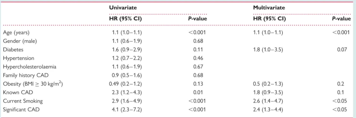
Table 2 Univariate and multivariate predictors of events
Univariate Multivariate
HR (95% CI) P-value HR (95% CI) P-value
Age (years) 1.1 (1.0 – 1.1) ,0.001 1.1 (1.0 – 1.1) ,0.001
Gender (male) 1.1 (0.6 – 1.9) 0.68
Diabetes 1.6 (0.9 – 2.9) 0.11 1.8 (1.0 – 3.5) 0.07
Hypertension 1.2 (0.7 – 2.2) 0.46 Hypercholesterolaemia 1.1 (0.6 – 1.9) 0.67 Family history CAD 0.9 (0.5 – 1.6) 0.68
Obesity (BMI≥ 30 kg/m2) 0.49 (0.2 – 1.2) 0.13 0.5 (0.2 – 1.3) 0.2
Known CAD 2.3 (1.2 – 4.3) 0.01 1.8 (0.9 – 3.5) 0.1
Current Smoking 2.9 (1.6 – 4.9) ,0.001 2.6 (1.4 – 4.7) ,0.05 Significant CAD 4.1 (2.3 – 7.2) ,0.001 2.4 (1.3 – 4.4) ,0.05
Smoking has been associated with inflammatory processes, and endothelial dysfunction which may increase plaque vulnerability resulting in a higher risk of intracoronary thrombus formation. In addition platelet function, antithrombotic/prothrombotic, and fibri-nolytic factors may be altered by smoking resulting in an increased thrombotic tendency which in turn may cause more frequent and
severe thrombus formation in response to plaque rupture.32–35
Finally, smoking results in activation of the sympathetic nervous system thereby increasing heart rate and myocardial contractility resulting in increased oxygen demand, while at the same time decreasing myocardial oxygen supply due to vasoconstriction of
the coronary arteries.36 This mismatch in oxygen demand/supply
may increase the myocardial vulnerability to ischaemia thereby
unfavourably altering myocardial response to thrombotic
occlusions.
Clinical implications
Further studies are needed to confirm our finding that the relative risk of events associated with significant CAD on CTA is signifi-cantly higher in smokers compared with non-smokers. Neverthe-less, our results do suggest that strategies aimed at preventing future cardiovascular events should be intensified in patients with significant CAD who smoke. This is further strengthened by the fact that smoking is a modifiable risk factor, and that smoking
cessation has been shown to improve survival.37,38
Interestingly, when regarding patients without significant CAD, the risk of events in smokers without significant CAD was similar to the risk observed in their non-smoking counterparts. On the basis of previous studies assessing effect of smoking on CAD, it is expected that new formation and progression of (non-significant) CAD should also be increased in patients without sig-nificant CAD who smoke. The similar event rates observed in the current study suggest that this effect may be more gradual. Longer FU studies are necessary to determine the influence of smoking status in patients without significant CAD.
Limitations
A limitation of the current study is that no exact data regarding quantification of smoking were available. This would have been of interest as several studies have suggested a dose – response relationship between smoking and the severity of CAD. In addition, the occurrence of passive smoking in the non-smoking subgroup was not systematically recorded. Because passive smoking has
also been associated with an increased risk of events,39–42 a
similar interaction as observed between significant CAD and active smoking may exist in passive smokers. Future studies are necessary to further study these concepts.
A general limitation of CTA imaging is the high radiation dose associated with traditional 64-slice CTA protocols, although the radiation dose of CTA has decreased substantially with the implementation of dose saving algorithms and novel acquisition
techniques.43–46 Importantly, low-dose CTA with prospective
ECG-triggering has recently been shown to reduce radiation burden while maintaining image quality and a high diagnostic
accu-racy.47Currently, the radiation burden with these novel acquisition
techniques is approaching≤2 mSv.48
Conclusion
The prognostic value of CTA was significantly influenced by smoking status. The event rates in patients with significant CAD were approximately four-fold higher in smokers compared with non-smokers. These results need to be confirmed in larger FU studies, but suggest that smoking cessation needs to be aggressively pursued, especially in smokers with significant CAD.
Funding
J.W. is financially supported by a research grant from the Netherlands Society of Cardiology (Utrecht, The Netherlands). P.A.K. is supported by a grant from the Swiss National Science, and has research grants from GE Healthcare (Milwaukee, WI, USA). J.J.B. has research grants from Medtronic, Boston Scientific, BMS medical imaging, St. Jude Medical, Biotronik, GE Healthcare, and Edwards Lifesciences. Conflict of interest: none declared.
References
1. Meijboom WB, Meijs MFL, Schuijf JD, Cramer MJ, Mollet NR, Van Mieghem CA, Nieman K, van Werkhoven JM, Pundziute G, Weustink A, de Vos AM, Pugliese F, Rensing BJ, Jukema JW, Bax JJ, Prokop M, Doevendans PA, Hunink MG, Krestin GP, de Feyter PJ. Diagnostic accuracy of 64-slice computed tomography coronary angiography a prospective multicenter, multivendor study. J Am Coll Cardiol 2008;52:2135 – 2144.
2. Gopal A, Nasir K, Ahmadi N, Gul K, Tiano J, Flores M, Young E, Witteman AM, Holland TC, Flores F, Mao SS, Budoff MJ. Cardiac computed tomographic angio-graphy in an outpatient setting: an analysis of clinical outcomes over a 40-month period. J Cardiovasc Comput Tomogr 2009;3:90 – 95.
3. Hadamitzky M, Freissmuth B, Meyer T, Hein F, Kastrati A, Martinoff S, Sheumig A, Hausleiter J. Prognostic value of coronary computed tomographic angiography for prediction of cardiac events in patients with suspected coronary artery disease. J Am Coll Cardiol Img 2009;2:404 – 411.
4. Ostrom MP, Gopal A, Ahmadi N, Nasir K, Yang E, Kakadiaris I, Flores F, Mao SS, Budoff MJ. Mortality incidence and the severity of coronary atherosclerosis assessed by computed tomography angiography. J Am Coll Cardiol 2008;52: 1335 – 1343.
5. van Werkhoven JM, Schuijf JD, Gaemperli O, Jukema JW, Boersma E, Wijns W, Stolzmann P, Alkadhi H, Valenta I, Stokkel MP, Kroft LJ, de Roos A, Pundziute G, Scholte A, van der Wall EE, Kaufmann PA, Bax JJ. Prognostic value of multislice computed tomography and gated single-photon emission com-puted tomography in patients with suspected coronary artery disease. J Am Coll Cardiol 2009;53:623 – 632.
6. Chow BJ, Wells GA, Chen L, Yam Y, Galiwango P, Abraham A, Sheth T, Dennie C, Beanlands RS, Ruddy TD. Prognostic value of 64-slice cardiac computed tomogra-phy severity of coronary artery disease, coronary atherosclerosis, and left ventri-cular ejection fraction. J Am Coll Cardiol 2010;55:1017 – 1028.
7. Min JK, Lin FY, Dunning AM, Delago A, Egan J, Shaw LJ, Berman DS, Callister TQ. Incremental prognostic significance of left ventricular dysfunction to coronary artery disease detection by 64-detector row coronary computed tomographic angiography for the prediciton of all-cause mortality: results from a two-centre study of 5330 patients. Eur Heart J 2010;31:1212 – 1219.
8. http://www.cdc.gov/tobacco/data_statistics/sgr/2004/index.htm(5 May 2009). 9. http://profiles.nlm.nih.gov/NN/B/B/X/S/_/nnbbxs.pdf(5 May 2009).
10. Shaw LJ, Raggi P, Callister TQ, Berman DS. Prognostic value of coronary artery calcium screening in asymptomatic smokers and non-smokers. Eur Heart J 2006; 27:968 – 975.
11. Schuijf JD, Wijns W, Jukema JW, Atsma DE, de Roos A, Lamb HJ, Stokkel MPM, Dibbets-Schneider P, Decramer I, De Bondt P, van der Wall EE, Vanhoenacker PK, Bax JJ. The relationship between non-invasive coronary angio-graphy with multi-slice computed tomoangio-graphy and myocardial perfusion imaging. J Am Coll Cardiol 2006;48:2508 – 2514.
12. Gaemperli O, Schepis T, Kalff V, Namdar M, Valenta I, Stefani L, Desbiolles L, Leschka S, Husmann L, Alkadhi H, Kaufmann PA. Validation of a new cardiac image fusion software for three-dimensional integration of myocardial perfusion SPECT and stand-alone 64-slice CT angiography. Eur J Nucl Med Mol Imaging 2007;34:1097 – 1106.
13. de Graaf FR, Schuijf JD, van Velzen JE, Kroft LJ, de Roos A, Reiber JH, Boersma E, Schalij MJ, Spano F, Jukema JW, van der Wall EE, Bax JJ. Diagnostic accuracy of
320-row multidetector computed tomography coronary angiography in the non-invasive evaluation of significant coronary artery disease. Eur Heart J 2010;31: 1908 – 1915.
14. Austen WG, Edwards JE, Frye RL, Gensini GG, Gott VL, Griffith LS, McGoon DC, Murphy ML, Roe BB. A reporting system on patients evaluated for coronary artery disease. Report of the Ad Hoc Committee for Grading of Coronary Artery Disease, Council on Cardiovascular Surgery, American Heart Association. Circulation 1975;51:5 – 40.
15. Thygesen K, Alpert JS, White HD. Universal definition of myocardial infarction. Eur Heart J 2007;28:2525 – 2538.
16. Goldenberg I, Jonas M, Tenenbaum A, Boyko V, Matetzky S, Shotan A, Behar S, Reicher-Reiss H. Current smoking, smoking cessation, and the risk of sudden cardiac death in patients with coronary artery disease. Arch Intern Med 2003; 163:2301 – 2305.
17. van Domburg RT, Meeter K, van Berkel DF, Veldkamp RF, van Herwerden LA, Bogers AJ. Smoking cessation reduces mortality after coronary artery bypass surgery: a 20-year follow-up study. J Am Coll Cardiol 2000;36:878 – 883. 18. Hasdai D, Garratt KN, Grill DE, Lerman A, Holmes DR Jr. Effect of smoking status
on the long-term outcome after successful percutaneous coronary revasculariza-tion. N Engl J Med 1997;336:755 – 761.
19. Ambrose JA, Barua RS. The pathophysiology of cigarette smoking and cardiovas-cular disease: an update. J Am Coll Cardiol 2004;43:1731 – 1737.
20. Strong JP, Richards ML. Cigarette smoking and atherosclerosis in autopsied men. Atherosclerosis 1976;23:451 – 476.
21. Wang XL, Tam C, McCredie RM, Wilcken DE. Determinants of severity of cor-onary artery disease in Australian men and women. Circulation 1994;89: 1974 – 1981.
22. Weintraub WS, Klein LW, Seelaus PA, Agarwal JB, Helfant RH. Importance of total life consumption of cigarettes as a risk factor for coronary artery disease. Am J Cardiol 1985;55:669 – 672.
23. Goel M, Wong ND, Eisenberg H, Hagar J, Kelly K, Tobis JM. Risk factor correlates of coronary calcium as evaluated by ultrafast computed tomography. Am J Cardiol 1992;70:977 – 980.
24. Loria CM, Liu K, Lewis CE, Hulley SB, Sidney S, Schreiner PJ, Williams OD, Bild DE, Detrano R. Early adult risk factor levels and subsequent coronary artery calcification: the CARDIA Study. J Am Coll Cardiol 2007;49:2013 – 2020. 25. Jockel KH, Lehmann N, Jaeger BR, Moebus S, Mohlenkamp S, Schmermund A,
Dragano N, Stang A, Gronemeyer D, Seibel R, Mann K, Volbracht L, Siegrist J, Erbel R. Smoking cessation and subclinical atherosclerosis—results from the Heinz Nixdorf Recall Study. Atherosclerosis 2009;203:221 – 227.
26. Howard G, Burke GL, Szklo M, Tell GS, Eckfeldt J, Evans G, Heiss G. Active and passive smoking are associated with increased carotid wall thickness. The Ather-osclerosis Risk in Communities Study. Arch Intern Med 1994;154:1277 – 1282. 27. Howard G, Wagenknecht LE, Burke GL, Diez-Roux A, Evans GW, McGovern P,
Nieto FJ, Tell GS. Cigarette smoking and progression of atherosclerosis: The Atherosclerosis Risk in Communities (ARIC) Study. J Am Med Assoc 1998;279: 119 – 124.
28. Herbert WH. Cigarette smoking and arteriographically demonstrable coronary artery disease. Chest 1975;67:49 – 52.
29. Waters D, Lesperance J, Gladstone P, Boccuzzi SJ, Cook T, Hudgin R, Krip G, Higginson L. Effects of cigarette smoking on the angiographic evolution of coron-ary atherosclerosis. A Canadian Coroncoron-ary Atherosclerosis Intervention Trial (CCAIT) Substudy. CCAIT Study Group. Circulation 1996;94:614 – 621. 30. Rosenberg L, Kaufman DW, Helmrich SP, Shapiro S. The risk of myocardial
infarc-tion after quitting smoking in men under 55 years of age. N Engl J Med 1985;313: 1511 – 1514.
31. Naghavi M, Libby P, Falk E, Casscells SW, Litovsky S, Rumberger J, Badimon JJ, Stefanadis C, Moreno P, Pasterkamp G, Fayad Z, Stone PH, Waxman S, Raggi P, Madjid M, Zarrabi A, Burke A, Yuan C, Fitzgerald PJ, Siscovick DS, de Korte CL, Aikawa M, Juhani Airaksinen KE, Assmann G, Becker CR,
Chesebro JH, Farb A, Galis ZS, Jackson C, Jang IK, Koenig W, Lodder RA, March K, Demirovic J, Navab M, Priori SG, Rekhter MD, Bahr R, Grundy SM, Mehran R, Colombo A, Boerwinkle E, Ballantyne C, Insull W Jr, Schwartz RS, Vogel R, Serruys PW, Hansson GK, Faxon DP, Kaul S, Drexler H, Greenland P, Muller JE, Virmani R, Ridker PM, Zipes DP, Shah PK, Willerson JT. From vulner-able plaque to vulnervulner-able patient: a call for new definitions and risk assessment strategies: Part I. Circulation 2003;108:1664 – 1672.
32. Hioki H, Aoki N, Kawano K, Homori M, Hasumura Y, Yasumura T, Maki A, Yoshino H, Yanagisawa A, Ishikawa K. Acute effects of cigarette smoking on platelet-dependent thrombin generation. Eur Heart J 2001;22:56 – 61. 33. Sambola A, Osende J, Hathcock J, Degen M, Nemerson Y, Fuster V, Crandall J,
Badimon JJ. Role of risk factors in the modulation of tissue factor activity and blood thrombogenicity. Circulation 2003;107:973 – 977.
34. Burke AP, Farb A, Malcom GT, Liang YH, Smialek J, Virmani R. Coronary risk factors and plaque morphology in men with coronary disease who died suddenly. N Engl J Med 1997;336:1276 – 1282.
35. Hung J, Lam JY, Lacoste L, Letchacovski G. Cigarette smoking acutely increases platelet thrombus formation in patients with coronary artery disease taking aspirin. Circulation 1995;92:2432 – 2436.
36. Quillen JE, Rossen JD, Oskarsson HJ, Minor RL Jr, Lopez AG, Winniford MD. Acute effect of cigarette smoking on the coronary circulation: constriction of epicardial and resistance vessels. J Am Coll Cardiol 1993;22:642 – 647.
37. van Berkel TF, Boersma H, Roos-Hesselink JW, Erdman RA, Simoons ML. Impact of smoking cessation and smoking interventions in patients with coronary heart disease. Eur Heart J 1999;20:1773 – 1782.
38. Gordon T, Kannel WB, McGee D, Dawber TR. Death and coronary attacks in men after giving up cigarette smoking. A report from the Framingham study. Lancet 1974;2:1345 – 1348.
39. Glantz SA, Parmley WW. Passive smoking and heart disease. Mechanisms and risk. J Am Med Assoc 1995;273:1047 – 1053.
40. Wells AJ. Passive smoking as a cause of heart disease. J Am Coll Cardiol 1994;24: 546 – 554.
41. Wells AJ. Heart disease from passive smoking in the workplace. J Am Coll Cardiol 1998;31:1 – 9.
42. Steenland K, Thun M, Lally C, Heath C Jr. Environmental tobacco smoke and cor-onary heart disease in the American Cancer Society CPS-II cohort. Circulation 1996;94:622 – 628.
43. Hausleiter J, Meyer T, Hadamitzky M, Huber E, Zankl M, Martinoff S, Kastrati A, Schomig A. Radiation dose estimates from cardiac multislice computed tomogra-phy in daily practice: impact of different scanning protocols on effective dose esti-mates. Circulation 2006;113:1305 – 1310.
44. Hsieh J, Londt J, Vass M, Li J, Tang X, Okerlund D. Step-and-shoot data acquisition and reconstruction for cardiac x-ray computed tomography. Med Phys 2006;33: 4236 – 4248.
45. Husmann L, Valenta I, Gaemperli O, Adda O, Treyer V, Wyss CA, Veit-Haibach P, Tatsugami F, von Schulthess GK, Kaufmann PA. Feasibility of low-dose coronary CT angiography: first experience with prospective ECG-gating. Eur Heart J 2008;29:191 – 197.
46. Rybicki FJ, Otero HJ, Steigner ML, Vorobiof G, Nallamshetty L, Mitsouras D, Ersoy H, Mather RT, Judy PF, Cai T, Coyner K, Schultz K, Whitmore AG, Di Carli MF. Initial evaluation of coronary images from 320-detector row computed tomography. Int J Cardiovasc Imaging 2008;24:535 – 546.
47. Herzog BA, Husmann L, Burkhard N, Gaemperli O, Valenta I, Tatsugami F, Wyss CA, Landmesser U, Kaufmann PA. Accuracy of low-dose computed tom-ography coronary angitom-ography using prospective electrocardiogram-triggering: first clinical experience. Eur Heart J 2008;29:3037 – 3042.
48. Herzog BA, Wyss CA, Husmann L, Gaemperli O, Valenta I, Treyer V, Landmesser U, Kaufmann PA. First Head-to-Head Comparison of Effective Radi-ation Dose from Low-Dose CT with Prospective ECG-Triggering vs. Invasive Coronary Angiography. Heart 2009;95:1656 – 1661.