1996 Oxford University Press Nucleic Acids Research, 1996, Vol. 24, No. 19 3829–3835
Structure, tissue distribution and genomic
organization of the murine RRM-type RNA binding
proteins TIA-1 and TIAR
Andreas R. P. Beck
1,2,4, Quintus G. Medley
1,2, Stephen O’Brien
1, Paul Anderson
1,3and
Michel Streuli
1,2,*
1Division of Tumor Immunology, Dana-Farber Cancer Institute, Boston, MA 02115, USA, 2Department of
Pathology and 3Department of Medicine, Harvard Medical School, Boston, MA 02115, USA and 4Laboratorium
für Biochemie I, ETH-Zentrum, 8092 Zürich, Switzerland
Received May 8, 1996; Revised and Accepted August 8, 1996 DDBJ/EMBL/GenBank accession nos U55861 and U55862
ABSTRACT
TIA-1 and TIAR are RNA binding proteins of the RNA recognition motif (RRM)/ribonucleoprotein (RNP) family that have been implicated as effectors of apoptotic cell death. We report the structures of murine TIA-1 and TIAR (mTIA-1 and mTIAR) deduced from cDNA cloning, the mRNA and protein tissue distribution of mTIA-1 and mTIAR, and the exon–intron structures of the mTIA-1 and mTIAR genes. Both mTIA-1 and mTIAR are comprised of three ∼100 amino acid N-terminal RRM domains and a ∼90 amino acid C-terminal auxiliary domain. This subfamily of RRM proteins is evolutionarily well conserved; mTIA-1 and mTIAR are 80% similar to each other, and 96 and 99% similar to hTIA-1 and hTIAR, respectively. The overall exon–intron structures of the mTIA-1 and mTIAR genes are also similar to each other, as well as to the human TIA-1 gene structure. While Northern blot analysis reveals that mTIA-1 and mTIAR mRNAs have a broad tissue distribution, mTIA-1 and mTIAR proteins are predominantly expressed in brain, testis and spleen. At least two isoforms of both mTIA-1 and mTIAR are generated by alternative splicing. Murine TIA-1 isoforms including or lacking the exon 5 encoded sequences are expressed at a ratio of ∼1:1, whereas mTIAR isoforms including or lacking the 5′-end of exon 3 sequences are expressed in a ∼1:6 ratio. Molecular characterization of murine TIA-1 and TIAR RNA binding proteins provides the basis for a genetic analysis of the functional roles of these proteins during mammalian development.
INTRODUCTION
RNA binding proteins of the RNA recognition motif (RRM)/ ribonucleoprotein (RNP) family function in diverse aspects of RNA metabolism including the biogenesis and translation of mRNA (1–4). RRM proteins contain one to four ∼100 amino acid RRM domains that have conserved hexamer and octamer peptide sequence motifs, referred to as the RNP 2 and RNP 1 motifs,
respectively, as well as various auxiliary domains (1,2). Several RRM proteins are known to regulate development. For instance, the Drosophila RRM proteins Sxl and tra2 control sex determination by sex-specific alternative splicing (5), the Drosophila squid gene product hrp40 is necessary for dorsoventral axis formation during oogenesis (6,7), the Drosophila orb gene product is required for the formation of the egg chamber and anteroposterior as well as dorsoventral patterning during oogenesis (8,9), the Drosophila
elav gene product is implicated in the development of the
embryonic nervous system (10), and a family of RRM proteins is likely to regulate human spermatogenesis (11,12). Previously we identified two highly related human RRM proteins, TIA-1 and TIAR, that contain three N-terminal RRM domains and a C-terminal auxiliary domain (13,14). TIA-1 and TIAR share 80% overall identity, and both proteins bind mRNA (15). The second RRM domains of TIA-1 and TIAR bind RNAs containing short stretches of uracil (15). Drosophila and C.elegans both contain a TIA-1/TIAR-like protein that is ∼47% identical to either human TIA-1 or TIAR, suggesting that these proteins have been evolutionarily conserved (16,17). Although the function of TIA-1 and TIAR is unknown, several findings implicate these RNA binding proteins as effectors of apoptosis: purified TIA-1 and TIAR induce apoptosis in appropriate permeabilized target cells (13,14), TIAR is translocated from the nucleus to the cytoplasm early during Fas-mediated apoptosis (18), and TIA-1 is a specific substrate for the Fas-activated protein serine/threonine (FAST) kinase (19). To develop a system to study RRM-type RNA binding proteins during mammalian development and apoptosis, we have characterized the cDNA and gene structures of the murine homologs of the human RNA binding proteins TIA-1 and TIAR, as well as determined their tissue distribution.
MATERIALS AND METHODS
cDNA cloning and sequencing
Mouse TIA-1 and TIAR cDNAs were isolated from a λ YES cDNA library derived from mRNA isolated from activated T-cells (20) by cross-hybridization with human TIA-1 (13) and TIAR (14) cDNA probes using standard techniques (21). DNA manipulations and DNA sequence determination using the chain
Nucleic Acids Research, 1996, Vol. 24, No. 19
3830
termination method were done according to standard procedures. The complete cDNA sequences of mTIA-1 and mTIAR appear in the EMBL/GenBank/DDBJ nucleotide sequence data bases (accession numbers U55862 and U55861, respectively). Characterization of mTIA-1 and mTIAR genomic DNA A mouse 129 SVJ genomic DNA library constructed in the λ FIX phage vector (Clontech) was screened with mTIA-1 and mTIAR cDNA probes, and positive phage clones were characterized by restriction mapping and Southern hybridization of restriction fragments with mTIA-1 and mTIAR cDNA probes essentially as previously described (22,23). The exact location of the exon–intron junctions was established by DNA sequencing of appropriate regions of the murine TIA-1 and TIAR genes.
Northern blot analysis
A tissue Northern blot containing about 2 µg poly(A)+ RNA per lane (Clontech) was probed with 32P-labeled mTIA-1 and mTIAR cDNA probes using the hybridization conditions recom-mended by the supplier. Murine TIA-1 specific mRNAs were detected using a mTIA-1 cDNA probe encoding amino acids 62–357 [including the exon 5 encoded TIA-1a peptide sequence (aa 93–103)] and 66 bp of 3′ non-translated region, and mTIAR specific mRNAs were detected using a cDNA probe encoding amino acids 112–357 [lacking the exon 3 encoded TIARa peptide sequence (aa 44–60)] and 37 bp of 3′ non-translated sequences. The absence of cross-hybridization between mTIA-1 and mTIAR under the conditions used for Northern blot hybridizations was confirmed by dot blot analysis (data not shown).
Plasmid constructions
The mTIA-1 expression plasmid pSRα.TIA-1a (containing the alternatively spliced exon 5 sequences) was generated by inserting TIA-1 cDNA encoding the TIA-1a isoform into the
EcoRI site of a modified version of the eukaryotic expression
vector pcDL-SRα296 (24), termed pSP65-SRα-PJ.Hygro, that
contains the hygromycin-β-phosphotransferase gene. The plasmid pMT.2.TIARb (lacking the alternatively spliced 5′ region of exon 3) was constructed by inserting the TIAR cDNA encoding the TIARb isoform into a modified version of the eukaryotic expression vector pMT.2 (25). cDNAs encoding the TIA-1b (lacking the alternatively spliced exon 5 sequence) and TIARa (containing the alternatively spliced 5′ region of exon 3) isoforms were generated by PCR using appropriate primers (mTIA-1 sense CGGGATCCATGGAGGACGAGATGCC, mTIA-1 anti-sense TACTGGCCAATCTGTTGTGC; mTIAR sense CGGGATC-CATGGAAGACGACGGACAGC, mTIAR anti-sense GCAGG-TGGTTTACGTGTGG) and the T cell cDNA library as template. DNA restriction fragments derived from the PCR-generated DNA were then used to replace the corresponding fragments in the pSRα.mTIA-1a and pMT.2.mTIARb plasmids to generate pSRα.mTIA-1b and pMT.2.mTIARa which encode the mTIA-1b and mTIARa isoforms, respectively.
Preparation of cell lysates, immunoprecipitations and immunoblotting
Tissues were isolated from Balb/c mice, washed with phosphate buffered saline, minced and homogenized in a dounce homogenizer
in buffer A (50 mM Tris–HCl pH 8.0, 1 mM EDTA, 1 mM phenylmethylsulfonyl fluoride, 10 µg/ml aprotinin and 10 µg/ml leupeptin). The homogenates were then adjusted to 150 mM NaCl, 1% NP-40 and incubated on ice for 20 min. Following incubation, the lysates were subjected to sonication, and then insoluble material was removed by centrifugation (20 000 g). For immunoprecipitations, cell lysates containing 2–4 mg protein (26) were precleared with Sepharose 4B beads, and then incubated with 5 µg mAb (the anti-TIA-1/TIAR mAbs ML29 or 6E3, or control isotype-matched mAb) and 25 µl protein G–Sepharose slurry (Pharmacia Biotech Inc.) for 3–5 h. The anti-TIA-1 and TIAR mAbs, 2G9 (13), 6E3 (18), 3E6 (18) and ML29 (18) were all originally obtained against human proteins. Immunoprecipitates were washed with buffer B (150 mM NaCl, 50 mM Tris–HCl pH 8.0 and 1% NP-40), and washed immuno-precipitated proteins were resolved by SDS–PAGE (10% gels) using reducing conditions. Proteins were then transferred to Immobilon P membrane (Millipore) and probed with anti-TIA-1 2G9 mAb (5 µg/ml) or anti-TIAR 3E6 mAb (1 µg/ml). Immunoblots were developed with protein A/G-horseradish peroxidase (Pierce) and the chemiluminescence reagent, luminol, essentially as described by the supplier (DuPont/NEN). To control for the specificity of the mAbs and to generate standards for the TIA-1 and TIAR isoforms, we produced recombinant mTIA-1a, mTIA-1b, mTIARa and mTIARb protein using the COS-7 transient transfection system. To this end, COS-7 (ATCC CRL 1651) cells were transfected with the expression vectors pSRα.mTIA-1a, pSRα.mTIA-1b, pMT.2.mTIARa and pMT.2.mTIARb using the DEAE–dextran method as described (21). Following transfection, cells were cultured for 48–56 h, washed with phosphate-buffered saline, and then cells were lysed in lysis buffer (1% NP-40, 150 mM NaCl, 50 mM Tris–HCl pH 7.5 containing 1 mM phenylmethylsulfonyl fluoride, 10 µg/ml aprotinin, 10 µg/ml leupeptin). Cell lysates from pSRα.mTIA-1a and pSRα.mTIA-1b transfected cells were pooled, as were cell lysates from pMT.2.mTIARa and pMT.2.mTIARb transfected cells, and then used as controls for immunoprecipitation and immunoblotting experiments. The relative amounts of mTIA-1 and mTIAR isoforms were determined by densitometric scanning of autoradiographs using a Ultrascan XL laser densitometer (Pharmacia).
RESULTS AND DISCUSSION
Structures of murine TIA-1 and TIAR deduced from cDNA sequences
To determine whether mice contain TIA-1 and TIAR RNA binding proteins, we screened a murine T-cell cDNA library using low stringency hybridization conditions with cDNA probes that contain the entire human TIA-1 and TIAR coding regions. Sequence analysis of the isolated cDNAs revealed the existence of murine homologs of human TIA-1 and TIAR. The deduced primary structure of the 386 amino acid mouse TIA-1 (mTIA-1) is 96% identical to the 386 amino acid human TIA-1 (hTIA-1) isoform, and the 392 amino acid mouse TIAR (mTIAR) sequence is 99% identical to the 392 amino acid human TIAR (hTIAR) isoform (Fig. 1A). Like their human counterparts, mTIA-1 and mTIAR consist of three RRM domains (RRM domain 1 to RRM domain 3) and a 90 or 88 amino acid C-terminal auxiliary domain, respectively (Fig. 1A and C). Furthermore, the mTIA-1 and mTIAR sequences share 80% overall identity, with the highest
3831
Nucleic Acids Research, 1994, Vol. 22, No. 1Nucleic Acids Research, 1996, Vol. 24, No. 19 3831
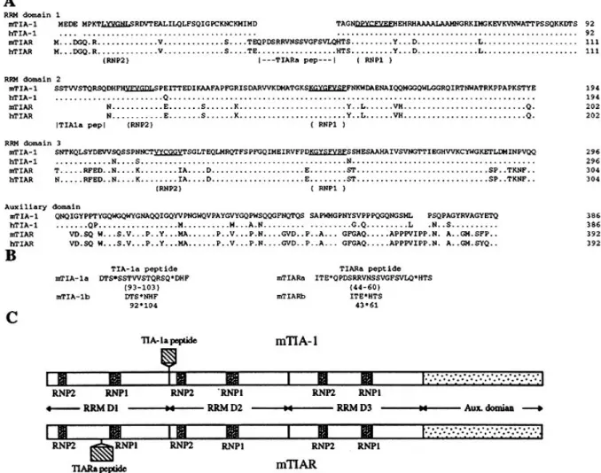
Figure 1. Comparison of the mTIA-1, mTIAR, hTIA-1 and hTIAR amino acid sequences. (A) The primary structure of mTIA-1 deduced from cDNA is shown using the standard single-letter code and is compared with the mouse mTIAR (this study), human hTIA-1 (13), and hTIAR (14) sequences. Dots in the hTIA-1, mTIAR and hTIAR sequences indicate identical amino acids as mTIA-1. Spaces in the sequences indicate gaps. The extent and boundaries of the RNP2, RNP1, TIA-1a and TIARa peptides are indicated below the sequence alignments. Locations of the RNP2 and RNP1 consensus sequences and RRM domains were identified by comparison with other RRM motif proteins (1). The predicted RRM domain and auxiliary domain junctions are based on sequence comparison and the exon–intron organization (Figs 4 and 5). Numbers indicate amino acid residues in the various proteins. (B) Deduced amino acid sequences of the two mTIA-1 and mTIAR isoforms. Shown are amino acid sequences containing the TIA-1a peptide (mTIA-1a isoform), lacking the mTIA-1a peptide (mTIA-1b isoform), containing the TIARa peptide (mTIARa isoform) or lacking the TIARa peptide (mTIARb isoform). The position of the sequence insertion or deletion is marked by a star. Numbering refers to the amino acid numbers shown in (A). (C) Schematic structure of mTIA-1 and mTIAR. The relative length and position of the alternatively used TIA-1a and TIARa peptides are indicated, as are the RNP2 and RNP1 consensus motif sequences in each of the three RRM domains (D1–D3).
degree of similarity residing in the RRM domain 3 (91% identity) and the lowest degree of similarity (50%) in the auxiliary domain. mTIA-1 and mTIAR are equally similar to the Drosophila
TIA-1-like protein rox8 (47% identity; 16) and a C.elegans TIA-like protein CM13B6 (43% identity; 17). The auxiliary domains of mTIA-1 and mTIAR do not have a significant degree of similarity with other known proteins.
Based on cDNA cloning and PCR analysis, two isoforms exist for both mTIA-1 and mTIAR. The two mTIA-1 isoforms differ from each other by the inclusion or exclusion of an 11 amino acid sequence (the TIA-1a peptide) located at the beginning of the second RRM domain (amino acids 93–103; Fig. 1), and are designated the TIA-1a isoform (containing the 11 amino acid TIA-1a peptide) and TIA-1b isoform (lacking the 11 amino acid sequence). The two mTIAR isoforms differ from each other by the inclusion or exclusion of a 17 amino acid sequence (the
TIARa peptide) located between the RNP2 and RNP1 motifs within RRM domain 1 (amino acids 44–60, Fig. 1), and are designated the TIARa (containing the 17 amino acid TIARa peptide) and TIARb (lacking the 17 amino acid sequence) isoforms. The mTIA-1 and mTIAR isoforms are generated by alternative splicing (see below), and are identical to their human counterparts in sequence and location (14,23). Thus the deduced primary structures and alternative splicing of TIA-1 and TIAR are highly conserved between humans and mice.
mTIA-1 and mTIAR mRNA tissue distribution
The expression of mTIA-1 and mTIAR mRNA was examined by Northern blot analysis using poly(A)+ RNA isolated from various mouse tissues and cDNA probes specific for either mTIA-1 or mTIAR. The Northern blot was sequentially probed for mTIA-1
Nucleic Acids Research, 1996, Vol. 24, No. 19
3832
Figure 2. mTIA-1 and mTIAR mRNA tissue expression. Northern blot analysis of poly(A)+ RNA isolated from different mouse tissues using a radiolabeled (A) mTIA-1 cDNA probe, (B) mTIAR cDNA probe or (C) β-actin cDNA probe. Size markers in kilobases (kb) are shown at the left.
expression and then for mTIAR expression. Multiple mTIA-1 mRNAs ranging in length from ∼3.5 to 9.5 kb are detected in all of the tissue mRNAs examined, with the exception of liver mRNA which has significantly lower levels of mTIA-1 mRNA (Fig. 2A). The predominant mTIA-1 mRNAs are ∼4, 4.4, 7 and 9.5 kb in length in most of the tissues analyzed, but the relative ratios of the various mTIA-1 mRNAs varies depending on the tissue mRNA analyzed. For instance, brain has comparable levels of the 4.0 and 9.5 kb mTIA-1 mRNA species, whereas testis has significantly more of the 4.0 kb mRNA than the 9.5 kb mRNA. The molecular basis for the various mTIA-1 mRNA forms is not known, but may include accumulation of mRNAs with retained introns and/or alternative length 3′ non-translated regions. Northern blot analysis of mTIA-1 mRNA isolated from cell lines that express mTIA-1 reveals only the 3.5 and 4.0 kb mRNA species, suggesting that these shorter mTIA-1 mRNAs are the mature isoforms encoding mTIA-1 (data not shown). mTIAR mRNA, like mTIA-1 mRNA, has a broad tissue distribution with multiple length mRNAs (Fig. 2B). In all of the tissue mRNAs examined (including liver), there are 1.6 and 4.5 kb mTIAR mRNA species, and in several tissues (i.e. spleen, lung and testis) there is an additional 2.3 kb form. In brain, the predominant mRNA isoform is 4.5 kb, whereas in testis it is 1.6 kb. Because brain and testis express mTIAR protein (see below) it is likely that both the 4.4 and 1.6 kb mTIAR mRNAs encode mTIAR. Control hybridization of the Northern blot with a β-actin cDNA probe indicates that approximately equal amounts of mRNA are present from each tissue (Fig. 2C). Thus, both mTIA-1 and mTIAR mRNAs are expressed in diverse tissues.
mTIA-1 and mTIAR tissue distribution
To determine the tissue distribution of mTIA-1 and mTIAR, as well as the relative expression levels of the mTIA-1 and mTIAR isoforms, cell lysates were prepared from mouse tissues and used for immunoprecipitation and immunoblotting analysis (Fig. 3). For this analysis, equal amounts of protein per lysate were used
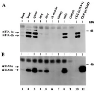
Figure 3. mTIA-1 and mTIAR protein tissue expression. Immunoblot analysis of (A) mTIA-1 and (B) mTIAR tissue distribution. Lysates prepared from different tissues (2–4 mg of total protein per immunoprecipitation), or from transfected COS cells, were immunoprecipitated with either the TIA-1/TIAR mAbs (A) ML29, or (B) 6E3, and the precipitated proteins were immunoblotted with (A) the TIA-1-specific mAb 2G9, or (B) the TIAR-specific mAb 3E6. COS(mTIA-1) and COS(mTIAR) indicates lysates prepared from COS cells transfected with cDNAs encoding mTIA-1 with and without alternatively spliced exon 5 (mTIA-1a and mTIA-1b), or cDNAs encoding mTIAR with and without the alternatively spliced 5′-end of exon 3 (mTIARa and mTIARb). Lysates containing the a and b isoforms of either mTIA-1 or mTIAR were pooled and then used in parallel with the tissue lysates for immunoprecipitations and immunoblots. Control indicates lysate prepared from brain and immuno-precipitated with a control isotype matched mAb. Analysis of the various lysate proteins following SDS–PAGE by Coomassie staining indicated the lysate proteins were intact for all samples. Molecular mass marker in kilodaltons (kDa) is shown at the right side, and the relative positions of the two mTIA-1 and mTIAR isoforms are shown at the left side.
for immunoprecipitation with either TIA-1/TIAR mAbs ML29 or 6E3, and then the immunoprecipitated proteins were analyzed by immunoblotting using either the TIA-1-specific mAb 2G9 (Fig. 3A) or the TIAR-specific mAb 3E6 (Fig. 3B). The specificities of the anti-hTIA-1 and anti-hTIAR mAbs were determined using COS cell-derived recombinant 42 kDa mTIA-1a, 40 kDa mTIA-1b, 42 kDa mTIARa and 40 kDa mTIARb proteins (e.g. Fig. 3, lanes 10 and 11). The 40 and 42 kDa isoforms of both mTIA-1 and mTIAR are predominantly expressed in brain, spleen and testis; and mTIAR is also expressed in liver and lung. mTIA-1 and mTIAR are not expressed, or only very weakly, in the other tissues tested such as heart, skeletal muscle and kidney (Fig. 3). The ratios of the mTIA-1a and mTIA-1b isoforms was ∼1:1 in all of the mTIA-1 containing tissues, and the ratios of mTIARa and mTIARb isoforms was ∼1:6. In contrast to the restricted distribution of the mTIA-1 protein, and to a lesser extent the mTIAR protein, mTIA-1 and mTIAR mRNAs are present in all of tissues tested (Fig. 2) suggesting that expression of mTIA-1 and mTIAR is subject to translational regulation. The functional significance of the largely coincident tissue distribution of mTIA-1 and mTIAR is unknown, but may suggest that these proteins serve complimentary or antagonistic functions during RNA metabolism. The differential expression of mTIA-1 and mTIAR in the various
3833
Nucleic Acids Research, 1994, Vol. 22, No. 1Nucleic Acids Research, 1996, Vol. 24, No. 19 3833
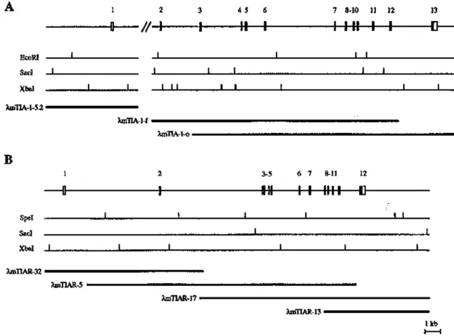
Figure 4. Exon–intron organization of the mouse mTIA-1 and mTIAR genes. The exon–intron organization of the (A) mTIA-1 and (B) mTIAR genes is schematically shown. The positions and relative sizes of the exons are indicated by closed boxes. For both the mTIA-1 and mTIAR genes, the initiating methionine residue is encoded within exon number 1. Open boxes represent 5′ and 3′ non-translated regions as determined by comparison with the cDNA-derived sequences of mTIA-1 and mTIAR. Alternatively spliced exons (mTIA-1 exon 5, and the 5′-end of mTIAR exon 3) are shown with stippled boxes. Introns are indicated by horizontal bars. The double slash indicates that the complete structure of mTIA-1 intron 1 has not been determined. Restriction maps of both mTIA-1 and mTIAR genes are shown, and restriction sites are indicated by vertical bars. The phage clones used for mapping and sequencing the exons are shown underneath the restriction maps of both mTIA-1 and mTIAR. Only the parts relevant to the structures of the mTIA-1 and mTIAR genes are shown for the phage clones λmTIA-5.2, λmTIAR-32 and λmTIAR-13.
tissues is also unclear, but other RNA binding proteins also implicated in development show a similar tissue distribution (27). Structure of the mTIA-1 gene
To establish the exon–intron organization of the mTIA-1 gene, a λgt11 murine genomic DNA library was screened using mTIA-1 cDNA probes containing the entire coding regions. Positive clones were characterized by restriction mapping, Southern blotting and DNA sequencing to establish the exon–intron junction of individual exons. Characterization of mTIA-1 genomic clone λmTIA-1-5.2 and the overlapping clones λmTIA-1-f and λmTIA-1-o revealed that the mTIA-1 gene contains 13 exons that span >20 kb (Fig. 4A). The exon–intron organization of the mTIA-1 gene, as well as the exon–intron junction sequences, are shown in Figures 4A and 5A, respectively. The mTIA-1 RRM domains 1, 2 and 3 are encoded by exons 1–4, 5–8 and 9–11, respectively; and the C-terminal auxiliary domain is encoded by exons 12 and 13. TIA-1 exon 5 encodes the alternatively used 11 amino acid TIA-1a peptide that distinguishes the mTIA-1a isoform from the mTIA-1b isoform. This exon–intron organization is conserved between the murine and human TIA-1 genes (23). The 11 amino acid TIA-1a peptide is directly N-terminal to the RNP2 motif consensus sequence of RNP domain 1. This location
is similar to an alternatively used 19 amino acid peptide of hnRNP D0 (28). hnRNP D0 contains two RRM domains, and the alternatively spliced 19 amino acid sequence affects RNA sequence binding specificity, suggesting that the alternative usage of the TIA-1a peptide may also modify RNA binding specificity (28).
Structure of the mTIAR gene
The exon–intron organization of the mTIAR gene was established in a similar manner as the mTIA-1 gene. Characterization of the overlapping mTIAR genomic clones λmTIAR-32, λmTIAR-5, λmTIAR-17 and λmTIAR-13 demonstrated that the mTIAR gene is comprised of 12 exons that span ∼20 kb (Fig. 4B). The exon–intron organization of the mTIAR gene, as well as the exon–intron junction sequences, are shown in Figures 4B and 5B, respectively. The mTIAR RRM domains 1, 2 and 3 are encoded by exons 1–4, 5–7 and 8–10, respectively; and the C-terminal auxiliary domain is encoded by exons 11 and 12. The 5′-end of TIAR exon 3 encodes the alternatively used 17 amino acid TIARa peptide sequence that distinguishes the mTIARa and mTIARb isoforms from each other. The 3′-end of exon 3 encodes amino acids 61–93, which are common to both mTIAR isoforms. Thus, the
Nucleic Acids Research, 1996, Vol. 24, No. 19
3834
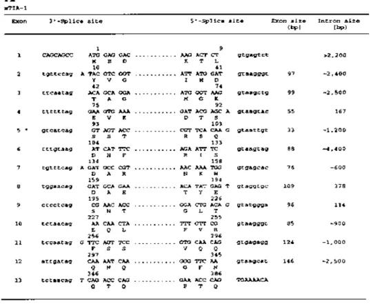
Figure 5. Exon–intron junctional sequences of the mTIA-1 and mTIAR genes. The nucleotide sequences flanking the 3′- and 5′-splice sites of the (A) mTIA-1 and (B) mTIAR genes are shown. Exon sequences are indicated by capital letters and intron sequences by lower case letters. The amino acid residues encoded by the junctional nucleotides of the exons are shown in single letter code, and the positions of the first and last amino acid encoded by each exon are indicated. The lengths of the exons and introns are shown on the right.
3835
Nucleic Acids Research, 1994, Vol. 22, No. 1Nucleic Acids Research, 1996, Vol. 24, No. 19 3835
mTIARa and mTIARb isoforms are generated by alternative 3′ splice acceptor site selection of intron 2. The TIARa peptide is located in a loop region in RNP domain 1 and may be important for RNA binding specificity (4,29).
The overall exon–intron organization of the mTIA-1 and mTIAR genes, as well as the hTIA-1 gene (23), are well conserved suggesting that the TIA-1 and TIAR genes were created by a gene duplication event that occurred prior to the divergence of mice and humans. However, the alternative usage of the TIA-1a peptide sequence (encoded by mTIA-1 exon 5) is specific for the TIA-1 gene, and the alternate usage of the TIARa peptide sequence (encoded by the 5′-end of mTIAR exon 3) is specific for the TIAR gene. It is unlikely that the mTIAR gene has a mTIA-1 exon-5-like exon, as the 133 bp mTIAR intron located between mTIAR exons 4 and 5 does not contain sequences that could encode an ∼11 amino acid peptide similar to the TIA-1a peptide. Sequences 5′ of mTIA-1 exon 3 also cannot be analogous to the TIARa peptide as alternative 3′ splice selection more 5′ of mTIA-1 exon 3 would result in the inclusion of a termination codon just 5′ of mTIA-1 exon 3. Thus, it is likely that alternative usage of the TIA-1a and/or TIARa peptide is a feature that was acquired (or lost) following the gene duplication event. The functional significance of different TIA-1 and TIAR isoforms generated by alternative splicing remains to be demonstrated, but is likely to regulate the RNA binding specificities of these proteins. The role of mTIA-1 and mTIAR in RNA metabolism remains to be established. In most tissues the expression of mTIA-1 and mTIAR is coincident (i.e. both proteins are expressed in brain, spleen and testis, and are absent in heart, skeletal muscle and kidney). This coincident expression pattern of mTIA-1 and mTIAR may suggest that these highly related proteins serve interdependent functions. However, in liver only mTIAR expression is observed, indicating that mTIAR can function independently of mTIA-1. There appears to be tissue-specific translational and/or transcriptional control of mTIA-1 and mTIAR expression. In skeletal muscle and kidney both mTIA-1 mRNA and mTIAR mRNA are expressed, but neither protein is detected. Furthermore, the absence of mTIA-1 mRNA in liver suggests that tissue-specific expression of these proteins can also be transcriptionally regulated. Given the role of RRM RNA binding proteins in Drosophila development (5–10), ongoing experiments with targeted disruptions of mTIA-1 and mTIAR should provide further insights into the functions of these mammalian RRM proteins.
ACKNOWLEDGEMENTS
We thank Drs Nancy Kedersha, Haruo Saito and Walter Blattler for critical review of the manuscript, Drs Stuart F. Schlossman and Kasper H. Winterhalter for encouragement and support, Dr S. Elledge for providing the murine cDNA library, and Dr Mark Boothby for help with isolating mTIA-1 cDNA. This work was supported by a Dana-Farber Cancer Institute/Apoptosis Technology Inc. drug discovery grant, National Institutes of Health Grants
AI33600 and CA67929, an ETH training fellowship to A.R.P.B, a Medical Research Council of Canada Fellowship to Q.G.M, and a Pew Scholar in the Biomedical Sciences Award to M.S.
REFERENCES
1 Burd,C.G. and Dreyfuss,G. (1994) Science, 265, 615–621.
2 Kenan,D.J., Query,C.C. and Keene,J.D. (1991) Trends Biochem. Sci., 16, 214–220.
3 Mattaj,I.W. (1993) Cell, 73, 837–840.
4 Nagai,K., Oubridge,C., Nobutoshi,I., Avis,J. and Evans,P. (1995) Trends Biochem. Sci., 20, 235–240.
5 Baker,B.S. (1989) Nature, 340, 521–524. 6 Kelley,R.L. (1993) Genes Dev., 7, 948–960.
7 Matunis,E.L., Kelley,R. and Dreyfuss,G. (1994) Proc. Natl. Acad. Sci. USA, 91, 2781–2784.
8 Lantz,V., Chang,J.S., Horabin,J.I., Bopp,D. and Schedl,P. (1994) Genes Dev., 8, 598–613.
9 Christerson,L.B. and McKearin,D.M. (1994) Genes Dev., 8, 614–628. 10 Yao,K.-M., Samson,M.-L., Reeves,R. and White,K. (1993) J. Neurobiol.,
24, 723–739.
11 Ma,K., Inglis,J.D., Sharkey,A., Bickmore,W.A., Hill,R.E., Prosser,E.J., Speed,R.M., Thomson,E.J., Jobling,M., Taylor,K., Wolfe,J., Cooke,H.J., Hargreave,T.B. and Chandley,A.C. (1993) Cell, 75, 1287–1295. 12 Reijo,R., Lee,T.-Y., Salo,P., Alagappan,R., Brown,L.G., Rosenberg,M.,
Rozen,S., Jaffe,T., Straus,D., Hovatta,O., de la Chapelle,A., Silber,S. and Page,D.C. (1995) Nature Genet., 10, 383–393.
13 Tian,Q., Streuli,M., Saito,H., Schlossman,S.F. and Anderson,P. (1991) Cell, 67, 629–639.
14 Kawakami,A., Tian,Q., Duan,X., Streuli,M., Schlossman,S.F. and Anderson,P. (1992) Proc. Natl. Acad. Sci. USA, 89, 8681–8685. 15 Dember,L.M., Kim,N.D., Liu,K.-Q. and Anderson,P. (1996) J. Biol.
Chem., 271, 2783–2788.
16 Brand,S. and Bourbon,H.-M. (1993) Nucleic Acids Res., 21, 3699–3704. 17 Wilson,R., Ainscough,R. anderson,K., Baynes,C., Berks,M., Bonfield,J.,
Burton,J., Connell,M., Copsey,T., Cooper,J. et al. (1994) Nature, 368, 32–38.
18 Taupin,J.-L., Tian,Q., Kedersha,N., Robertson,M. and Anderson,P. (1995) Proc. Natl. Acad. Sci. USA, 92, 1629–1633.
19 Tian,Q., Taupin,J.-L., Elledge,S., Robertson,M. and Anderson,P. (1995) J. Exp. Med., 182, 865–874.
20 Elledge,S.J., Mulligan,J.T., Ramer,S.W., Spottswood,M. and Davis,R.W. (1991) Proc. Natl. Acad. Sci. USA, 88, 1731–1735.
21 Ausubel,F.M., Brent,R., Kingston,R.E., Moore,D.D., Seidman,J.G., Smith,J.A. and Struhl,K. (1987–1997) Current Protocols in Molecular Biology. John Wiley & Sons, New York.
22 Hall,L.R., Streuli,M., Schlossman,S.F. and Saito,H. (1988) J. Immunol., 141, 2781–2787.
23 Kawakami,A., Tian,Q., Streuli,M., Poe,M., Edelhoff,S., Disteche,C.M. and Anderson,P. (1994) J. Immunol., 152, 4937–4945.
24 Takebe,Y., Seiki,M., Fujisawa,J.-I., Hoy,P., Yokota,K., Arai,K.-I., Yoshida,M. and Arai,N. (1988) Mol. Cell. Biol., 8, 466–472. 25 Bonthron,D.T., Handin,R.I., Kaufman,R.J., Wasley,L.C., Orr,E.C.,
Mitsock,L.M., Ewenstein,B., Loscalzo,J., Ginsburg,D. and Orkin,S.H. (1986) Nature, 324, 270–273.
26 Bradford,M.M. (1976) Anal. Biochem., 72, 248–254.
27 Han,J.R., Yiu,G.K. and Hecht,N.B. (1995) Proc. Natl. Acad. Sci. USA, 92, 9550–9554.
28 Kajita,Y., Nakayama,J., Aizawa,M. and Ishikawa,F. (1995) J. Biol. Chem., 270, 22167–22175.
29 Oubridge,C., Ito,N., Evans,P.R., Teo,C.-H. and Nagai,K. (1994) Nature, 372, 432–438.